Embed Size (px)
Citation preview

British Medical Bulletin 2002;63: 39–57 The British Council 2002
Sensory organ development in the inner ear:molecular and cellular mechanisms
Jane Bryant, Richard J Goodyear and Guy P RichardsonSchool of Biological Sciences, University of Sussex, Brighton, UK
The molecular mechanisms underlying the specification of sensory organs in theinner ear and the development of hair and supporting cells within these organsare described. The different organs are all derived from a common pro-sensoryregion, and may be specified by their proximity to the boundaries betweencompartments – broad domains within the otocyst defined by the asymmetricexpression patterns of transcription factors. Activation of Notch may specify thepro-sensory region, and lateral inhibition mediated by Notch signallinginfluences whether cells of common lineage in a sensory patch differentiate aseither hair cells or supporting cells. The transcription factors Math1 and Brn3.1are required for hair cell differentiation, and supporting cells express negativeregulators of neurogenesis, Hes1 and Hes5. Retinoic acid and thyroid hormoneinfluence early aspects and timing of hair cell differentiation, respectively.Development of the hair cell’s mechanosensory hair bundle involves interactionsbetween the cytoskeleton, cell-surface adhesion molecules, receptors andassociated extracellular matrix.
There are six distinct sensory organs in the mammalian inner ear: thethree cristae of the semicircular canals, the two maculae of the saccule andutricle, and the organ of Corti of the cochlea (Fig. 1). The cristae and themaculae are vestibular organs that respond to angular and linearacceleration, respectively. The organ of Corti is the organ of hearing.These three types of organ differ in their function, and in the fine detailsof their cellular architecture, but they all conform to the same basic plan.They are relatively simple epithelia composed of two basic cell types, thesensory hair cells and their surrounding, non-sensory supporting cells.These epithelia lie upon a sheet of extracellular matrix, a basal lamina,and also have a prominent extracellular structure, a cupula, an otoconialmembrane or a tectorial membrane, associated with their apical surface.The supporting cells sit on the basal lamina, and their lateral membranessurround the hair cells, projecting up to the surface of the epithelium. Thehair cells do not contact the basal lamina, and they are isolated from oneanother by the supporting cells. At the apical surface of the epithelium, thesupporting cell processes form tight and adherens junctions with each
Correspondence to:Dr Guy P Richardson,
School of BiologicalSciences, University of
Sussex, Falmer, Brighton BN1 9QG, UK

40
other and with the hair cells. The hair cells have a highly specializedbundle of modified microvilli on their apical surface, a hair bundle, and itis this feature that enables them to detect mechanical stimuli and trans-duce them into electrical signals.
New developments in hearing and balance
British Medical Bulletin 2002;63
Fig. 1 Diagram illustrating the main steps in the development of the inner ear (a–d), andthe structure of the three types of sensory organ (e–g). The inner ear develops from theotic placode (a) an ectodermal thickening that invaginates to form the otic pit (b) whichin turns pinches off from the ectoderm to form the otic vesicle (c). The different sensoryorgans are derived from a common pro-sensory patch (black) in the ventromedial wall ofthe otocyst (c). A complex series of morphogenetic events transforms the otic vesicle (c)into the labyrinth (d) containing three cristae (e), two maculae (f), and an organ of Corti(g). Abbreviations: G, VIIIth ganglion; AA, anterior ampulla; LA, lateral ampulla; PA,posterior ampulla;, U, utricle; S, saccule; C, cochlea; CUP, cupula; OM, otoconialmembrane; TM, tectorial membrane.

41
These sensory organs of the inner ear are all derived during developmentfrom the wall of the otocyst, a hollow, pear-shaped structure that forms,just after neural tube closure, from a thickening of the head ectoderm thatlies adjacent to the rhombencephalon and is known as the otic placode.The neurones that innervate the hair cells in each organ of the ear are alsoderived from the otocyst, by a process of delamination, prior to theformation the sensory organs. Although these neurones become criticallydependent on trophic support from the hair cells1,2, there is little evidencethat they influence the development of hair and supporting cells. In thisbrief review, we will focus specifically on the sequence of molecular andcellular events that leads to the differentiation of hair and supporting cellswithin these epithelial organs, rather than the process of synaptogenesis.We will describe the origin of the different sensory organs, the lineagerelationships of hair and supporting cells, and how the two cell typescome to adopt different fates. The roles of retinoic acid and thyroidhormone will be discussed, and recent evidence indicating that a complexinterplay between the cytoskeleton, components of the hair cell surfaceand the associated extracellular matrix controls the process of hair bundledevelopment will be reviewed.
Much of our understanding about the development of the inner earcomes from a number of different systems, and not just from the experi-ments done with mammalian species. Studies on the developing chick innerear, and more recently that of the zebrafish, provide considerableinformation on basic mechanisms and principles that are likely to beapplicable to the process of hair and supporting cell development in bothmice and humans. For certain aspects, detailed data are only available forthe chick and, when necessary, these will be used to illustrate how thesensory patches in the inner ear are formed.
Generation and specification of sensory organs
Histological studies3 originally suggested that the different sensoryorgans of the chick inner ear are all derived from a single patch of cellsin the ventromedial wall of the otocyst. Molecular studies have recentlyrevived support for this suggestion, and have revealed that there is a pro-sensory area in the ventromedial region of the otocyst that can bedefined by the expression patterns of Serrate14, Lunatic fringe4 andBEN5. Serrate1 (known as Jagged1 in mammals) is a transmembraneligand for Notch, a membrane receptor that is involved in many aspectsof development6. Lunatic fringe is a protein that modulates interactionbetween Notch and its ligands7. BEN is a cell–cell adhesion molecule ofthe Ig superfamily8. Notch is widely expressed throughout the otocyst9,and Serrate1 may serve to maintain a high level of Notch activation
Sensory organ development in the inner ear
British Medical Bulletin 2002;63

42
within the pro-sensory patch, endowing this region with the capacity toform sensory organs, and preventing the premature differentiation ofhair cells10. The role of Lunatic fringe in the pro-sensory patch isuncertain. Lfng–/– mice do not have any inner ear abnormalities11, and itmay only act as a weak inhibitor of Notch signalling10. As a cell–celladhesion molecule, BEN may serve to stop the cells of the pro-sensorypatch from mixing with cells in other regions of the otocyst, i.e. thosethat are destined to become non-sensory parts of the ear5. Althoughthese studies provide evidence for the existence of a common pro-sensory patch and reveal molecular constituents with potential roles, itis not yet known what signals determine the formation of this region, orfrom where these signals emanate.
A boundary model has been proposed to account for how the differentsensory organs of the inner ear are specified12. This model postulatesthat the different sensory organs of the inner ear form at, or in proximityto, the boundaries between a small number of different compartments.These compartments are defined by the asymmetric expression patternsof a few genes and may, for example, correspond to the ventral anddorsal halves, or the anterior and posterior segments of the otocyst.Genes for transcription factors such as Pax2, Dlx5, Otx1 and Hmx3 areexpressed in such broad domains in the otocyst, and data fromknockout mice are consistent with the boundary model (see Brigande etal13 and Cantos et al14 for reviews). For example, anterior and posteriorcristae along with their associated canals are missing in Dlx5–/– mice15.Also Pax2-/- mice fail to form a cochlear duct16. Compartment boundaryintersection points may, therefore, define where a sensory organ formswithin the ventromedial pro-sensory patch, and whether it becomes acrista, a macula, or a hearing organ.
The expression of BMP4 in spatially discrete regions within theventromedial pro-sensory patch marks the first appearance of individualsensory organs17,18. BMP4 is a member of the TGF-β family of secretedgrowth factors and, in the chick otocyst, BMP4 is expressed in all of thesensory organs as they first emerge17. However, BMP4 is not a goodmarker for every sensory organ in the mouse otocyst18, and it is onlyexpressed in some of the organs in the zebrafish ear19. Although BMP4could autonomously regulate the development of hair and supportingcells within the organs within which it is expressed, it may actually playa major role in controlling the development of the accessory structuresthat form adjacent to the sensory patches. For example, the BMP4antagonist Noggin severely disrupts semicircular canal formation buthas comparatively little effect on the development and differentiation ofhair cells20,21. FGF10 is also expressed by the presumptive sensoryepithelia of the mouse otocyst, and it too may operate in a paracrinefashion, signalling through the IIIb isoform of FGFR-2 to control the
New developments in hearing and balance
British Medical Bulletin 2002;63

43
development of adjacent non-sensory tissues22. A number of other genesare also expressed in sensory patches at the very early stages of theirdevelopment. These include BMP5 and BMP723, p75NGFR24,25, Msx117,NT318,24, bmp2b19, and MsxC19,26. Some of these, like MsxC, are onlyexpressed in certain sensory patches, and may specify the type of organthat develops.
Lineage and birth of hair and supporting cells
Hair and supporting cells in the mammalian inner ear are born over a briefperiod of development after the sensory patches have been specified27,28.The cells then differentiate and remain mitotically inactive. In the cochleaof the mouse, the cyclin-dependent kinase inhibitor, p27Kip1, is expressed bycells of the organ of Corti as soon as they withdraw from the cell cycle29,30.The pattern of p27Kip1 expression precisely delineates the region of thecochlear duct within which the hair and supporting cells of the organ ofCorti will differentiate. The expression of p27Kip1 is down-regulated in haircells as they begin overt cytodifferentiation, but in supporting cells itpersists into adulthood29. In p27Kip1–/– mice, cell proliferation within theorgan of Corti continues, and an excess of hair and supporting cells isfound29,31. These data indicate p27Kip1 negatively regulates cell proliferationin the organ of Corti. However, other molecules must also control thisprocess as many hair and supporting cells do leave the cell cycle anddifferentiate in the cochleae of p27Kip1 knockout mice.
In the hearing organs of both mammals27 and birds32, hair and sup-porting cells at any one place within the structure are born simultaneously,suggesting they may share a common lineage. Retroviral tracing studies inthe chick auditory organ have provided firm experimental evidence forthis suggestion33,34. Furthermore, it has been shown that the potential tobecome either a hair or a supporting cell is retained by a progenitor celluntil it has passed through its final mitotic division, as two-cell cloneswere found that contained either two supporting cells, two hair cells orboth cell types33.
The differentiation of hair and supporting cells
The decision to become either a hair cell or a supporting cell mostprobably involves lateral inhibition35,36, a process by which cells ofcommon origin adopt different fates. Lateral inhibition is usuallymediated by Notch signalling, and a number of studies have now shownthat the products of the neurogenic genes of the Notch signallingpathway play a key role in mediating the differentiation of hair and
Sensory organ development in the inner ear
British Medical Bulletin 2002;63

44
New developments in hearing and balance
British Medical Bulletin 2002;63
Table 1 Expression patterns of transcription factors and components of the Notch signalling pathway observedduring sensory organ development in the inner ear, and the effects of experimentally manipulating their expression
Name of Nature of protein Expression pattern Mutant/experimental Referencesgene or phenotypeprotein
Math1 bHLH transcription Mouse vestibule at E12, cochlea at E13.5; Complete absence of hair cells 30, 42, 50,factor expressed transiently by hair cells and in the inner ear of Math1–/– 54
down-regulated by P3 mice by P1
Brn3.1 POU domain Mouse vestibule E12.5, cochlea at E14.5; Loss of hair cells by P14 in 46–48transcription factor expressed specifically by hair cells Brn3.1-/- mice
GATA3 C4 zinc finger Mouse auditory epithelium at E14; 49transcription factor down-regulates in hair cells, then
in supporting cells
Hes1 bHLH transcription Greater and lesser epithelial ridges Increase in inner hair cells 52, 53factor of rat (E17.5) and mouse (E18.5) and utricular hair cells in
cochlea; supporting cells of rat Hes1–/– miceutricle by E17.5
Hes5 bHLH transcription Mouse cochlea at E15, restricted to Increased in outer hair cells 53, 54factor Deiter’s cells by E17 and macular hair cells in
Hes5–/– mice
Notch1 Transmembrane Wide-spread in epithelium of otocyst Increase in OHCs in Notch1+/– 9, 37–40,receptor from early stages, becomes restricted mice. Increased rows of IHCs and 42, 43
to supporting cells as they differentiate OHCs in rat cochlear cultures treated with Notch1 antisense oligonucleotides
Delta1 Transmembrane Mouse vestibular hair cells at E12.5, Large increase in hair cells 9, 10, 38, ligand for Notch mouse cochlear hair cells at 14.5. In numbers in zebrafish dominant 39
sensory patches of chick otocyst negative DeltaAdx2 mutants. from E3.5 onwards, and zebrafish Retroviral expression of dominantotocyst at 14 hpf negative Dl1dn in chick down-
regulates Ser1 expression
Serrate1 Transmembrane ligand In all sensory organs of the mouse Extra IHCs but loss of third row 4, 9, 38, 39,(Jagged1) for Notch inner ear by E12.5, and becomes OHCs along with loss of cristae 43–45
restricted to supporting cells as hair in mice with dominant missense cells differentiate In pro-sensory patch Jag1 mutations. Extra rows of of chick otocyst from E2.5 (stage 19) IHCs and OHCs in rat cochlear
cultures treated with Jag1 antisense oligonucleotides
Serrate2 Transmembrane ligand In hair cells of mouse vestibule at Extra rows of IHCs and OHCs in 37, 40, 43(Jagged2) for Notch E13.5, and cochlea at E14.5. In rat IHCs Jag2 null mutant mice
at E18, and OHCs at E20. In zebrafish from 18 hpf
Lunatic Glycosyl transferase In sensory organs of mouse inner ear by No obvious phenotype in inner 4, 11, 18fringe that modifies Notch E11.5, restricted to supporting cells in ear of Lfng deficient mice, but
extracellular domain organ of Corti by E16. In pro-sensory the extra rows of IHCs seen inpatch of chick otocyst by E2.5 Jag2 null mutant are suppressed
on a Lfng null mutant background
Numb Intracellular protein In all cells of chick sensory patch at E3, 10that blocks Notch restricted to hair cells at E12activation
Abbreviations: IHC, inner hair cell; OHC, outer hair cell; E, embryonic day; P, postnatal day; hpf, hours post-fertilisation.
45
supporting cells in the inner ear9,10,37–45. The expression patterns of thesegenes in the ear, and the phenotypes of the different mutants that havebeen examined, are summarised in Table 1. In addition, a number oftranscription factors have been shown to be involved in the process ofhair and supporting-cell differentiation46–54, some of which may directlyinteract with the Notch signalling pathway. These are also listed in Table1. The expression patterns of both the neurogenic genes and thetranscription factors, the consequences of experimentally manipulatingtheir expression, are now described and discussed in detail.
Math1
Math1, a mouse homologue of the Drosophila proneural gene atonal,encodes a basic, helix-loop-helix (bHLH) transcription factor. It isexpressed in the primordium of the organ of Corti after the cells in thisregion have withdrawn from the cell cycle and have begun to expressp27Kip1, but before hair cells have started to express myosin VIIa, amarker of overt differentiation30. Math1 is initially expressed by thinbands of cells that span the entire thickness of the epithelium, andexpression becomes restricted to hair cells located at the lumenal surfaceof the epithelium as they differentiate30,54. These thin bands of Math1-expressing cells that initially span the thickness of the epithelium may bebi-potential progenitor cells54. Alternatively, they may be vertical stacksof hair cells that subsequently undergo movement within the thicknessof the epithelium and spread out along the longitudinal axis of thecochlea as it elongates30.
Hair cells are absent from the inner ears of Math1–/– mice by birth50,and the ectopic expression of Math1 in the non-sensory cells of thegreater epithelial ridge that lie adjacent to the organ of Corti results inthe formation of supernumerary hair cells51. These results indicate thatMath1 is both ‘necessary and sufficient’ for hair cell differentiation30.Early markers of overt hair cell differentiation, myosin VI and calretinin,are never expressed during inner ear development in Math1–/– mice50.The sensory epithelia are thinner and non-stratified, but have anoverlying extracellular matrix suggesting the supporting cells may havedifferentiated. An apparent overcrowding of supporting-cell nuclei anda failure to observe apoptosis in the sensory epithelia of Math1–/– micewas originally interpreted as indicating that a fate switch had occurred,leading to an overproduction of supporting cells50. However, apoptoticcells have recently been reported in the organ of Corti of Math1–/– mice,so the hair cells may be produced, fail to express any known markers ofdifferentiation, and then die30. Sensory epithelia, albeit eventually devoidof hair cells, do form in Math1–/– mice, so it is unlikely that Math1 is
Sensory organ development in the inner ear
British Medical Bulletin 2002;63
46
acting as a true proneural gene like its invertebrate homologue atonal50.A proneural gene required for sensory organ specification in the innerear has, therefore, yet to be discovered.
Delta1, Jagged2 and Notch1
Delta1 and Jagged2 (known as Serrate2 in chick) are expressed by haircells in the mouse cochlea approximately 1 day after the onset of Math1expression38,40. In Jagged2 null mutant mice, there is an increase in thelinear density of inner and outer hair cells in the cochlea, with a nearlycomplete duplication of the normal, single row of inner hair cells, andmany stretches where there are four instead of three rows of outer haircells40. Delta mutants have not been studied in the mouse, but a dominantnegative allele of the zebrafish deltaA gene, deltaAdx2, results in a 5–6-foldincrease in hair cell numbers in the inner ear, and a loss of most of thesupporting cells41. Notch expression is initially widely distributedthroughout the inner ear epithelium and the sensory patches, and becomesrestricted to the supporting-cell layer as the hair cells differentiate9,39,40,42,43.Notch1–/– mice are early embryonic lethals, but in heterozygotes withpresumably reduced levels of Notch1, a significant increase is observed inthe numbers of regions along the cochlea where there are four instead ofthree rows of outer hair cells11. Treating rat cochlear cultures withantisense Notch oligonucleotides causes the production of extra rows ofboth inner and outer hair cells43. The expression of Delta1 and Jagged2 inhair cells, and the overproduction of hair cells seen in correspondingmutants and with antisense oligonucleotides to Notch1, all indicate thathair cells use Notch signalling to inhibit laterally their neighbouring cellsand thereby prevent them from adopting the same fate.
Serrate1/Jagged1
Serrate1/Jagged1 is expressed throughout the pro-sensory patchinitially4,9,38, but becomes progressively restricted to the supporting cellsas they differentiate38,39. The expression of Serrate1/Jagged1 bysupporting cells may not appear to be consistent with the idea that haircells inhibit their neighbours. However, there is evidence from the chickthat hair cells express Numb, a protein that blocks Notch signalling10.These hair cells would, therefore, be deaf to signals delivered by Serrate1from adjacent supporting cells. Furthermore, there is also evidence fromthe chick inner ear that the expression of Serrate 1 in supporting cells ispositively regulated by Notch signalling via lateral induction10. Thiswould serve to increase the level of Notch activation amongst the
New developments in hearing and balance
British Medical Bulletin 2002;63
47
supporting cells, and ensure that they do not differentiate as hair cells.The overproduction of hair cells in late embryonic rat cochlear culturescaused by Jagged1 antisense oligonucleotides43 is consistent with this, asreduced Notch activation should lead to an excess of hair cells.However, in mouse mutants44,45, Jagged1 mutations that are assumed tobe dominant negative lead to the complete loss of some sensory organs(cristae) and only perturb hair cell patterning in others (cochlea). Thiscould be because Serrate1/Jagged1 has an early role in specifying sensory
Sensory organ development in the inner ear
British Medical Bulletin 2002;63
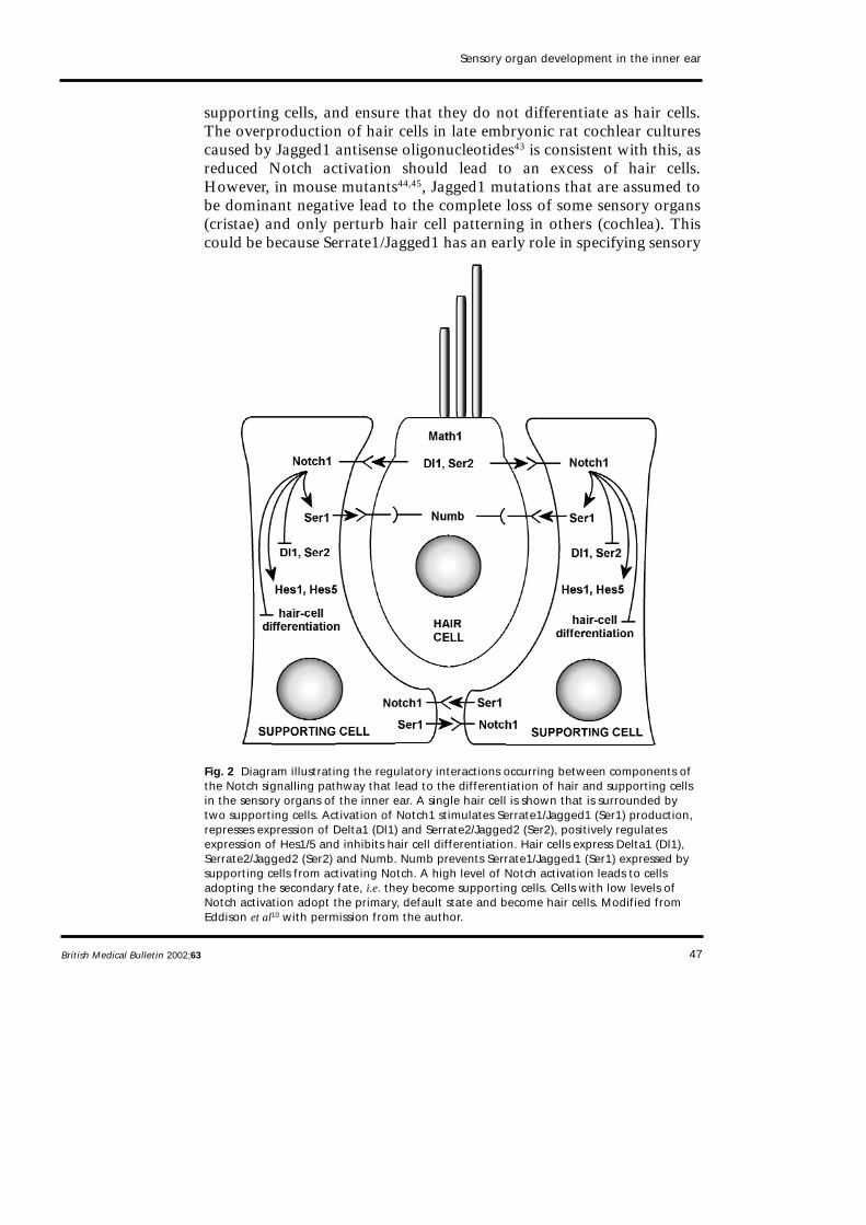
Fig. 2 Diagram illustrating the regulatory interactions occurring between components ofthe Notch signalling pathway that lead to the differentiation of hair and supporting cellsin the sensory organs of the inner ear. A single hair cell is shown that is surrounded bytwo supporting cells. Activation of Notch1 stimulates Serrate1/Jagged1 (Ser1) production,represses expression of Delta1 (Dl1) and Serrate2/Jagged2 (Ser2), positively regulatesexpression of Hes1/5 and inhibits hair cell differentiation. Hair cells express Delta1 (Dl1),Serrate2/Jagged2 (Ser2) and Numb. Numb prevents Serrate1/Jagged1 (Ser1) expressed bysupporting cells from activating Notch. A high level of Notch activation leads to cellsadopting the secondary fate, i.e. they become supporting cells. Cells with low levels ofNotch activation adopt the primary, default state and become hair cells. Modified fromEddison et al10 with permission from the author.
48
patches and/or preventing their premature differentiation, and a laterrole in regulating the development of hair and supporting cells withinthe patch44. Whilst this may partially explain why the mutations, whichare effective throughout development, and antisense treatment, whichhas only been applied at relatively late developmental stages, havedifferent effects, it does not explain why the different organs in the earsof Jagged1 mutant mice respond differently to the same mutation.However, the loss of cristae may occur secondarily to the truncation ofthe canals, which in turn may be due to diminished BMP4 signallingfrom the sensory patch resulting from reduced Notch activation.
The expression patterns of the neurogenic genes described above andthe phenotypes of the different mutants that have been examined aregenerally in accordance with the theory that hair cells laterally inhibittheir neighbours and force them to become supporting cells. However,the situation is clearly more complex than originally envisioned35,36.Although a stochastic fluctuation in Notch signalling in a field of cellsinitially expressing uniform levels of Notch and Delta can theoreticallylead, as a result of negative feedback, to the production of regularmosaics of hair and supporting cells55,56, Delta1 is not uniformlyexpressed38, there are two other Notch ligands operating in the ear, andthe signalling pathway can be modulated at many levels, making thesimple scenario, although attractive, less likely. Furthermore, an activere-arrangement of hair and supporting cells relative to one another maycontribute to the formation of a precise cellular mosaic, and refine theimperfect patterns that are initially observed during the early stages ofdevelopment57, indicating that lateral inhibition with feedback is not theonly mechanism contributing to pattern formation in the sensoryepithelia. The expression patterns for the neurogenic genes and theexperimental findings described above are largely consistent with asystem of regulatory interactions (see Fig. 2) recently proposed by Lewisand colleagues10.
Hes1 and Hes5
Hes1 and Hes5, mammalian homologues of the Drosophila hairy andenhancer of Split genes, are expressed in patterns that are bothcomplementary and overlapping in the developing cochlea and utricle ofthe mammal52,53. The products of these genes act as negative regulatorsof neurogenesis in vertebrates58. Expression of Hes5 is first observed inthe supporting cells of the organ of Corti after the expression of Math1has begun in differentiating hair cells, and probably slightly after theonset of expression of Delta1 and Jagged254. The expression of Hes5may be positively regulated by Notch activation as it is much reduced in
New developments in hearing and balance
British Medical Bulletin 2002;63
49
the supporting cells (Deiter’s cells) of Jag2–/– mice54. Additional innerhair cells are observed in Hes1–/– mutant mice, and additional outer haircells are observed in Hes5–/– mutants52,53, generally consistent with theexpression patterns of these genes and their proposed role as negativeregulators of hair cell differentiation. Hes1 may directly antagonize theactivity of Math1, as co-transfecting cells in cochlear cultures with Hes1and Math1, blocks the effects of ectopic Math1 expression52, reducingthe production of supernumerary hair cells.
Brn3.1 and GATA3
Brn3.1 (also called Brn3c) is a POU domain transcription factor that isspecifically expressed by hair cells within the adult mouse inner ear46,47.Brn3.1 is expressed by postmitotic hair cells at approximately the sametime as Jagged2 and Delta1, 1 day before the hair cell markers myosinVI and myosin VIIa48. In Brn3.1–/– mice, hair cells are generated andexpress myosin VI, myosin VIIa and calretinin, but they never developsensory hair bundles and are lost from the inner ear by P1446,47. Theectopic overexpression of Brn3.1 does not lead to the production of haircells51, indicating Brn3.1 is only required for the later aspects of hair celldifferentiation. GATA3 is another transcription factor that may beinvolved in cell differentiation within the sensory patches. All epithelialcells in the dorsal wall of the cochlear duct express GATA3, and itsexpression selectively decreases in both hair and supporting cells as theydifferentiate, indicating it may act as a negative regulator of hair andsupporting-cell differentiation49.
Roles of thyroid hormone and retinoic acid in hair celldifferentiation
Nuclear receptors for thyroid hormone and retinoic acid are expressedin the developing sensory epithelia of the inner ear and their respectiveligands are known to play roles in hair cell development (see Raz andKelley59 for recent review). Retinoic acid is produced by the embryonic,but not adult, organ of Corti, and treating cochlear cultures from early,but not late, embryonic stages of development with retinoic acid leadsto the production of extra rows of inner and outer hair cells60. TheRARα/RXRα heterodimer is the most likely hair cell receptor forretinoic acid, and application of the RARα antagonist Ro-41-5253 tocochlear cultures leads to a reduction in the number of hair cells thateventually develop61. However Ro-41-5253 does not block the initialdifferentiation of hair cells, as judged by the expression of Brn3.1 and
Sensory organ development in the inner ear
British Medical Bulletin 2002;63

50
myosin VI61, so it has been suggested that retinoic acid induces certainearly aspects of hair cell differentiation rather than determining the sizeof the pro-sensory cell population61.
The three functional thyroid hormone receptors, TRα1, TRβ1 andTRβ2, and the non-ligand binding TRα2 are expressed in the sensoryepithelia of the inner ear from early stages, with the expression of TRβ1and TRβ2 being restricted to the cochlear epithelium62. Chemicallyinduced hypothyroidism results in a delay in the maturation of mostelements in the organ of Corti, and the same effect is observed intransgenic mice that lack all known thyroid hormone receptors63. TRβis essential for the development of hearing64 and in Thrb–/– mice there isa delay in the onset of the fast potassium conductance (IK,f) in inner haircells, but the onset of outer hair cell motility is unaffected65. AlthoughIK,f is eventually expressed by inner hair cells in Thrb–/– mice, hearingdoes not recover, possibly due to a permanent impairment of thetectorial membrane63. In Thra–/–/Thrb–/– compound null mutant miceboth the onset of IK,f and outer hair cell motility is delayed63, and a recentstudy has shown that the gene encoding prestin, the outer hair cellmotor66, is regulated by thyroid hormone67.
Development of the sensory hair bundle
The sensory hair bundle, a precisely determined array of actin-filled stereocilialocated on the hair cell’s apical surface, is a distinguishing characteristic of thehair cell and is essential for the process of mechanotransduction. Thedevelopment of the hair bundle is a complex process and, for the auditoryorgan of the avian inner ear, the morphological details have been describedthoroughly (see Tilney and Tilney68 for review). For the mammal, this processhas been best characterized in the mouse vestibular system69,70, and thecochlea of the hamster71 and the rat72. For the mouse cochlea, information issomewhat limited.
Although there are some inter-species and inter-organ differences, thegeneral features of the process of hair bundle development can bedescribed as follows. First, small stereocilia sprout up over the entireapical surface of the hair cell, clustering around a centrally locatedkinocilium to form a short bundle of uniform height. Second, thekinocilium migrates to one side of the cell and the stereocilia nearest thekinocilium begin to elongate and generate the staircase pattern in whichadjacent rows of stereocilia become arranged in increasing height withthe tallest row lying next to the now eccentrically placed kinocilium. Theposition of the kinocilium, therefore, defines the planar polarity of thehair cell and its bundle. Third, rootlets project down from thestereocilia, anchoring them into the cuticular plate, and the excess
New developments in hearing and balance
British Medical Bulletin 2002;63

51
supernumerary stereocilia that have not been incorporated into theranked rows of the bundle are re-absorbed. Finally, the stereocilia withinthe bundle grow to their final width and height, and the bundle achievesits mature shape and form. In the chick auditory organ, increases in thewidth and height of stereocilia occur over different time periods68,whereas in mammals both processes, thickening and lengthening, occurconcurrently71. Also, differences in bundle height in the avian hearingorgan may be governed by regulating the period over which the growthof stereocilia occurs at a constant rate68, whereas in the mammaliancochlea they may be generated by differences in growth rate71.
Surprisingly little is known about the molecular basis of hair bundledevelopment in any species. Clearly, it will involve molecules andpathways that direct the assembly of the actin cytoskeleton, like theRho-GTPases along with their upstream effectors and their downstreamtargets, and two recent reviews have suggested how this may beaccomplished73,74. However, thus far, studies on mouse mutants andhuman deafness genes have provided the greatest insight into themolecular processes that may be involved in hair bundle development.
Mice with mutations in the genes that underlie deaf-blindness in threeof the genetic forms of the human Usher type I syndrome, USHIB,USHID and USHIF, exhibit defects in hair-bundle development. Thesegenes encode the unconventional myosin, myosin VIIa (shaker-1mouse75), and two cell adhesion molecules, cadherin 23 (waltzermouse76) and protocadherin 15 (Ames waltzer mouse77). Bundles withranked rows of stereocilia form in all three mouse mutants, but by theearly stages of postnatal development, they show varying degrees ofdisruption and are often fragmented into several smaller units75–77,suggesting there may be defects in the mechanisms of inter-stereociliaryadhesion. The USHIC gene encodes a PDZ domain protein that ispresent in stereocilia and could act as an interface between thecytoskeleton and the plasma membrane78, although it is not yet knownhow mutations in this gene affect hair bundle development. Interactionsbetween the actin cytoskeleton, unconventional myosins, PDZ domainproteins and cell-surface adhesion molecules may, therefore, play apivotal role in the development of hair bundle integrity. Mutations intwo other unconventional myosins, cause non-syndromic humanhereditary deafness and cause defects in hair bundle development in thecorresponding mouse mutants. Defects in myosin VI (Snell’s waltzermouse79) lead to the fusion of stereocilia and the formation of giantbundles in the early postnatal period80. Myosin VI is unusual as it is aminus-end directed actin motor81, unlike most other myosins that movetowards the plus, or barbed, ends of actin filaments. It is not found instereocilia and it has been proposed80 that it tethers the apical membranearound the base of each stereocilium to the rootlet or the cuticular plate,
Sensory organ development in the inner ear
British Medical Bulletin 2002;63

52
thereby stabilising the stereocilium. Defects in myosin XV (shaker-2mouse82) lead to the production of hair bundles that are abnormallyshort, although of normal shape and with height-ranked rows ofstereocilia. Abnormally long actin filament bundles, cytocauds, arefound in these hair cells that project many microns from the base of thecell83, suggesting a mis-regulated deployment of a limited supply of actinmonomers may account for the reduction in bundle height observed.
Integrins are cell surface receptors for extracellular matrix moleculesand a recent study84 has shown that the α8 integrin subunit specificallylocalizes to the apical pole of developing utricular hair cells along withfocal adhesion kinase (FAK), and that the extracellular matrix moleculesfibronectin and collagen type IV are associated with the apical surfaceof the developing epithelium. Transgenic inactivation of the α8 integrinsubunit leads to the malformation of a subset of utricular hair bundles,a loss of FAK from the apical pole of the hair cell and a disappearanceof fibronectin from the apical epithelial surface. The growth of sensoryhair bundles may, therefore, involve reciprocal interactions between hairbundle receptors and matrix molecules associated with the apicalsurface of the sensory epithelia.
Conclusions and key points for clinicians
The evidence reviewed suggests the following sequence of molecularevents occurs during the differentiation of sensory organs in the innerear (Fig. 3). The different sensory organs all originate from a singlecommon patch in the otocyst that can be defined by the expression ofSerrate1, Lunatic fringe, and BEN. Notch is present throughout theotocyst, and is maintained in an activated state within the pro-sensorypatch by Serrate1. Notch activation can also laterally induceSerrate1/Jagged1, re-inforcing the expression of Serrate1/Jagged1 in cellswith activated Notch. Notch activation may make the patch sensory-competent, and prevent the premature differentiation of hair cells.Discrete regions within this pro-sensory patch then become specified toform different types of organs, cristae, maculae or a cochlea, possibly byvirtue of their position relative to compartment boundaries within theinner ear. Hair and supporting cells share a common lineage, andexpress p27Kip1, a cyclin-dependent kinase inhibitor, as soon as theywithdraw from the cell cycle. Once postmitotic, the hair cells expressMath1, followed by Delta1, Jagged2/Serrate2 and Brn3.1 At some stageduring this process, possibly during the final round of cell division, thepresumptive hair cells acquire Numb, thereby rendering them deaf toJagged1/Serrate1 signalling from the supporting cells. This decreasesNotch activation in the presumptive hair cells, further promoting their
New developments in hearing and balance
British Medical Bulletin 2002;63

53
Sensory organ development in the inner ear
British Medical Bulletin 2002;63
Fig. 3 Diagram summarizing the key steps in the production of a sensory organ. (1) Aproliferative pro-sensory region is defined by the expression Jagged1/Serrate1 (JAG1),Lunatic fringe (LFNG) and BEN. Notch1 (NOTCH) is expressed throughout the epithelialwall of the otocyst. (2) Cells within a future sensory organ withdraw from the cell cycleand express p27Kip1 (grey cytoplasm). Hair cell progenitors may inherit Numb at this stage,via asymmetric division. Shortly after, a subset of cells begins to express Math1 (blacknuclei). (3) Math1 expressing cells express Jagged2/Serrate2 (JAG2) and Delta1 as theydifferentiate as hair cells, forcing their neighbours to become supporting cells. (4) Haircells down-regulate Notch1 and Jagged1/Serrate1, and express Brn3.1 followed by anumber of hair cell markers including myosin VI, myosin VIIa and calretinin. Notch 1,Jagged1/Serrate1 and Lunatic fringe become restricted to the supporting cells, and thesecells begin to express Hes1/5 (hatched nuclei).
54
differentiation along the hair cell pathway. Delta1 and Jagged2/Serrate2signalling from hair cells increases the levels of Notch activation insupporting cells, preventing them from expressing Delta1 andJagged2/Serrate2, and inhibiting hair cell differentiation, probably bypositively regulating the expression of Hes1/5. Levels of Notchactivation, therefore, determine whether a cell becomes a hair cell (lowlevels) or a supporting cell (high levels); intermediate levels maymaintain cells as pro-sensory progenitors. Retinoic acid may control anearly event in the differentiation of hair cells, but probably does notdetermine the size of the pro-sensory cell population. Brn3.1 is requiredfor the later stages of hair cell differentiation, including the appearanceof a sensory hair bundle, the full and complete development of whichmay involve interactions between the cytoskeleton, cell adhesionmolecules, cell-surface receptors and surface associated extracellularmatrix. Finally, thyroid hormone controls the timing of certain aspectsof hair cell differentiation, including the expression of ion channels and,in cochlear outer hair cells, the motor protein, prestin.
Acknowledgements
Jane Bryant is an MRC postgraduate student. Richard Goodyear and GuyRichardson are supported with funds from The Wellcome Trust. The authorswould like to thank Stuart Johnson for his help with figure preparation.
References
1 Rubel EW, Fritzsch B. Auditory system development: primary auditory neurons and their targets.Annu Rev Neurosci 2002; 25: 51–101
2 Fritzsch B, Pirvola U, Ylikowski J. Making and breaking the innervation of the inner ear:neurotrophic support during ear development and its clinical implications. Cell Tissue Res 1999;295: 369–82
3 Knowlton VY. Correlation of the development of membranous and bony labyrinths, acousticganglia, nerves, and brain centers of chick embryos. J Morphol 1967; 121: 179–208
4 Cole LK, Le Roux I, Nunes B, Laufer E, Lewis J, Wu DK. Sensory organ generation in the chickinner ear: contributions of bone morphogenetic protein 4, Serrate1, and Lunatic Fringe. J CompNeurol 2000; 424: 509–20
5 Goodyear RJ, Kwan T, Oh S-H, Raphael Y, Richardson GP. The cell adhesion molecule BEN definesa prosensory patch in the developing avian otocyst. J Comp Neurol 2001; 434: 275–88
6 Bray S. Notch signalling in Drosophila: three ways to use a pathway. Semin Cell Dev Biol 1998; 9:591–7
7 Moloney DJ, Panin VM, Johnston SH et al. Fringe is a glycosyl transferase that modifies Notch.Nature 2000; 406: 369–75
8 Pourquié O, Corbel C, Le Caer J-P, Rossier J, Le Douarin NM. BEN, a surface glycoprotein of theimmunoglobulin superfamily, is expressed in a variety of developing systems. Proc Natl Acad SciUSA 1992; 89: 5261–5
9 Adam J, Myat A, Le Roux I et al. Cell fate choices and the expression of Notch, Delta and Serratehomologues in the chick inner ear: parallels with Drosophila sense-organ development.Development 1998; 125: 4645–54
New developments in hearing and balance
British Medical Bulletin 2002;63

55
10 Eddison M, Le Roux I, Lewis J. Notch signalling in the development of the inner ear: lessons fromDrosophila. Proc Natl Acad Sci USA 2000; 97: 1692–9
11 Zhang N, Martin GV, Kelley MW, Gridley T. A mutation in the Lunatic fringe gene suppresses theeffects of a Jagged2 mutation on inner hair cell development in the cochlea. Curr Biol 2000; 10: 659–62
12 Fekete DM. Cell fate specification in the inner ear. Curr Opin Neurobiol 1996; 6: 533–4113 Brigande JV, Kiernan AE, Gao X, Iten LE, Fekete DM. Molecular genetics of pattern formation in
the inner ear: Do compartment boundaries play a role. Proc Natl Acad Sci USA 2000; 97: 11700–614 Cantos R, Cole LK, Acampora D, Simeone A, Wu DK. Patterning of the mammalian cochlea. Proc
Natl Acad Sci USA 2000; 97: 117071315 Acampora D, Merlo GR, Paleari L et al. Craniofacial, vestibular and bone defects in mice lacking
the Distal-less-related gene Dlx5. Development 1999; 126: 3795–80916 Torres M, Gomez-Pardo E, Gruss P. Pax2 contributes to inner ear patterning and optic nerve
trajectory. Development 1996; 122: 3381–9117 Wu DK, Oh S-H. Sensory organ generation in the chick inner ear. J Neurosci 1996; 16: 6454–6218 Morsli H, Choo D, Ryan A, Johnson R, Wu DK. Development of the mouse inner ear and origin
of its sensory organs. J Neurosci 1998; 18: 3327–3519 Mowbray C, Hammerschmidt M, Whitfield TT. Expression of BMP signalling pathway members
in the developing zebrafish inner ear and lateral line. Mech Dev 2001; 108: 179–8420 Chang W, Numes FD, De Jesus-Escobar JM, Harland R, Wu DK. Ectopic noggin blocks sensory
and nonsensory organ morphogenesis in the chicken inner ear. Dev Biol 1999; 216: 369–8121 Gerlach LM, Hutson MR, Germiller JA, Nguyen-Luu D, Victor JC, Barald KF. Addition of the
BMP4 antagonist, noggin, disrupts avian inner ear development. Development 2000; 127: 45–5422 Pirvola U, Spencer-Dene B, Xing-Qun L et al. FGF/FGFR-2(IIIb) signalling is essential for inner ear
morphogenesis. J Neurosci 2000; 20: 6125–3423 Oh S-H, Johnson R, Wu DK. Differential expression of bone morphogenetic proteins in the
developing vestibular and auditory sensory organs. J Neurosci 1996; 16: 6463–7524 Von Bartheld CS, Patterson SL, Heuer JG, Wheeler EF, Bothwell M, Rubel EW. Expression of nerve
growth factor (NGF) receptors in the developing inner ear of chick and rat. Development 1991;113: 455–70
25 Pirvola U, Arumäe U, Moshnyakov M, Palgi J, Saarma M, Ylikoski J. Coordinated expression andfunction of neurotrophins and their receptors in the rat inner ear during target innervation. HearRes 1994; 75: 131–44
26 Ekker M, Akimenko MA, Bremiller R, Westerfield M. Regional expression of three homeoboxtranscripts in the inner ear of zebrafish embryos. Neuron 1992; 9: 27–35
27 Ruben RJ. Development of the inner ear of the mouse: a radioautographic study of terminal mitoses.Acta Otolaryngol Suppl 1967; 220: 1–44
28 Sans A, Chat M. Analysis of temporal and spatial patterns of rat vestibular hair cell differentiationby tritiated thymidine radioautography. J Comp Neurol 1982; 206: 1–8
29 Chen P, Segil N. p27Kip1 links cell proliferation to morphogenesis in the developing organ of Corti.Development 1999; 126: 1581–90
30 Chen P, Johnson JE, Zoghbi HY, Segil N. The role of Math1 in inner ear development: uncouplingthe establishment of the sensory primordium from hair cell fate determination. Development 2002;129: 2495–505
31 Lowenheim H, Furness DN, Kil J et al. Gene disruption of p27Kip1 allows cell proliferation in thepostnatal and adult organ of Corti. Proc Natl Acad Sci USA 1999; 96: 4084–8
32 Katayama A, Corwin JT. Cell production in the chicken cochlea. J Comp Neurol 1989; 81: 129–3533 Fekete DM, Muthukumar S, Karagogeos D. Hair and supporting cells share a common progenitor
in the avian inner ear. J Neurosci 1998; 18: 7811–2134 Lang H, Fekete DM. Lineage analysis in the chicken inner ear shows differences in clonal dispersion
for epithelial, neuronal, and mesenchymal cells. Dev Biol 2001; 234: 120–3735 Lewis J. Rules for the production of sensory hair cells. Ciba Found Symp 1991; 160: 25–3936 Corwin JT, Jones JE, Katayama A, Kelley MW, Warchol ME. Hair cell regeneration: the identities
of progenitor cells, potential triggers and instructive cues. Ciba Found Symp 1991; 160: 103–2037 Haddon C, Jiang Y-J, Smithers L, Lewis J. Delta-Notch signalling and the patterning of sensory cell
differentiation in the zebrafish ear: evidence from the mind bomb mutant. Development 1998; 125:4637–44
Sensory organ development in the inner ear
British Medical Bulletin 2002;63
56
38 Morrison A, Hodgetts C, Gossler A, Hrabé de Angelis M, Lewis J. Expression of Delta1 andSerrate1 (Jagged1) in the mouse inner ear. Mech Dev 1999 84: 169–72
39 Stone JS, Rubel EW. Delta1 expression during avian hair cell regeneration. Development 1999; 126:961–73
40 Lanford PJ, Lan Y, Jiang R et al. Notch signalling pathway mediates hair cell development inmammalian cochlea. Nat Genet 1999; 21: 289–92
41 Riley B, Chiang M-Y, Farmer L, Heck R. The deltaA gene of zebrafish mediates lateral inhibitionof hair cells in the inner ear and is regulated by pax2.1 Development 1999; 126: 5669–78
42 Shailam R, Lanford PJ, Dolinsky CM, Norton C, Gridley T, Kelley MW. Expression of proneuraland neurogenic genes in the embryonic mammalian vestibular system. J Neurocytol 1999; 28:809–19
43 Zine A, Van de Water TR, de Ribaupierre F. Notch signalling regulates the pattern of auditory haircell differentiation in mammals. Development 2000; 127: 3373–83
44 Kiernan AE, Ahituv N, Fuchs H et al. The Notch ligand Jagged1 is required for inner ear sensorydevelopment. Proc Natl Acad Sci USA 2001; 98: 3873–8
45 Tsai H, Hardisty RE, Rhodes C et al. The mouse slalom mutant demonstrates a role for Jagged1 inneuroepithelial patterning in the organ of Corti. Hum Mol Genet 2001; 10: 507–12
46 Erkman L, McEvilly RJ, Luo L et al. Role of transcription factors Brn-3.1 and Brn-3.2 in auditoryand visual system development. Nature 1996; 381: 603–6
47 Xiang M, Gan L, Li D et al. Essential role of POU-domain transcription factor Brn-3c in auditoryand vestibular hair cell development. Proc Natl Acad Sci USA 1997; 94: 9445–50
48 Xiang M, Gao W-Q, Hasson T, Shin JJ. Requirement for Brn-3c in maturation and survival, but notin fate determination of inner ear hair cells. Development 1998; 125: 3935–46
49 Rivolta MN, Holley MC. GATA3 is downregulated during hair cell differentiation in the mousecochlea. J Neurocytol 1998; 27: 637–47
50 Bermingham NA, Hassan BA, Price SD et al. Math1: an essential gene for the generation of innerear hair cells. Science 1999; 284: 1837–41
51 Zheng JL, Gao W-Q. Overexpression of Math1 induces robust production of extra hair cells inpostnatal rat inner ears. Nat Neurosci 2000; 3: 580–6
52 Xheng JL, Shou J, Guillemot F, Kageyama F, Gao W-Q. Hes1 is a negative regulator of inner earhair cell differentiation. Development 2000; 127: 4551–60
53 Zine A, Aubert A, Qiu J et al. Hes1 and Hes5 activities are required for the normal development ofthe hair cells in the mammalian inner ear. J Neurosci 2001; 21: 4712–20
54 Lanford PJ, Shailam R, Norton CR, Gridley T, Kelley MW. Expression of Math1 and Hes5 in thecochlea of wild type and Jag2 mutant mice. J Assoc Res Otolaryngol 2000; 1: 161–71
55 Collier JR, Monk NA, Maini PK, Lewis JA. Pattern formation by lateral inhibition: a mathematicalmodel of Delta-Notch intercellular signalling. J Theor Biol 1996; 183: 429–46
56 Pickles JO, van Heumen WRA. Lateral interactions account for the pattern of the hair cell array inthe chick basilar papilla. Hear Res 2000; 145: 65–74
57 Goodyear R, Richardson G. Pattern formation in the basilar papilla: evidence for cellrearrangement. J Neurosci 1997; 17: 6289–301
58 Ohtsuka T, Ishibashi M, Gradwohl G, Nakanishi S, Guillemot F, Kageyama R. Hes1 and Hes5 asnotch effectors in mammalian neuronal differentiation. EMBO J 1999; 18: 2196–207
59 Raz Y, Kelley MW. Effects of retinoid and thyroid receptors during development of the inner ear.Semin Cell Dev Biol 1997; 8: 257–64
60 Kelley MW, Xu XM, Wagner MA, Warchol ME, Corwin JT. The developing organ of Corticontains retinoic acid and forms supernumerary hair cells in response to exogenous retinoic acid inculture. Development 1993; 119: 1041–53
61 Raz Y, Kelley MW. Retinoic acid signalling is necessary for the development of the organ of Corti.Dev Biol 1999; 213: 180–93
62 Bradley DJ, Towle HC, Young III WS. α and β thyroid hormone receptor (TR) gene expressionduring auditory neurogenesis: evidence for TR isoform-specific transcriptional regulation in vivo.Proc Natl Acad Sci USA 1994; 91: 439–43
63 Rüsch A, Ng L, Goodyear R et al. Retardation of cochlear maturation and impaired hair cellfunction caused by deletion of all known thyroid hormone receptors. J Neurosci 2001; 21:9792–800
New developments in hearing and balance
British Medical Bulletin 2002;63

57
64 Forrest D, Erway LC, Ng L, Altschuler R, Curran T. Thyroid hormone receptor β is essential fordevelopment of auditory function. Nat Genet 1996; 13: 354–7
65 Rüsch A, Erway LC, Oliver D, Vennstrom B, Forrest D. Thyroid hormone receptor β-dependentexpression of a potassium conductance in inner hair cells at the onset of hearing. Proc Natl AcadSci USA 1998; 95: 15758–62
66 Zheng J, Shen W, He DZZ, Long KB, Madison LD, Dallos P. Prestin is the motor protein of cochlearhair outer hair cells. Nature 2000; 405: 149–55
67 Weber T, Zimmerman U, Winter H et al. Thyroid hormone is a critical determinant for theregulation of the cochlear motor protein prestin. Proc Natl Acad Sci USA 2002; 99: 2901–6
68 Tilney LG, Tilney MS. Actin filaments, stereocilia, and hair cells: how cells count and measure.Annu Rev Cell Biol 1992; 8: 257–74
69 Mbiene J-P, Sans A. Differentiation and maturation of the sensory hair bundles in the fetal andpostnatal vestibular receptors of the mouse: a scanning electron microscope study. J Comp Neurol1996; 254: 271–8
70 Denman-Johnson K, Forge A. Establishment of hair bundle polarity and orientation in thedeveloping vestibular system of the mouse. J Neurocytol 1999; 28: 821–35
71 Kaltenbach JA, Falzarano PR, Simpson TH. Postnatal development of the hamster cochlea. II.Growth and differentiation of stereocilia bundles. J Comp Neurol 1994; 350: 187–98
72 Zine A, Romand R. Development of the auditory receptors of the rat: a SEM study. Brain Res 1996;721: 49–58
73 Kollmar R. Who does the hair cell’s do? Rho GTPase and hair-bundle morphogenesis. Curr OpinNeurobiol 1999; 9: 394–8
74 Muller U, Littlewood-Evans A. Mechanisms that regulate mechanosensory hair cell differentiation.Trends Cell Biol 2001; 11: 334–42
75 Self T, Mahoney M, Fleming J, Walsh J, Brown SDM, Steel KP. Shaker-1 mutations reveal roles formyosin VIIA in both development and function of cochlear hair cells. Development 1996; 125:557–66
76 Di Palma F, Holme RH, Bryda EC et al. Mutations in Cdh23, encoding a new type of cadherin,cause stereocilia disorganization in waltzer, the mouse model for Usher syndrome type 1D. NatGenet 2001; 27: 103–7
77 Alagraman KN, Murcia CL, Kwon HY, Pawlowski KS, Wright CG, Woychik B. The mouse Ameswaltzer hearing-loss mutant is caused by a mutation of Pdch15, a novel protocadherin gene. NatGenet 2001; 27: 99–102
78 Verpy E, Leibovici M, Zwaenpoel I et al. A defect in harmonin, a PDZ domain-containing proteinexpressed in the inner ear sensory hair cells, underlies Usher syndrome type 1c. Nat Genet 2000;26: 51–5
79 Avraham KB, Hasson T, Steel KP et al. The mouse Snells waltzer deafness gene encodes anunconventional myosin required for structural integrity of the inner ear hair cells. Nat Genet 1995;11: 369–75
80 Self T, Sobe T, Copeland NG, Jenkins NA, Avraham KB, Steel KP. Role of myosin VI in thedifferentiation of cochlear hair cells. Dev Biol 1999; 214: 331–41
81 Wells AL, Lin AW, Chen LQ et al. Myosin VI is an actin-based motor that moves backwards. Nature1999; 401: 505–8
82 Probst FJ, Fridell RA, Raphael Y et al. Correction of deafness in shaker-2 mice by an unconventionalmyosin in a BAC transgene. Science 1998; 280: 1444–7
83 Beyer LA, Odeh H, Probst FJ et al. Hair cells in the inner ear of the pirouette and shaker 2 mutantmice. J Neurocytol 2000; 29: 227–40
84 Littlewood-Evans A, Muller U. Stereocilia defects in the sensory hair cells of the inner ear in micedeficient in integrin α8β1. Nat Genet 2000; 24: 424–8
Sensory organ development in the inner ear
British Medical Bulletin 2002;63