Embed Size (px)
Citation preview
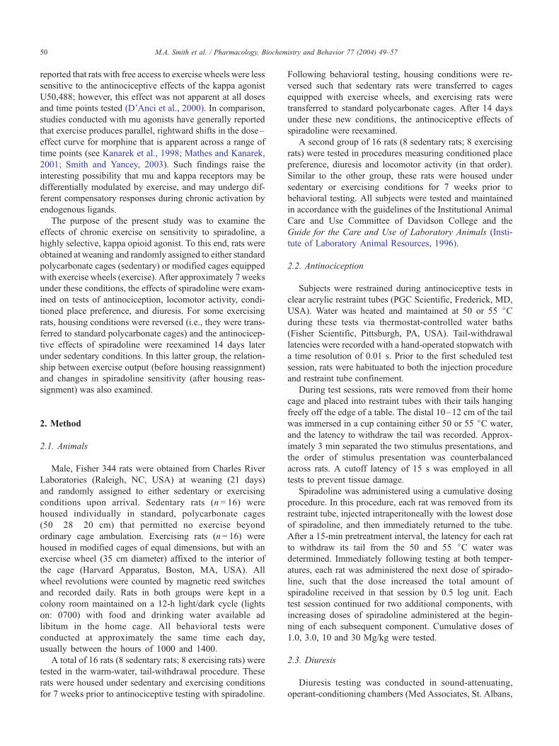
www.elsevier.com/locate/pharmbiochembehPharmacology, Biochemistry and Behavior 77 (2004) 49–57
Sensitivity to the effects of a kappa opioid in rats with free access to
exercise wheels: differential effects across behavioral measures
Mark A. Smitha,b,c,*, Jacob M. McCleanb,c, Paul A. Bryanta
aDepartment of Psychology, Davidson College, Davidson, NC 28035, USAbProgram in Neuroscience, Davidson College, Davidson, NC 28035, USA
cCenter for Interdisciplinary Studies, Davidson College, Davidson, NC 28035, USA
Received 15 June 2003; received in revised form 16 September 2003; accepted 17 September 2003
Abstract
It is well established that chronic exercise decreases sensitivity to mu opioid agonists; however, it is less clear what effects it has on kappa
opioids. The purpose of the present study was to examine sensitivity to the effects of the selective, kappa opioid spiradoline in rats with free
access to exercise wheels. Rats were obtained at weaning and randomly assigned to either standard polycarbonate cages (sedentary) or
modified cages equipped with exercise wheels (exercise). After approximately 7 weeks under these conditions, sensitivity to the effects of
spiradoline on tests of antinociception, locomotor activity, conditioned place preference, and diuresis were examined in both groups of rats.
Sedentary rats were more sensitive than exercising rats to the antinociceptive effects of spiradoline, and this effect was observed at both low
and high nociceptive intensities. In contrast, exercising rats were more sensitive than sedentary rats to the diuretic effects of spiradoline, and
slightly more sensitive to spiradoline’s effects in the conditioned place preference procedure. No differences in sensitivity were observed to
the effects of spiradoline on locomotor activity. Sensitivity to the antinociceptive effects of spiradoline nonsignificantly increased in
exercising rats that were reassigned to sedentary housing conditions, and changes in spiradoline sensitivity were correlated with exercise
output in individual subjects. Collectively, these data suggest that exercise alters sensitivity to the behavioral effects of kappa opioids, but that
the direction and magnitude of this effect depends on the behavioral measure examined.
D 2003 Elsevier Inc. All rights reserved.
Keywords: Antinociception; Conditioned place preference; Diuresis; Exercise; Locomotor activity; Kappa; Opioid; Rat; Spiradoline
1. Introduction
Acute exercise stimulates the release of endogenous
opioid peptides and produces analgesia that can be reversed
by the opioid antagonist naloxone (Janal, 1996; O’Connor
and Cook, 1999; Koltyn, 2000). Chronic exercise leads to
the sustained release of these peptides and to subsequent
decreases in sensitivity to morphine and other mu opioid
agonists (Kanarek et al., 1998; Mathes and Kanarek, 2001;
Lett et al., 2002). It has been proposed that these decreases
in sensitivity may reflect the development of cross-tolerance
between endogenous opioid peptides released during exer-
cise and exogenously administered mu agonists (Mathes and
Kanarek, 2001; Lett et al., 2002). In support of this
0091-3057/$ – see front matter D 2003 Elsevier Inc. All rights reserved.
doi:10.1016/j.pbb.2003.09.021
* Corresponding author. Department of Psychology, Davidson College,
Davidson, NC 28035-7037, USA. Tel.: +1-704-894-2470; fax: +1-704-894-
2512.
E-mail address: [email protected] (M.A. Smith).
hypothesis, Houghten et al. (1986) reported that rats with
free access to exercise wheels had fewer beta-endorphin
binding sites than sedentary rats, an effect that was pre-
sumed to reflect a compensatory down-regulation of opioid
receptors during exercise. Such reductions in opioid recep-
tor density have been reported in opioid-treated rats (Rogers
and el-Fakahany, 1986; Tao et al., 1987; Bhargava and
Gulati, 1990; Diaz et al., 1995), and are believed to
contribute to the reductions in opioid sensitivity (i.e.,
tolerance) observed during chronic opioid administration
(Paronis and Holtzman, 1992; Smith and Picker, 1998;
Stafford et al., 2001).
The effects of exercise on the kappa opioid receptor
system have not been as well characterized as its effects on
the mu receptor system. Previous studies (e.g., Aravich et al.,
1993; Fontana et al., 1994) have reported that exercise
stimulates the release of dynorphin, the principle endogenous
ligand for the kappa receptor, but measures of kappa receptor
density and sensitivity have not been taken. A recent study
M.A. Smith et al. / Pharmacology, Biochemistry and Behavior 77 (2004) 49–5750
reported that rats with free access to exercise wheels were less
sensitive to the antinociceptive effects of the kappa agonist
U50,488; however, this effect was not apparent at all doses
and time points tested (D’Anci et al., 2000). In comparison,
studies conducted with mu agonists have generally reported
that exercise produces parallel, rightward shifts in the dose–
effect curve for morphine that is apparent across a range of
time points (see Kanarek et al., 1998; Mathes and Kanarek,
2001; Smith and Yancey, 2003). Such findings raise the
interesting possibility that mu and kappa receptors may be
differentially modulated by exercise, and may undergo dif-
ferent compensatory responses during chronic activation by
endogenous ligands.
The purpose of the present study was to examine the
effects of chronic exercise on sensitivity to spiradoline, a
highly selective, kappa opioid agonist. To this end, rats were
obtained at weaning and randomly assigned to either standard
polycarbonate cages (sedentary) or modified cages equipped
with exercise wheels (exercise). After approximately 7 weeks
under these conditions, the effects of spiradoline were exam-
ined on tests of antinociception, locomotor activity, condi-
tioned place preference, and diuresis. For some exercising
rats, housing conditions were reversed (i.e., they were trans-
ferred to standard polycarbonate cages) and the antinocicep-
tive effects of spiradoline were reexamined 14 days later
under sedentary conditions. In this latter group, the relation-
ship between exercise output (before housing reassignment)
and changes in spiradoline sensitivity (after housing reas-
signment) was also examined.
2. Method
2.1. Animals
Male, Fisher 344 rats were obtained from Charles River
Laboratories (Raleigh, NC, USA) at weaning (21 days)
and randomly assigned to either sedentary or exercising
conditions upon arrival. Sedentary rats (n = 16) were
housed individually in standard, polycarbonate cages
(50� 28� 20 cm) that permitted no exercise beyond
ordinary cage ambulation. Exercising rats (n= 16) were
housed in modified cages of equal dimensions, but with an
exercise wheel (35 cm diameter) affixed to the interior of
the cage (Harvard Apparatus, Boston, MA, USA). All
wheel revolutions were counted by magnetic reed switches
and recorded daily. Rats in both groups were kept in a
colony room maintained on a 12-h light/dark cycle (lights
on: 0700) with food and drinking water available ad
libitum in the home cage. All behavioral tests were
conducted at approximately the same time each day,
usually between the hours of 1000 and 1400.
A total of 16 rats (8 sedentary rats; 8 exercising rats) were
tested in the warm-water, tail-withdrawal procedure. These
rats were housed under sedentary and exercising conditions
for 7 weeks prior to antinociceptive testing with spiradoline.
Following behavioral testing, housing conditions were re-
versed such that sedentary rats were transferred to cages
equipped with exercise wheels, and exercising rats were
transferred to standard polycarbonate cages. After 14 days
under these new conditions, the antinociceptive effects of
spiradoline were reexamined.
A second group of 16 rats (8 sedentary rats; 8 exercising
rats) were tested in procedures measuring conditioned place
preference, diuresis and locomotor activity (in that order).
Similar to the other group, these rats were housed under
sedentary or exercising conditions for 7 weeks prior to
behavioral testing. All subjects were tested and maintained
in accordance with the guidelines of the Institutional Animal
Care and Use Committee of Davidson College and the
Guide for the Care and Use of Laboratory Animals (Insti-
tute of Laboratory Animal Resources, 1996).
2.2. Antinociception
Subjects were restrained during antinociceptive tests in
clear acrylic restraint tubes (PGC Scientific, Frederick, MD,
USA). Water was heated and maintained at 50 or 55 jCduring these tests via thermostat-controlled water baths
(Fisher Scientific, Pittsburgh, PA, USA). Tail-withdrawal
latencies were recorded with a hand-operated stopwatch with
a time resolution of 0.01 s. Prior to the first scheduled test
session, rats were habituated to both the injection procedure
and restraint tube confinement.
During test sessions, rats were removed from their home
cage and placed into restraint tubes with their tails hanging
freely off the edge of a table. The distal 10–12 cm of the tail
was immersed in a cup containing either 50 or 55 jC water,
and the latency to withdraw the tail was recorded. Approx-
imately 3 min separated the two stimulus presentations, and
the order of stimulus presentation was counterbalanced
across rats. A cutoff latency of 15 s was employed in all
tests to prevent tissue damage.
Spiradoline was administered using a cumulative dosing
procedure. In this procedure, each rat was removed from its
restraint tube, injected intraperitoneally with the lowest dose
of spiradoline, and then immediately returned to the tube.
After a 15-min pretreatment interval, the latency for each rat
to withdraw its tail from the 50 and 55 jC water was
determined. Immediately following testing at both temper-
atures, each rat was administered the next dose of spirado-
line, such that the dose increased the total amount of
spiradoline received in that session by 0.5 log unit. Each
test session continued for two additional components, with
increasing doses of spiradoline administered at the begin-
ning of each subsequent component. Cumulative doses of
1.0, 3.0, 10 and 30 Mg/kg were tested.
2.3. Diuresis
Diuresis testing was conducted in sound-attenuating,
operant-conditioning chambers (Med Associates, St. Albans,
M.A. Smith et al. / Pharmacology, Biochemistry and Behavior 77 (2004) 49–57 51
VT, USA). One day prior to testing, all rats were habituated to
the chambers for 2 h. Drug and saline (control) sessions were
conducted over the next two consecutive days, with the order
of drug and saline administration counterbalanced across rats.
During these sessions, each rat was administered either 10
mg/kg spiradoline or saline, and urine was collected over 2
h in steel pans located beneath the grid floor of the chamber.
All rats were normally hydrated at the beginning of the
session, but no food or water was available during the session.
2.4. Conditioned place preference
The conditioned place preference procedure employed a
three-compartment place preference chamber (Med Associ-
ates). The chamber consisted of two choice compartments
(25� 20� 20 cm) separated by a neutral center compartment
(13� 20� 20 cm). One choice compartment was painted
white and had a wire-mesh floor covering pine bedding; the
other choice compartment was painted black and had a steel-
rod floor covering corncob bedding. The center compartment
was painted gray and had a solid PVC floor with no
underlying bedding. Each choice compartment was separated
from the center compartment by a guillotine door. All
behavioral activity was recorded by a video camera mounted
1.5 m above the chamber.
On the day prior to the first conditioning trial, each rat was
given a 15-min habituation session during which it had free
access to the entire chamber. The habituation session began
by placing the rat in the center compartment and opening the
guillotine doors leading to the two choice compartments.
Entrance into a compartment was recordedwhen the head and
both forepaws passed completely through a doorway. The
amount of time spent in each of the three compartments was
measured over the 15-min session. Neither group showed a
significant bias toward one choice compartment over the
other during this session.
Conditioning took place over the next eight consecutive
days. During conditioning, rats were injected with either 10
mg/kg spiradoline or saline and placed into one of the two
choice compartments for 30 min. Rats were confined to the
appropriate compartment for the duration of the trial by
closing the guillotine door leading to the center compart-
ment. Drug and saline administration alternated daily such
that each rat received four conditioning trials with the
saline-paired compartment and four conditioning trials with
the drug-paired compartment. For half of the rats, the black
compartment served as the drug-paired compartment; for the
other half, the white compartment served as the drug-paired
compartment.
Place preference was assessed in each rat on the day
immediately following the last conditioning trial. Similar to
the initial habituation session, this session began by placing
the rat in the center compartment and opening both guillo-
tine doors. Rats were given free access to the entire chamber
for 15 min, and the amount of time spent in each of the three
compartments was measured.
2.5. Locomotor activity
Locomotor activity was measured in an open-field, loco-
motor-activity chamber measuring 50� 50� 40 cm. The
interior of the chamber was made of plywood and painted
with white latex paint. The lid of the chamber was made of
transparent Plexiglas, which allowed all activity to be
recorded from a video camera suspended 1.5 m above the
chamber. Thick black lines were drawn on the lower surface
of the chamber with black ink that could easily be observed
from the camera mounted above. These lines divided the
lower surface of the chamber into a grid of 25 squares, each
measuring 10� 10 cm. Awire-mesh screen was suspended 2
cm above the lower surface, and served as the floor of the
chamber during behavioral testing.
One day immediately prior to testing, rats were habitu-
ated to the testing environment by being placing into the
activity chamber for 10 min. Drug and saline (control) tests
were conducted over the next two consecutive days, with
the order of drug and saline administration counterbalanced
across rats. During drug tests, each rat was initially injected
with the lowest dose of spiradoline and then returned to its
home cage. Following a 15-min pretreatment interval, the
rat was placed into the activity chamber for 130 s, and the
number of locomotor activity counts was recorded (see Data
Analysis) The first 10 s of this interval served as an
acclimation period, and only data from the final 120 s were
used in the statistical analysis. Immediately following the
observation period, the rat was removed from the chamber,
injected with the next dose of spiradoline, and returned to its
home cage. Fifteen minutes later, the rat was again placed
into the chamber and locomotor activity was measured.
Each test session continued for two additional components,
with increasing doses of spiradoline administered at the
beginning of each subsequent component. Cumulative doses
of 0.3, 1.0, 3.0 and 10 Mg/kg were tested. Saline control
tests were conducted in an identical manner, with the
exception that saline was administered at the beginning of
each component in lieu of spiradoline.
2.6. Drugs
Spiradoline mesylate was obtained from Sigma Chemical
(St. Louis, MO, USA) and dissolved in distilled water. In all
tests, spiradoline was administered intraperitoneally in a
volume of 1.0–2.0 ml/kg of body weight.
2.7. Data analysis
In antinociceptive tests, tail-withdrawal latencies were
converted to percent antinociceptive effect using the follo-
wing equation: % antinociceptive effect=[(observed� base-
baseline)/(15 s� baseline)]� 100. For each dose–effect
curve, the dose of spiradoline required to produce 50% of
the maximal possible effect (A50) was computed for each rat
mathematically (least squares method) by log-linear interpo-
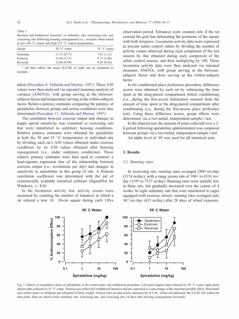
Table 1
Baseline tail-withdrawal latenciesa of sedentary rats, exercising rats, and
exercising rats following housing reassignment (i.e., reversal) when tested
at low (50 jC water) and high (55 jC water) temperatures
Group 50 jC water 55 jC water
Sedentary 11.31 (0.77) 7.81 (1.11)
Exercise 11.64 (1.17) 9.73 (1.06)
Reversal 12.08 (0.94) 8.56 (0.92)
a All data reflect the mean (S.E.M) of eight rats as measured in
seconds.
M.A. Smith et al. / Pharmacology, Biochemistry and Behavior 77 (2004) 49–5752
lation (Procedure 8, Tallarida and Murray, 1987). These A50
values were then analyzed via repeated measures analysis of
variance (ANOVA), with group serving as the between-
subjects factor and temperature serving as the within-subjects
factor. Relative potency estimates comparing the potency of
spiradoline between groups and across conditions were also
determined (Procedure 11, Tallarida and Murray, 1987).
The correlation between exercise output and changes in
kappa opioid sensitivity was examined in exercising rats
that were transferred to sedentary housing conditions.
Relative potency estimates were obtained for spiradoline
at both the 50 and 55 jC temperatures in individual rats
by dividing each rat’s A50 values obtained under exercise
conditions by its A50 values obtained after housing
reassignment (i.e., under sedentary conditions). These
relative potency estimates were then used to construct a
least-squares regression line of the relationship between
exercise output (i.e., revolutions per day) and changes in
sensitivity to spiradoline in this group of rats. A Pearson
correlation coefficient was determined with the aid of
commercially available statistical software (SigmaPlot for
Windows, v. 8.0).
In the locomotor activity test, activity counts were
measured by counting the number of instances in which a
rat entered a new 10� 10-cm square during each 120-s
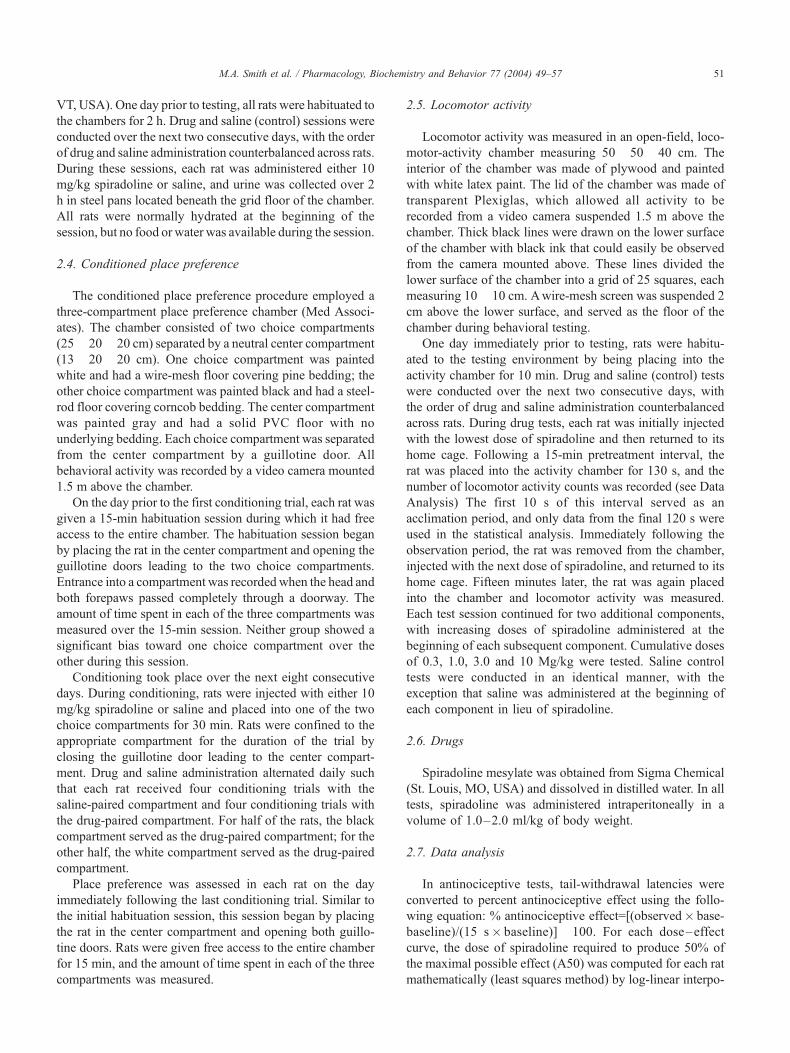
Fig. 1. Effects of cumulative doses of spiradoline in the warm-water, tail-withdraw
depicts data collected in 55 jC water. Vertical axes reflect tail-withdrawal latencies
axes reflect doses in milligram per kilogram of body weight. Vertical lines on data
data point. Data are shown from sedentary rats, exercising rats, and exercising ra
observation period. Entrances were counted only if the rat
crossed the grid line delineating the perimeter of the square
with both forepaws. Locomotor activity data were expressed
as percent saline control values by dividing the number of
activity counts observed during each component of the test
session by that obtained during each component of the
saline control session, and then multiplying by 100. These
locomotor activity data were then analyzed via repeated
measures ANOVA, with group serving as the between-
subjects factor and dose serving as the within-subjects
factor.
In the conditioned place preference procedure, difference
scores were obtained for each rat by subtracting the time
spent in the drug-paired compartment before conditioning
(i.e., during the free-access habituation session) from the
amount of time spent in the drug-paired compartment after
conditioning (i.e., during the free-access place preference
test). Using these difference scores, group effects were
determined via a two-tailed, independent-sample t test.
In the diuresis test, the amount of urine collected over a 2-
h period following spiradoline administration was compared
between groups via a two-tailed, independent-sample t test.
An alpha level of .05 was used for all statistical tests.
3. Results
3.1. Running rates
In exercising rats, running rates averaged 2809 rev/day
(3174 m/day), with a range across rats of 1061 to 6316 rev/
day (1199 to 7137 m/day). Running rates were initially low
in these rats, but gradually increased over the course of 4
weeks. In eight sedentary rats that were transferred to cages
equipped with exercise wheels, running rates averaged only
367 rev/day (415 m/day) after 28 days of wheel exposure.
al procedure. Left panel depicts data collected in 50 jC water; right panel
and are expressed as a percentage of the maximal possible effect. Horizontal
points represent the S.E.M.; where not indicated, the S.E.M. fell within the
ts 14 days after housing reassignment (reversal).
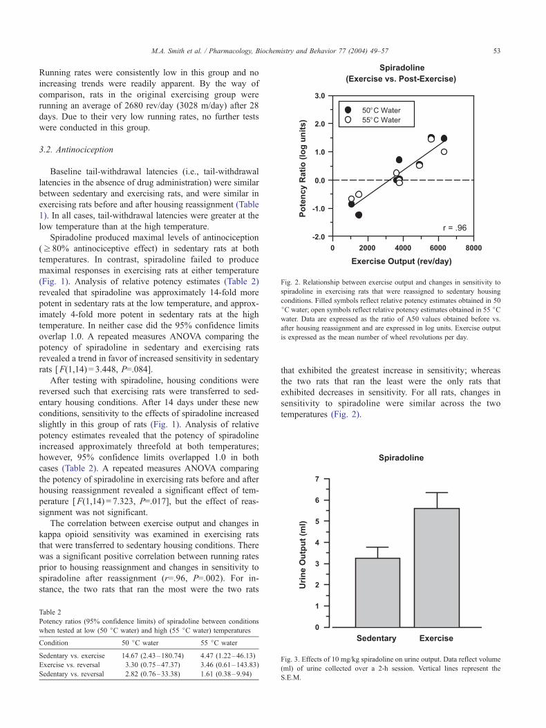
Fig. 2. Relationship between exercise output and changes in sensitivity to
spiradoline in exercising rats that were reassigned to sedentary housing
conditions. Filled symbols reflect relative potency estimates obtained in 50
jC water; open symbols reflect relative potency estimates obtained in 55 jCwater. Data are expressed as the ratio of A50 values obtained before vs.
after housing reassignment and are expressed in log units. Exercise output
is expressed as the mean number of wheel revolutions per day.
M.A. Smith et al. / Pharmacology, Biochemistry and Behavior 77 (2004) 49–57 53
Running rates were consistently low in this group and no
increasing trends were readily apparent. By the way of
comparison, rats in the original exercising group were
running an average of 2680 rev/day (3028 m/day) after 28
days. Due to their very low running rates, no further tests
were conducted in this group.
3.2. Antinociception
Baseline tail-withdrawal latencies (i.e., tail-withdrawal
latencies in the absence of drug administration) were similar
between sedentary and exercising rats, and were similar in
exercising rats before and after housing reassignment (Table
1). In all cases, tail-withdrawal latencies were greater at the
low temperature than at the high temperature.
Spiradoline produced maximal levels of antinociception
(z 80% antinociceptive effect) in sedentary rats at both
temperatures. In contrast, spiradoline failed to produce
maximal responses in exercising rats at either temperature
(Fig. 1). Analysis of relative potency estimates (Table 2)
revealed that spiradoline was approximately 14-fold more
potent in sedentary rats at the low temperature, and approx-
imately 4-fold more potent in sedentary rats at the high
temperature. In neither case did the 95% confidence limits
overlap 1.0. A repeated measures ANOVA comparing the
potency of spiradoline in sedentary and exercising rats
revealed a trend in favor of increased sensitivity in sedentary
rats [F(1,14) = 3.448, P=.084].
After testing with spiradoline, housing conditions were
reversed such that exercising rats were transferred to sed-
entary housing conditions. After 14 days under these new
conditions, sensitivity to the effects of spiradoline increased
slightly in this group of rats (Fig. 1). Analysis of relative
potency estimates revealed that the potency of spiradoline
increased approximately threefold at both temperatures;
however, 95% confidence limits overlapped 1.0 in both
cases (Table 2). A repeated measures ANOVA comparing
the potency of spiradoline in exercising rats before and after
housing reassignment revealed a significant effect of tem-
perature [F(1,14) = 7.323, P=.017], but the effect of reas-
signment was not significant.
The correlation between exercise output and changes in
kappa opioid sensitivity was examined in exercising rats
that were transferred to sedentary housing conditions. There
was a significant positive correlation between running rates
prior to housing reassignment and changes in sensitivity to
spiradoline after reassignment (r=.96, P=.002). For in-
stance, the two rats that ran the most were the two rats
Table 2
Potency ratios (95% confidence limits) of spiradoline between conditions
when tested at low (50 jC water) and high (55 jC water) temperatures
Condition 50 jC water 55 jC water
Sedentary vs. exercise 14.67 (2.43–180.74) 4.47 (1.22–46.13)
Exercise vs. reversal 3.30 (0.75–47.37) 3.46 (0.61–143.83)
Sedentary vs. reversal 2.82 (0.76–33.38) 1.61 (0.38–9.94)
that exhibited the greatest increase in sensitivity; whereas
the two rats that ran the least were the only rats that
exhibited decreases in sensitivity. For all rats, changes in
sensitivity to spiradoline were similar across the two
temperatures (Fig. 2).
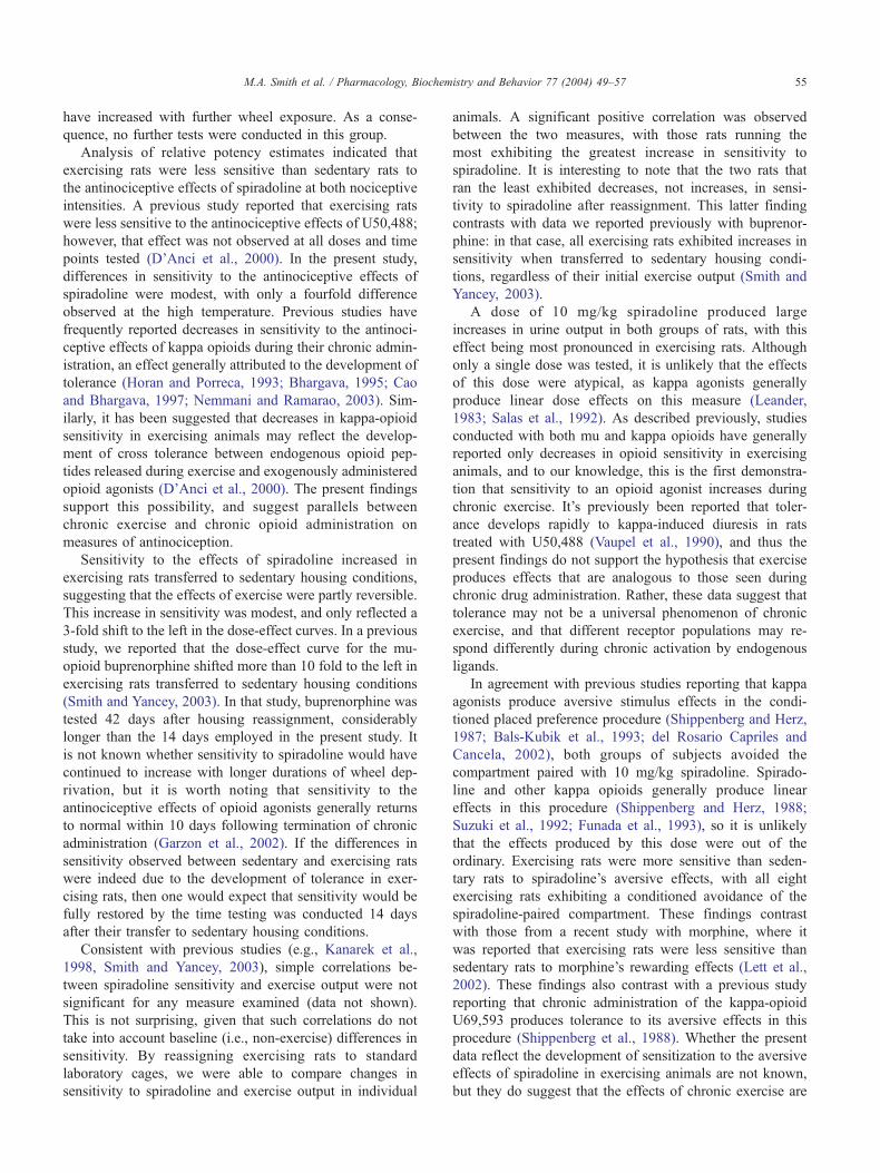
Fig. 3. Effects of 10 mg/kg spiradoline on urine output. Data reflect volume
(ml) of urine collected over a 2-h session. Vertical lines represent the
S.E.M.
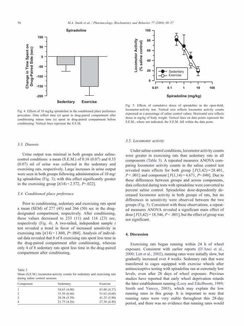
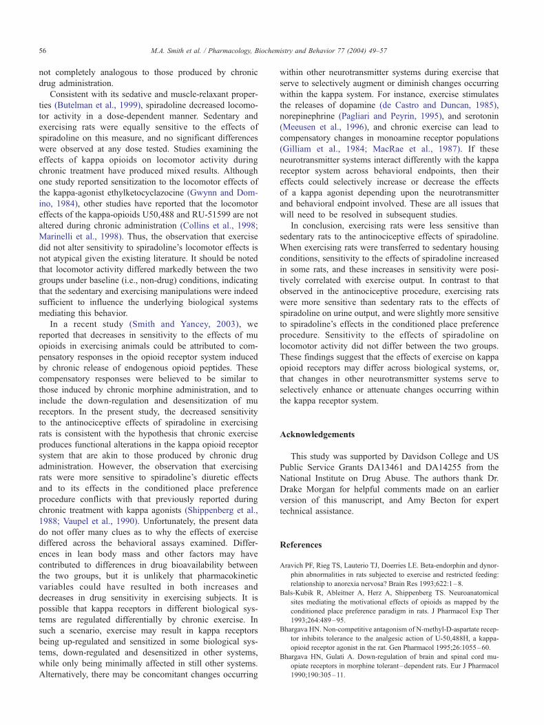
Fig. 4. Effects of 10 mg/kg spiradoline in the conditioned place preference
procedure. Data reflect time (s) spent in drug-paired compartment after
conditioning minus time (s) spent in drug-paired compartment before
conditioning. Vertical lines represent the S.E.M.
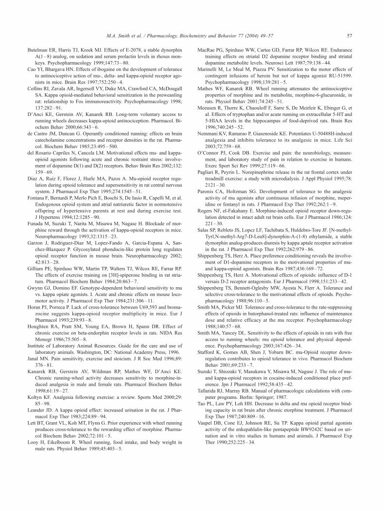
Fig. 5. Effects of cumulative doses of spiradoline in the open-field,
locomotor-activity test. Vertical axis reflects locomotor activity counts
expressed as a percentage of saline control values. Horizontal axis reflects
doses in mg/kg of body weight. Vertical lines on data points represent the
S.E.M.; where not indicated, the S.E.M. fell within the data point.
M.A. Smith et al. / Pharmacology, Biochemistry and Behavior 77 (2004) 49–5754
3.3. Diuresis
Urine output was minimal in both groups under saline-
control conditions: a mean (S.E.M.) of 0.16 (0.07) and 0.33
(0.07) ml of urine was collected in the sedentary and
exercising rats, respectively. Large increases in urine output
were seen in both groups following administration of 10 mg/
kg spiradoline (Fig. 3); with this effect significantly greater
in the exercising group [t(14) = 2.572, P=.022].
3.4. Conditioned place preference
Prior to conditioning, sedentary and exercising rats spent
a mean (SEM) of 277 (45) and 266 (50) sec in the drug-
designated compartment, respectively. After conditioning,
these values decreased to 233 (11) and 116 (23) sec,
respectively (Fig. 4). A two-tailed, independent sample t
test revealed a trend in favor of increased sensitivity in
exercising rats [t(14) = 1.860, P=.084]. Analysis of individ-
ual data revealed that 8 of 8 exercising rats spent less time in
the drug-paired compartment after conditioning, whereas
only 4 of 8 sedentary rats spent less time in the drug-paired
compartment after conditioning.
Table 3
Mean (S.E.M.) locomotor-activity counts for sedentary and exercising rats
during saline control sessions
Component Sedentary Exercise
1 54.63 (4.00) 63.00 (4.37)
2 31.50 (6.66) 51.63 (4.68)
3 28.38 (5.59) 41.25 (5.90)
4 21.75 (4.26) 27.38 (4.49)
3.5. Locomotor activity
Under saline-control conditions, locomotor activity counts
were greater in exercising rats than sedentary rats in all
components (Table 3). A repeated measures ANOVA com-
paring locomotor activity counts in the saline control test
revealed main effects for both group [F(3,42) = 28.401,
P < .001] and component [F(1,14) = 4.671, P=.048]. Due to
these differences between groups and across components,
data collected during tests with spiradoline were converted to
percent saline control. Spiradoline dose-dependently de-
creased locomotor activity in both groups of rats, but no
differences in sensitivity were observed between the two
groups (Fig. 5). Consistent with these observations, a repeat-
ed measures ANOVA revealed a significant main effect of
dose [F(3,42) = 18.346, P < .001], but the effect of group was
not significant.
4. Discussion
Exercising rats began running within 24 h of wheel
exposure. Consistent with earlier reports (D’Anci et al.,
2000; Lett et al., 2002), running rates were initially slow, but
gradually increased over 4 weeks. Sedentary rats that were
transferred to cages equipped with exercise wheels after
antinociceptive testing with spiradoline ran at extremely low
levels, even after 28 days of wheel exposure. Previous
studies have reported that early wheel deprivation retards
the later establishment running (Looy and Eikelboom, 1989;
Smith and Yancey, 2003), which may explain the low
running rates in this group. It is important to note that
running rates were very stable throughout this 28-day
period, and there was no evidence that running rates would
M.A. Smith et al. / Pharmacology, Biochemistry and Behavior 77 (2004) 49–57 55
have increased with further wheel exposure. As a conse-
quence, no further tests were conducted in this group.
Analysis of relative potency estimates indicated that
exercising rats were less sensitive than sedentary rats to
the antinociceptive effects of spiradoline at both nociceptive
intensities. A previous study reported that exercising rats
were less sensitive to the antinociceptive effects of U50,488;
however, that effect was not observed at all doses and time
points tested (D’Anci et al., 2000). In the present study,
differences in sensitivity to the antinociceptive effects of
spiradoline were modest, with only a fourfold difference
observed at the high temperature. Previous studies have
frequently reported decreases in sensitivity to the antinoci-
ceptive effects of kappa opioids during their chronic admin-
istration, an effect generally attributed to the development of
tolerance (Horan and Porreca, 1993; Bhargava, 1995; Cao
and Bhargava, 1997; Nemmani and Ramarao, 2003). Sim-
ilarly, it has been suggested that decreases in kappa-opioid
sensitivity in exercising animals may reflect the develop-
ment of cross tolerance between endogenous opioid pep-
tides released during exercise and exogenously administered
opioid agonists (D’Anci et al., 2000). The present findings
support this possibility, and suggest parallels between
chronic exercise and chronic opioid administration on
measures of antinociception.
Sensitivity to the effects of spiradoline increased in
exercising rats transferred to sedentary housing conditions,
suggesting that the effects of exercise were partly reversible.
This increase in sensitivity was modest, and only reflected a
3-fold shift to the left in the dose-effect curves. In a previous
study, we reported that the dose-effect curve for the mu-
opioid buprenorphine shifted more than 10 fold to the left in
exercising rats transferred to sedentary housing conditions
(Smith and Yancey, 2003). In that study, buprenorphine was
tested 42 days after housing reassignment, considerably
longer than the 14 days employed in the present study. It
is not known whether sensitivity to spiradoline would have
continued to increase with longer durations of wheel dep-
rivation, but it is worth noting that sensitivity to the
antinociceptive effects of opioid agonists generally returns
to normal within 10 days following termination of chronic
administration (Garzon et al., 2002). If the differences in
sensitivity observed between sedentary and exercising rats
were indeed due to the development of tolerance in exer-
cising rats, then one would expect that sensitivity would be
fully restored by the time testing was conducted 14 days
after their transfer to sedentary housing conditions.
Consistent with previous studies (e.g., Kanarek et al.,
1998, Smith and Yancey, 2003), simple correlations be-
tween spiradoline sensitivity and exercise output were not
significant for any measure examined (data not shown).
This is not surprising, given that such correlations do not
take into account baseline (i.e., non-exercise) differences in
sensitivity. By reassigning exercising rats to standard
laboratory cages, we were able to compare changes in
sensitivity to spiradoline and exercise output in individual
animals. A significant positive correlation was observed
between the two measures, with those rats running the
most exhibiting the greatest increase in sensitivity to
spiradoline. It is interesting to note that the two rats that
ran the least exhibited decreases, not increases, in sensi-
tivity to spiradoline after reassignment. This latter finding
contrasts with data we reported previously with buprenor-
phine: in that case, all exercising rats exhibited increases in
sensitivity when transferred to sedentary housing condi-
tions, regardless of their initial exercise output (Smith and
Yancey, 2003).
A dose of 10 mg/kg spiradoline produced large
increases in urine output in both groups of rats, with this
effect being most pronounced in exercising rats. Although
only a single dose was tested, it is unlikely that the effects
of this dose were atypical, as kappa agonists generally
produce linear dose effects on this measure (Leander,
1983; Salas et al., 1992). As described previously, studies
conducted with both mu and kappa opioids have generally
reported only decreases in opioid sensitivity in exercising
animals, and to our knowledge, this is the first demonstra-
tion that sensitivity to an opioid agonist increases during
chronic exercise. It’s previously been reported that toler-
ance develops rapidly to kappa-induced diuresis in rats
treated with U50,488 (Vaupel et al., 1990), and thus the
present findings do not support the hypothesis that exercise
produces effects that are analogous to those seen during
chronic drug administration. Rather, these data suggest that
tolerance may not be a universal phenomenon of chronic
exercise, and that different receptor populations may re-
spond differently during chronic activation by endogenous
ligands.
In agreement with previous studies reporting that kappa
agonists produce aversive stimulus effects in the condi-
tioned placed preference procedure (Shippenberg and Herz,
1987; Bals-Kubik et al., 1993; del Rosario Capriles and
Cancela, 2002), both groups of subjects avoided the
compartment paired with 10 mg/kg spiradoline. Spirado-
line and other kappa opioids generally produce linear
effects in this procedure (Shippenberg and Herz, 1988;
Suzuki et al., 1992; Funada et al., 1993), so it is unlikely
that the effects produced by this dose were out of the
ordinary. Exercising rats were more sensitive than seden-
tary rats to spiradoline’s aversive effects, with all eight
exercising rats exhibiting a conditioned avoidance of the
spiradoline-paired compartment. These findings contrast
with those from a recent study with morphine, where it
was reported that exercising rats were less sensitive than
sedentary rats to morphine’s rewarding effects (Lett et al.,
2002). These findings also contrast with a previous study
reporting that chronic administration of the kappa-opioid
U69,593 produces tolerance to its aversive effects in this
procedure (Shippenberg et al., 1988). Whether the present
data reflect the development of sensitization to the aversive
effects of spiradoline in exercising animals are not known,
but they do suggest that the effects of chronic exercise are
M.A. Smith et al. / Pharmacology, Biochemistry and Behavior 77 (2004) 49–5756
not completely analogous to those produced by chronic
drug administration.
Consistent with its sedative and muscle-relaxant proper-
ties (Butelman et al., 1999), spiradoline decreased locomo-
tor activity in a dose-dependent manner. Sedentary and
exercising rats were equally sensitive to the effects of
spiradoline on this measure, and no significant differences
were observed at any dose tested. Studies examining the
effects of kappa opioids on locomotor activity during
chronic treatment have produced mixed results. Although
one study reported sensitization to the locomotor effects of
the kappa-agonist ethylketocyclazocine (Gwynn and Dom-
ino, 1984), other studies have reported that the locomotor
effects of the kappa-opioids U50,488 and RU-51599 are not
altered during chronic administration (Collins et al., 1998;
Marinelli et al., 1998). Thus, the observation that exercise
did not alter sensitivity to spiradoline’s locomotor effects is
not atypical given the existing literature. It should be noted
that locomotor activity differed markedly between the two
groups under baseline (i.e., non-drug) conditions, indicating
that the sedentary and exercising manipulations were indeed
sufficient to influence the underlying biological systems
mediating this behavior.
In a recent study (Smith and Yancey, 2003), we
reported that decreases in sensitivity to the effects of mu
opioids in exercising animals could be attributed to com-
pensatory responses in the opioid receptor system induced
by chronic release of endogenous opioid peptides. These
compensatory responses were believed to be similar to
those induced by chronic morphine administration, and to
include the down-regulation and desensitization of mu
receptors. In the present study, the decreased sensitivity
to the antinociceptive effects of spiradoline in exercising
rats is consistent with the hypothesis that chronic exercise
produces functional alterations in the kappa opioid receptor
system that are akin to those produced by chronic drug
administration. However, the observation that exercising
rats were more sensitive to spiradoline’s diuretic effects
and to its effects in the conditioned place preference
procedure conflicts with that previously reported during
chronic treatment with kappa agonists (Shippenberg et al.,
1988; Vaupel et al., 1990). Unfortunately, the present data
do not offer many clues as to why the effects of exercise
differed across the behavioral assays examined. Differ-
ences in lean body mass and other factors may have
contributed to differences in drug bioavailability between
the two groups, but it is unlikely that pharmacokinetic
variables could have resulted in both increases and
decreases in drug sensitivity in exercising subjects. It is
possible that kappa receptors in different biological sys-
tems are regulated differentially by chronic exercise. In
such a scenario, exercise may result in kappa receptors
being up-regulated and sensitized in some biological sys-
tems, down-regulated and desensitized in other systems,
while only being minimally affected in still other systems.
Alternatively, there may be concomitant changes occurring
within other neurotransmitter systems during exercise that
serve to selectively augment or diminish changes occurring
within the kappa system. For instance, exercise stimulates
the releases of dopamine (de Castro and Duncan, 1985),
norepinephrine (Pagliari and Peyrin, 1995), and serotonin
(Meeusen et al., 1996), and chronic exercise can lead to
compensatory changes in monoamine receptor populations
(Gilliam et al., 1984; MacRae et al., 1987). If these
neurotransmitter systems interact differently with the kappa
receptor system across behavioral endpoints, then their
effects could selectively increase or decrease the effects
of a kappa agonist depending upon the neurotransmitter
and behavioral endpoint involved. These are all issues that
will need to be resolved in subsequent studies.
In conclusion, exercising rats were less sensitive than
sedentary rats to the antinociceptive effects of spiradoline.
When exercising rats were transferred to sedentary housing
conditions, sensitivity to the effects of spiradoline increased
in some rats, and these increases in sensitivity were posi-
tively correlated with exercise output. In contrast to that
observed in the antinociceptive procedure, exercising rats
were more sensitive than sedentary rats to the effects of
spiradoline on urine output, and were slightly more sensitive
to spiradoline’s effects in the conditioned place preference
procedure. Sensitivity to the effects of spiradoline on
locomotor activity did not differ between the two groups.
These findings suggest that the effects of exercise on kappa
opioid receptors may differ across biological systems, or,
that changes in other neurotransmitter systems serve to
selectively enhance or attenuate changes occurring within
the kappa receptor system.
Acknowledgements
This study was supported by Davidson College and US
Public Service Grants DA13461 and DA14255 from the
National Institute on Drug Abuse. The authors thank Dr.
Drake Morgan for helpful comments made on an earlier
version of this manuscript, and Amy Becton for expert
technical assistance.
References
Aravich PF, Rieg TS, Lauterio TJ, Doerries LE. Beta-endorphin and dynor-
phin abnormalities in rats subjected to exercise and restricted feeding:
relationship to anorexia nervosa? Brain Res 1993;622:1–8.
Bals-Kubik R, Ableitner A, Herz A, Shippenberg TS. Neuroanatomical
sites mediating the motivational effects of opioids as mapped by the
conditioned place preference paradigm in rats. J Pharmacol Exp Ther
1993;264:489–95.
Bhargava HN. Non-competitive antagonism of N-methyl-D-aspartate recep-
tor inhibits tolerance to the analgesic action of U-50,488H, a kappa-
opioid receptor agonist in the rat. Gen Pharmacol 1995;26:1055–60.
Bhargava HN, Gulati A. Down-regulation of brain and spinal cord mu-
opiate receptors in morphine tolerant–dependent rats. Eur J Pharmacol
1990;190:305–11.
M.A. Smith et al. / Pharmacology, Biochemistry and Behavior 77 (2004) 49–57 57
Butelman ER, Harris TJ, Kreek MJ. Effects of E-2078, a stable dynorphin
A(1–8) analog, on sedation and serum prolactin levels in rhesus mon-
keys. Psychopharmacology 1999;147:73–80.
Cao YI, Bhargava HN. Effects of ibogaine on the development of tolerance
to antinociceptive action of mu-, delta- and kappa-opioid receptor ago-
nists in mice. Brain Res 1997;752:250–4.
Collins RI, Zavala AR, Ingersoll VY, Duke MA, Crawford CA, McDougall
SA. Kappa opioid-mediated behavioral sensitization in the preweanling
rat: relationship to Fos immunoreactivity. Psychopharmacology 1998;
137:282–91.
D’Anci KE, Gerstein AV, Kanarek RB. Long-term voluntary access to
running wheels decreases kappa-opioid antinociception. Pharmacol. Bi-
ochem Behav 2000;66:343–6.
de Castro JM, Duncan G. Operantly conditioned running: effects on brain
catecholamine concentrations and receptor densities in the rat. Pharma-
col. Biochem Behav 1985;23:495–500.
del Rosario Capriles N, Cancela LM. Motivational effects mu- and kappa-
opioid agonists following acute and chronic restraint stress: involve-
ment of dopamine D(1) and D(2) receptors. Behav Brain Res 2002;132:
159–69.
Diaz A, Ruiz F, Florez J, Hurle MA, Pazos A. Mu-opioid receptor regu-
lation during opioid tolerance and supersensitivity in rat central nervous
system. J Pharmacol Exp Ther 1995;274:1545–51.
Fontana F, Bernardi P, Merlo Pich E, Boschi S, De Iasio R, Capelli M, et al.
Endogenous opioid system and atrial natriuretic factor in normotensive
offspring of hypertensive parents at rest and during exercise test.
J Hypertens 1994;12:1285–90.
Funada M, Suzuki T, Narita M, Misawa M, Nagase H. Blockade of mor-
phine reward through the activation of kappa-opioid receptors in mice.
Neuropharmacology 1993;32:1315–23.
Garzon J, Rodriguez-Diaz M, Lopez-Fando A, Garcia-Espana A, San-
chez-Blazquez P. Glycosylated phonducin-like protein long regulates
opioid receptor function in mouse brain. Neuropharmacology 2002;
42:813–28.
Gilliam PE, Spirduso WW, Martin TP, Walters TJ, Wilcox RE, Farrar RP.
The effects of exercise training on [3H]-spiperone binding in rat stria-
tum. Pharmacol Biochem Behav 1984;20:863–7.
Gwynn GJ, Domino EF. Genotype-dependent behavioral sensitivity to mu
vs. kappa opiate agonists. I. Acute and chronic effects on mouse loco-
motor activity. J Pharmacol Exp Ther 1984;231:306–11.
Horan PJ, Porreca P. Lack of cross-tolerance between U69,593 and broma-
zocine suggests kappa-opioid receptor multiplicity in mice. Eur J
Pharmacol 1993;239:93–8.
Houghten RA, Pratt SM, Young EA, Brown H, Spann DR. Effect of
chronic exercise on beta-endorphin receptor levels in rats. NIDA Res
Monogr 1986;75:505–8.
Institute of Laboratory Animal Resources. Guide for the care and use of
laboratory animals. Washington, DC: National Academy Press, 1996.
Janal MN. Pain sensitivity, exercise and stoicism. J R Soc Med 1996;89:
376–81.
Kanarek RB, Gerstein AV, Wildman RP, Mathes WF, D’Anci KE.
Chronic running-wheel activity decreases sensitivity to morphine-in-
duced analgesia in male and female rats. Pharmacol Biochem Behav
1998;61:19–27.
Koltyn KF. Analgesia following exercise: a review. Sports Med 2000;29:
85–98.
Leander JD. A kappa opioid effect: increased urination in the rat. J Phar-
macol Exp Ther 1983;224:89–94.
Lett BT, Grant VL, Koh MT, Flynn G. Prior experience with wheel running
produces cross-tolerance to the rewarding effect of morphine. Pharma-
col Biochem Behav 2002;72:101–5.
Looy H, Eikelboom R. Wheel running, food intake, and body weight in
male rats. Physiol Behav 1989;45:403–5.
MacRae PG, Spirduso WW, Cartee GD, Farrar RP, Wilcox RE. Endurance
training effects on striatal D2 dopamine receptor binding and striatal
dopamine metabolite levels. Neurosci Lett 1987;79:138–44.
Marinelli M, Le Meal M, Piazza PV. Sensitization to the motor effects of
contingent infusions of heroin but not of kappa agonist RU-51599.
Psychopharmacology 1998;139:281–5.
Mathes WF, Kanarek RB. Wheel running attenuates the antinociceptive
properties of morphine and its metabolite, morphine-6-glucuronide, in
rats. Physiol Behav 2001;74:245–51.
Meeusen R, Thorre K, Chaouloff F, Sarre S, De Meirleir K, Ebinger G, et
al. Effects of tryptophan and/or acute running on extracellular 5-HT and
5-HIAA levels in the hippocampus of food-deprived rats. Brain Res
1996;740:245–52.
Nemmani KV, Ramarao P, Giasenoside KE. Potentiates U-50488H-induced
analgesia and inhibits tolerance to its analgesic in mice. Life Sci
2003;72:759–68.
O’Connor PJ, Cook DB. Exercise and pain: the neurobiology, measure-
ment, and laboratory study of pain in relation to exercise in humans.
Exerc Sport Sci Rev 1999;27:119–66.
Pagliari R, Peyrin L. Norepinephrine release in the rat frontal cortex under
treadmill exercise: a study with microdialysis. J Appl Physiol 1995;78:
2121–30.
Paronis CA, Holtzman SG. Development of tolerance to the analgesic
activity of mu agonists after continuous infusion of morphine, meper-
idine or fentanyl in rats. J Pharmacol Exp Ther 1992;262:1–9.
Rogers NF, el-Fakahany E. Morphine-induced opioid receptor down-regu-
lation detected in intact adult rat brain cells. Eur J Pharmacol 1986;124:
221–30.
Salas SP, Rebleto JS, Lopez LF, Tachibata S, Huldebro-Tore JF. [N-methyl-
Tyrl,N-methyl-Arg7-D-Leu8]-dynorphin-A-(1-8) ethylamide, a stable
dymorphin analog-produces diuresis by kappa aptale receptor activation
in the rat. J Pharmacol Exp Ther 1992;262:979–86.
Shippenberg TS, Herz A. Place preference conditioning reveals the involve-
ment of D1-dopamine receptors in the motivational properties of mu-
and kappa-opioid agonists. Brain Res 1987;436:169–72.
Shippenberg TS, Herz A. Motivational effects of opioids: influence of D-1
versais D-2 receptor antagonists. Eur J Pharmacol 1998;151:233–42.
Shippenberg TS, Bennett-Oglesby MW, Ayesta N, Flerr A. Tolerance and
selective cross-tolerance to the motivational effects of opioids. Psycho-
pharmacology 1988;96:110–5.
Smith MA, Picker MJ. Tolerance and cross-tolerance to the rate-suppressing
effects of opioids in butorphanol-treated rats: influence of maintenance
dose and relative efficacy at the mu receptor. Psychopharmacology
1988;140:57–68.
Smith MA, Yancey DL. Sensitivity to the effects of opioids in rats with free
access to running wheels: mu opioid tolerance and physical depend-
ence. Psychopharmacology 2003;167:426–34.
Stafford K, Gomes AB, Shen J, Yoburn BC. mu-Opioid receptor down-
regulation contributes to opioid tolerance in vivo. Pharmacol Biochem
Behav 2001;69:233–7.
Suzuki T, Shiozaki Y, Masukawa Y, Misawa M, Naguse J. The role of mu-
and kappa-opioid receptors in cocaine-induced conditioned place pref-
erence. Jpn J Pharmacol 1992;58:435–42.
Tallarida RJ, Murray RB. Manual of pharmacologic calculations with com-
puter programs. Berlin: Springer; 1987.
Tao PL, Law PY, Loh HH. Decrease in delta and mu opioid receptor bind-
ing capacity in rat brain after chronic etorphine treatment. J Pharmacol
Exp Ther 1987;240:809–16.
Vaupel DB, Cone EJ, Johnson RE, Su TP. Kappa opioid partial agonists
activity of the enkepahlalin-like pentapeptide BW9242C based on uri-
nation and in vitro studies in humans and animals. J Pharmacol Exp
Ther 1990;252:225–34.