Embed Size (px)
Citation preview

RESEARCH ARTICLE Open Access
Sensitivity to gene dosage and geneexpression affects genes with copy numbervariants observed among neuropsychiatricdiseasesMaria Yamasaki1*, Takashi Makino2, Seik-Soon Khor3, Hiromi Toyoda3,4, Taku Miyagawa5, Xiaoxi Liu6,Hitoshi Kuwabara7, Yukiko Kano8,9, Takafumi Shimada10, Toshiro Sugiyama8, Hisami Nishida11, Nagisa Sugaya12,Mamoru Tochigi13, Takeshi Otowa14, Yuji Okazaki15, Hisanobu Kaiya16, Yoshiya Kawamura17, Akinori Miyashita18,Ryozo Kuwano18,19, Kiyoto Kasai20, Hisashi Tanii21, Tsukasa Sasaki22, Makoto Honda5 and Katsushi Tokunaga3,4
Abstract
Background: Copy number variants (CNVs) have been reported to be associated with diseases, traits, andevolution. However, it is hard to determine which gene should have priority as a target for further functionalexperiments if a CNV is rare or a singleton. In this study, we attempted to overcome this issue by using twoapproaches: by assessing the influences of gene dosage sensitivity and gene expression sensitivity. Dosage sensitivegenes derived from two-round whole-genome duplication in previous studies. In addition, we proposed a cross-sectional omics approach that utilizes open data from GTEx to assess the effect of whole-genome CNVs on geneexpression.
Methods: Affymetrix Genome-Wide SNP Array 6.0 was used to detect CNVs by PennCNV and CNV Workshop. Afterquality controls for population stratification, family relationship and CNV detection, 287 patients with narcolepsy,133 patients with essential hypersomnia, 380 patients with panic disorders, 164 patients with autism, 784 patientswith Alzheimer disease and 1280 healthy individuals remained for the enrichment analysis.
(Continued on next page)
© The Author(s). 2020 Open Access This article is licensed under a Creative Commons Attribution 4.0 International License,which permits use, sharing, adaptation, distribution and reproduction in any medium or format, as long as you giveappropriate credit to the original author(s) and the source, provide a link to the Creative Commons licence, and indicate ifchanges were made. The images or other third party material in this article are included in the article's Creative Commonslicence, unless indicated otherwise in a credit line to the material. If material is not included in the article's Creative Commonslicence and your intended use is not permitted by statutory regulation or exceeds the permitted use, you will need to obtainpermission directly from the copyright holder. To view a copy of this licence, visit http://creativecommons.org/licenses/by/4.0/.The Creative Commons Public Domain Dedication waiver (http://creativecommons.org/publicdomain/zero/1.0/) applies to thedata made available in this article, unless otherwise stated in a credit line to the data.
* Correspondence: [email protected] of Health Data Science Research, Healthy Aging InnovationCenter, Tokyo Metropolitan Geriatric Medical Center, Tokyo, JapanFull list of author information is available at the end of the article
Yamasaki et al. BMC Medical Genomics (2020) 13:55 https://doi.org/10.1186/s12920-020-0699-9

(Continued from previous page)
Results: Overall, significant enrichment of dosage sensitive genes was found across patients with narcolepsy, panicdisorders and autism. Particularly, significant enrichment of dosage-sensitive genes in duplications was observedacross all diseases except for Alzheimer disease. For deletions, less or no enrichment of dosage-sensitive genes withdeletions was seen in the patients when compared to the healthy individuals. Interestingly, significant enrichmentsof genes with expression sensitivity in brain were observed in patients with panic disorder and autism. Whileduplications presented a higher burden, deletions did not cause significant differences when compared to thehealthy individuals. When we assess the effect of sensitivity to genome dosage and gene expression at the sametime, the highest ratio of enrichment was observed in the group including dosage-sensitive genes and genes withexpression sensitivity only in brain. In addition, shared CNV regions among the five neuropsychiatric diseases werealso investigated.
Conclusions: This study contributed the evidence that dosage-sensitive genes are associated with CNVs amongneuropsychiatric diseases. In addition, we utilized open data from GTEx to assess the effect of whole-genome CNVson gene expression. We also investigated shared CNV region among neuropsychiatric diseases.
Keywords: Copy number variants, Ohnolog, Two-round whole-genome duplication, Gene dosage sensitivity, Geneexpression sensitivity, Neuropsychiatric diseases
BackgroundCopy number variants (CNVs) have been reported to beassociated with diseases, traits, and evolution [1–5].With new technologies for detecting CNVs, research inthese fields have progressed rapidly [6–8]. However,when it comes to clinical applications, several issues stillremain. One is that although rare CNVs have been re-ported to be associated with diseases [2, 3], the rarity ofthe CNVs makes them difficult to study for elucidatingthe pathogenicity of the diseases. This is similar to thesituation of whole- or exome-sequencing analysis, fromwhich a large number of rare mutations or singletonshave been discovered [9]. Secondly, CNVs often span afew megabases and cover several genes [2, 3]; this makesit hard to determine which gene should have priority asa target for further functional experiments. Recent megabiobank projects [10–12] and international data-sharingconsortia [13] have enabled the first issue to be over-come; however, the second issue remains unresolved. Inthis study, we attempted to overcome the second issueby using two approaches: by assessing the influences ofgene dosage sensitivity and gene expression sensitivity.Gene dosage sensitivity has been of increasing interest
because it might provide a clue for elucidating thepathogenicity of diseases. As such, ClinGen Dosage Sen-sitivity Map (https://www.ncbi.nlm.nih.gov/projects/dbvar/clingen/) has started to accumulate data ondosage-sensitive genes. The notion of gene dosage sensi-tivity derived from the gene balance hypothesis, whichwas suggested in earlier studies [14–16]. In short, thegene balance hypothesis states that a stoichiometric bal-ance is maintained among all of the complex gene prod-ucts in a pathway, so a copy number change in a singlegene of a pathway would be deleterious. Thus, genesunder this hypothesis are thought to be dosage-sensitive
genes. Copy number alterations, for example, CNVs, indosage-sensitive genes are harmful and might affect theonset of diseases (Fig. 1(a) and (b)). In contrast, copynumber alterations in dosage-insensitive genes are notharmful and might have no effect on the onset of dis-eases. As such, dosage-sensitive genes may provide use-ful information in the research of diseases.Recently, dosage-sensitive genes were effectively iden-
tified based on a particular hypothesis and applied tonarrow down susceptible genes. In 1970, Susumu Ohnoproposed a hypothesis that over the course of evolution,vertebrates experienced genome-wide duplication twice;this is otherwise known as two-round whole-genome du-plication (2R-WGD) [18]. Genes that existed at the ageof 2R-WGD are called ohnologs, named in honor ofOhno. Studies of ohnologs revealed that the pattern ofretained ohnologs was not random and that the cur-rently existing ohnologs are in gene balance as dosage-sensitive genes (Fig. 1(c)) [17, 19]. Several studies havedemonstrated the importance and applicability of thedosage sensitivity of ohnologs using CNVs from data-bases or previously reported pathogenic CNVs [20–23].Firstly, the susceptibility region for Down syndrome on21q22.13 was reported to be enriched with ohnologs[19]. Human monogenic disease genes in the OnlineMendelian Inheritance in Man (OMIN) (https://www.omim.org) database and previous literature were alsofound to be enriched in ohnologs [20, 22]. Similarly, pre-viously reported pathogenic genes involved in neuro-psychiatric diseases were frequently uncovered to beohnologs [21, 23].Previous studies about application of ohnologs limited
their survey to target CNVs from databases or previouslyreported pathogenic CNVs. They did not assess the in-fluence of dosage-sensitive ohnologs on small CNVs or
Yamasaki et al. BMC Medical Genomics (2020) 13:55 Page 2 of 13

the CNVs detected in each patient. In this study, CNVs> 100 kb in size that were observed in individuals withneuropsychiatric diseases were investigated to assess theburden of the dosage-sensitive ohnologs on thesediseases.Expression quantitative trait locus (eQTL) analysis has
been the focus of attention because it allows the assess-ment of whether single nucleotide polymorphisms(SNPs) or variants affect the expression of genes [24].However, eQTL analysis does not examine whether analteration in the gene expression level is deleterious ornot. We propose a concept for genes with expressionsensitivity (Fig. 2(a) and (b)): modification of the expres-sion level of genes with expression sensitivity is deleteri-ous, while modification of the expression level of geneswithout expression sensitivity is not deleterious. CNVsare one possible cause of expression level changes be-cause CNVs themselves can change the gene dosage, soour assumption is that CNVs in genes with expressionsensitivity might be deleterious. We utilized open datafrom the Genotype-Tissue Expression (GTEx) project(https://www.gtexportal.org/home/datasets) [24] in orderto simulate this concept. Genes with expression sensitiv-ity and, in other words, stable expression in a certain tis-sue were taken to be genes that are expressed in thetissue and that do not have any eQTL SNPs in the tis-sue. In contrast, genes without expression sensitivityand, in a different way, unstable expression in a certaintissue were taken to be genes that are expressed in thetissue and that have at least one eQTL SNP in the tissue.
Using this definition, we assessed the effect of genes withexpression sensitivity in the CNVs observed amongneuropsychiatric diseases.
MethodsSubjectsThe participants in this study were 425 patients withnarcolepsy [25], 171 patients with essential hypersomnia(EHS) [26], 595 patients with panic disorders [27], 246patients with autism [28], 1032 patients with Alzheimerdisease [29] and 2135 healthy individuals. All subjectswere genotyped in our previous studies, and their datawere included for analysis in this study. Ethical approvalwas obtained from the local institutional review boardsof all participating organizations. Age and gender werenot matched between the participants with neuropsychi-atric diseases and the healthy individuals. All individualsprovided written informed consent for their inclusion inthis study.
Genotyping and quality controlsGenomic DNA from all participants was genotyped for906,622 SNPs using the Affymetrix Genome-Wide SNPArray 6.0 (Thermo Fisher Scientific, Waltham, MA) (S1Fig). Genotype calling was done using the Birdseed algo-rithm in Affymetrix Power Tools software (ThermoFisher Scientific, Waltham, MA). Quality control proce-dures were performed using PLINK v1.07 (http://zzz.bwh.harvard.edu/plink/). Samples with a call rate < 97%were excluded. For SNP quality control, SNPs with a
Fig. 1 The concept of dosage-sensitive genes and their definitions according to a previous study [17] and our pilot result in S6 Fig. (a) Copynumber alterations in dosage-sensitive genes are harmful, but (b) copy number alterations in dosage-insensitive genes are not harmful. (c)According to a previous report [17], all genes can be classified into four groups: ohnologs with small-scale duplications (SSD); ohnologs withoutSSD; non-ohnologous duplicates; and singletons
Yamasaki et al. BMC Medical Genomics (2020) 13:55 Page 3 of 13

minor allele frequency < 0.05, a Hardy-Weinberg equilib-rium p < 0.001 for either the patient group or the healthycontrol group, and a SNP call rate < 99% were excluded.Samples with a reported family relationship with otherparticipants or a mean probability of being identity-by-descent (PIHAT, calculated in PLINK) value > 0.185were also excluded. Outliers in the principal componentanalysis using EIGENSOFT (http://www.hsph.harvard.edu/alkes-price/software/) were also excluded to elimin-ate population stratification. In the principal componentanalysis, data from 91 Japanese in Tokyo, Japan (JPT),90 Han Chinese in Beijing, China (CHB), 180 Utah resi-dents with Northern and Western European ancestry(CEU), and 180 Yoruba in Ibadan, Nigeria (YRI), ob-tained from the HapMap Project, were also included[30]. Data from the HapMap populations and thepresent sample sets were combined using common SNPsamong all populations after the quality control steps de-scribed above.
CNV detection and quality controlsPennCNV (http://www.openbioinformatics.org/) [31] andCNV Workshop (http://cnv.sourceforge.net) were utilized
to detect CNVs (Supporting Information and S1 Fig) [32].PennCNV employs the Hidden Markov Model. Briefly,PennCNV requires external reference values for the B al-lele frequency and log R ratio because its algorithm apply-ing the Hidden Markov Model identifies CNV regionsusing the degree of deviation from these references. Weused an in-house reference comprising some of thehealthy individuals in our sample set.After CNV detection by PennCNV, samples with a low
detection signal were removed from the subsequent ana-lyses. Samples with a log R ratio standard deviation>|0.3|, B allele frequency drift > 0.01 and CNV call count> 100 were excluded [33, 34]. CNVs with < 10 detectionprobes and a size < 30 kb were excluded. After the qual-ity controls for population stratification, family relation-ships and CNV detection, 287 patients with narcolepsy,133 patients with EHS, 380 patients with panic disor-ders, 164 patients with autism, 784 patients with Alzhei-mer disease and 1280 healthy individuals remained.Gene coordinates were converted from hg18 to hg19
using Liftover (https://genome.ucsc.edu/cgi-bin/hgLift-Over). Artifact regions that tend to cause false-positiveCNV detection, centromeric and telomeric regions (±
Fig. 2 The concept of genes with expression sensitivity only in brain and their definitions, proposed in this study. (a) Expression alterations ingenes with expression sensitivity are deleterious, while (b) expression alterations in genes without expression sensitivity are not deleterious. (c)Genes with expression sensitivity only in brain were defined using GTEx as genes that have stable or low variable expression in brain andunstable or high variable expression in other tissues
Yamasaki et al. BMC Medical Genomics (2020) 13:55 Page 4 of 13

500 kb) and immunoglobulin regions (±500 kb), were re-moved based on a previous study and software tutorial[3, 31].
Dosage-sensitive genesAccording to a previous study, all genes can be classifiedinto four groups based on evolutionary features and esti-mation methodology (Fig. 1(c)) [19]. Briefly, genes weredivided into singletons and duplicates by means of aBLAST search. Next, among duplicates, genes were clas-sified into ohnologs and non-ohnologous duplicates bycomparison with other species. Ohnologs were separatedbased on whether or not they had undergone small-scaleduplication (SSD) after 2R-WGD; these were labelledohnologs with SSD or ohnologs without SSD. Of thesefour categories, ohnologs without SSD and singletonswere defined as dosage-sensitive genes because genesunder the gene balance hypothesis tend not to haveundergone SSD after 2R-WGD, and because singletonswere found to be less likely to have CNVs in our data (S2Fig). The classification of human ohnologs was done basedon a previous paper by Makino, T et al. using coordinatesin Ensembl 73 [19]. According to previous reports, thenumbers of dosage-sensitive genes and dosage-insensitivegenes are 11,927 and 8360, respectively.Another classification of ohnologs from OHNOLOGS
(http://ohnologs.curie.fr) was utilized by Singh, PP et al.to identify ohnologs [35]. This previous study did notclassify ohnologs based on past SSD, so the dosage-sensitive genes were not clearly defined. Here, we used agene list from OHNOLOGS to validate the results.OHNOLOGS provides results from three different criteriafor the identification of ohnologs, and a list of ohnologs de-fined using the strictest criteria was utilized in our analysis.
Genes with expression sensitivity only in brainGenes with expression sensitivity only in brain were fo-cused on in this study. We made the assumption that ifgenes with expression sensitivity in any tissue are dis-rupted, expression balance might be affected not only inbrain, but also in other tissues, and it might contributeto neuropsychiatric diseases as well as diseases related toany of the other tissues. However, if genes with expres-sion sensitivity only in brain are disrupted, it might onlyaffect expression balance in brain and contribute toneuropsychiatric diseases. Therefore, in this study, geneswith expression sensitivity only in brain were analyzedto evaluate the genetic background of neuropsychiatricdiseases.Genes with expression sensitivity only in brain were
defined as those that had stable or low variable expres-sion in brain and unstable or high variable expression inother tissues (Fig. 2(c)). The GTEx project (https://www.gtexportal.org/home/datasets) [24] was utilized to
simulate genes with expression sensitivity in brain. Inthe database, data from 26 different tissues, including 10different brain tissues, were registered. (I) (Tissue)_Ana-lysis.v6p.egenes.txt which is a list of genes expressed ineach tissue and (II) (Tissue)_Analysis.v6p.signif_snpgene_pairs.txt which is a list of eQTL SNPs within each genewere utilized. Genes with stable expression in a certain tis-sue were taken to be those that are expressed in that tissueand are thus listed in (I), and those that did not have anyeQTL SNPs in the tissue and are thus not listed in (II).Genes with expression sensitivity in brain were defined asgenes with stable expression in the 10 different tissuesfrom brain. In contrast, genes with unstable expression ina certain tissue were defined as genes that are expressedin that tissue and they are included in (I), and those thathave at least one eQTL SNP in the tissue and they are in-cluded in (II). Genes with unstable expression in other tis-sues were defined as genes with unstable expression in the16 different tissues other than brain. Finally, overlappinggenes with expression sensitivity in brain and unstable ex-pression in other tissues were defined as genes with ex-pression sensitivity only in brain.
Statistical analysisThe frequency of CNVs was calculated among the patientsof each disease and healthy individuals. CNVs with a size> 100 kb and a frequency < 1% were used in enrichmenttests. In the tests, the average number of genes overlappedby CNVs were compared between the cases and the con-trols for the following categories:(1) dosage-sensitive genes(2); genes with expression sensitivity only in brain; and (3)a combined category of [1, 2]. In more detail, enrichmenttests were conducted using the --cnv-count, −-cnv-subsetand --cnv-enrichment test options in PLINK to assesswhether a subset of genes was enriched relative to allgenes.
Inspection of regions detected only in the patientsRegions that were found only in the patients of the fiveneuropsychiatric diseases were examined. CNVs with asize of > 100 kb and a frequency of < 1% were examined.To narrow down the possible candidate regions, the fol-lowing criteria were used: (i) previously reported regions;(ii) shared regions among the five neuropsychiatric dis-eases; or (iii) regions with more than six dosage-sensitivegenes. Previously reported regions were taken from thefollowing papers and database: a paper by Itsara, A et al.[36] and a list of candidate genes from the Autism Data-base (AutDB; http://autism.mindspec.org/autdb/) [37]for autism; a paper by Howe, A et al. [38] for panic dis-orders; a paper by Lane, J et al. [39] for sleep disorders;a paper by Ripke, S et al. [40] for schizophrenia; and apaper by Van Cauwenberghe, C et al. [41] for Alzheimerdisease. Regions with (i) and (ii), or with (ii) and (iii)
Yamasaki et al. BMC Medical Genomics (2020) 13:55 Page 5 of 13
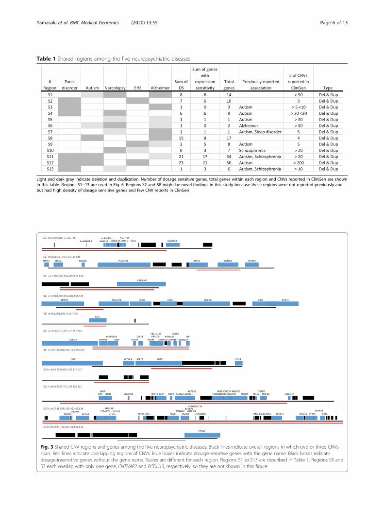
Table 1 Shared regions among the five neuropsychiatric diseases
Light and dark gray indicate deletion and duplication. Number of dosage sensitive genes, total genes within each region and CNVs reported in ClinGen are shownin this table. Regions S1~13 are used in Fig. 6. Regions S2 and S8 might be novel findings in this study because these regions were not reported previously andbut had high density of dosage sensitive genes and few CNV reports in ClinGen
Fig. 3 Shared CNV regions and genes among the five neuropsychiatric diseases. Black lines indicate overall regions in which two or three CNVsspan. Red lines indicate overlapping regions of CNVs. Blue boxes indicate dosage-sensitive genes with the gene name. Black boxes indicatedosage-insensitive genes without the gene name. Scales are different for each region. Regions S1 to S13 are described in Table 1. Regions S5 andS7 each overlap with only one gene, CNTNAP2 and PCDH15, respectively, so they are not shown in this figure
Yamasaki et al. BMC Medical Genomics (2020) 13:55 Page 6 of 13

were listed as shared CNVs among the neuropsychiatricdiseases in Table 1 and Fig. 3. The disease-specific re-gions, defined as those with (iii) and without (i) or witha p < 0.05 on Fisher’s exact test, are listed in S7 Table.
ResultsEnrichment of dosage-sensitive genesThe average number of dosage-sensitive genes was com-pared between the patients of each disease and thehealthy individuals. According to the data from previousreports, 11,927 dosage-sensitive genes and 8360 dosage-insensitive genes were included in the analysis. Overall,in comparison to healthy individuals, a significant en-richment of dosage-sensitive genes was found among in-dividuals with narcolepsy, panic disorders, or autism(Fig. 4 and S1–5 Tables). The similar enrichments inpanic disorders and autism were also observed usingohnologs estimated by Singh PP et al. (S3 Fig). Inaddition, the weaker enrichment was also found using adifferent CNV detection algorithm (S4 and S5 Figs). Indetail, reproducibility of significant enrichments of dos-age sensitive genes is partly limited between software,however we could see tendency of enrichment acrossdiseases. Overall, dosage-sensitive genes were signifi-cantly enriched in the patients with narcolepsy, panicdisorders, or autism. Of note, significant enrichment ofdosage-sensitive genes with duplications were observedin all diseases except for Alzheimer disease (Fig. 4).Among the five diseases, patients with panic disorders orautism showed higher enrichment of dosage-sensitive
genes with duplications when compared to individualswith narcolepsy or essential hypersomnia (EHS). For de-letions, less or no enrichment of dosage-sensitive geneswith deletions was seen in the patients when comparedto the healthy individuals (Fig. 4).
Enrichment of genes with expression sensitivity only inbrainTo demonstrate the effect of CNVs on gene expression,the average number of genes with expression sensitivityonly in brain among the CNVs of the five neuropsychi-atric diseases were compared between the cases and thecontrols. In our analysis, we proposed the idea of defin-ing expression sensitivity by utilizing the GTEx database.We defined 11,926 genes as genes with expression sensi-tivity only in brain from the 39,769 expressed genes inany tissue in the GTEx database (Fig. 2 (c)). Significantenrichment of genes with expression sensitivity only inbrain was observed among patients with panic disordersand autism; in contrast, patients with narcolepsy did notshow significant enrichment of genes with expression sen-sitivity only in brain (Fig. 5). While duplications presenteda higher burden, deletions did not cause significant differ-ences when compared to the healthy individuals. An en-richment of genes with expression sensitivity only in brainwas also seen using a different CNV detection algorithm(S6 Fig). Significant enrichments of genes with expressionsensitivity is not partly concordant between software,however we could see slight tendency of enrichmentacross diseases.
Fig. 4 Enrichment of dosage-sensitive genes in CNVs observed in five neuropsychiatric diseases according to the definitions by Makino, T et al.[17]. The y axis shows the relative ratio of the average number of genes spanned by CNVs in comparison to that in healthy controls. CNVs weredetected using PennCNV. Asterisks indicate significant enrichment when compared to the healthy individuals in each category
Yamasaki et al. BMC Medical Genomics (2020) 13:55 Page 7 of 13

Combined effect of dosage-sensitive genes and geneswith expression sensitivity only in brainTo assess the effect of sensitivity to genome dosage andgene expression at the same time, enrichment tests werealso performed. Genes were categorized into four groupsbased on the combinations of dosage-sensitive genes andgenes with expression sensitivity only in brain: (a)dosage-sensitive genes and genes with expression sensi-tivity only in brain, including 5590 genes; (b) dosage-sensitive genes and genes without expression sensitivityonly in brain, including 5527 genes; (c) dosage-insensitive genes and genes with expression sensitivityonly in brain, including 3179 genes; and (d) dosage-insensitive genes and genes without expression sensitiv-ity only in brain, including 4480 genes. After convertingthe genome coordinates to Ensembl 73, 9169 and 10,007genes were mapped as genes with and without expres-sion sensitivity only in brain, respectively.Among the four categories of genes, the group includ-
ing dosage-sensitive genes and genes with expressionsensitivity only in brain showed the highest enrichmentamong the CNVs from the patients when compared tohealthy individuals (Fig. 6). The group including dosage-sensitive genes and genes without expression sensitivityonly in brain showed the second highest enrichment,followed by the group including dosage-insensitive genesand genes with expression sensitivity only in brain, then
the group including dosage-insensitive genes and geneswithout expression sensitivity only in brain. A similartendency was observed in CNVs detected by anothersoftware (S6 Table). When the ratio of the average num-ber of genes was compared between cases and controlsamong all tests for all diseases, the highest ratio of en-richment was observed in the group including dosage-sensitive genes and genes with expression sensitivity onlyin brain.
Inspection of regions detected only in the patientsShared CNV regions among the five neuropsychiatricdiseases were also investigated. Table 1 and Fig. 3 showthe previously implicated regions or regions with morethan six dosage-sensitive genes and regions associatedwith at least two different diseases. With the use of genedosage sensitivity, regions S2 (chr1: 28,571,570-29,134,846) and S8 (chr11: 67,016,451-67,257,824) might repre-sent novel findings because these regions have not beenreported previously and have few reported CNVs in theClinGen database (Table 1). In particular, region S8 wassignificantly enriched in dosage-sensitive genes whencompared to the genome-wide ratio of dosage-sensitivegenes and dosage-insensitive genes (p = 0.013 on Fisher’sexact test), and while region S2 was not significantlyenriched (p = 0.540 on Fisher’s exact test), it had a higherdensity than the genome-wide average (density of
Fig. 5 Enrichment of genes with expression sensitivity only in brain in CNVs observed in five neuropsychiatric diseases. The y axis shows therelative ratio of the average number of genes spanned by CNVs in comparison to that in healthy controls. CNVs were detected using PennCNV.Asterisks indicate significant enrichment when compared to the healthy individuals in each category
Yamasaki et al. BMC Medical Genomics (2020) 13:55 Page 8 of 13

dosage-sensitive genes at region S2: 70.0%; genome-wideaverage of dosage-sensitive genes: 58.7%). Disease-specificregions were also examined, including regions with morethan six dosage-sensitive genes; regions that have not beenreported previously; or regions with a p < 0.05 on Fisher’sexact test. All diseases except for narcolepsy had disease-specific regions that fulfilled the above criteria.
DiscussionIn this paper, the phenotypic impact of sensitivity togene dosage was demonstrated using CNVs detected inpatients of five neuropsychiatric disorders. These resultswere concordant with a previous study using intolerancescore [8]. The previous study utilized a ranking systemthat scored genes according to their tolerance to func-tional variation and demonstrated that CNVs among pa-tients with schizophrenia had higher genic intolerancescores than that of healthy controls. In other words,CNVs in schizophrenia occurred within genes intoler-ance to functional variations. This is similar to our re-sults, because it was known that genes with dosagesensitivity hardly experienced copy number alteration byCNV [19].Duplications appeared to have a substantial impact on
disease onset, as we saw from the significant enrichmentof dosage-sensitive genes with duplications in all diseasesexcept for Alzheimer disease. Until recently, duplicationswere often deprioritized and analyzed after deletions,perhaps because duplications are thought to be lessharmful than deletions [3]. However, a recent study sug-gested that duplications exhibit more diversity, weakerselective constraint, and a four-fold greater chance of af-fecting genes than deletions, indicating that they possess
signatures of adaptive evolution [5]. Indeed, several stud-ies have observed that high-copy CNVs were associatedwith human traits [4, 42–45]. It might be because oncedeletions occurred, they disappeared quickly or their fre-quencies were reduced in a population because theywere lethal or extremely harmful and disadvantageous.However, when duplications occurred, they wereretained within the diversity of a population becausethey were not lethal nor sufficiently harmful or disad-vantageous to have had their frequencies reduced. Fromthe viewpoint of the total number of dosage-sensitivegenes affected by CNVs, the accumulative impact of du-plications might be larger than that of deletions. Thisstudy provides another example that demonstrates theimportance of duplications in the onset of diseases.Genes with sensitive expression only in brain were
identified utilizing publically available GTEx data, andthe effects of CNVs on the expression of these geneswere evaluated. Similar to the gene balance hypothesis,expression balance might also be maintained; thus, alter-ations in the gene expression level of genes with expres-sion sensitivity are deleterious, while such alterations ingenes without expression sensitivity are not. Indeed, it isalready reported that dosage change of expressed genesin brain are less frequent than those of other genes andare controlled by tighter transcriptional regulation [46].Also, according to an analysis using intolerance score,genes highly expressed in the brain showed the most in-tolerance to CNV [8].CNVs could be one cause ofchange at the expression level because CNVs modifygene dosage, so it is possible that CNVs in genes withexpression sensitivity might contribute to the onset ofdiseases.
Fig. 6 Combined enrichment of dosage-sensitive genes and genes with expression sensitivity only in brain in CNVs observed in fiveneuropsychiatric diseases in comparison to those in healthy individuals. Human genes were classified into four groups based on thecombinations of dosage-sensitive genes and genes with expression sensitivity only in brain: dosage-sensitive genes and genes with expressionsensitivity only in brain (DS & ES); dosage-sensitive genes and genes without expression sensitivity only in brain (DS & -ES); dosage-insensitivegenes and genes with expression sensitivity only in brain (DinS & ES); and dosage-insensitive genes and genes without expression sensitivity onlyin brain (DinS & -ES). Asterisks indicate significant enrichment when compared to the healthy individuals in each category
Yamasaki et al. BMC Medical Genomics (2020) 13:55 Page 9 of 13

Our finding of no significant enrichment of genes withexpression sensitivity only in brain among individualswith narcolepsy suggests that the genetic background ofnarcolepsy may not be related to brain function, at leastnot in relation to CNVs. Previous studies of narcolepsyshowed an autoimmune etiology for the disease andorexin (hypocretin) deficiency among patients with nar-colepsy. HLA-DQB1*06:02 was a major genetic factor forthe onset of narcolepsy [47–51], and the T cell receptoralpha gene (TRA) and purinergic receptor P2Y, G-protein coupled, 11 gene (P2RY11) were also reported tobe associated with narcolepsy [52, 53]. Pathway analysisof CNVs in patients with narcolepsy found enrichmentof immune-related pathway [54]. Additionally, thehypocretin-1 level was found to be reduced or undetect-able in the cerebrospinal fluid of narcoleptic patients[55], and postmortem examination showed a marked re-duction of hypocretin-producing neurons in the hypo-thalamus [56]. Nevertheless, regarding CNVs, our resultof no significant enrichment of genes with expressionsensitivity only in brain demonstrated that overall, nar-colepsy might be an autoimmune disease rather than abrain-related disease.In this study, we proposed a method of assessing the
influence of CNVs on gene expression. Analysis of eQTLenables the evaluation of whether SNPs or variants affectthe gene expression level [24]; however, these analysisdoes not assess whether alterations of the gene expres-sion level are deleterious or not. Here, genes with ex-pression sensitivity were able to evaluate influence ofchange at expression-level. In addition to expressionsensitivity, it is necessary to assess the impact of CNVson gene expression [57] because CNVs contribute to 18to 99% of the expression level of genes [58, 59]. Indeed,previous studies analyzed expression-level of CNV locususing human brain tissue from healthy individuals andpatients with neuropsychiatric diseasaes [60, 61]. Yet, ithas been difficult to know the effect of each CNVs ongene expression easily because there is no gene expres-sion reference panel for CNVs like there is for SNPs. Inthis study, we proposed a cross-sectional omics ap-proach using publicly available data. To the best of ourknowledge, this is a novel method for assessing the influ-ence of CNVs on gene expression.The shared regions among five neuropsychiatric dis-
eases were assessed. With the use of gene dosage sensi-tivity, regions S2 and S8 may be novel findings (Table 1and Fig. 3). Region S8 was shared between autism andAlzheimer disease, and within region S8, the carnosinesynthase 1 gene (CARNS1) was highly expressed inbrain. This gene was known to catalyze the formation ofcarnosine and homocarnosine. Base on ARCHS4 data-base, top predicted biological process in Gene Ontologyis fatty acid elongation [62]. Increased expression of fatty
acid synthesis in model of autism was demonstrated pre-viously [63]. Correlation between deficient biosynthesisof fatty acid and cognitive impairment in Alzheimer’sdisease was reported [64]. This gene might be involvedpathogenesis of these diseases. The protein tyrosinephosphatase receptor type C-associated protein gene(PTPRCAP) was reported to contain one of the top dif-ferentially methylated probes in autism [65]. A geneticlink between autism and Alzheimer disease has previ-ously been reported, and this provides additional evi-dence for a shared background between autism andAlzheimer disease [66].Disease-specific regions of five neuropsychiatric dis-
eases were evaluated. For panic disorders, region P3spanned seven dosage-sensitive genes (S7 Table). One ofthem was the solute carrier family 17 member 7 gene(SLC17A7); this gene is highly expressed in brain, and itwas reported that expression of this gene resulted in theuptake of glutamate as a vesicular glutamate transporter[67]. Reduced expression of this gene leads to reduceduptake of glutamate and an increased amount of glutam-ate in brain. Previously, a high amount of glutamate wasreported to be associated with panic attacks [68]. There-fore, it seems that SLC17A7 within this deletion may bea candidate causative gene in patients with panic disor-ders. Region P2 (chr14:50,043,390-50,311,552) spannedeight dosage-sensitive genes. Among these genes, theribosomal protein S29 gene (RPS29) showed a marginalsignificant association in a genome-wide associationstudy with posttraumatic stress disorder [69]. Anothergene, the kelch domain containing 1 gene (KLHDC1),was reported to show a moderate significant associationwith bipolar disease [70]. Four individuals with panicdisorders had duplications in region P1 (chr4:39,500,375-39,784,412; p = 0.0027 on Fisher’s exact test). Theubiquitin-conjugating enzyme gene (UBE2K) and smallintegral membrane protein 14 gene (SMIM14) wereoverlapped in all duplications among these four individ-uals. UBE2K was reported to be correlated with positivesymptoms of psychosis in schizophrenia and bipolar pa-tients [71]. By thoroughly inspecting dosage-sensitivegenes within CNVs, it seems possible to narrow downcandidate genes.We found several regions with more than six dosage-
sensitive ohnologs in Alzheimer disease. No increase inthe average number of CNVs per person was observedin Alzheimer disease. Nevertheless, when we further in-vestigated each CNV that occurred only among patients,Alzheimer disease seemed to associate with dosage-sensitive genes. This result was concordant with the re-sults of a recent paper [23]. In particular, a duplicationin region AD6 (chr11: 104,756,445-107,834,208) was ob-served in one case in this study (S7 Table), and it over-lapped with the contactin 5 gene (CNTN5) and ELMO
Yamasaki et al. BMC Medical Genomics (2020) 13:55 Page 10 of 13

domain-containing 1 gene (ELMOD1), which were re-ported as candidate genes in the recent paper. Inaddition, when we investigated CNVs with a size < 100kb, the amyloid beta precursor protein gene (APP) over-lapped with a deletion from one patient with Alzheimerdisease in this study. Previously, duplications of APPwere reported to be causative variants for early onset fa-milial Alzheimer disease [72–74]. Although a deletionwas observed in this study, APP was a dosage-sensitivegene, so it is possible that not only a gain, but also a lossof gene dosage might have contributed to the onset ofdisease in this patient. Therefore, both the total burdenof CNVs and inspection of dosage-sensitive genesaround CNVs are useful strategies for identifying suscep-tibility genes.The scope of our study was limited to ohnologs that
underwent 2R-WGD and were very ancient. However,recent human-specific segmental duplications are alsoknown to contribute to disease onset or phenotypic di-versity [75]. It is important to inspect CNVs from bothviewpoints. In addition, we only evaluated the effect ofCNVs on neighboring genes. It was reported that amonggenes with expression influenced by CNVs, 53% of theexpression is affected by CNVs that are distant from thetarget genes [1]. However, our analysis did not considerthe distant effects of CNVs. Also the method we pro-posed in this study simplify expression among brain anddid not consider different gene expression of region inbrain and dynamics of gene expression through life spanso on.
ConclusionsIn this study, we demonstrated the impact of sensitivityto gene dosage and gene expression using CNVs identi-fied in patients with neuropsychiatric diseases. A novelapproach was proposed, and the effect of CNVs on geneexpression was globally assessed. These results will helpelucidate the pathogenicity of diseases more clearly thanbefore.
Supplementary informationSupplementary information accompanies this paper at https://doi.org/10.1186/s12920-020-0699-9.
Additional file 1: Figure S1. Overview of the study. CNVs identifiedfrom each detection method were filtered and analyzed independently.Figure S2. CNV frequency and proportion of dosage-sensitive genesoverlapped by CNVs. The x axis shows the frequency of CNVs in each pa-tient and healthy individual. The y axis shows the proportion of ohnologswith SSD, singletons, ohnologs without SSD, or non-ohnologous dupli-cates. Figure S3. Enrichment of ohnologs in CNVs observed in fiveneuropsychiatric diseases according to the definitions by Singh, PP et al.Figure S4. Enrichment of dosage-sensitive genes in CNVs observed infive neuropsychiatric diseases with the use of another software, CNVWorkshop. Figure S5. Enrichment of ohnologs in CNVs observed in fiveneuropsychiatric diseases with the use of another software, CNV Work-shop. Figure S6. Enrichment of genes with expression sensitivity only in
brain in CNVs observed in five neuropsychiatric diseases with the use ofanother software, CNV Workshop. Table S1. Enrichment of dosage-sensitive ohnologs in individuals with narcolepsy using CNVs detected byPennCNV. Table S2. Enrichment of dosage-sensitive ohnologs in individ-uals with autism using CNVs detected by PennCNV. Table S3. Enrich-ment of dosage-sensitive ohnologs in individuals with panic disordersusing CNVs detected by PennCNV. Table S4. Enrichment of dosage-sensitive ohnologs in individuals with essential hypersomnia using CNVsdetected by PennCNV. Table S5. Enrichment of dosage-sensitive ohno-logs in individuals with Alzheimer disease using CNVs detected byPennCNV. Table S6. Combined enrichment of dosage-sensitive genesand genes with expression sensitivity only in brain in CNVs observed infive neuropsychiatric diseases with the use of another software, CNVWorkshop. Table S7. Disease-specific regions for five neuropsychiatricdiseases.
Abbreviations2R-WGD: Two-round whole-genome duplication; APP: Amyloid betaprecursor protein gene; AutDB: Autism Database; CARNS1: Carnosinesynthase 1 gene; CEU: Utah residents with Northern and Western Europeanancestry; CHB: Han Chinese in Beijing, China; CNTN5: Contactin 5 gene;CNVs: Copy number variants; EHS: Essential hypersomnia; ELMOD1: ELMOdomain-containing 1 gene; EQTL: Expression quantitative trait locus;GTEx: Genotype-Tissue Expression; HLA-DQB1: Major histocompatibilitycomplex, class II, DQ beta 1; JPT: Japanese in Tokyo, Japan; KLHDC1: Kelchdomain containing 1 gene; OMIN: Online Mendelian Inheritance in Man;P2RY11: Purinergic receptor P2Y, G-protein coupled, 11 gene;PIHAT: Probability of being identity-by-descent; PTPRCAP: Protein tyrosinephosphatase receptor type C-associated protein gene; RPS29: Ribosomalprotein S29 gene; SLC17A7: Solute carrier family 17 member 7 gene;SMIM14: Small integral membrane protein 14 gene; SNPs: Single nucleotidepolymorphisms; SSD: Small-scale duplication; TRA: T cell receptor alpha gene;UBE2K: Ubiquitin-conjugating enzyme gene; YRI: Yoruba in Ibadan, Nigeria
AcknowledgementsThis work was supported by the Japan Agency for Medical Research andDevelopment (AMED) under grant number JP17km0405205h0002 and18km0405205h0003 to Katsushi Tokunaga.
Authors’ contributionsMY analyzed and interpreted the patient data regarding CNV and ohnologsand wrote the manuscript. TM1 provided list of ohnologs and has beeninvolved in revising the manuscript critically for important intellectualcontent. SSK provided supportive advice regarding data analysis,interpretation and writing the manuscript. HT1, TM2 and MH werecontributors in collection of individuals with sleep disorders. XL, HK1, YK1,TS1, TS2, HN and KK were contributors in acquisition of individuals withautism. NS, MT, TO, YO, HK2, HT2 and TS3 provided support to collectpatients with panic disorder and healthy individuals. YK2, AM and RKcontributed in acquisition of individuals with Alzheimer disease and healthyindividuals. KT contributed this study in writing the manuscript and funding.All authors read and approved the final manuscript.
FundingThis study was supported by Japan Agency for Medical Research andDevelopment, AMED-17km0405205h0002.The funding body played no role in the design of the study and collection,analysis, and interpretation of data and in writing the manuscript.
Availability of data and materialsThe datasets used in the current study are available from National BioscienceDatabase Center (https://biosciencedbc.jp) and from the authors with thereasonable request.
Ethics approval and consent to participateThis study was approved by the Human Genome, Gene Analysis ResearchEthics Committee of the University of Tokyo, the National Center ofNeurology and Psychiatry Ethics Committee and the Research EthicsCommittee of TMIMS (Tokyo Metropolitan Institute of Medical Science).
Yamasaki et al. BMC Medical Genomics (2020) 13:55 Page 11 of 13

All individuals provided written informed consent for their inclusion in thisstudy.
Consent for publicationNot applicable.
Competing interestsThe authors declare that they have no competing interests.
Author details1Department of Health Data Science Research, Healthy Aging InnovationCenter, Tokyo Metropolitan Geriatric Medical Center, Tokyo, Japan.2Laboratory of Evolutionary Genomics, Graduate School of Life Sciences,Tohoku University, Sendai, Japan. 3Genome Medical Science Project(Toyama), National Center for for Global Health and Medicine, Tokyo, Japan.4Department of Human Genetics, Graduate School of Medicine, TheUniversity of Tokyo, Tokyo, Japan. 5Department of Psychiatry and BehavioralSciences, Tokyo Metropolitan Institute of Medical Science, Tokyo, Japan.6RIKEN Center for Integrative Medical Sciences, Kanagawa, Japan.7Department of Psychiatry, Hamamatsu University School of Medicine,Hamamatsu, Japan. 8Department of Child and Adolescent Psychiatry,Hamamatsu University School of Medicine, Shizuoka, Japan. 9Department ofChild Psychiatry, Graduate School of Medicine, The University of Tokyo,Tokyo, Japan. 10Division for Counseling and Support, The University of Tokyo,Tokyo, Japan. 11Asunaro Hospital for Child and Adolescent Psychiatry, Mie,Japan. 12Unit of Public Health and Preventive Medicine, School of Medicine,Yokohama City University, Kanagawa, Japan. 13Department ofNeuropsychiatry, Teikyo University Hospital, Tokyo, Japan. 14Department ofNeuropsychiatry, NTT Medical Center Tokyo, Tokyo, Japan. 15Department ofPsychiatry, Koseikai Michinoo Hospital, Nagasaki, Japan. 16Panic DisorderResearch Center, Warakukai Med Corp, Tokyo, Japan. 17Department ofPsychiatry, Shonan Kamakura General Hospital, Kanagawa, Japan.18Department of Molecular Genetics, Bioresource Science Branch, Center forBioresources, Brain Research Institute, Niigata University, Niigata, Japan.19Asahigawaso Research Institute, Asahigawaso Medical-Welfare Center,Okayama, Japan. 20Department of Neuropsychiatry, Graduate School ofMedicine, The University of Tokyo, Tokyo, Japan. 21Center for Physical andMental Health, Mie University, Tsu, Mie, Japan. 22Division of Physical andHealth Education, Graduate School of Education, The University of Tokyo,Tokyo, Japan.
Received: 25 October 2018 Accepted: 24 February 2020
References1. Redon R, Ishikawa S, Fitch KR, Feuk L, Perry GH, Andrews TD, et al. Global
variation in copy number in the human genome. Nature. 2006;444(7118):444–54.
2. Glessner JT, Wang K, Cai G, Korvatska O, Kim CE, Wood S, et al. Autismgenome-wide copy number variation reveals ubiquitin and neuronal genes.Nature. 2009;459(7246):569–73.
3. Cooper GM, Coe BP, Girirajan S, Rosenfeld JA, Vu TH, Baker C, et al. A copynumber variation morbidity map of developmental delay. Nat Genet. 2011;43(9):838–46.
4. Falchi M, El-Sayed Moustafa JS, Takousis P, Pesce F, Bonnefond A,Andersson-Assarsson JC, et al. Low copy number of the salivary amylasegene predisposes to obesity. Nat Genet. 2014;46(5):492–7.
5. Sudmant PH, Mallick S, Nelson BJ, Hormozdiari F, Krumm N, Huddleston J,et al. Global diversity, population stratification, and selection of humancopy-number variation. Science. 2015;349(6253):aab3761.
6. Handsaker RE, Van Doren V, Berman JR, Genovese G, Kashin S, Boettger LM,et al. Large multiallelic copy number variations in humans. Nat Genet. 2015;47(3):296–303.
7. Zarrei M, MacDonald JR, Merico D, Scherer SW. A copy number variationmap of the human genome. Nat Rev Genet. 2015;16(3):172–83.
8. Ruderfer DM, Hamamsy T, Lek M, Karczewski KJ, Kavanagh D, Samocha KE,et al. Patterns of genic intolerance of rare copy number variation in 59,898human exomes. Nat Genet. 2016;48(10):1107–11.
9. Richards S, Aziz N, Bale S, Bick D, Das S, Gastier-Foster J, et al. Standards andguidelines for the interpretation of sequence variants: a joint consensusrecommendation of the American College of Medical Genetics and
Genomics and the Association for Molecular Pathology. Genet Med. 2015;17(5):405–24.
10. Sudlow C, Gallacher J, Allen N, Beral V, Burton P, Danesh J, et al. UKbiobank: an open access resource for identifying the causes of a wide rangeof complex diseases of middle and old age. PLoS Med. 2015;12(3):e1001779.
11. Yamaguchi-Kabata Y, Nariai N, Kawai Y, Sato Y, Kojima K, Tateno M, et al.iJGVD: an integrative Japanese genome variation database based on whole-genome sequencing. Hum Genome Var. 2015;2:15050.
12. Lek M, Karczewski KJ, Minikel EV, Samocha KE, Banks E, Fennell T, et al.Analysis of protein-coding genetic variation in 60,706 humans. Nature. 2016;536(7616):285–91.
13. Terry SF. The global alliance for genomics & health. Genet Test MolBiomarkers. 2014;18(6):375–6.
14. Blakeslee AF, Belling J, Farnham ME. Chromosomal duplication andMendelian phenomena in Datura mutants. Science. 1920;52(1347):388–90.
15. Bridges CB. Sex in relation to chromosomes and genes. Am Nat. 1925;59(661):127–37.
16. Papp B, Pal C, Hurst LD. Dosage sensitivity and the evolution of genefamilies in yeast. Nature. 2003;424(6945):194–7.
17. Makino T, McLysaght A, Kawata M. Genome-wide deserts for copy numbervariation in vertebrates. Nat Commun. 2013;4:2283.
18. Ohno S. Evolution by gene duplication. Berlin: Springer; 1970.19. Makino T, McLysaght A. Ohnologs in the human genome are dosage
balanced and frequently associated with disease. Proc Natl Acad Sci U S A.2010;107(20):9270–4.
20. Chen WH, Zhao XM, van Noort V, Bork P. Human monogenic disease geneshave frequently functionally redundant paralogs. PLoS Comput Biol. 2013;9(5):e1003073.
21. McLysaght A, Makino T, Grayton HM, Tropeano M, Mitchell KJ, Vassos E,et al. Ohnologs are overrepresented in pathogenic copy number mutations.Proc Natl Acad Sci U S A 2014;111(1):361–366.
22. Singh PP, Affeldt S, Malaguti G, Isambert H. Human dominant disease genesare enriched in paralogs originating from whole genome duplication. PLoSComput Biol. 2014;10(7):e1003754.
23. Sekine M, Makino T. Inference of causative genes for Alzheimer's diseasedue to dosage imbalance. Mol Biol Evol. 2017;34(9):2396–407.
24. Consortium GT. The genotype-tissue expression (GTEx) project. Nat Genet.2013;45(6):580–5.
25. Toyoda H, Miyagawa T, Koike A, Kanbayashi T, Imanishi A, Sagawa Y, et al. Apolymorphism in CCR1/CCR3 is associated with narcolepsy. Brain Behaviorand Immunity. 2015;49:148–55.
26. Khor SS, Miyagawa T, Toyoda H, Yamasaki M, Kawamura Y, Tanii H, et al.Genome-wide association study of HLA-DQB1*06:02 negative essentialhypersomnia. PeerJ. 2013;1:e66.
27. Otowa T, Kawamura Y, Nishida N, Sugaya N, Koike A, Yoshida E, et al. Meta-analysis of genome-wide association studies for panic disorder in theJapanese population. Transl Psychiatry. 2012;2:e186.
28. Liu X, Kawamura Y, Shimada T, Otowa T, Koishi S, Sugiyama T, et al.Association of the oxytocin receptor (OXTR) gene polymorphisms withautism spectrum disorder (ASD) in the Japanese population. J Hum Genet.2010;55(3):137–41.
29. Miyashita A, Wen YN, Kitamura N, Matsubara E, Kawarabayashi T, Shoji M,et al. Lack of genetic association between TREM2 and late-onset Alzheimer'sdisease in a Japanese population. J Alzheimers Dis. 2014;41(4):1031–8.
30. International HapMap C. The international HapMap project. Nature. 2003;426(6968):789–96.
31. Wang K, Li M, Hadley D, Liu R, Glessner J, Grant SF, et al. PennCNV: anintegrated hidden Markov model designed for high-resolution copynumber variation detection in whole-genome SNP genotyping data.Genome Res. 2007;17(11):1665–74.
32. Gai X, Perin JC, Murphy K, O'Hara R, D'Arcy M, Wenocur A, et al. CNVworkshop: an integrated platform for high-throughput copy numbervariation discovery and clinical diagnostics. BMC Bioinformatics. 2010;11:74.
33. Zhang X, Du R, Li S, Zhang F, Jin L, Wang H. Evaluation of copy numbervariation detection for a SNP array platform. BMC Bioinformatics. 2014;15:50.
34. Mace A, Tuke MA, Beckmann JS, Lin L, Jacquemont S, Weedon MN, et al.New quality measure for SNP array based CNV detection. Bioinformatics.2016;32(21):3298–305.
35. Singh PP, Arora J, Isambert H. Identification of Ohnolog genes originating fromwhole genome duplication in early vertebrates, based on Synteny comparisonacross multiple genomes. PLoS Comput Biol. 2015;11(7):e1004394.
Yamasaki et al. BMC Medical Genomics (2020) 13:55 Page 12 of 13

36. Itsara A, Cooper GM, Baker C, Girirajan S, Li J, Absher D, et al. Populationanalysis of large copy number variants and hotspots of human geneticdisease. Am J Hum Genet. 2009;84(2):148–61.
37. Basu SN, Kollu R, Banerjee-Basu S. AutDB: a gene reference resource forautism research. Nucleic Acids Res. 2009;37(Database issue):D832–6.
38. Howe AS, Buttenschon HN, Bani-Fatemi A, Maron E, Otowa T, Erhardt A,et al. Candidate genes in panic disorder: meta-analyses of 23 commonvariants in major anxiogenic pathways. Mol Psychiatry. 2016;21(5):665–79.
39. Lane JM, Liang JJ, Vlasac I, Anderson SG, Bechtold DA, Bowden J, et al.Genome-wide association analyses of sleep disturbance traits identify newloci and highlight shared genetics with neuropsychiatric and metabolictraits. Nat Genet. 2017;49(2):274–81.
40. Ripke S, Neale BM, Corvin A, Walters JTR, Farh KH, Holmans PA, et al.Biological insights from 108 schizophrenia-associated genetic loci. 2014;511(7510):Nature, 421–427.
41. Van Cauwenberghe C, Van Broeckhoven C, Sleegers K. The geneticlandscape of Alzheimer disease: clinical implications and perspectives.Genetics in Medicine. 2016;18(5):421–30.
42. Gonzalez E, Kulkarni H, Bolivar H, Mangano A, Sanchez R, Catano G, et al.The influence of CCL3L1 gene-containing segmental duplications on HIV-1/AIDS susceptibility. Science. 2005;307(5714):1434–40.
43. McKinney C, Merriman ME, Chapman PT, Gow PJ, Harrison AA, Highton J,et al. Evidence for an influence of chemokine ligand 3-like 1 (CCL3L1) genecopy number on susceptibility to rheumatoid arthritis. Ann Rheum Dis.2008;67(3):409–13.
44. Fellermann K, Stange DE, Schaeffeler E, Schmalzl H, Wehkamp J, Bevins CL,et al. A chromosome 8 gene-cluster polymorphism with low human beta-defensin 2 gene copy number predisposes to Crohn disease of the colon.Am J Hum Genet. 2006;79(3):439–48.
45. Hollox EJ, Huffmeier U, Zeeuwen PLJM, Palla R, Lascorz J, Rodijk-Olthuis D,et al. Psoriasis is associated with increased beta-defensin genomic copynumber. Nat Genet. 2008;40(1):23–5.
46. Henrichsen CN, Vinckenbosch N, Zollner S, Chaignat E, Pradervand S, SchutzF, et al. Segmental copy number variation shapes tissue transcriptomes. NatGenet. 2009;41(4):424–9.
47. Juji T, Satake M, Honda Y, Doi Y. HLA antigens in Japanese patients withnarcolepsy. All the patients were DR2 positive. Tissue Antigens. 1984;24(5):316–9.
48. Langdon N, Welsh KI, van Dam M, Vaughan RW, Parkes D. Genetic markersin narcolepsy. Lancet. 1984;2(8413):1178–80.
49. Matsuki K, Juji T, Tokunaga K, Naohara T, Satake M, Honda Y. Humanhistocompatibility leukocyte antigen (HLA) haplotype frequencies estimatedfrom the data on HLA class I, II, and III antigens in 111 Japanesenarcoleptics. J Clin Invest. 1985;76(6):2078–83.
50. Miyagawa T, Hohjoh H, Honda Y, Juji T, Tokunaga K. Identification of atelomeric boundary of the HLA region with potential for predisposition tohuman narcolepsy. Immunogenetics. 2000;52(1–2):12–8.
51. Yamasaki M, Miyagawa T, Toyoda H, Khor SS, Liu X, Kuwabara H, et al.Evaluation of polygenic risks for narcolepsy and essential hypersomnia. JHum Genet. 2016;61(10):873–8.
52. Hallmayer J, Faraco J, Lin L, Hesselson S, Winkelmann J, Kawashima M, et al.Narcolepsy is strongly associated with the T-cell receptor alpha locus. NatGenet. 2009;41(6):708–11.
53. Kornum BR, Kawashima M, Faraco J, Lin L, Rico TJ, Hesselson S, et al.Common variants in P2RY11 are associated with narcolepsy (vol 43, pg 66,2011). Nat Genet. 2011;43(10):1040.
54. Yamasaki M, Miyagawa T, Toyoda H, Khor SS, Koike A, Nitta A, et al.Genome-wide analysis of CNV (copy number variation) and theirassociations with narcolepsy in a Japanese population. J Hum Genet. 2014;59(5):235–40.
55. Mignot E, Lammers GJ, Ripley B, Okun M, Nevsimalova S, Overeem S, et al.The role of cerebrospinal fluid hypocretin measurement in the diagnosis ofnarcolepsy and other hypersomnias. Arch Neurol-Chicago. 2002;59(10):1553–62.
56. Nishino S, Ripley B, Overeem S, Lammers GJ, Mignot E. Hypocretin (orexin)deficiency in human narcolepsy. Lancet. 2000;355(9197):39–40.
57. Gamazon ER, Stranger BE. The impact of human copy number variation ongene expression. Brief Funct Genomics. 2015;14(5):352–7.
58. Stranger BE, Forrest MS, Dunning M, Ingle CE, Beazley C, Thorne N, et al.Relative impact of nucleotide and copy number variation on geneexpression phenotypes. Science. 2007;315(5813):848–53.
59. Fehrmann RS, Karjalainen JM, Krajewska M, Westra HJ, Maloney D, SimeonovA, et al. Gene expression analysis identifies global gene dosage sensitivity incancer. Nat Genet. 2015;47(2):115–25.
60. Mehta D, Iwamoto K, Ueda J, Bundo M, Adati N, Kojima T, et al.Comprehensive survey of CNVs influencing gene expression in the humanbrain and its implications for pathophysiology. Neurosci Res. 2014;79:22–33.
61. Ye T, Lipska BK, Tao R, Hyde TM, Wang L, Li C, et al. Analysis of copynumber variations in brain DNA from patients with schizophrenia and otherpsychiatric disorders. Biol Psychiatry. 2012;72(8):651–4.
62. Lachmann A, Torre D, Keenan AB, Jagodnik KM, Lee HJ, Wang L, et al.Massive mining of publicly available RNA-seq data from human and mouse.Nat Commun. 2018;9(1):1366.
63. Chen J, Wu W, Fu Y, Yu S, Cui D, Zhao M, et al. Increased expression of fattyacid synthase and acetyl-CoA carboxylase in the prefrontal cortex andcerebellum in the valproic acid model of autism. Exp Ther Med. 2016;12(3):1293–8.
64. Astarita G, Jung KM, Berchtold NC, Nguyen VQ, Gillen DL, Head E, et al.Deficient liver biosynthesis of docosahexaenoic acid correlates withcognitive impairment in Alzheimer's disease. PLoS One. 2010;5(9):e12538.
65. Loke YJ, Hannan AJ, Craig JM. The role of epigenetic change in autismspectrum disorders. Front Neurol. 2015;6.
66. Malishkevich A, Amram N, Hacohen-Kleiman G, Magen I, Giladi E, Gozes I.Activity-dependent neuroprotective protein (ADNP) exhibits striking sexualdichotomy impacting on autistic and Alzheimer's pathologies. TranslPsychiatry. 2015;5:e501.
67. Takamori S, Rhee JS, Rosenmund C, Jahn R. Identification of a vesicularglutamate transporter that defines a glutamatergic phenotype in neurons.Nature. 2000;407(6801):189–94.
68. Zwanzger P, Zavorotnyy M, Gencheva E, Diemer J, Kugel H, Heindel W, et al.Acute shift in glutamate concentrations following experimentally inducedpanic with cholecystokinin Tetrapeptide-a 3T-MRS study in healthy subjects.Neuropsychopharmacol. 2013;38(9):1648–54.
69. Xie PX, Kranzler HR, Yang C, Zhao HY, Farrer LA, Gelernter J. Genome-wideassociation study identifies new susceptibility loci for posttraumatic stressdisorder. Biol Psychiatry. 2013;74(9):656–63.
70. Feng T, Zhu XF. Genome-wide searching of rare genetic variants in WTCCCdata. Hum Genet. 2010;128(3):269–80.
71. Bousman CA, Chana G, Glatt SJ, Chandler SD, May T, Lohr J, et al. PositiveSymptoms of Psychosis Correlate With Expression of Ubiquitin ProteasomeGenes in Peripheral Blood. Am J Med Genet B. 2010;153b(7):1336–41.
72. Rovelet-Lecrux A, Hannequin D, Raux G, Le Meur N, Laquerriere A, Vital A,et al. APP locus duplication causes autosomal dominant early-onsetAlzheimer disease with cerebral amyloid angiopathy. Nat Genet. 2006;38(1):24–6.
73. Sleegers K, Brouwers N, Gijselinck I, Theuns J, Goossens D, Wauters J, et al.APP duplication is sufficient to cause early onset Alzheimer's dementia withcerebral amyloid angiopathy. Brain. 2006;129:2977–83.
74. McNaughton D, Knight W, Guerreiro R, Ryan N, Lowe J, Poulter M, et al.Duplication of amyloid precursor protein (APP), but not prion protein(PRNP) gene is a significant cause of early onset dementia in a large UKseries. Neurobiol Aging. 2012;33(2):426 e13–21.
75. Nuttle X, Giannuzzi G, Duyzend MH, Schraiber JG, Narvaiza I, Sudmant PH,et al. Emergence of a Homo sapiens-specific gene family and chromosome16p11.2 CNV susceptibility. Nature. 2016;536(7615):205–9.
Publisher’s NoteSpringer Nature remains neutral with regard to jurisdictional claims inpublished maps and institutional affiliations.
Yamasaki et al. BMC Medical Genomics (2020) 13:55 Page 13 of 13