Embed Size (px)
Citation preview
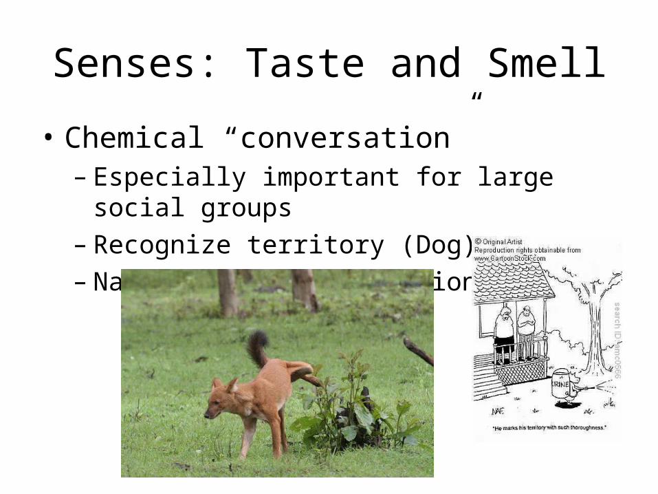
Senses: Taste and Smell
• Chemical “conversation”– Especially important for large social groups– Recognize territory (Dog)– Navigate during migration (Salmon)
Continued
• Gustation- taste• Olfaction- smell• Both are dependent on chemoreceptors• Land animals – taste is detection of chemicals within a solution– Smell is a detection of chemicals that travel through air
• No difference in aquatic animals
Taste in Humans
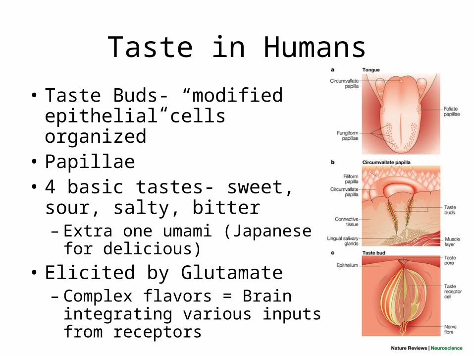
• Taste Buds- “modified epithelial cells organized”
• Papillae• 4 basic tastes- sweet, sour, salty,
bitter– Extra one umami (Japanese for
delicious)• Elicited by Glutamate – Complex flavors = Brain integrating
various inputs from receptors
Continued
• Chemoreceptors have channels present within their plasma membranes that ions diffuse through.
• Salt = Na+
• Sourness = H+
• Bitterness = K+
• Ions diffuse into the cell – cell depolarizes• Depolarization causes cell to release
neurotransmitter on a sensory neuron
Smell in Humans
• Olfactory receptor cells- neurons in nose– Send impulses to the olfactory bulb of the brain
• Odorant receptors (specific proteins) that get bind to the odorous substances that diffuse into the nose– Signal transduction pathway opened
• G protein, enzyme adenylyl cyclase, 2nd messenger cyclic AMP– 2nd messenger cyclic AMP opens channels in the plasma
membrane that are permeable to both Na+ and CA 2+ ions– Influx in ions depolarizes the membrane = ACTIVE POTENTIAL
Continued
• More than a 1000 Ors= 3% of human genes• Receptors in Brain for Taste and Smell are
independent yet they interact.
Vision in Invertebrates
• Ocellus- provides information about information about light intensity and direction– Doesn’t create images
• Compound eyes (insects)– Contain several thousand light detectors=
ommatidia• Each has its own focusing lens
• Humans can see flashes that occur at 50 flashes per second, while insects can see them at 330 times per second
Vision in Human
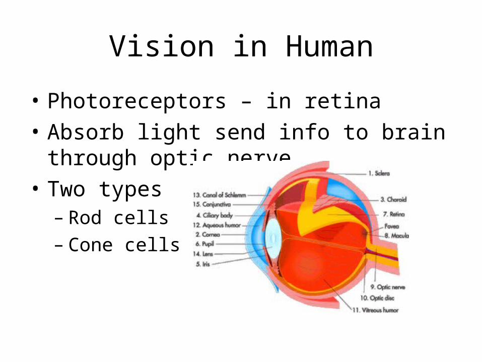
• Photoreceptors – in retina• Absorb light send info to brain through optic
nerve • Two types– Rod cells– Cone cells
Differences
• Rod cells– More sensitive to light, function better in dim light– absorb all wavelengths= monochrome vision– cone cells provide better visual acuity– widely dispersed throughout the cell= wider vision
• Cone cells– Less sensitive to light, function better in bright light– sensitive to red, green, and blue= color vision– concentrated near the fovea= acute area of vision
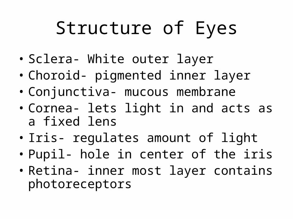
Structure of Eyes
• Sclera- White outer layer• Choroid- pigmented inner layer• Conjunctiva- mucous membrane• Cornea- lets light in and acts as a fixed lens• Iris- regulates amount of light• Pupil- hole in center of the iris• Retina- inner most layer contains
photoreceptors
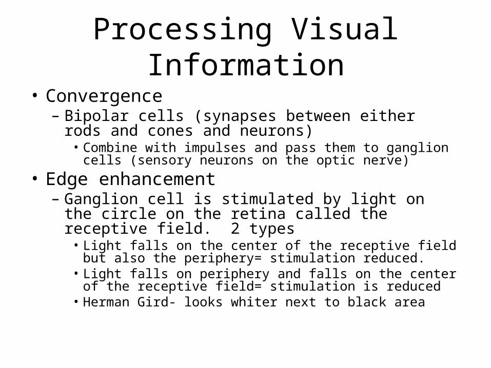
Processing Visual Information• Convergence– Bipolar cells (synapses between either rods and cones
and neurons)• Combine with impulses and pass them to ganglion cells
(sensory neurons on the optic nerve)• Edge enhancement– Ganglion cell is stimulated by light on the circle on the
retina called the receptive field. 2 types• Light falls on the center of the receptive field but also the
periphery= stimulation reduced.• Light falls on periphery and falls on the center of the
receptive field= stimulation is reduced• Herman Gird- looks whiter next to black area
Continued
• Contralateral Processing– Left and right optic
nerves meet at optic chiasma.
– Impulses cross over to opposite optic nerves
– Allows for brain to deduce distance and size