Embed Size (px)
Citation preview
Journ
alof
Cell
Scie
nce
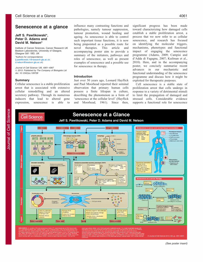
Senescence at a glance
Jeff S. Pawlikowski*,Peter D. Adams andDavid M. Nelson*
Institute of Cancer Sciences, Cancer Research UKBeatson Laboratories, University of Glasgow,Glasgow G61 1BD, UK
*Authors for correspondence
Journal of Cell Science 126, 4061–4067
� 2013. Published by The Company of Biologists Ltd
doi: 10.1242/jcs.109728
SummaryCellular senescence is a stable proliferation
arrest that is associated with extensive
cellular remodelling and an altered
secretory pathway. Through its numerous
inducers that lead to altered gene
expression, senescence is able to
influence many contrasting functions and
pathologies, namely tumour suppression,
tumour promotion, wound healing and
ageing. As senescence is able to control
such important tissue functions, it is now
being pinpointed as a possible route for
novel therapies. This article and
accompanying poster aim to provide a
summary of the initiators, pathways and
roles of senescence, as well as present
examples of senescence and a possible use
for senescence in therapy.
IntroductionJust over 50 years ago, Leonard Hayflick
and Paul Moorhead reported their seminal
observation that primary human cells
possess a finite lifespan in culture,
describing the phenomenon as a form of
‘senescence at the cellular level’ (Hayflick
and Moorhead, 1961). Since then,
significant progress has been made
toward characterizing how damaged cells
establish a stable proliferation arrest, a
process that we now refer to as cellular
senescence, and research has focused
on identifying the molecular triggers,
mechanisms, phenotypes and functional
impact of engaging the senescence
programme (Adams, 2009; Campisi and
d’Adda di Fagagna, 2007; Kuilman et al.,
2010). Here, and in the accompanying
poster, we concisely summarise recent
advances in our mechanistic and
functional understanding of the senescence
programme and discuss how it might be
exploited for therapeutic purposes.
Cell senescence is a stable state of
proliferation arrest that cells undergo in
response to a variety of detrimental stimuli
to limit the propagation of damaged and
stressed cells. Considerable evidence
supports a functional role for senescence
(See poster insert)
Cell Science at a Glance 4061
Journ
alof
Cell
Scie
nce
in tumour suppression and wound healing,but also possibly in promoting tissue
ageing. To date, a diverse array ofcellular stressors have been identified astriggers of senescence.
One of the first reported molecular
triggers of senescence was telomereattrition, the progressive shortening of thelinear ends of chromosomes that occurs
with repeated rounds of cell division(Harley et al., 1990). Consequently,senescence that occurs because of
telomere shortening (and probablycoupled with oxidative damage) isreferred to as replicative senescence (RS).Senescence induction also occurs in
response to the activation of oncogenes,termed oncogene-induced senescence(OIS) (Serrano et al., 1997). In addition,
DNA-damaging agents and oxidative stresshave been identified as potent initiators ofsenescence (d’Adda di Fagagna, 2008;
Saretzki and Von Zglinicki, 2002). Eachof these molecular triggers is fully capableof driving senescence by engaging an
integrated network of effector pathwaysthat collectively culminate in theestablishment of a stable proliferationarrest and the expression of the
senescence-associated secretory phenotype(SASP), an array of chemokines, cytokines,extracellular matrix remodelling
enzymes and other soluble andinsoluble factors secreted by senescentcells (see Box 1).
Effectors of the senescence program
It is now well established that the p53and p16INK4a-pRb tumour suppressor
pathways are the master regulators ofsenescence and serve to initiate a state ofstable proliferation arrest. Indeed, bypass
of senescence in primary human cellsrequires inactivation of both the p53 andp16INK4a-pRb pathways (Bond et al.,
1999; Hahn et al., 2002; Shay et al.,1991), as reviewed extensively elsewhere(Ben-Porath and Weinberg, 2005; Campisi,2005). In culture, senescent cells
frequently display an enlarged, flattenedmorphology that is accompanied byexpression of p16INK4a and elevated
lysosomal activity (senescence-associatedb-galactosidase; SA b-gal) (Campisi andd’Adda di Fagagna, 2007). Recent studies
have identified additional effectormechanisms involved in establishingsenescence that are further discussed
here, including the DNA-damageresponse, chromatin remodelling andautophagy (see Poster).
DNA-damage responseA common feature of many senescence
triggers is the ability to produce DNAdamage. Not surprisingly, the DNA-damage response (DDR) has emerged as
a crucial senescence effector mechanism.Replicative senescent cells accumulatemarkers of DNA damage, includingphosphorylated c-H2AX, CHK1, CHK2,
SMC1 and RAD17, and inactivation of theDDR enables replicative senescent cells toresume DNA replication (d’Adda di
Fagagna et al., 2003; Takai et al., 2003).Similarly, oncogenic activation engagesthe DDR by driving hyper-replication,
resulting in improperly terminatedreplication forks and DNA double-strandbreaks (Bartkova et al., 2006; Di Micco
et al., 2006). More recent findings suggestthat when DNA damage occurs attelomeres, it cannot be effectivelyrepaired, resulting in the presence of
chronic DNA damage foci, a persistentDDR and senescence induction (Fumagalliet al., 2012; Hewitt et al., 2012).
Intriguingly, oncogenic activation alsoinduces telomeric lesions, stochastictelomere attrition and senescence (Hewitt
et al., 2012; Suram et al., 2012).Inactivation of CHK2 not only abolishesoncogenic H-RAS-induced senescence, but
also promotes cellular transformation,
further illustrating the significance of anintact DDR for the establishment of OIS(Di Micco et al., 2006).
In addition, a persistent DDR is requiredfor robust expression of the SASP (see Box1). Indeed, inactivation of the DDR
mediators ATM, CHK2 or NBS1attenuates the SASP in radiation-inducedsenescent cells (Rodier et al., 2009).
Thus, a sustained DDR is crucial forestablishment of proliferation arrest andSASP expression, the two phenotypichallmarks of senescence.
Chromatin remodellingUpon senescence induction, cells undergoprofound chromatin remodelling, which is
also emerging as an important mediator ofthe senescence program, and its moststriking manifestation is the formation offacultative heterochromatin structures
termed senescence-associatedheterochromatic foci (SAHF). Originallyreported by Scott Lowe’s laboratory,
SAHF appear microscopically as largeDNA foci when senescent cells arestained with 49,6-diamidino-2-
phenylindole (DAPI) (Narita et al., 2003).There are ,30–50 foci per nucleus, andeach focus arises from the compaction of
Box 1. Senescence-associated secretory phenotype
In addition to the establishment of a stable proliferation arrest, the other hallmark of
senescence is the senescence-associated secretory phenotype (SASP). Senescent cells
remain metabolically active and express and secrete a broad spectrum of soluble and
insoluble proteins, as well as other factors collectively termed the SASP. SASP factors can
be classified into several defined categories including interleukins, chemokines and other
inflammatory factors, proteases and regulators, growth factors and regulators, receptors and
ligands, and extracellular matrix components (Coppe et al., 2010). The SASP is at least
partially dependent upon a persistent DNA-damage signal, because depletion of the DDR
mediators ATM, CHK2 or NBS1 in senescent cells attenuates secretion of the cytokine
interleukin 6 (IL6) (Rodier et al., 2009). In addition, SASP factors including several chemokine
receptor 2 (CXCR2) ligands (e.g. IL8) reinforce the senescence proliferation arrest by
enhancing the DDR, underscoring the role of the SASP as not only a downstream phenotype
of senescence, but also an integral effector mechanism (Acosta et al., 2008; Kuilman et al.,
2008). Functionally, secretion of SASP factors into the extracellular milieu by senescent cells
can elicit a variety of autocrine and paracrine responses. Non-cell-autonomous functions of
the SASP include the attenuation of fibrosis in response to chemical-induced or physical
injury, modulation of the immune response and transmission of senescence to normal
neighbouring cells adjacent to senescent lesions (Acosta et al., 2013; Jun and Lau, 2010;
Krizhanovsky et al., 2008; Lujambio et al., 2013; Nelson et al., 2012). Indeed, a number of
reports ascribe a role for the SASP in the tumour suppressive function of senescence through
the initiation of immune surveillance and clearance of senescent cells in vitro and in vivo
(Kang et al., 2011; Krizhanovsky et al., 2008; Xue et al., 2007). By contrast, secretion of the
SASP by senescent cells can also facilitate detrimental non-cell-autonomous effects,
including enhancement of cell growth, induction of the epithelial-to-mesenchymal transition
(EMT) and invasiveness and the promotion of tumorigenesis (Coppe et al., 2008; Krtolica
et al., 2001; Yoshimoto et al., 2013). Consequently, the SASP reflects both a definitive and
essential feature of senescence that can also potentiate tissue dysfunction and cancer in
certain contexts.
Journal of Cell Science 126 (18)4062
Journ
alof
Cell
Scie
nce
an individual chromosome (Funayamaet al., 2006; Zhang et al., 2007).
Although SAHF probably reflect aheterochromatic state, pericentric andtelomeric heterochromatin domains arelargely excluded from SAHF (Chandra
et al., 2012; Funayama et al., 2006;Narita et al., 2003; Zhang et al., 2007).
SAHF exhibit enrichment of histone H3
lysine 9 dimethylation and trimethylation(H3K9me2/3) and are devoid of theeuchromatic histone H3 lysine 9
acetylation (H3K9ac) and histone H3lysine 4 methylation (H3K4me) marks(Narita et al., 2006; Narita et al., 2003).Recent data indicate that SAHF result
from the spatial reorganization of pre-existing domains of repressive histonemodifications rather than through the
acquisition of new repressive histonemarks (Chandra et al., 2012). This studyfurther showed that, whereas H3K9me2 is
distributed across the entire SAHF focus,H3K9me3 is restricted to the SAHF coreand is enveloped by a ring of H3K27me3.SAHF are also composed of additional
heterochromatin components, includingthe histone H2A variant macroH2A andthe heterochromatin protein 1 (HP1) and
high-mobility group A (HMGA) proteins(Narita et al., 2006; Narita et al., 2003;Zhang et al., 2005).
Functionally, SAHF probably mediatessenescence in two ways. First, SAHF islikely to restrict the expression of
proliferation genes, such as cyclin A2, agene required for cell cycle progressionthat is silenced and physically incorporatedinto SAHF (Narita et al., 2003; Zhang et al.,
2007). Likewise, actively transcribedchromatin regions are largely excludedfrom SAHF (Funayama et al., 2006;
Narita et al., 2003). Recent findings alsosupport a role for SAHF in dampening theDDR and preserving cell viability following
oncogene-induced replication stress (DiMicco et al., 2011). This study revealedthat the DDR mediators c-H2AX, RAD50,NBS1 and activated ATM are physically
excluded from SAHF in OIS fibroblasts.Furthermore, inhibition of the H3K9me3methyltransferase SUV39, or depletion of
SUV39 or HP1 disrupts heterochromatinformation in oncogene-expressing cells,increases the DDR and leads to the
accumulation of DDR markers at DAPI-rich regions, and ultimately results incell death (Di Micco et al., 2011).
Thus, chromatin remodelling reinforcessenescence-mediated proliferation arrestand constrains the DDR.
Autophagy
More recently, evidence has emergedindicating that autophagy has an
important role as a molecular mediator ofcell senescence, including in theestablishment of cell cycle exit and SASP
expression. Autophagy is a program inwhich intracellular proteins, smallorganelles and other cytoplasmic
constituents are degraded by lysosomesfor their subsequent use by the cell assubstrates in various metabolic and
synthetic processes (Mizushima, 2007).Intriguingly, autophagic activity increasesmarkedly during OIS in primary humanfibroblasts (Young et al., 2009). Depletion
of either ATG5 or ATG7, genes requiredfor autophagy, attenuates SA b-galactosidase activity, a molecular marker
of a senescence-specific expansion of thelysosomal compartment, and delaysexpression of SASP components, IL6 and
IL8, in OIS fibroblasts. Remarkably,reduction of ATG5 or ATG7 proteinlevels also enables cells to bypass H-RAS-induced proliferation arrest,
indicating that autophagy is an essentialcontributor to the establishment of OIS.
Additionally, senescent cells typically
exhibit an enlarged morphology, containmore protein per cell than proliferatingcells and maintain active protein synthesis
(Young et al., 2009). To accommodate thissignificant energetic demand, OIS cellsupregulate autophagy to increase
nascent protein synthesis through theformation of a novel subcellular structuretermed the TOR-autophagy spatial-coupling compartment (TASCC) (Narita
et al., 2011). Findings from this studyindicated that the recruitment of the mTORcomplex to the TASCC is dependent on
amino acids and Rag GTPase, anddisruption of mTOR recruitment to theTASCC restricts IL6 and IL8 expression
during OIS. As a component of the SASP(see Text Box 1), IL6 is involved in theactivation of the senescence inflammatorytranscriptome and is required for the
proper establishment of OIS (Kuilmanet al., 2008). Consequently, the spatialassociation of mTOR-regulated autophagy
and protein synthesis in senescent cellsappears to facilitate the synthesis of at leasta subset of SASP proteins and thus
represents a crucial effector mechanismin the establishment of OIS. Whetherautophagy plays an essential role in all
modes of senescence remains to beelucidated, because different studies havefound that inhibition of mTOR, which is
known to activate autophagy, alternatelydelays or potentiates aspects of senescence
(Cao et al., 2011; Demidenko et al., 2009;Iglesias-Bartolome et al., 2012; Kennedyet al., 2011; Wall et al., 2013).
Interestingly, an autophagy and/orlysosomal pathway has been shown toprocess chromatin in senescent cells,
leading to depletion in total histonecontent. Depletion of histones was shownto correlate with naevus maturation, an
established histopathological parameterassociated with proliferation arrest andclinical benignancy, therefore linkingchromatin remodelling via an autophagic
pathway with senescence and tumoursuppression (Ivanov et al., 2013).
The diverse roles of senescence
Since the first description of senescence, it
is becoming apparent that it has diversefunctions and impinges on fundamentalbiological processes that can have
opposing effects, such as tumoursuppression, tissue repair, ageing andtumour promotion.
To form a tumour, cancer cells typicallymust acquire an unrestrained growthpotential, a trait that is suppressed by
senescence (Hanahan and Weinberg,2000). Indeed, many of the same triggersthat initiate cell transformation are alsoable to elicit a senescence response.
Consequently, most, if not all, cancershave mutations in the p53 and p16INK4a-pRb pathways, allowing for a bypass or
escape from senescence. In addition, manypremalignant tissues contain senescentcells (discussed below).
Senescence has also been shown toregulate tissue repair using a mousemodel of liver damage (Krizhanovsky
et al., 2008). Upon the induction of liverdamage by chemical treatment, hepaticstellate cells proliferate and secrete
extracellular matrix (ECM), which resultsin the formation of a fibrotic scar. Thesecells soon senesce and display a SASP thatresults in the downregulation of ECM
components and the increased expressionof matrix metalloproteinases (MMPs),which can also degrade ECM proteins.
Senescent hepatic cells also secretechemokines to attract natural killer cellsto resolve fibrosis once the healing has
taken place to allow the restoration ofnormal tissue function. Furthermore, whensenescence is inhibited in hepatic stellate
cells severe fibrosis entails after acute liverinjury, underscoring the importance ofsenescence in facilitating a portion of the
Journal of Cell Science 126 (18) 4063
Journ
alof
Cell
Scie
nce
wound-healing response (Krizhanovsky
et al., 2008).
Cells with senescent properties have
been shown to increase with age in a
variety of mammalian tissues (Dimri et al.,
1995; Herbig et al., 2006; Jeyapalan et al.,
2007; Krishnamurthy et al., 2004;
Sedelnikova et al., 2004; Wang et al.,
2009), and, moreover, the accumulation of
senescent cells is also related to age-
associated tissue pathologies, such as
osteoarthritis, atherosclerosis and liver
cirrhosis (Minamino and Komuro, 2007;
Price et al., 2002; Wiemann et al., 2002).
Experimentally, inactivation of p16INK4a-
pRb in a prematurely aged mouse model is
able to prolong cell renewal and transgenic
expression of telomerase, an enzyme that
maintains telomere length and extends
longevity in cancer-resistant mice (Baker
et al., 2008; Janzen et al., 2006;
Krishnamurthy et al., 2006; Molofsky
et al., 2006; Tomas-Loba et al., 2008).
The contribution of senescence to
ageing has been explained by the ‘theory
of antagonistic pleiotropy’, which
stipulates that a biological process can be
both beneficial and deleterious, depending
on the age of the organism (Williams,
1957). Specifically, a subset of genes
responsible for negative effects are
selected for if they can confer a
reproductive advantage early in life,
whereas they confer harmful effects in
aged individuals. Antagonistic pleiotropy
is based on the fact that most organisms
evolve in environments with fatal extrinsic
hazards: a so-called ‘survival of the fittest’.
However, age-associated phenotypes are
not under the control of natural selection
and, therefore, processes that promote
fitness in young individuals can be
detrimental in aged organisms (Rodier
and Campisi, 2011; Williams, 1957).
According to this view, senescence is
beneficial in young organisms through its
ability to promote tumour suppression and
wound healing, but it has detrimental
effects on old organisms. This is in line
with the finding that cellular senescence is
associated with age-related phenotypes and
the removal of senescent cells is able to
prevent or delay tissue dysfunction and
extend healthspan (Baker et al., 2011).
Nonetheless, the notion of senescence as
an example of antagonistic pleiotropy has
also been challenged recently because
there is little evidence that the positive
effects of senescence on survival
predominate at young ages and the
negative effects predominate at late ages
(Giaimo and d’Adda di Fagagna, 2012).
The theory of antagonistic pleiotropycan explain the tumour-promoting roleof senescence. Although seemingly
paradoxical, senescence might promotecancer owing to the fact that senescentcells are able to promote malignancy in the
cells around them. Through the SASP (seeBox 1), senescent cells secrete a largeamount of cytokines and chemokines into
their surrounding environment (Coppeet al., 2010). Because the number ofsenescent cells increases with age, thesecretion of these factors can persist and
stimulate the formation of a tumour. It ispossible that a large, persistent amountof SASP signalling promotes the pro-
tumorigenic effects, whereas theirsecretion in lower amounts during acuteSASP promotes the tumour suppressive
effects of senescence. Whatever the maincontribution of SASP might be, either pro-or anti-tumorigenic, it appears to be clearly
context dependent, as a result of manyfactors such as the interplay oftumour suppressors and the tissuemicroenvironment (Lujambio et al., 2013).
Senescence in cancer and normaltissue homeostasis
Senescence was initially considered an
artefact of in vitro cell culture shock, butthis view has now changed owing to manygroups observing senescent cells in
premalignant tissues (Sherr and DePinho,2000). Analyses of human and murinetissues have shown the presence of
senescent cells in lung adenomas,pancreatic intraductal neoplasia (PanINlesions), prostatic intraepithelial neoplasia(PIN lesions) and melanocytic naevi, all of
which are premalignant neoplasms (Braiget al., 2005; Chen et al., 2005; Colladoet al., 2005; Michaloglou et al., 2005) (see
Poster). It has also been shownthat senescence is largely abolished inthe corresponding malignant lesion –
lung carcinomas, pancreatic ductaladenocarcinomas, prostate carcinomasand melanomas, respectively (Chen et al.,
2005; Collado et al., 2005; Gray-Schopferet al., 2006).
Mouse models have been used to altertumour suppressor genes such as PTEN or
oncogenes such as NRAS to inducesenescence that is associated with thedevelopment of pre-malignant lesions
without signs of apoptosis (Braig et al.,2005; Chen et al., 2005). Upon inactivationof senescence through deletion of
senescence modulators such as p53 orchromatin modulators, fully developed
malignancy then occurred, highlightingthe role of senescence in tumoursuppression. Senescence has also beenobserved in melanocytic naevi in mice
with an activated mutant form of theoncogene Braf (BRAFV600E) expressedspecifically in their melanocytes (Dhomen
et al., 2009). Appropriately, the majority ofbenign human naevi have also been foundto have a BRAFV600E mutation (Pollock
et al., 2003). However, the efficiency ofsenescence in acting as a tumoursuppression mechanism in murinenaevi harbouring BRAFV600E-expressing
melanocytes is reduced compared withhuman naevi because these mice typicallydevelop melanomas within a year
(Dhomen et al., 2009). Nonetheless, therather long latency of tumours in thesemice suggests that oncogenic BRAF alone
is not sufficient for the induction ofmelanoma and that additional events arerequired to bypass senescence and
for subsequent disease progression.Indeed, additional genetic alterations inoncogenes or tumour suppressors havebeen shown to accelerate progression into
melanoma (Damsky et al., 2011; Dankortet al., 2009; Delmas et al., 2007; Vredeveldet al., 2012).
In addition to the role of senescence intumour suppression and in ageing, there isalso in vivo evidence for its role in normal
cellular differentiation and development,such as the terminal differentiation ofmegakaryocytes (Besancenot et al., 2010).The proliferative arrest that is observed
in mature megakaryocytes resembles asenescence arrest, because many markersof senescence are upregulated in this state.
Interestingly, senescence is not observed inmalignant megakaryocytes, which mightbe the underlying reason for their
cancerous phenotype.
Senescence therapies
As discussed above, the bypass or escape
of a senescence response in premalignantlesions is required for tumour progression(Bennecke et al., 2010; Braig et al., 2005;
Chen et al., 2005; Collado and Serrano,2010; Dankort et al., 2009; Dhomen et al.,2009). Therefore, if senescence can
be either maintained or reactivated,progression towards a malignant statemight be slowed or malignancy even
averted, suggesting that senescence couldbe a therapy target. Supporting evidencefrom murine models shows that the
Journal of Cell Science 126 (18)4064
Journ
alof
Cell
Scie
nce
reactivation of tumour suppressor genes
such as p53 can induce senescence andtumour regression in a liver tumour model(Ventura et al., 2007; Xue et al., 2007).
Similarly, tumour regression due to theinactivation of Myc can be associated withcell senescence, functionally supporting atherapeutic role for senescence (Wu et al.,
2007).
The outcome of conventional cancertherapy is enhanced when a senescence
response is present (Schmitt et al., 2002; tePoele et al., 2002). In addition, otherstrategies for pro-senescence therapy havebeen proposed, including inhibition of
telomerase activity and alteration of CDKor CDK regulators (Campaner et al., 2010;Chen et al., 2005; Harley, 2008; Kennedy
et al., 2011; Nardella et al., 2011; Puyolet al., 2010; Ventura et al., 2007; Wallet al., 2013; Xue et al., 2007) (see Poster).
Moreover, it has been shown that
senescent cells can be cleared by theimmune system owing to the SASP. Forexample, reactivation of p53 in a mouse
model of hepatocellular carcinoma that isdriven by RAS expression results intumour regression that was associated
with a strong SASP, which allows fortumour clearance (Kang et al., 2011; Xueet al., 2007). However, the clearance of
senescent cells by the immune system isnot universal because precancerous lesionssuch as naevi can persist for decadeswithout any sign of malignant
transformation (Michaloglou et al., 2005).The targeting of SASP might also providea possible route for pro-senescence cancer
therapy by enhancing cell clearance.However, it is important to rememberthat even though senescence acts
primarily as a tumour suppressionmechanism, it can also promotetumorigenesis (Krtolica et al., 2001).
Therefore, pro-senescence therapy willhave to be applied in a highly specificmanner in the clinic and might requireinitial genotyping before use. Our
expanding knowledge of the senescencemechanism will help to advance pro-senescence cancer therapy, so that it can
reach the clinic as a cancer therapy in thenear future.
ConclusionsIn the 50 years since a role of senescencein tumour suppression and ageing was firstproposed, the senescence phenotype has
become much clearer. Once thought to bemerely a phenomenon found in vitro,senescence is now being recognised as a
relevant cellular mechanism in vivo. A
major hurdle that the senescence field must
overcome is labelling senescence with a
strict definition. Because there are a
multitude of distinctive initiators,
pathways and markers of senescence,
unambiguously defining a cell as
senescent can be difficult. Senescence is
increasingly referred to not as an
irreversible growth arrest, but as a stable
growth arrest.
Additional functions and signalling
mechanisms of senescence have also
recently been uncovered, including links
to autophagy, the inflammatory response,
chromatin structure, as well as its
regulation by miRNAs, which have been
shown to induce senescence by regulating
key effectors of senescence pathways
(Christoffersen et al., 2010; Feliciano
et al., 2011). Powerful high-throughput
analyses at the genomic and epigenomic
level will continue to progress the field to a
better understanding of senescence at the
molecular level and further elucidate its
potential in tumour suppression, ageing,
wound healing and tumour promotion, as
well as other roles that probably remain to
be discovered. Further research into these
mechanisms might address unanswered
questions in the senescence field such as
how to control the effects of senescence in
such a way that it could be used in cancer
therapy.
Funding
The work done in the Peter Adams lab is
funded by Cancer Research UK [grant
number C10652/A10250]; and the National
Institutes of Health [grant number R01
CA129334-01]. Deposited in PMC for
release after 12 months.
A high-resolution version of the poster is available for
downloading in the online version of this article at
jcs.biologists.org. Individual poster panels are available
as JPEG files at http://jcs.biologists.org/lookup/suppl/
doi:10.1242/jcs.109728/-/DC1.
ReferencesAcosta, J. C., O’Loghlen, A., Banito, A., Guijarro,
M. V., Augert, A., Raguz, S., Fumagalli, M., Da Costa,
M., Brown, C., Popov, N. et al. (2008). Chemokine
signaling via the CXCR2 receptor reinforces senescence.
Cell 133, 1006-1018.
Acosta, J. C., Banito, A., Wuestefeld, T., Georgilis, A.,
Janich, P., Morton, J. P., Athineos, D., Kang, T.-W.,
Lasitschka, F., Andrulis, M. et al. (2013). A complex
secretory program orchestrated by the inflammasome
controls paracrine senescence. Nat. Cell Biol. doi:
10.1038/ncb2784.
Adams, P. D. (2009). Healing and hurting: molecular
mechanisms, functions, and pathologies of cellular
senescence. Mol. Cell 36, 2-14.
Baker, D. J., Perez-Terzic, C., Jin, F., Pitel, K. S.,
Niederlander, N. J., Jeganathan, K., Yamada, S.,
Reyes, S., Rowe, L., Hiddinga, H. J. et al. (2008).Opposing roles for p16Ink4a and p19Arf in senescenceand ageing caused by BubR1 insufficiency. Nat. Cell Biol.
10, 825-836.
Baker, D. J., Wijshake, T., Tchkonia, T., LeBrasseur,
N. K., Childs, B. G., van de Sluis, B., Kirkland, J. L. and
van Deursen, J. M. (2011). Clearance of p16Ink4a-positivesenescent cells delays ageing-associated disorders. Nature
479, 232-236.
Bartkova, J., Rezaei, N., Liontos, M., Karakaidos, P.,
Kletsas, D., Issaeva, N., Vassiliou, L.-V. F., Kolettas, E.,
Niforou, K., Zoumpourlis, V. C. et al. (2006).Oncogene-induced senescence is part of thetumorigenesis barrier imposed by DNA damagecheckpoints. Nature 444, 633-637.
Ben-Porath, I. and Weinberg, R. A. (2005). The signalsand pathways activating cellular senescence. Int. J.
Biochem. Cell Biol. 37, 961-976.
Bennecke, M., Kriegl, L., Bajbouj, M., Retzlaff, K.,
Robine, S., Jung, A., Arkan, M. C., Kirchner, T. and
Greten, F. R. (2010). Ink4a/Arf and oncogene-inducedsenescence prevent tumor progression during alternativecolorectal tumorigenesis. Cancer Cell 18, 135-146.
Besancenot, R., Chaligne, R., Tonetti, C., Pasquier, F.,
Marty, C., Lecluse, Y., Vainchenker, W.,
Constantinescu, S. N. and Giraudier, S. (2010). Asenescence-like cell-cycle arrest occurs duringmegakaryocytic maturation: implications forphysiological and pathological megakaryocyticproliferation. PLoS Biol. 8.
Bond, J. A., Haughton, M. F., Rowson, J. M., Smith,
P. J., Gire, V., Wynford-Thomas, D. and Wyllie, F. S.
(1999). Control of replicative life span in human cells:barriers to clonal expansion intermediate between M1senescence and M2 crisis. Mol. Cell. Biol. 19, 3103-3114.
Braig, M., Lee, S., Loddenkemper, C., Rudolph, C.,
Peters, A. H. F. M., Schlegelberger, B., Stein, H.,
Dorken, B., Jenuwein, T. and Schmitt, C. A. (2005).Oncogene-induced senescence as an initial barrier inlymphoma development. Nature 436, 660-665.
Campaner, S., Doni, M., Hydbring, P., Verrecchia, A.,
Bianchi, L., Sardella, D., Schleker, T., Perna, D.,
Tronnersjo, S., Murga, M. et al. (2010). Cdk2suppresses cellular senescence induced by the c-myconcogene. Nat. Cell Biol. 12, 54-59.
Campisi, J. (2005). Senescent cells, tumor suppression,and organismal aging: good citizens, bad neighbors. Cell
120, 513-522.
Campisi, J. and d’Adda di Fagagna, F. (2007). Cellularsenescence: when bad things happen to good cells. Nat.
Rev. Mol. Cell Biol. 8, 729-740.
Cao, K., Graziotto, J. J., Blair, C. D., Mazzulli, J. R.,
Erdos, M. R., Krainc, D. and Collins, F. S. (2011).Rapamycin reverses cellular phenotypes and enhancesmutant protein clearance in hutchinson-gilford progeriasyndrome cells. Sci. Transl. Med. 3, 89ra58.
Chandra, T., Kirschner, K., Thuret, J.-Y., Pope, B. D.,Ryba, T., Newman, S., Ahmed, K., Samarajiwa, S. A.,
Salama, R., Carroll, T. et al. (2012). Independence ofrepressive histone marks and chromatin compactionduring senescent heterochromatic layer formation. Mol.
Cell 47, 203-214.
Chen, Z., Trotman, L. C., Shaffer, D., Lin, H.-K.,
Dotan, Z. A., Niki, M., Koutcher, J. A., Scher, H. I.,
Ludwig, T., Gerald, W. et al. (2005). Crucial role of p53-dependent cellular senescence in suppression of Pten-deficient tumorigenesis. Nature 436, 725-730.
Christoffersen, N. R., Shalgi, R., Frankel, L. B., Leucci,
E., Lees, M., Klausen, M., Pilpel, Y., Nielsen, F. C.,
Oren, M. and Lund, A. H. (2010). p53-independentupregulation of miR-34a during oncogene-inducedsenescence represses MYC. Cell Death Differ. 17, 236-245.
Collado, M. and Serrano, M. (2010). Senescence intumours: evidence from mice and humans. Nat. Rev.
Cancer 10, 51-57.
Collado, M., Gil, J., Efeyan, A., Guerra, C.,
Schuhmacher, A. J., Barradas, M., Bengurıa, A.,
Zaballos, A., Flores, J. M., Barbacid, M. et al. (2005).Tumour biology: senescence in premalignant tumours.Nature 436, 642.
Journal of Cell Science 126 (18) 4065
Journ
alof
Cell
Scie
nce
Coppe, J.-P., Patil, C. K., Rodier, F., Sun, Y., Munoz,
D. P., Goldstein, J., Nelson, P. S., Desprez, P.-Y. and
Campisi, J. (2008). Senescence-associated secretory
phenotypes reveal cell-nonautonomous functions of
oncogenic RAS and the p53 tumor suppressor. PLoS
Biol. 6, 2853-2868.
Coppe, J.-P., Desprez, P.-Y., Krtolica, A. and Campisi,
J. (2010). The senescence-associated secretory phenotype:
the dark side of tumor suppression. Annu. Rev. Pathol. 5,
99-118.
d’Adda di Fagagna, F. (2008). Living on a break: cellular
senescence as a DNA-damage response. Nat. Rev. Cancer
8, 512-522.
d’Adda di Fagagna, F., Reaper, P. M., Clay-Farrace,
L., Fiegler, H., Carr, P., Von Zglinicki, T., Saretzki, G.,
Carter, N. P. and Jackson, S. P. (2003). A DNA damage
checkpoint response in telomere-initiated senescence.
Nature 426, 194-198.
Damsky, W. E., Curley, D. P., Santhanakrishnan, M.,
Rosenbaum, L. E., Platt, J. T., Gould Rothberg, B. E.,
Taketo, M. M., Dankort, D., Rimm, D. L., McMahon,
M. et al. (2011). b-catenin signaling controls metastasis in
Braf-activated Pten-deficient melanomas. Cancer Cell 20,
741-754.
Dankort, D., Curley, D. P., Cartlidge, R. A., Nelson, B.,
Karnezis, A. N., Damsky, W. E. J., Jr, You, M. J.,
DePinho, R. A., McMahon, M. and Bosenberg, M.
(2009). Braf(V600E) cooperates with Pten loss to induce
metastatic melanoma. Nat. Genet. 41, 544-552.
Delmas, V., Beermann, F., Martinozzi, S., Carreira, S.,
Ackermann, J., Kumasaka, M., Denat, L., Goodall, J.,
Luciani, F., Viros, A. et al. (2007). Beta-catenin induces
immortalization of melanocytes by suppressing p16INK4a
expression and cooperates with N-Ras in melanoma
development. Genes Dev. 21, 2923-2935.
Demidenko, Z. N., Zubova, S. G., Bukreeva, E. I.,
Pospelov, V. A., Pospelova, T. V. and Blagosklonny,
M. V. (2009). Rapamycin decelerates cellular senescence.
Cell Cycle 8, 1888-1895.
Dhomen, N., Reis-Filho, J. S., da Rocha Dias, S.,
Hayward, R., Savage, K., Delmas, V., Larue, L.,
Pritchard, C. and Marais, R. (2009). Oncogenic Braf
induces melanocyte senescence and melanoma in mice.
Cancer Cell 15, 294-303.
Di Micco, R., Fumagalli, M., Cicalese, A., Piccinin, S.,
Gasparini, P., Luise, C., Schurra, C., Garre’, M.,
Nuciforo, P. G., Bensimon, A. et al. (2006). Oncogene-
induced senescence is a DNA damage response triggered
by DNA hyper-replication. Nature 444, 638-642.
Di Micco, R., Sulli, G., Dobreva, M., Liontos, M.,
Botrugno, O. A., Gargiulo, G., dal Zuffo, R., Matti, V.,
d’Ario, G., Montani, E. et al. (2011). Interplay between
oncogene-induced DNA damage response and
heterochromatin in senescence and cancer. Nat. Cell
Biol. 13, 292-302.
Dimri, G. P., Lee, X., Basile, G., Acosta, M., Scott, G.,
Roskelley, C., Medrano, E. E., Linskens, M., Rubelj, I.,
Pereira-Smith, O. et al. (1995). A biomarker that
identifies senescent human cells in culture and in aging
skin in vivo. Proc. Natl. Acad. Sci. USA 92, 9363-9367.
Feliciano, A., Sanchez-Sendra, B., Kondoh, H. and
Lleonart, M. E. (2011). MicroRNAs regulate key effector
pathways of senescence. J. Aging Res. 2011, 205378.
Fumagalli, M., Rossiello, F., Clerici, M., Barozzi, S.,
Cittaro, D., Kaplunov, J. M., Bucci, G., Dobreva, M.,
Matti, V., Beausejour, C. M. et al. (2012). Telomeric
DNA damage is irreparable and causes persistent DNA-
damage-response activation. Nat. Cell Biol. 14, 355-365.
Funayama, R., Saito, M., Tanobe, H. and Ishikawa,
F. (2006). Loss of linker histone H1 in cellular
senescence. J. Cell Biol. 175, 869-880.
Giaimo, S. and d’Adda di Fagagna, F. (2012). Is cellular
senescence an example of antagonistic pleiotropy? Aging
Cell 11, 378-383.
Gray-Schopfer, V. C., Cheong, S. C., Chong, H., Chow,
J., Moss, T., Abdel-Malek, Z. A., Marais, R., Wynford-
Thomas, D. and Bennett, D. C. (2006). Cellular
senescence in naevi and immortalisation in melanoma: a
role for p16? Br. J. Cancer 95, 496-505.
Hahn, W. C., Dessain, S. K., Brooks, M. W., King,
J. E., Elenbaas, B., Sabatini, D. M., DeCaprio, J. A. and
Weinberg, R. A. (2002). Enumeration of the simian virus
40 early region elements necessary for human celltransformation. Mol. Cell. Biol. 22, 2111-2123.
Hanahan, D. and Weinberg, R. A. (2000). The hallmarksof cancer. Cell 100, 57-70.
Harley, C. B. (2008). Telomerase and cancer therapeutics.Nat. Rev. Cancer 8, 167-179.
Harley, C. B., Futcher, A. B. and Greider, C. W.
(1990). Telomeres shorten during ageing of humanfibroblasts. Nature 345, 458-460.
Hayflick, L. and Moorhead, P. S. (1961). The serialcultivation of human diploid cell strains. Exp. Cell Res.
25, 585-621.
Herbig, U., Ferreira, M., Condel, L., Carey, D. and
Sedivy, J. M. (2006). Cellular senescence in agingprimates. Science 311, 1257.
Hewitt, G., Jurk, D., Marques, F. D. M., Correia-Melo,
C., Hardy, T., Gackowska, A., Anderson, R., Taschuk,M., Mann, J. and Passos, J. F. (2012). Telomeres arefavoured targets of a persistent DNA damage response inageing and stress-induced senescence. Nat. Commun. 3,708.
Iglesias-Bartolome, R., Patel, V., Cotrim, A.,
Leelahavanichkul, K., Molinolo, A. A., Mitchell, J. B.
and Gutkind, J. S. (2012). mTOR inhibition preventsepithelial stem cell senescence and protects fromradiation-induced mucositis. Cell Stem Cell 11, 401-414.
Ivanov, A., Pawlikowski, J., Manoharan, I., van Tuyn,
J., Nelson, D. M., Rai, T. S., Shah, P. P., Hewitt, G.,Korolchuk, V. I., Passos, J. F. et al. (2013). Lysosome-mediated processing of chromatin in senescence. J. Cell
Biol. 202, 129-143.
Janzen, V., Forkert, R., Fleming, H. E., Saito, Y.,Waring, M. T., Dombkowski, D. M., Cheng, T.,
DePinho, R. A., Sharpless, N. E. and Scadden, D. T.
(2006). Stem-cell ageing modified by the cyclin-dependent kinase inhibitor p16INK4a. Nature 443, 421-426.
Jeyapalan, J. C., Ferreira, M., Sedivy, J. M. and
Herbig, U. (2007). Accumulation of senescent cells inmitotic tissue of aging primates. Mech. Ageing Dev. 128,36-44.
Jun, J.-I. and Lau, L. F. (2010). The matricellular proteinCCN1 induces fibroblast senescence and restricts fibrosisin cutaneous wound healing. Nat. Cell Biol. 12, 676-685.
Kang, T.-W., Yevsa, T., Woller, N., Hoenicke, L.,
Wuestefeld, T., Dauch, D., Hohmeyer, A., Gereke, M.,Rudalska, R., Potapova, A. et al. (2011). Senescencesurveillance of pre-malignant hepatocytes limits livercancer development. Nature 479, 547-551.
Kennedy, A. L., Morton, J. P., Manoharan, I., Nelson,D. M., Jamieson, N. B., Pawlikowski, J. S., McBryan,
T., Doyle, B., McKay, C., Oien, K. A. et al. (2011).Activation of the PIK3CA/AKT pathway suppressessenescence induced by an activated RAS oncogene topromote tumorigenesis. Mol. Cell 42, 36-49.
Krishnamurthy, J., Torrice, C., Ramsey, M. R.,
Kovalev, G. I., Al-Regaiey, K., Su, L. and Sharpless,N. E. (2004). Ink4a/Arf expression is a biomarker ofaging. J. Clin. Invest. 114, 1299-1307.
Krishnamurthy, J., Ramsey, M. R., Ligon, K. L.,
Torrice, C., Koh, A., Bonner-Weir, S. and Sharpless,
N. E. (2006). p16INK4a induces an age-dependent declinein islet regenerative potential. Nature 443, 453-457.
Krizhanovsky, V., Yon, M., Dickins, R. A., Hearn, S.,Simon, J., Miething, C., Yee, H., Zender, L. and Lowe,
S. W. (2008). Senescence of activated stellate cells limitsliver fibrosis. Cell 134, 657-667.
Krtolica, A., Parrinello, S., Lockett, S., Desprez, P. Y.and Campisi, J. (2001). Senescent fibroblasts promoteepithelial cell growth and tumorigenesis: a link betweencancer and aging. Proc. Natl. Acad. Sci. USA 98, 12072-12077.
Kuilman, T., Michaloglou, C., Vredeveld, L. C. W.,Douma, S., van Doorn, R., Desmet, C. J., Aarden, L. A.,
Mooi, W. J. and Peeper, D. S. (2008). Oncogene-inducedsenescence relayed by an interleukin-dependentinflammatory network. Cell 133, 1019-1031.
Kuilman, T., Michaloglou, C., Mooi, W. J. and Peeper,
D. S. (2010). The essence of senescence. Genes Dev. 24,2463-2479.
Lujambio, A., Akkari, L., Simon, J., Grace, D.,
Tschaharganeh, D. F., Bolden, J. E., Zhao, Z.,Thapar, V., Joyce, J. A., Krizhanovsky, V. et al.
(2013). Non-cell-autonomous tumor suppression by p53.
Cell 153, 449-460.
Michaloglou, C., Vredeveld, L., Soengas, M. S.,
Denoyelle, C., Kuilman, T., van der Horst, C.,
Majoor, D., Shay, J., Mooi, W. and Peeper, D. S.
(2005). BRAF(E600)-associated senescence-like cell
cycle arrest of human naevi. Nature 436, 720-724.
Minamino, T. and Komuro, I. (2007). Vascular cell
senescence: contribution to atherosclerosis. Circ. Res. 100,
15-26.
Mizushima, N. (2007). Autophagy: process and function.
Genes Dev. 21, 2861-2873.
Molofsky, A. V., Slutsky, S. G., Joseph, N. M., He, S.,
Pardal, R., Krishnamurthy, J., Sharpless, N. E. and
Morrison, S. J. (2006). Increasing p16INK4a expression
decreases forebrain progenitors and neurogenesis during
ageing. Nature 443, 448-452.
Nardella, C., Clohessy, J. G., Alimonti, A. and
Pandolfi, P. P. (2011). Pro-senescence therapy for
cancer treatment. Nat. Rev. Cancer 11, 503-511.
Narita, M., Nu,nez, S., Heard, E., Narita, M., Lin,
A. W., Hearn, S. A., Spector, D. L., Hannon, G. J. and
Lowe, S. W. (2003). Rb-mediated heterochromatin
formation and silencing of E2F target genes during
cellular senescence. Cell 113, 703-716.
Narita, M., Narita, M., Krizhanovsky, V., Nunez, S.,
Chicas, A., Hearn, S. A., Myers, M. P. and Lowe, S. W.
(2006). A novel role for high-mobility group a proteins in
cellular senescence and heterochromatin formation. Cell
126, 503-514.
Narita, M., Young, A. R. J., Arakawa, S., Samarajiwa,
S. A., Nakashima, T., Yoshida, S., Hong, S., Berry,
L. S., Reichelt, S., Ferreira, M. et al. (2011). Spatial
coupling of mTOR and autophagy augments secretory
phenotypes. Science 332, 966-970.
Nelson, G., Wordsworth, J., Wang, C., Jurk, D.,
Lawless, C., Martin-Ruiz, C. and von Zglinicki,
T. (2012). A senescent cell bystander effect: senescence-
induced senescence. Aging Cell 11, 345-349.
Pollock, P. M., Harper, U. L., Hansen, K. S., Yudt,
L. M., Stark, M., Robbins, C. M., Moses, T. Y.,
Hostetter, G., Wagner, U., Kakareka, J. et al. (2003).
High frequency of BRAF mutations in nevi. Nat. Genet.
33, 19-20.
Price, J. S., Waters, J. G., Darrah, C., Pennington, C.,
Edwards, D. R., Donell, S. T. and Clark, I. M. (2002).
The role of chondrocyte senescence in osteoarthritis.
Aging Cell 1, 57-65.
Puyol, M., Martın, A., Dubus, P., Mulero, F., Pizcueta,
P., Khan, G., Guerra, C., Santamarıa, D. and
Barbacid, M. (2010). A synthetic lethal interaction
between K-Ras oncogenes and Cdk4 unveils a
therapeutic strategy for non-small cell lung carcinoma.
Cancer Cell 18, 63-73.
Rodier, F. and Campisi, J. (2011). Four faces of cellular
senescence. J. Cell Biol. 192, 547-556.
Rodier, F., Coppe, J. P., Patil, C. K., Hoeijmakers,
W. A. M., Munoz, D. P., Raza, S. R., Freund, A.,
Campeau, E., Davalos, A. R. and Campisi, J. (2009).
Persistent DNA damage signalling triggers senescence-associated inflammatory cytokine secretion. Nat. Cell
Biol. 11, 973-979.
Saretzki, G. and Von Zglinicki, T. (2002). Replicative
aging, telomeres, and oxidative stress. Ann. N. Y. Acad.
Sci. 959, 24-29.
Schmitt, C. A., Fridman, J. S., Yang, M., Lee, S.,
Baranov, E., Hoffman, R. M. and Lowe, S. W. (2002).
A senescence program controlled by p53 and p16INK4a
contributes to the outcome of cancer therapy. Cell 109,
335-346.
Sedelnikova, O. A., Horikawa, I., Zimonjic, D. B.,
Popescu, N. C., Bonner, W. M. and Barrett, J. C.
(2004). Senescing human cells and ageing mice
accumulate DNA lesions with unrepairable double-strand
breaks. Nat. Cell Biol. 6, 168-170.
Serrano, M., Lin, A. W., McCurrach, M. E., Beach,
D. and Lowe, S. W. (1997). Oncogenic ras provokes
premature cell senescence associated with accumulation
of p53 and p16INK4a. Cell 88, 593-602.
Shay, J. W., Pereira-Smith, O. M. and Wright, W. E.
(1991). A role for both RB and p53 in the regulation of
human cellular senescence. Exp. Cell Res. 196, 33-39.
Journal of Cell Science 126 (18)4066
Journ
alof
Cell
Scie
nce
Sherr, C. J. and DePinho, R. A. (2000). Cellularsenescence: mitotic clock or culture shock? Cell 102,407-410.Suram, A., Kaplunov, J., Patel, P. L., Ruan, H., Cerutti,
A., Boccardi, V., Fumagalli, M., Di Micco, R., Mirani,
N., Gurung, R. L. et al. (2012). Oncogene-inducedtelomere dysfunction enforces cellular senescence inhuman cancer precursor lesions. EMBO J. 31, 2839-2851.Takai, H., Smogorzewska, A. and de Lange, T. (2003).DNA damage foci at dysfunctional telomeres. Curr. Biol.
13, 1549-1556.te Poele, R. H., Okorokov, A. L., Jardine, L.,
Cummings, J. and Joel, S. P. (2002). DNA damage isable to induce senescence in tumor cells in vitro and invivo. Cancer Res. 62, 1876-1883.Tomas-Loba, A., Flores, I., Fernandez-Marcos, P. J.,
Cayuela, M. L., Maraver, A., Tejera, A., Borras, C.,
Matheu, A., Klatt, P., Flores, J. M. et al. (2008).Telomerase reverse transcriptase delays aging in cancer-resistant mice. Cell 135, 609-622.Ventura, A., Kirsch, D. G., McLaughlin, M. E.,
Tuveson, D. A., Grimm, J., Lintault, L., Newman, J.,
Reczek, E. E., Weissleder, R. and Jacks, T. (2007).Restoration of p53 function leads to tumour regression invivo. Nature 445, 661-665.Vredeveld, L. C. W., Possik, P. A., Smit, M. A., Meissl,
K., Michaloglou, C., Horlings, H. M., Ajouaou, A.,
Kortman, P. C., Dankort, D., McMahon, M. et al.(2012). Abrogation of BRAFV600E-induced senescenceby PI3K pathway activation contributes tomelanomagenesis. Genes Dev. 26, 1055-1069.Wall, M., Poortinga, G., Stanley, K. L., Lindemann,
R. K., Bots, M., Chan, C. J., Bywater, M. J., Kinross,K. M., Astle, M. V., Waldeck, K. et al. (2013). ThemTORC1 inhibitor everolimus prevents and treats Em-Myc lymphoma by restoring oncogene-inducedsenescence. Cancer Discov. 3, 82-95.Wang, C., Jurk, D., Maddick, M., Nelson, G., Martin-Ruiz, C. and von Zglinicki, T. (2009). DNA damageresponse and cellular senescence in tissues of aging mice.Aging Cell 8, 311-323.Wiemann, S. U., Satyanarayana, A., Tsahuridu, M.,Tillmann, H. L., Zender, L., Klempnauer, J.,
Flemming, P., Franco, S., Blasco, M. A., Manns,
M. P. et al. (2002). Hepatocyte telomere shortening andsenescence are general markers of human liver cirrhosis.FASEB J. 16, 935-942.Williams, G. C. (1957). Pleiotrpy, Natural Selection, andthe evolution of senescence. Evolution 11, 398-411.Wu, C.-H., van Riggelen, J., Yetil, A., Fan, A. C.,Bachireddy, P. and Felsher, D. W. (2007). Cellularsenescence is an important mechanism of tumor regressionupon c-Myc inactivation. Proc. Natl. Acad. Sci. USA 104,13028-13033.
Xue, W., Zender, L., Miething, C., Dickins, R. A.,
Hernando, E., Krizhanovsky, V., Cordon-Cardo,
C. and Lowe, S. W. (2007). Senescence and tumour
clearance is triggered by p53 restoration in murine liver
carcinomas. Nature 445, 656-660.
Yoshimoto, S., Loo, T. M., Atarashi, K., Kanda, H.,
Sato, S., Oyadomari, S., Iwakura, Y., Oshima, K.,
Morita, H., Hattori, M. et al. (2013). Obesity-induced
gut microbial metabolite promotes liver cancer through
senescence secretome. Nature 499, 97-101.
Young, A. R. J., Narita, M., Ferreira, M., Kirschner,
K., Sadaie, M., Darot, J. F. J., Tavare, S., Arakawa, S.,
Shimizu, S., Watt, F. M. et al. (2009). Autophagy
mediates the mitotic senescence transition. Genes Dev. 23,
798-803.
Zhang, R., Poustovoitov, M. V., Ye, X., Santos, H. A.,
Chen, W., Daganzo, S. M., Erzberger, J. P.,
Serebriiskii, I. G., Canutescu, A. A., Dunbrack, R. L.
et al. (2005). Formation of MacroH2A-containing
senescence-associated heterochromatin foci and
senescence driven by ASF1a and HIRA. Dev. Cell 8,
19-30.
Zhang, R., Chen, W. and Adams, P. D. (2007).
Molecular dissection of formation of senescence-
associated heterochromatin foci. Mol. Cell. Biol. 27,
2343-2358.
Journal of Cell Science 126 (18) 4067


![SatCom For Net-Centric Warfare May 2008 MilsatMagazine · 2008. 5. 20. · Nicholas Yuran SMC History Office SPECIAL THANKS TO: General Hamel General Pawlikowski General Armor [Ret.]](https://img.pdfslide.us/doc/110x75/60da57ec0f3c36643a00b1d7/satcom-for-net-centric-warfare-may-2008-2008-5-20-nicholas-yuran-smc-history.jpg)