Embed Size (px)
Citation preview

Seminar of Cell Seminar of Cell Culture Culture
TechniquesTechniquesTapodi Antal Tapodi Antal
Department of Biochemistry and Department of Biochemistry and Medicinal Chemistry, Faculty of Medicinal Chemistry, Faculty of
Medicine, University of Pecs, Medicine, University of Pecs, Hungary Hungary

ContentContentss
II. Cells. Cells Types Types IIII. Introduction . Introduction toto Cell Cell
Culture LabCulture Lab IIIIII. Techniques. Techniques

I. I. CellCell Types Types
PrimPrimaarryy cultures cultures Secondary culturesSecondary cultures
NormalNormal ImmortalizedImmortalized
SpontaneousSpontaneous TransformationTransformation
TransfectTransfectionion Somatic Cell FusionSomatic Cell Fusion
(Hybridomas, (Hybridomas, Hybrids)Hybrids)
Cell linesCell lines Adherent Adherent SuspensionSuspension
Cells from ATCC Cells from ATCC and ETCC and ETCC

1. 1. PrimerPrimeryy CCulturesultures Tissue preparationTissue preparation
from young animal, or from young animal, or isolation of isolation of cells from cells from blood, blood, intraperitoneal intraperitoneal fluid, etc.fluid, etc.
Tissue dissociation Tissue dissociation Dissection thenDissection then
HomogenizHomogenizationation with with Knife Knife or or BlenderBlender
Enzymatic Enzymatic DigestionDigestion ((collagenase, papain, collagenase, papain, trypsine)/cleaving of trypsine)/cleaving of DNA of damaged cell DNA of damaged cell with DNasewith DNase
DissociationDissociation of cells in of cells in medium and selection of medium and selection of organicorganic cell typescell types
Knife Blender
CO2 Incubator

2. 2. Secondary culturesSecondary cultures
Normal cell linesNormal cell lines They were spontaneousThey were spontaneouslyly
immortalized.(e.g.: immortalized.(e.g.: Cardio-mCardio-myyocytocyteses from from rat)rat)
ImmortalizedImmortalized TransfectedTransfected with some with some
sort of oncogene; SV40 sort of oncogene; SV40 (Simian virus)Large T (Simian virus)Large T antigen antigen
(T IDBL)(T IDBL) Tumor cells (e.g.: Human Tumor cells (e.g.: Human
cervix carcinomas: HeLa) cervix carcinomas: HeLa) Hybridomas Hybridomas
H9c2
HeLa

HybridomasHybridomas
Cell fusion of Cell fusion of
HGPRTHGPRT and TK and TK-/--/- myeloma and myeloma and B-B-cells from cells from immunized animal immunized animal
Selection of Selection of hybridomas in hybridomas in HATHAT ((HHypoxanthine, ypoxanthine, AAminopterine and minopterine and TThymidine)hymidine) medium medium

Metabolic pathways relevant to hybrid selection in medium containing hypoxanthine, aminopterin and thymidine (HAT medium).
When the main synthetic pathways are blocked with the folic acid analogue aminopterin (*), the cell must depend on the “salvage” enzymes HGPRT and TK (thymidine kinase). HGPRT (-) cells cannot grow in HAT medium unless they are fused with HGPRT (+) cells.
Hybrid selectionHybrid selection

Effect of HAT-medium Effect of HAT-medium SelectionSelection

5-Amino Imidazole- 4-Carboxy Ribonucleotide * 5-Formido-Imidazole- 4-Carboxamine Ribo- nucleotide PRPP PP
Hypoxanthine Inosine Monophosphate Hypoxanthine Guanine Phosphoribosyl Transferase (HGPRT) Guanine Guanosine Monophosphate (GMP) PRPP PP Thymidine GDP dGDP Thymidine kinase RNA GTP dGTP
dTMP dTDP d TTP DNA * Thymidylate Synthetase UDP dUTP dUMP dCTP dATP

Production of Polyclonal Production of Polyclonal and Monoclonal antibodiesand Monoclonal antibodies
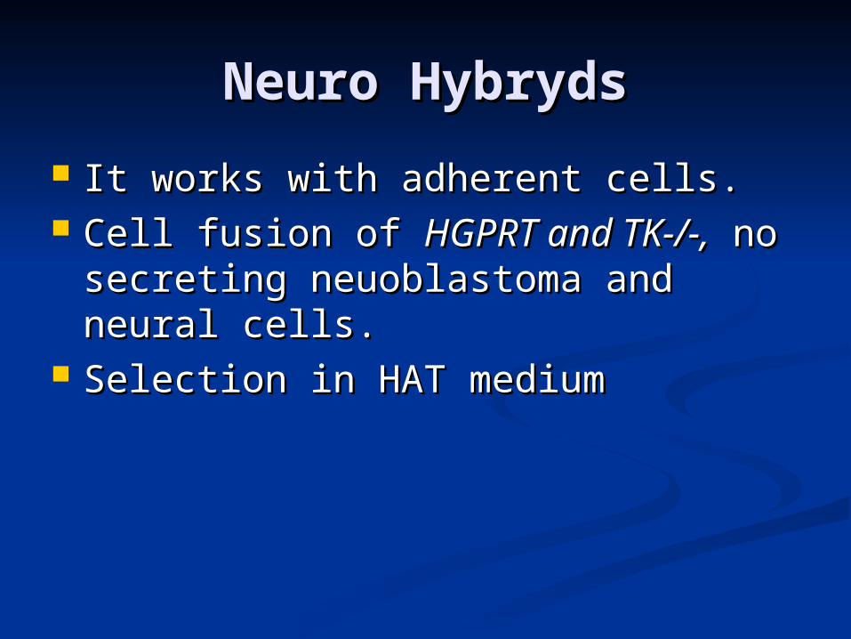
Neuro HybrydsNeuro Hybryds
It works with adherent cells.It works with adherent cells. Cell fusion of Cell fusion of HGPRTHGPRT and TK and TK-/--/-,, no no
secreting neuoblastomasecreting neuoblastoma and and neural neural cells.cells.
Selection in HAT mediumSelection in HAT medium

Cell linesCell lines
Adherent (WRL-68, Adherent (WRL-68, HepG2, HeLa etc.)HepG2, HeLa etc.)
Suspension Suspension (Jurkat)(Jurkat)
Cells from ATCC Cells from ATCC and ETCCand ETCC
JurkatWRL-68
HeLa HepG2

Online Order of Cell Online Order of Cell LinesLines
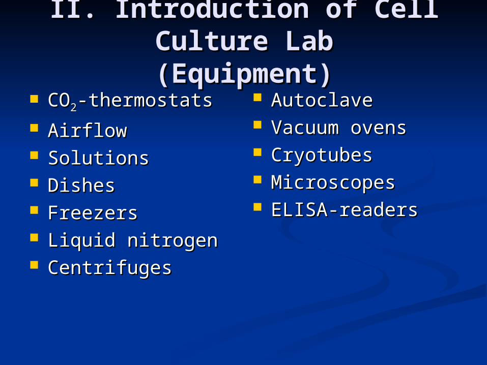
II. Introduction of Cell II. Introduction of Cell Culture LabCulture Lab(E(Equipmentquipment))
COCO22-thermostats-thermostats AirflowAirflow SolutionsSolutions DishesDishes FreezersFreezers Liquid nitrogen Liquid nitrogen CentrifugesCentrifuges
AutoclaveAutoclave Vacuum ovensVacuum ovens Cryotubes Cryotubes Microscopes Microscopes ELISA-readersELISA-readers

COCO22 Incubators Incubators
Water Jacketed COWater Jacketed CO22 incubatorincubator
3 Gas/CO3 Gas/CO22 Incubator Incubator with RH Control with RH Control Precise control of Precise control of
Oxygen levels Oxygen levels combined with COcombined with CO22, N, N22 and RH ensure and RH ensure accurate conditions accurate conditions for applications such for applications such as, hypoxic cell studies as, hypoxic cell studies and cancer research.and cancer research.

Laminar Flow BoxLaminar Flow Box
HEPA filter rated HEPA filter rated at 99.99% efficient at 99.99% efficient for 0.3 micron for 0.3 micron particulates. The particulates. The HEPA filtered air is HEPA filtered air is then directed then directed verticvertically across ally across the work surface. the work surface.
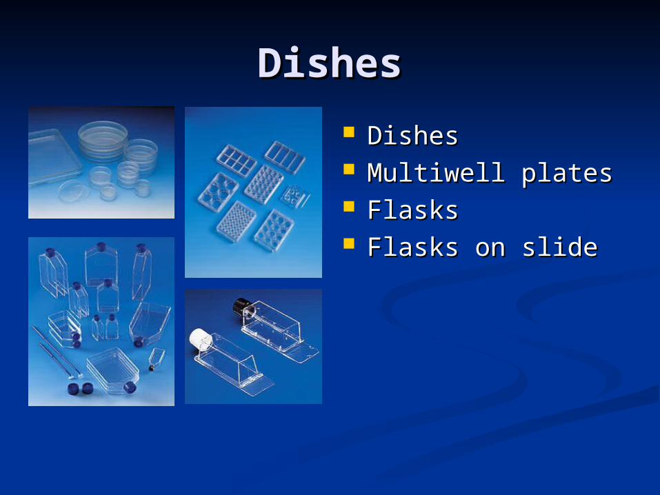
DishesDishes
DishesDishes Multiwell platesMultiwell plates FlasksFlasks Flasks on slideFlasks on slide

FreezersFreezers

CentrifugesCentrifuges

AutoclavesAutoclaves

Vacuum OvensVacuum Ovens

MicroscopesMicroscopes

ELISA readersELISA readers

FACSFACS

II. Introduction of Cell II. Introduction of Cell Culture LabCulture Lab(Culture) (Culture)
Growth of the cells in adequatGrowth of the cells in adequatee media media with serumwith serum (FCS(FCS/FBS/FBS) and antibiotics ) and antibiotics and and antimycotics (chemically defined serum-antimycotics (chemically defined serum-free media)free media)
Environment:Environment: Temperature: 37°CTemperature: 37°C (34 °C, 41 °C) (34 °C, 41 °C) HighHigh humidityhumidity 5% CO5% CO22
Split: Trypsin-EDTASplit: Trypsin-EDTA Count of Cells (Thrypan Blue)Count of Cells (Thrypan Blue)
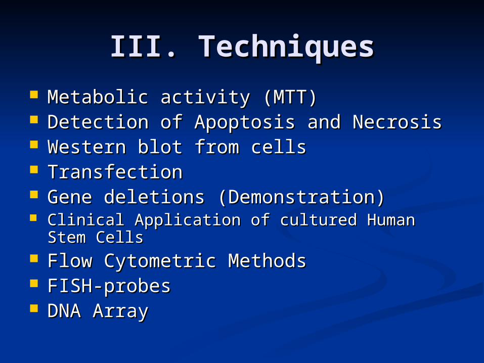
III. TechniquesIII. Techniques Metabolic activity (MTT)Metabolic activity (MTT) Detection of Apoptosis and NecrosisDetection of Apoptosis and Necrosis Western blot from cells Western blot from cells TransfectionTransfection Gene deletions (Demonstration)Gene deletions (Demonstration) Clinical Application of cultured Human Stem Clinical Application of cultured Human Stem
CellsCells Flow Cytometric Methods Flow Cytometric Methods FISH-probes FISH-probes DNA ArrayDNA Array

Metabolic activityMetabolic activity(MTT, viability assay)(MTT, viability assay)
Seed the cellsSeed the cells into 96-well plates at a starting into 96-well plates at a starting density of 10 cell/well and culture overnight in density of 10 cell/well and culture overnight in humidified 5 % COhumidified 5 % CO22 atmosphere at 37 °C. atmosphere at 37 °C.
Treat the cellsTreat the cells modifying the their viability the modifying the their viability the following day.following day.
Remove mediumRemove medium from the wells containing from the wells containing 0,5% 0,5% water suluble mitocondrial dye, water suluble mitocondrial dye, (3-(4,5-(3-(4,5-dimethylthiazol-2-yl]-2,5-diphenyl-tetrazolium dimethylthiazol-2-yl]-2,5-diphenyl-tetrazolium bromide (bromide (MTTMTT+)+)
IncubateIncubate 3 hours and 3 hours and solubilizesolubilize the water insoluble the water insoluble blue formasan dye by 10% SDS in 10mM HClblue formasan dye by 10% SDS in 10mM HCl
DetermineDetermine the optical density by an ELISA reder the optical density by an ELISA reder at 550 nm wavelengthat 550 nm wavelength
4
0
20
40
60
80
100
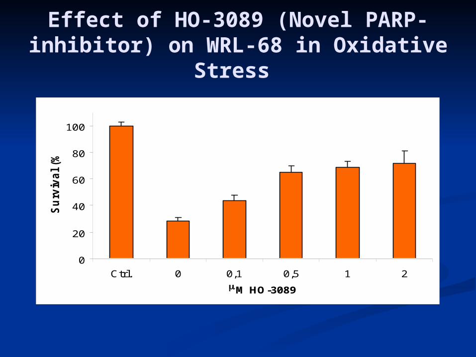
Ctrl. 0 0,1 0,5 1 2
M HO-3089
Su
rviv
al
(%)
Effect of HO-3089 (Novel PARP-inhibitor) on WRL-68 in Oxidative
Stress

Detection of Apoptosis and Detection of Apoptosis and NecrosisNecrosis
Activity of CaspaseActivity of Caspase 3 3 and Caspase 8and Caspase 8 Release of Cytochrome c and AIFRelease of Cytochrome c and AIF Fluorescence dyesFluorescence dyes
Hoechst 33342Hoechst 33342 Annexin VAnnexin V PropidPropidiium iodideum iodide RhodamineRhodamine
DNA LadderingDNA Laddering Induction and protection Induction and protection PARP PARP

Apoptosis signallingApoptosis signalling

Activation and inhibition of Activation and inhibition of ApoptosisApoptosis
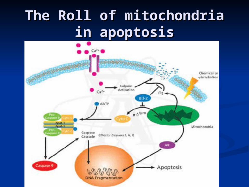
The Roll of mitochondria in The Roll of mitochondria in apoptosisapoptosis

Caspase CascadeCaspase Cascade

Fluorescent dyesFluorescent dyes I. I. Hoechst 33342:blueHoechst 33342:blue
Selective nuclear dyeSelective nuclear dye Chromatin Chromatin
condensation, condensation, fragnentationfragnentation
Rhodamine 110: Rhodamine 110: greengreen
Bis-L-asparic acide Bis-L-asparic acide amide (substrate by amide (substrate by caspase 3): greencaspase 3): green
TMRE: polarization TMRE: polarization of mitochondria: redof mitochondria: red

Fluorescent dyesFluorescent dyes II. II.
Propidium iodidePropidium iodide: : Late-stage Late-stage apoptotic and apoptotic and necrotic cells: necrotic cells: redred
YO-PRO-1YO-PRO-1: Viable : Viable cell nuclei cell nuclei greengreen
Annexin VAnnexin V: early-: early-stage apoptotic stage apoptotic cells: cells: greengreen

To investigate the DNA To investigate the DNA fragmentation, the fragmentation, the extracted DNA has to extracted DNA has to run on 1,5% agarose gel.run on 1,5% agarose gel.
DNA fragments show DNA fragments show ‘ladder-pattern’.‘ladder-pattern’.
DNA LadderingDNA Laddering

DNA LadderingDNA Laddering

Detection of Apoptosis and Detection of Apoptosis and NecrosisNecrosis
Activity of CaspaseActivity of Caspase 3 3 and Caspase 8and Caspase 8 Release of Cytochrome c and AIFRelease of Cytochrome c and AIF Fluorescence dyesFluorescence dyes
Hoechst 33342Hoechst 33342 Annexin VAnnexin V PropidPropidiium iodideum iodide RhodamineRhodamine
DNA LadderingDNA Laddering Induction and protection Induction and protection PARP PARP

Induction and Protection of Induction and Protection of ApoptosisApoptosis
Induction: Induction: Hydrogen peroxideHydrogen peroxide EtoposideEtoposide Death domainDeath domains: s: TNFTNF, , FASFAS, TRAIL, TRAIL BADBAD
Protection:Protection: BCL-2 familyBCL-2 family IAPIAP Inhibition of PARPInhibition of PARP HSP27,70,90HSP27,70,90

PARP(poly-ADP-rybose-PARP(poly-ADP-rybose-polymerase)polymerase)
Nuclear enzymeNuclear enzyme Structure of PARPStructure of PARP 1st activator of PARP are ssDNA-1st activator of PARP are ssDNA-
breaksbreaks The roll of PARP in necrosis and The roll of PARP in necrosis and
apoptosis or repair-mechanismapoptosis or repair-mechanism The roll of PARGThe roll of PARG

-R-P-P-R-R-P-P-R-R-P-P-R-R-P-P-R
Ad
PARP Glu
AdN
CONH2
Ad
R-P-P-R-R-P-P-R
Ad Ad
+
Nic
Nic-R-P-P-R
Ad
(NAD+)
Reaction catalyzed by
PARP

NF-B/I-B
NF-B
módosult génexpresszió
MAP-kináz kaszkád
fokozott tirozinfoszforiláció
glutation /redox státusz
proteinfoszfatáz
gátlás
ROS
PARP-gátlók
PARP
(ADP-R)n
NAD+
NicA
ATP
CisplatinTaxol
ROS
Lipid peroxidáció
Protein oxidáció PARP-gátlók

III. TechniquesIII. Techniques Metabolic activity (MTT)Metabolic activity (MTT) Detection of Apoptosis and NecrosisDetection of Apoptosis and Necrosis Western blot from cells Western blot from cells TransfectionTransfection Gene deletions (Demonstration)Gene deletions (Demonstration) Clinical Application of cultured Human Clinical Application of cultured Human
Stem CellsStem Cells FISH-probesFISH-probes Flow Cytometric Methods Flow Cytometric Methods DNA ArrayDNA Array

TransfectionTransfection I. I.
Expression vector Expression vector systemssystems pcDNApcDNA pEGFPpEGFP
pEGFP with NLS
pEGFP without NLS

TransfectionTransfection II. II.
RNAiRNAisiRNAsiRNAstRNA or Dicer RNAistRNA or Dicer RNAishRNA Using vectors for shRNA Using vectors for RNAi analysis RNAi analysis
siRNA cassettesiRNA cassette

Proposed mechanism for Proposed mechanism for how siRNA workshow siRNA works
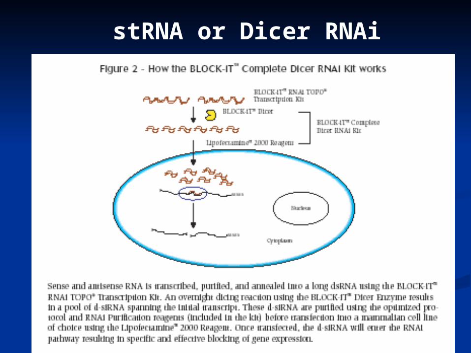
stRNA or Dicer RNAi

Gene Gene deletiondeletion
(Demonstratio(Demonstration)n)

Clinical Application of Clinical Application of cultured Human Stem Cellscultured Human Stem Cells
Not only can human embryonic Not only can human embryonic stem cells be cultured in the stem cells be cultured in the laboratory.laboratory.
But cells may be manipulated to But cells may be manipulated to produce cultures and produce cultures and Characteristics of particular tissue.Characteristics of particular tissue.
Possibility by damage and ageing Possibility by damage and ageing (Parkinson’s disease, diabetes)(Parkinson’s disease, diabetes)

Epithelial Stem Cell Epithelial Stem Cell identification and isolationidentification and isolation
First methods involved in the First methods involved in the separation of an epithelial cell type separation of an epithelial cell type from other cells will be examined, from other cells will be examined, followed by ways in which the followed by ways in which the proliferative capacity of such a cell type proliferative capacity of such a cell type can be assessed.can be assessed.
Secondly, methods used for the Secondly, methods used for the maintenance of primery stem cells in maintenance of primery stem cells in culture and ways of caracterizing stem culture and ways of caracterizing stem cells using immunocytochemistry will cells using immunocytochemistry will be described.be described.

FISH FISH (Fluorescence in situ (Fluorescence in situ
Hybridization)Hybridization) Application of FISH-probes Application of FISH-probes
Prenatal, Postnatal and Preimplantation Prenatal, Postnatal and Preimplantation GeneticsGenetics
Oncology, Cytology & Pathology Oncology, Cytology & Pathology Hematological Cancer Hematological Cancer Etc. Etc.
Equipments:Equipments: Fluorescence MicroscopeFluorescence Microscope Dye adequat filter setsDye adequat filter sets Sample and Reference DNASample and Reference DNA

Detection of Bladder Detection of Bladder CancerCancer
The probe was The probe was designed to detect designed to detect aneuploidy for aneuploidy for chromosomes 3, 7, 17 chromosomes 3, 7, 17 and loss of the 9p21 and loss of the 9p21 locus via fluorescence locus via fluorescence in situin situ hybridization hybridization (FISH) in urine (FISH) in urine specimens from specimens from subjects with subjects with transitional cell transitional cell carcinoma of the carcinoma of the bladder. bladder.
two copies of chromosome 3 (red), four copies of chromosome 7 (green), five copies of chromosome 17 (aqua) and one copy of p16 gene (gold)
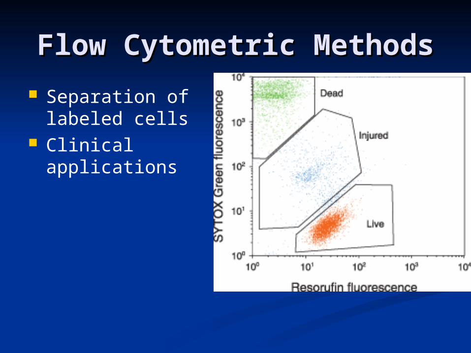
Flow Cytometric MethodsFlow Cytometric Methods
Separation of labeled cells
Clinical applications

DNA Array techniqueDNA Array technique
Mr. Péter JakusMr. Péter Jakus

Cell suspension by Cell suspension by NMRNMR
Dr. Zoltán BerenteDr. Zoltán Berente