Embed Size (px)
Citation preview

SEMESTER COURSE PLAN
(RPS)
Course:
HISTOLOGY
Coordinator of Course Convenor Team
Dra. Sri Wahyuni, M.Kes
BIOLOGY EDUCATION
FACULTY OF TEACHER TRAINING AND EDUCATION
UNIVERSITY OF MUHAMMADIYAH MALANG
2017
BIOLOGY EDUCATION FTTE
Document : RPS (Semester Course Plan)
Course Name : HISTOLOGY
Unit of Credit : 3 Credits
Coordinator of Course Convenor Team : Dra. Sri Wahyuni, M. Kes
Coordinator Course Cluster : Dr. Rr. Eko Susetyarini, M. Si
Team Teaching /sharing MK/Team of LS : 1. Dra. Sri Wahyuni, M. Kes
2. Drs. Muizzudin, M. Kes
3. Dr. Nurul Mahmudati, M. Kes
4. Dr. Rr. Eko Susetyarini, M. Si
5. Drs. Nurwidodo, M. Kes
Published by: Biology Education, 2017
BIOLOGY EDUCATION FTTE

CONTENTS
Page
Cover 1
Managing Editor 2
Content 3
Learning Objectives 4
Learning Objective Map 5
Semester Course Plan 6

SEMESTER COURSE PLAN
BIOLOGY EDUCATION3a1
FACULTY OF TEACHER TRAINING AND EDUCATION UNIVERSITY OF MUHAMMADIYAH
MALANG
COURSE NAME CODE Course Cluster UNIT OF CREDIT
(UoC)
SEMESTER Date of
Compilation HISTOLOGY3a2
BIO 033309 3a3
Course of Expertise
in Biology
3 UoC 3a5
Theory = 2 UoC
Practice = 1 UoC
4 3a4
December 1,
2016
Learning Objectives
(CP)
Course Outline Developer Coordinator of Course
Cluster
Head of Biology Education
Program
Dra. Sri Wahyuni, M.Kes 3
a6
Ttd
Dr. Ainur Rofieq, M. M.Kes
Ttd
Dr. Yuni Pantiwanti, MM, M.Pd
3b
(Attachment 2)
Learning Objectives (Code S, KU, KK, P)
S8 Intergrating value, norm, and ethic in academic
S9 Showing responsibility on the job of expertise independently.
KU1 Having ability to apply logical, critical, systematic, and innovative in the context of developing or
implementing science and technology based on the field of expertise.
KU2 Showing independent, qualified, and measurable work.
KK3 Having the ability to apply biological concepts and pedagogical technology in term of planning,
implementing, and evaluating the teaching and learning process by considering on Science and
Technology (IPTEKS) based on the issues occur at school (classroom, laboratory).
KK8 Having the ability to implement biological concepts and pedagogical technology using the
development of Science and Technology to create teaching and learning products in support of the
implementation of teaching and learning in biology.
P1 Having the ability to master theoretical concepts of Science and Technology in biology also its
application into teaching and learning process.
P8 Having the ability to master factual knowledge about function and usage of technology,

particularly information technology and communication that is relevant to the development of
educational quality.
Learning Objectives-Course (Code M)
M1 Having the ability to identify main tissue of organ formulation and organ system of body with
discipline and responsibility (P1, S9)
M2 Having the ability to create modes of tissue based on norms and ethics in academic. (S8, KU1,
KK3, P1, P8)
M3 Having the ability to analyse histology-based research with discipline and responsibility
considering on norms and ethics in academic. (S8, S9, KU1,KU2, KK3, KK8, P1, )
SUB-CPMK (Code L)
L1 Explaining the scope of Histology, Tool, and Methods of Leaning Histology (M1,M2)
L2 Explaining cell structure and methods to create histology microscope object tool (M1,M2)
L3 Identifying Epithel and Gland Tissue (M1)
L4 Identifying General Connective Tissue (4a) (M1)
L5 Identifying Special Connective Tissue (4b) (M1)
L6 Identifying Cantilever Tissue (4c) (M1)
L7 Identifying Muscular Tissue (M1)
L8 Identifying Nervous Tissue (M1)
L9 Describing organ formulation tissue that forms an orgam system (M1, M2)
L10 Creating Model of Tissue (M2)
L11 Analysing histology-based research article (M3)
Short Description
about the Course
DESCRIPTION
This course presents material and basic concept also the theory of structure and function of four main body tissues
(epithel tissue, connective tissue, muscular tissue, and nervous tissue) and the concept of component in organ
formulation tissue that forms digestive system, respiration, excretion, reproduction, circulation, coordination and
endocrine as well as their use in real life.
Learning Material/ Topics

Topic of Discussion Biology Functional Structure, Animal Structure
Sub Topic of Analysis
a. Concept in Histology,
b. Cell Structure
c. Histotechnique
d. Structure of Epithel Tissue
e. Structure of Connective Tissue
f. Structure of Muscular Tissue
g. Structure of Nervous Tissue
h. Histology of Organ System (Organology)
i. Method in Histology
j. Research in Histology
Topic of
Discussion
I. Scope, Tool, and Method to Learn Histology
1 Definition of Histology
2 Scope of Histology
3 Tool and Method to Learn Histology
II. Cell Structure and Method to Create Object of Microscope Tool of Histology
1. Cell Ultrastructure, organella cell
2. Kinds of Cell to Form Tissue
3. Method in Histology
4. Method to Create Microscope Tool of Histology
5. Method of Colouring
III. Epithel and Gland Tissue
1. Definition
2. Characteristic of Epithel Tissue
3. Closed epithel tissue
4. Gland Epithel Tissue
5. Exocrine gland

6. Endocrine Tissue
7. Kinds of epithel tissue
8. Function of epithel tissue
IV. General Connective Tissue
1. Definition
2. Characteristic of Connective Tissue
3. Classification of Connective Tissue
4. Embrional Connective Tissue
5. Adult Connective Tissue
6. Dense Solid Connective Tissue
7. Irregular Solid Connective Tissue
8. Loose Connective Tissue
V. Special Connecyive Tissue
1. Blood Tissue
2. Bone Marrow
3. Fat Tissue
VI. Cantilever Tissue
1. Cartilage Tissue
2. Bone Tissue
VII. Muscular Tissue
1 Smooth Muscle
2 Striated Muscle
3 Cardiac Muscle
VIII. Nervous Tissue
1. Central Nerve
2. Peripheral Nerve
IX. Organ and Organ System
1.Relation between organ tissue and organ system
2. Histology of respiration organ
3. Circulation and excretion organ, reproduction, digestion, endocrine)
X. Images of Tissue
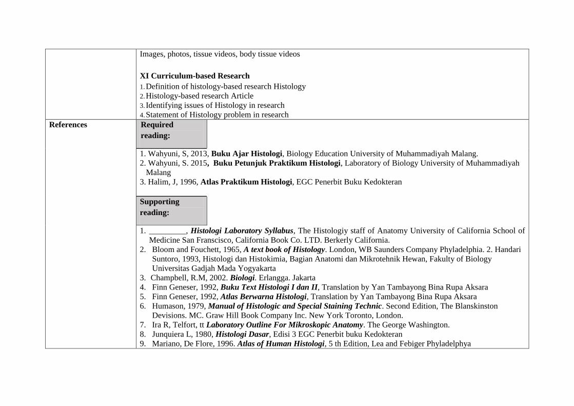
Images, photos, tissue videos, body tissue videos
XI Curriculum-based Research
1. Definition of histology-based research Histology
2. Histology-based research Article
3. Identifying issues of Histology in research
4. Statement of Histology problem in research
References Required
reading:
1. Wahyuni, S, 2013, Buku Ajar Histologi, Biology Education University of Muhammadiyah Malang.
2. Wahyuni, S. 2015, Buku Petunjuk Praktikum Histologi, Laboratory of Biology University of Muhammadiyah
Malang
3. Halim, J, 1996, Atlas Praktikum Histologi, EGC Penerbit Buku Kedokteran
Supporting
reading:
1. _________, Histologi Laboratory Syllabus, The Histologiy staff of Anatomy University of California School of
Medicine San Franscisco, California Book Co. LTD. Berkerly California.
2. Bloom and Fouchett, 1965, A text book of Histology. London, WB Saunders Company Phyladelphia. 2. Handari
Suntoro, 1993, Histologi dan Histokimia, Bagian Anatomi dan Mikrotehnik Hewan, Fakulty of Biology
Universitas Gadjah Mada Yogyakarta
3. Champbell, R.M, 2002. Biologi. Erlangga. Jakarta
4. Finn Geneser, 1992, Buku Text Histologi I dan II, Translation by Yan Tambayong Bina Rupa Aksara
5. Finn Geneser, 1992, Atlas Berwarna Histologi, Translation by Yan Tambayong Bina Rupa Aksara
6. Humason, 1979, Manual of Histologic and Special Staining Technic. Second Edition, The Blanskinston
Devisions. MC. Graw Hill Book Company Inc. New York Toronto, London.
7. Ira R, Telfort, tt Laboratory Outline For Mikroskopic Anatomy. The George Washington.
8. Junquiera L, 1980, Histologi Dasar, Edisi 3 EGC Penerbit buku Kedokteran
9. Mariano, De Flore, 1996. Atlas of Human Histologi, 5 th Edition, Lea and Febiger Phyladelphya
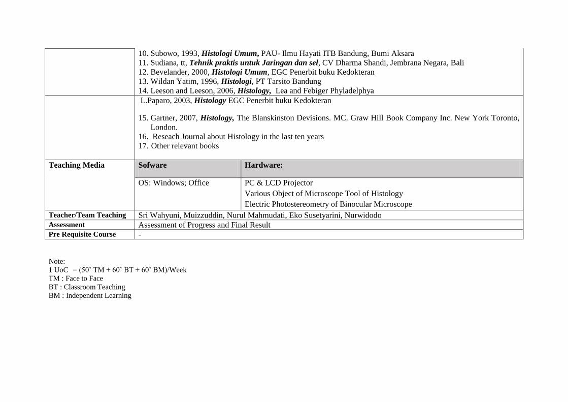
10. Subowo, 1993, Histologi Umum, PAU- Ilmu Hayati ITB Bandung, Bumi Aksara
11. Sudiana, tt, Tehnik praktis untuk Jaringan dan sel, CV Dharma Shandi, Jembrana Negara, Bali
12. Bevelander, 2000, Histologi Umum, EGC Penerbit buku Kedokteran
13. Wildan Yatim, 1996, Histologi, PT Tarsito Bandung
14. Leeson and Leeson, 2006, Histology, Lea and Febiger Phyladelphya
L.Paparo, 2003, Histology EGC Penerbit buku Kedokteran
15. Gartner, 2007, Histology, The Blanskinston Devisions. MC. Graw Hill Book Company Inc. New York Toronto,
London.
16. Reseach Journal about Histology in the last ten years
17. Other relevant books
Teaching Media Sofware Hardware:
OS: Windows; Office PC & LCD Projector
Various Object of Microscope Tool of Histology
Electric Photostereometry of Binocular Microscope
Teacher/Team Teaching Sri Wahyuni, Muizzuddin, Nurul Mahmudati, Eko Susetyarini, Nurwidodo
Assessment Assessment of Progress and Final Result
Pre Requisite Course -
Note:
1 UoC = (50’ TM + 60’ BT + 60’ BM)/Week
TM : Face to Face
BT : Classroom Teaching
BM : Independent Learning

MAP OF LEARNING OBJECTIVE IN HISTOLOGY
KA/ Sub CPMK (2)
Explaining cell structure and creation of histology laboratory for microscope object
(Week ke 3-4)
KA/ Sub CPMK (1) Explaining scope of Histology, Tool, and Way to Learn Histology
(Week 1-2)
KA/ Sub CPMK (7) Describing tissue of organ formulation that forms organ system
(respiration, circulation and excretion, reproduction, digestion, and
endocrine)
(Week ke 13)
KA/ Sub CPMK (3) Identifying epithel and
gland tissue (Week 5-6) W. 5
KA/Sub CPMK (5) Identifying Muscular Tissue
(Week 10)
KA/Sub CPMK (4) Identifying Connective
tissue (Week 7-9)
KA/ Sub CPMK (6) Identifying Nervous
Tissue
(Week 11-12)
KA/ Sub CPMK (8)
Creating Model of Tissue
(Week 14)
Final Test (Week 16)
Mid Test (Week 8)
KA// Sub CPMK (9)
Conducting analysis on histology-based research
article (Week 15)
Learning Objectives of Histology
Identifying main tissue of formulation organ and body system organ,
(M1) creating model of tissue based on ethic and academic norm (M2)
also histology-based research with discipline and responsibility (M3)
Diagram of analysis result on Histology learning

Wee
k
KA/SubCPMK
(as the
expected
outcome) 3c
Indicator KA
(3c)
BK/Core Material
3d
Method of
Teaching
(Estimated
Time) 3e, 3f
Assessment
Reference
3i Form 3g Criteria/&indicator
3g
Weight
3h
1 2 3 4 5 6 7 8 9
1 Learning Contract (Explaining Course Outline and Agreement with Students)
2 Explaining
Sope, Tool, and
Ways of
Leaning
Histology
1. Explaining definition of histologi
2. Explaining scope of histology
3. Explaining some tools and ways of
learning histology
I. Scope, Tool, and
Method of
Learning Histology
1. Definition of
Histology
2. Scope of
Histology
3. Tool and Ways of
Learning
Histology
Lecture,
discussion, Q&
A, demo, and
assignment
[TM:1x(2x50”)]
[BT :1x(2x60”)]
[BM:1x(2x60”)]
Written test
(Essay)*
Assignment
1
Accuracy and depth in
explaining scope, tool,
and ways of learning
Histology
Accuracy, depth, and
order of finishing the
assignment
5%
Reference
1-15
3&4 Explaining cell
structure and
method of
creating
laboratory
object for
microscopic
observation of
Histology
1. Explaining ultrastructure &
organella cell
2. Explaining kinds of cell to
formulate tissue
3. Explaining some Methods in
Histology
4. Explaining methods of creating
laboratory object for microscopic
observation
5. Explaining methods of colouring
II. Cell Structure and
Methods of Creating
laboratory object for
microscopic
observation in
Histology
1. Ultrastructure
cell, irri ogy
cel
2. Kinds of Cells
that Create Tissue
Lecture, Think
Pair Share,
Team Work (10
students),
assignment
[TM:2x(2x50”)]
[BT :2x(2x60”)]
[BM:2x(2x60”)]
Written test
(Essay)*
Assignment
2
Accuracy and depth of
explaining structure of
cell and method of
creating laboratory
object for microscopic
observation in
histology
Accuracy, depth, and
order of finishing the
assignment
5%
Reference
1-15

Wee
k
KA/SubCPMK
(as the
expected
outcome) 3c
Indicator KA
(3c)
BK/Core Material
3d
Method of
Teaching
(Estimated
Time) 3e, 3f
Assessment
Reference
3i Form 3g Criteria/&indicator
3g
Weight
3h
1 2 3 4 5 6 7 8 9
3. Methods in
Histology
4. Methods of
Creating
laboratory object
for microscopic
observation
5. Methods of
Colouring
5 Identifying
Epithel and
Gland Tissue
1. Explaining kinds of epithel tissue
2. Explaining kinds of epithel gland
3. Comparing epithel and gland tissue
4. Identifying kind of glands
5. Identifying structure of epithel
tissue
III Epithel and Gland
Tissue
1. Definition
2. Characteristic of
Epithel Tissue
3. Closed epithel
tissue
4. Gland epithel
tissue
5. Functions of
epithel tissue
Book analysis,
lecture, Q & A,
demo,
presentation,
assignment
[TM:1x(2x50”)]
[BT :1x(2x60”)]
BM :1x(2x60”)]
Laboratory
Work*
Written test
(Essay)*
Assignment
3
Accuracy and depth in
identifying Epithel and
Gland Tissue
Accuracy,
completeness, and
order of finishing the
assignment
5%
Reference
1-15
6 Identifying
general
connective
tissue
1. Explaining definition of connective
tissue
2. Identifying characteristics of
general connective tissue
3. Classifying connective tissue
IV. General Connective
Tissue
1. Definition
2. Characteristics of
Connective Tissue
3. Classification of
Connective Tissue
4. Connective Embrional
Jigsaw
[TM:1x(2x50”)]
[BT :1x(2x60”)]
BM :1x(2x60”)]
Praktikum*
Written Test
(Essay)
Assignmen
t 4a
Accuracy and depth in
identifying general
connective tissue
Accuracy,
completeness, and
order of finishing the
assignment
Reference
1-15

Wee
k
KA/SubCPMK
(as the
expected
outcome) 3c
Indicator KA
(3c)
BK/Core Material
3d
Method of
Teaching
(Estimated
Time) 3e, 3f
Assessment
Reference
3i Form 3g Criteria/&indicator
3g
Weight
3h
1 2 3 4 5 6 7 8 9 Tissue
5. Adult Connective
Tissue
6. Regular Solid
Connective Tissue
7. Irregular Solid
Connective Tissue
8. Loose Connective
Tissue
7. Identifying
Special
Connective
Tissue
1. Explaining kinds of Special
Connective Tissue
2. Identifying blood tissue and bone
marrow
3. Identifying fat tissue.
Lymphoreticular and pigment.
V. Special Connective
Tissue
1. Blood Tissue
2. Bone Marrow
3. Fat Tissue
4. Lymphoreticular
tissue
5. Pigment Tissue
Brainstorming to
Work in a team,
Simulation,
Assignment
[TM:1x(2x50”)]
[BT :1x(2x60”)]
BM :1x(2x60”)]
Laboratory
Work*
Written
Test
(Essay)*
Assignmen
t 4b
Accuracy and depth in
identifying Special
Connective Tissue
Accuracy,
completeness, and
order of finishing the
assignment
Reference
1-15
8 Mid Test: Evaluation in Mid Semester (Formative Evaluation-it is aimed at giving rooms of improvement to the larning process
considering on the assessment conducted) 15%
9 Identifying
Cantilever
Tissue
1. Explaining kinds of cantilever tissue
2. Identifying cartilage tissue
3. Identifying bone tissue
4. Comparing cartilage of hyaline,
elastic, and fibrosa
VI. Cantilever
Tissue
1. Cartilage
Tissue
2. Bone Tissue
Demo,
Simulation,
Lecture,
Assignment
[TM:1x(2x50”)][
BT :1x(2x60”)]
BM :1x(2x60”)]
Lab Work*
Written
Test
(Essay)
Assignment
4c
Accuracy and depth in
identifying Cantilever
Tissue
Accuracy,
completeness, and
order of finishing the
assignment
7.5%
Reference
1-15

Wee
k
KA/SubCPMK
(as the
expected
outcome) 3c
Indicator KA
(3c)
BK/Core Material
3d
Method of
Teaching
(Estimated
Time) 3e, 3f
Assessment
Reference
3i Form 3g Criteria/&indicator
3g
Weight
3h
1 2 3 4 5 6 7 8 9
10 Identifying
Muscular
Tissue
1. Identifying muscular tissue
2. Comparing smooth muscle, striated
muscle, and cardiac
VII. Muscular
Tissue
1. Smooth
Muscle
2. Striated
Muscle
3. Cardiac
Muscle
Discussion,
Presentation,
Lecture,
[TM: 1x(2x50”)]
Lab Work*
Written
Test
(Essay)
Assignment
5b
Accuracy and depth in
identifying Muscular
Tissue
Accuracy,
completeness, and
order of finishing the
assignment
Reference
1-15
11-
12
Identifying
Nervous Tissue
1. Identifying kinds of nervous tissue
2. Identifying central nervous tissue
3. Identifying peripheral nerve tissue
VIII. Nervous Tissue
1. Central Nerve
2. Peripheral
Nerve
Discussion,
Presentation,
Lecture, Demo,
Simulation,
Assignment
[TM:2x(2x50”)]
[BT :2x(2x60”)]
[BM:2x(2x60”)]
Lab Work*
Written
Test
(Essay)
Assignme
nt 5b
Accuracy and depth in
identifying Nervous
Tissue
Accuracy,
completeness, and
order of finishing the
assignment
.
7.5 %
Reference
1-15
13 Describing
organ tissue
that forms orga
system
1. Describing the relation of organ and
organ system
.2. Describing one of body organ system
(chosen)
IX. Organ and Organ
System
1. The relation of organ
tissue and organ
system
2. Histology of
respiration,
circulation,
extretion,
reproduction,
digestion, and
endocrine organ)
Lecture,
discussion,
Presentation
[TM:1x(2x50”)]
[BT :1x(2x60”)]
[BM:1x(2x60”)]
Laboratory
Work*
Written
Test
essay*
Accuracy and depth in
describing the chosen
organ system
Reference
1-15

Wee
k
KA/SubCPMK
(as the
expected
outcome) 3c
Indicator KA
(3c)
BK/Core Material
3d
Method of
Teaching
(Estimated
Time) 3e, 3f
Assessment
Reference
3i Form 3g Criteria/&indicator
3g
Weight
3h
1 2 3 4 5 6 7 8 9
14 Creating Model
of Tissue
1. Creating Model of Tissue Images, Photos, Videos
of Tissue, Videos of
Body Tissue
Assignment/
Project
[TM:1x(2x50”)]
[BT :1x(2x60”)]
[BM:1x(2x60”)]
Non test
Product
Assessme
nt
Assignme
nt 6
Product Quality of
Tissue Model
10%
Reference
1-15
15 Analysing
histology-based
research article
1. Finding histology-based research
problem
2. Stating histology-based research
problem
Histology-based
research articles
Assignment,
Journal analysis,
Presentation
[TM:1x(2x50”)]
[BT :1x(2x60”)]
[BM:1x(2x60”)]
Presentati
on
Assignme
nt 7
Depth analysis of
research articles
5%
16 Final Semester Evaluation (It is aimed at knowing the expected learning objectives of students) 20%
Discipline and responsible during the lecture (Attendance & Performace during lecture)
Non Test
Observati
on
Dicsipline
Responsible 20%
notes:
1 credit = (50’ TM + 60’ BT + 60’ BM)/week BM = Independent Learning T = Theory (knowledge aspect)
TM = Face to face (Lecture) PS P = Practice (work skills)
BT = Structured Learning PL 1 credit of practice= (100’ TM + 70’BT& Assistenceh)/week

HISTOLOGY LABORATORY WORK PLAN
FORMAT OF LAB WORK PLAN
COURSE : Histology
SEMESTER : 4 Unit of Credit: 1 UoC (100 minutes), 70 minutes for assistance, consultation, and working on a report. Total
170 minutes
THE OBJECTIVES OF LAB WORKS: students are able to identify and describe tissue LAB WORK JOB DESCRIPTION:
a. Lab work object: animal/human tissue
b. Do’s and Don’ts: identifying and describing parts in detil and accurate from epithel, gland, general connective, special connective, cantilever, and muscular tissue
c. Method/procedure of lab work: Pengamatan dan projek, alat yang digunakan mikroskop, preparat. Procedure of lab work: take a microscope, set an object, observation, drawing, taking picture, taking notes, writing a report, consultation to an instructure/assistant
d. Description on lab work report: Lab work report is presented in a report format which describes about: the objectives of observation, name of object, types of colouring, hand drawing of observation result, caption of the images, pictures of observation with magnification in comparison to the pictures used in the literature and being discussed.
ASSESSMENT CRITERIA: a. Accuracy
b. Depth
c. Order d. Creativity
Assessment indicator is stated in the rubric

Me
etin
g
The expected
outcome of
expertise
Indicator
Core Material
(Topic of Discussion)
Learning
Method
Assessment
Main
Reference Form Criteria & Indicator Weigh
t
1 2 3 4 5 6 7 8 9
1 Contract of Lab Work & Rules
2 Identifying
Epithel tissue
1. Recognizing kinds of epithel
tissue using the light of
microscope
2. Skilfully identifying
structure of epithel tissue
I. Jaringan Epithel
-E. Squamous simplex
-E. Cuboid simplex
-E. Columnair simplek
-E. Trantitional
-E. Squamous complex
-E. Cuboid complex
-E. Pseudocomplek
Lab work
(TM:1x100”)
(A&PL: 1x70’)
Pretest
(Essay)
Observatio
n
Assignmen
t of writing
a report
Accuracy upon answering
pretest question
Skillful to recognize epithel
tissue
Accuracy, completeness,
depth, and order to write a
report
5%
Guidelines
s on
Histology
Lab Work
3 Identifying
gland tissue
1. Recognizing kinds of glands
using the light of microscope
2. Skillfully identifying glands
structures
II. Glands
-Endocrine & Exocrine
-Mucus, serus, seromucus
-merokrin, apokrin,
holokrin
-Unicellular &
multicellular
Lab work
(TM:1x100”)
(A&PL: 1x70’)
Pretest
(Essay)
Observatio
n
tugas
pembuatan
laporan
Accuracy upon answering
pretest question
Skillful to recognize and
identify glands
Accuracy, completeness,
depth, and order to write a
report
6%
Guidelines
s on
Histology
Lab Work
4 Identifying
general
connective
tissue
1. Recognizing kinds of
general connective tissue
using the light of
microscope
2. Skillfully identifying general
connective tissue
III.General Connective
Tissue
- Regular solid connective
tissue (transverse &
longitudinal tendon) sbt.
Elastic, sbt collagen
- Irregular solid
connective tissue, tunica
albugenia
- Loose connective tissue,
Lab work
(TM:1x100”)
(A&PL: 1x70’)
Pretest
(Essay)
Observatio
n
Assignmen
t Writing a
report
Accuracy upon answering
pretest question
Skillful to recognize and
identify tissue
Accuracy, completeness,
depth, and order to write a
report
6%
Guidelines
s on
Histology
Lab Work

Me
etin
g
The expected
outcome of
expertise
Indicator
Core Material
(Topic of Discussion)
Learning
Method
Assessment
Main
Reference Form Criteria & Indicator Weigh
t
1 2 3 4 5 6 7 8 9
subcutan,
- Embrional connective
tissue (mesenkim,
mukus)
5 Identifying
Special
Connective
Tissue
1. Recognizing kinds of special
connective tissue using the
light of microscope
2. Skillfully identifying special
connective tissue
IV. Special Connective
Tissue
- Lymphoreticular
- Fat tissue (signet ring
cell
- Pigmented connective
tissue (eyeball choroid)
- Blood, Blood vessel &
bone marrow
Lab work
(TM:1x100”)
(A&PL: 1x70’)
Pretest
(Essay)
Observatio
n
Assignmen
t Writing a
report
Accuracy upon answering
pretest question
Skillful to recognize and
identify special connective
tissue
Accuracy, completeness,
depth, and order to write a
report
6%
Guidelines
s on
Histology
Lab Work
6 Identifying
Cantilever
Tissue
1. Recognizing kinds of
cantilever tissue using the
light of microscope
2. Skillfully identifying
cantilever tissue
III. V. Cantilever Tissue
- Cartilage tissue
- (hyaline, elastic,
Fibrosa)
- Bone tissue (young
adult, primary
ossification, secondary)
Lab work
(TM:1x100”)
(A&PL: 1x70’)
Pretest
(Essay)
Observatio
n
Assignmen
t Writing a
report
Accuracy upon answering
pretest question
Skillful to recognize and
identify cantilever tissue
Accuracy, completeness,
depth, and order to write a
report
6%
Guidelines
s on
Histology
Lab Work

Me
etin
g
The expected
outcome of
expertise
Indicator
Core Material
(Topic of Discussion)
Learning
Method
Assessment
Main
Reference Form Criteria & Indicator Weigh
t
1 2 3 4 5 6 7 8 9
7 Identifying
Muscular
Tissue
1. Recognizing muscular tissue
using the light of
microscope
2. Skillfully identifying
muscular tissue
VI. Jaringan Otot
Smooth Muscle
Striated Muscle
Cardiac Muscle
Lab Work
(TM:1x100”)
(A&PL: 1x70’)
Pretest
(Essay)
Observatio
n
Assignmen
t Writing a
report
Accuracy upon answering
pretest question
Skillful to recognize and
identify muscular tissue
Accuracy, completeness,
depth, and order to write a
report
6%
Guidelines
s on
Histology
Lab Work
Me
etin
g
The expected
outcome of
expertise
Indicator Core Material
(Topic of Discussion)
Teaching Method Assessment Main
Reference Form Criteria &
Indicator
Weight
1 2 3 4 5 6 7 8 9
8. Identifying
Nervous
Tissue
1. Recognizing
nervous tissue using
the light of
microscope
2. Skillfully
identifying nervous
tissue
VII. Jaringan syaraf
- Central nerve, peripheral nerve
- serebrum serebellum, medulla
spinalis
- ganglion spinal, ganglio
otonom, nerve endings
Lab work
(TM:1x100”)
(A&PL: 1x70’)
Pretest
(Essay)
Observati
on
Assignme
nt Writing
a report
Accuracy upon
answering pretest
question
Skillful to
recognize and
identify nervous
tissue
Accuracy,
completeness,
depth, and order
to write a report
6%
Guidelines
s on
Histology
Lab Work
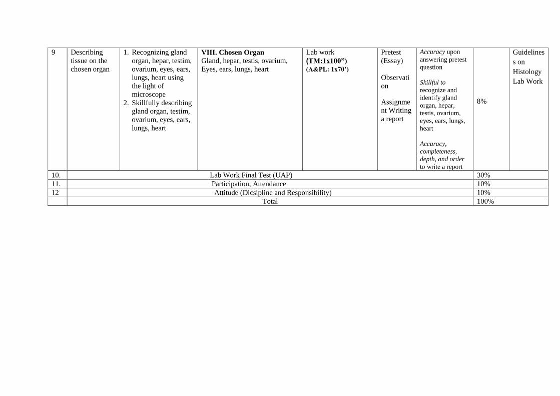
9 Describing
tissue on the
chosen organ
1. Recognizing gland
organ, hepar, testim,
ovarium, eyes, ears,
lungs, heart using
the light of
microscope
2. Skillfully describing
gland organ, testim,
ovarium, eyes, ears,
lungs, heart
VIII. Chosen Organ
Gland, hepar, testis, ovarium,
Eyes, ears, lungs, heart
Lab work
(TM:1x100”) (A&PL: 1x70’)
Pretest
(Essay)
Observati
on
Assignme
nt Writing
a report
Accuracy upon
answering pretest
question
Skillful to
recognize and
identify gland
organ, hepar,
testis, ovarium,
eyes, ears, lungs,
heart
Accuracy,
completeness,
depth, and order
to write a report
8%
Guidelines
s on
Histology
Lab Work
10. Lab Work Final Test (UAP) 30%
11. Participation, Attendance 10%
12 Attitude (Dicsipline and Responsibility) 10%
Total 100%