Embed Size (px)
Citation preview
Can J Gastroenterol Vol 23 No x Month 2009 1
Sellar inflammatory mass with inflammatory bowel disease
Hugh J Freeman MD, John Maguire MD
Departments of Medicine (Gastroenterology) and Anatomical Pathology (Neuropathology), University of British Columbia, Vancouver, British Columbia
Correspondence and reprints: Dr Hugh Freeman, Division of Gastroenterology, 2211 Wesbrook Mall, Vancouver, British Columbia V6T 1W5. Telephone 604-822-7216, fax 604-822-7236, e-mail [email protected]
Received for publication January 16, 2009. Accepted January 21, 2009
An inflammatory sellar mass or pseudotumour of the pitu-itary sella is very rare; however, a dramatic clinical
response to medical treatment with corticosteroids may occur (1). The differential diagnosis of inflammatory sellar lesions is difficult, relies primarily on histological analysis and, in addi-tion to infectious causes (eg, tuberculosis), includes other non-neoplastic disorders such as lymphocytic hypophysitis, granulomatous inflammation (ie, sarcoidosis) and Wegener’s granulomatosis (2). Clinical effects related to the sellar lesion may include either neurological or endocrinological features, or both. Interestingly, however, these non-neoplastic disorders may rarely be associated with intestinal disease and positive cytoplasmic-staining antineutrophil cytoplasmic antibody (c-ANCA) (eg, Wegener’s granulomatosis with intestinal involvement) (3-5).
The present report documents the unusual clinical simul-taneous occurrence of two apparently distinct clinicopatho-logical disorders – specifically, a sellar inflammatory mass associated with an intermittently symptomatic inflammatory process characteristic of Crohn’s disease involving the colon alone for more than a decade. Interestingly, while the two sites for this concomitant inflammatory process were quite separate, their relationship may be more than coincidental, with poten-tial therapeutic implications for the future management of inflammatory bowel disease.
CASE PRESENTATION
A 32-year-old First Nations man first developed waxing and waning headaches that coincided with intermittent diplo-
pia in 1997. An ophthalmologist diagnosed a right sixth cranial nerve paresis. In August 2000, his headaches progressively became more severe, and were associated with diplopia on right-sided gaze. He became photophobic, with nausea and vomiting. Computed tomography and magnetic resonance imaging scans at a community hospital showed an enhancing clivus lesion with increased tissue density in the sphenoid sinus. Although cultures were negative, ceftriaxone, flagyl {metronidaz-ole?} and gentamicin were administered for four weeks. His headache improved but never completely resolved. Another magnetic resonance imaging scan scan showed a persistent clivus lesion, but the sphenoid density resolved.
In January 2001, recurrent, localized, right frontal and retro-orbital headaches developed, with nausea, vomiting, photo-phobia and horizontal diplopia on rightward gaze. There were no other neurological symptoms or evidence of altered endo-crine function. Transphenoidal biopsy showed sphenoid sinus mucosa along with bone and soft tissue from the clivus. The sphenoid sinus biopsy revealed a polypoidal lesion with pre-dominantly chronic inflammation. The bone showed reactive changes, with areas of increased osteoclastic activity. Sinusoidal mucosa and fragments of lamellar bone were seen. The mucosal
brief communication
©2009 Pulsus Group Inc. All rights reserved
HJ Freeman, J Maguire. Sellar inflammatory mass with inflammatory bowel disease. Can J Gastroenterol 2009;23(x): xxx-xxx.
Inflammatory bowel disease may be associated with different intracra-nial disorders. An inflammatory sellar mass is very rare but includes a variety of noninfectious causes including lymphocytic hypophysitis, granulomatous inflammation and Wegener’s granulomatosis. A 32-year-old man was diagnosed as having an inflammatory sellar mass associated with an extensive colonic inflammatory process clinically characteristic of Crohn’s disease. The concurrent onset of these inflammatory disorders in distinctly separate sites may reflect their common embryological origin or represent an unusual form of meta-static Crohn’s disease. Further studies are needed to determine if less overt or focal sellar inflammatory processes occur in inflammatory bowel disease, particularly in Crohn’s disease because their occurrence may be critically relevant for long-term management.
Key Words: Crohn’s disease; First Nations; Granulomatous inflam-mation; Inflammatory bowel disease; Metastatic Crohn’s disease; Pituitary sella
2
1
Freeman and Maguire
Can J Gastroenterol Vol 23 No x Month 20092
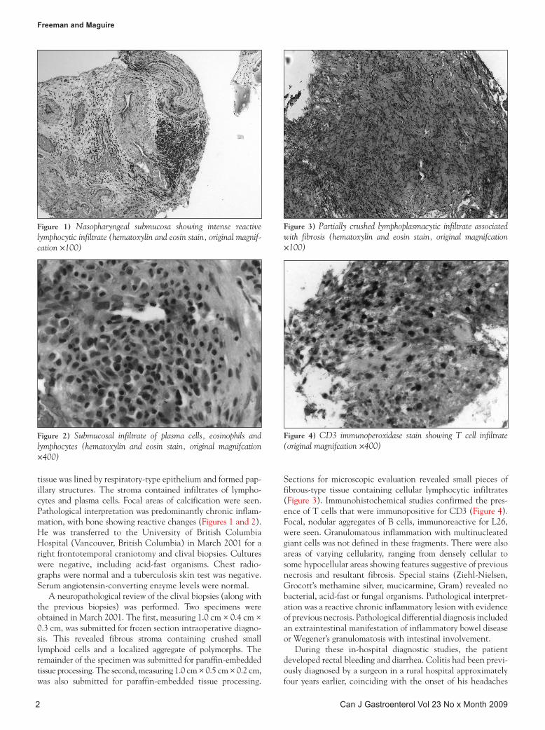
tissue was lined by respiratory-type epithelium and formed pap-illary structures. The stroma contained infiltrates of lympho-cytes and plasma cells. Focal areas of calcification were seen. Pathological interpretation was predominantly chronic inflam-mation, with bone showing reactive changes (Figures 1 and 2). He was transferred to the University of British Columbia Hospital (Vancouver, British Columbia) in March 2001 for a right frontotemporal craniotomy and clival biopsies. Cultures were negative, including acid-fast organisms. Chest radio-graphs were normal and a tuberculosis skin test was negative. Serum angiotensin-converting enzyme levels were normal.
A neuropathological review of the clival biopsies (along with the previous biopsies) was performed. Two specimens were obtained in March 2001. The first, measuring 1.0 cm × 0.4 cm × 0.3 cm, was submitted for frozen section intraoperative diagno-sis. This revealed fibrous stroma containing crushed small lymphoid cells and a localized aggregate of polymorphs. The remainder of the specimen was submitted for paraffin-embedded tissue processing. The second, measuring 1.0 cm × 0.5 cm × 0.2 cm, was also submitted for paraffin-embedded tissue processing.
Sections for microscopic evaluation revealed small pieces of fibrous-type tissue containing cellular lymphocytic infiltrates (Figure 3). Immunohistochemical studies confirmed the pres-ence of T cells that were immunopositive for CD3 (Figure 4). Focal, nodular aggregates of B cells, immunoreactive for L26, were seen. Granulomatous inflammation with multinucleated giant cells was not defined in these fragments. There were also areas of varying cellularity, ranging from densely cellular to some hypocellular areas showing features suggestive of previous necrosis and resultant fibrosis. Special stains (Ziehl-Nielsen, Grocott’s methamine silver, mucicarmine, Gram) revealed no bacterial, acid-fast or fungal organisms. Pathological interpret-ation was a reactive chronic inflammatory lesion with evidence of previous necrosis. Pathological differential diagnosis included an extraintestinal manifestation of inflammatory bowel disease or Wegener’s granulomatosis with intestinal involvement.
During these in-hospital diagnostic studies, the patient developed rectal bleeding and diarrhea. Colitis had been previ-ously diagnosed by a surgeon in a rural hospital approximately four years earlier, coinciding with the onset of his headaches
Figure 1) Nasopharyngeal submucosa showing intense reactive lymphocytic infiltrate (hematoxylin and eosin stain, original magnif-cation ×100)
Figure 3) Partially crushed lymphoplasmacytic infiltrate associated with fibrosis (hematoxylin and eosin stain, original magnifcation ×100)
Figure 2) Submucosal infiltrate of plasma cells, eosinophils and lymphocytes (hematoxylin and eosin stain, original magnifcation ×400)
Figure 4) CD3 immunoperoxidase stain showing T cell infiltrate (original magnifcation ×400)
Sellar inflammatory mass in IBD
Can J Gastroenterol Vol 23 No x Month 2009 3
and diplopia. His symptoms had previously responded to inter-mittent courses of steroid enemas, most recently required approximately three years earlier. Fecal cultures and fecal para-site studies were negative. Colonoscopy showed patchy inflam-matory change throughout the colon with normal rectosigmoid mucosa, most consistent with Crohn’s colitis. Numerous aph-thoid and larger punched-out, linear and serpinginous ulcers were seen in the proximal colon, along with extensive pseudo-polypoid changes. Biopsies in the proximal colon confirmed the presence of chronic inflammatory changes but no granulo-matous inflammation was detected.
Other studies during the patient’s hospitalization included a normal hemogram, with a hemoglobin of 123 {g/L?} and a white blood cell count of 9900 {9.9×10⁹/L?}. The patient’s erythrocyte sedimentation rate was 27 mm/h. Electrolytes, renal function, protein, albumin and liver chemistry tests were normal. Thyroid and complement studies were normal. Antinuclear antibodies were negative and ANCA (including c-ANCA studies because of the suggestion of Wegener’s dis-ease) were negative (4,5). The patient was initially treated with oral prednisone 100 mg daily. His neurological and intes-tinal symptoms resolved and his steroids were gradually tapered and discontinued.
Over the next five years, he was treated with intermittent pulse steroid therapy for recurrent headache, diplopia and occasional bouts of concomitant bloody diarrhea in his local rural hospital. On each occasion, his symptoms completely resolved, with no recurrence of headache or diplopia. From 2006 to 2008, however, the patient was seen in four different hospitals in Vancouver and the lower mainland (British Columbia) for intermittent bloody diarrhea. Colonoscopies performed by six different gastroenterologists each confirmed the same endoscopic and histological findings believed to be most characteristic of Crohn’s disease.
DISCUSSIONThe present report documents an unusual sellar inflammatory mass associated with an extensive colonic inflammatory
process clinically most characteristic of Crohn’s disease. Most often, the majority of sellar masses represent neoplastic lesions. However, as in the present report, inflammatory sellar masses may also rarely occur and include lymphocytic hypophysitis, idiopathic giant cell hypophysitis and granulomatous hypophy-sitis. The latter include several diverse conditions, such as tuberculosis, sarcoidosis, Wegener’s granulomatosis, syphilis and mycotic infections – all excluded in the present report. The appearance of a sellar inflammatory mass has been previously noted in Crohn’s disease (6,7). In one of these reports (6), it was hypothesized that the inflammatory mass – being granuloma-tous – may represent a form of extraintestinal granuloma in Crohn’s disease or so-called ‘metastatic’ Crohn’s disease (8). The latter is an uncommon entity reported elsewhere to occur in different sites separate from the gastrointestinal tract includ-ing skin (8,9), muscle (10), genital tissues (11) and bone (12). Owing to the common embryological derivation of the pituitary gland and the gastrointestinal tract, an alternative and intriguing hypothesis may involve inflammatory processes developing in apparently separate but embryologically related sites.
Although the present experience appears to be unusual, it is conceivable that a more focal lymphocytic inflammatory pro-cess could occur in the sella with potentially significant clinical sequelae. Although overt neurological changes were evident in the present case, more subtle endocrine effects could occur with deficiencies in one or more pituitary hormones. Isolated adrenocorticotropic hormone deficiency has been associated with Crohn’s disease, with the implication that the role for corticosteroids may extend beyond simply controlling the inflammatory process (13). Other pituitary hormone deficien-cies may occur, including deficiencies of growth hormone and gonadotropins (14). These may, for example, have an import-ant role to play in growth failure or sexual immaturity often seen with childhood Crohn’s disease (15). Clearly, the hypo-thalamus and pituitary axis may be more significantly altered in patients with inflammatory bowel disease than is currently appreciated. Future neuroendocrine studies are needed to fur-ther elucidate these important clinical changes.
REFERENCES1. Hansen I, Petrossians P, Thiry A, et al. Extensive inflammatory
pseudotumor of the pituitary. J Clin Endocrinol Metab 2001;86:4603-10.
2. Glezer A, Paraiba DB, Bronstein MD. Rare sellar lesions. Endocrinol Metab Clin North Am 2008;37:195-211.
3. Sokol RJ, Farrell MK, McAdams AJ. An unusual presentation of Wegener’s granulomatosis mimicking inflammatory bowel disease. Gastroenterology 1984;876:426-32.
4. Weir A, Taylor-Robinson SD, Poole S, Pignatelli M, Walters JFR, Calam J. Cytoplasmic antineutrophil cytoplasmic antibody positive vasculitis associated with ulcerative colitis. Am J Gastroenterol 1997;92:506-8.
5. Freeman HJ. Inflammatory bowel disease with cytoplasmic-staining antineutrophil cytoplasmic antibody and extensive colitis. Can J Gastroenterol 1998;12:279-82.
6. de Bruin WI, van’t Verlaat JW, Graamans K, de Bruin TW. Sellar granulomatous mass in a pregnant woman with active Crohn’s disease. Neth J Med 1991;39:136-41.
7. Ransley PG. Crohn’s disease of Rathke’s pouch? Guys Hosp Rep 1974;123:187-96.
8. McCallum DI, Gray WM. Metastatic Crohn’s disease. Br J Dermatol 1976;95:551-4.
9. Sutphen JL, Cooper PH, Mackel SE, Nelson DL. Metastatic cutaneous Crohn’s disease. Gastroenterology 1984;86:941-4.
10. Tweedie JH, McCann BG. Crohn’s disease of the thigh and forearm. Gut 1984;25:213-4.
11. Freeman HJ, Kwong R, Sacks SL. Granulomatous vaginal ulceration due to metastatic cutaneous Crohn’s disease. Can J Gastroenterol 1995;9:183-6.
12. Freeman HJ, Owen D, Millan M. Granulomatous osteonecrosis in Crohn’s disease. Can J Gastroenterol 2000;14:951-4.
13. Kalambokis G, Vassiliou V, Vergos T, Christou L, Tsatsoulis A, Tsianos EV. Isolated ACTH deficiency associated with Crohn’s disease. J Endocrinol Invest 2004;27:961-4.
14. Farthing MJ, Campbell CA, Walker-Smith J, Edwards CR, Rees LH, Dawson AM. Nocturnal growth hormone and gonadotrophin secretion in growth retarded children with Crohn’s disease. Gut 1981;22:933-8.
15. Newby EA, Sawczenko A, Thomas AG, Wilson D. Interventions for growth failure in childhood Crohn’s disease. Cochrane Database Syst Rev 2005:CD003873.
3


![w[1].c. Sellar and r.j. Yeatman - 1066 and All That - V1.0](https://img.pdfslide.us/doc/110x75/543b8de1afaf9f52578b49db/w1c-sellar-and-rj-yeatman-1066-and-all-that-v10.jpg)