Embed Size (px)
Citation preview
Hindawi Publishing CorporationInternational Journal of Surgical OncologyVolume 2012, Article ID 647256, 9 pagesdoi:10.1155/2012/647256
Clinical Study
Sellar and Parasellar Metastatic Tumors
Tamer Altay, Khaled M. Krisht, and William T. Couldwell
Department of Neurosurgery, University of Utah, 175 N. Medical Drive East, Salt Lake City,UT 84132, USA
Correspondence should be addressed to William T. Couldwell, [email protected]
Received 3 June 2011; Accepted 11 August 2011
Academic Editor: Russell Lonser
Copyright © 2012 Tamer Altay et al. This is an open access article distributed under the Creative Commons Attribution License,which permits unrestricted use, distribution, and reproduction in any medium, provided the original work is properly cited.
The sellar and parasellar (SPS) region is a complex area rich in vital neurovascular structures and as such may be the location offirst manifestation of a systemic malignancy. Metastases to this region are rare; breast cancer is the most common source amongthose that metastasize to the SPS region. Ophthalmoplegia, headache, retroorbital or facial pain, diabetes insipidus, and visualfield defects are the most commonly reported symptoms. Lack of specific clinical and radiological features renders SPS metastasesdifficult to differentiate from the other frequently encountered lesions in this area, especially when there is no known history of aprimary disease. Currently accepted management is multimodality therapy that includes biopsy and/or palliative surgical resection,radiation, and chemotherapy. Although no significant survival benefits have been shown by the surgical series, surgical resectionmay improve quality of life. Here we review the relevant literature and present six illustrative cases from our own institution.
1. Introduction
Metastatic lesions comprise approximately 1% of the tumorsin the sellar/parasellar (SPS) area for which patients undergotranssphenoidal surgery (TSS) [1, 2]; however, it has beenreported in autopsy series that the rate of metastasis to theseareas could be as high as 28% [3]. Breast and lung cancerare the two most common types of malignant tumors thatmetastasize to the SPS region, with respective rates of 40%and 33% [4]. Metastases of prostate [5], renal cell [6], gas-trointestinal [7], thyroid [8, 9], and pancreatic cancers [10],and lymphoma [11], leukemia [12], melanoma [13], andplasmocytoma [14] have also been reported.
Despite the advancement in the imaging modalities, tu-mors that have metastasized to the SPS areas may still bedifficult to differentiate from pituitary adenoma on radio-graphic studies [2, 14, 15]. Thickening of the pituitary stalkand invasion of the cavernous sinus may be suggestive of suchlesions, but invasion of the cavernous sinus may commonlyoccur with pituitary adenomas. This distinction is also clini-cally challenging, although there are very few symptoms thatsuggest a metastatic lesion.
Management options are multimodal and vary depend-ing on whether a primary source is known or on the likely
differential diagnoses based on the clinical and radiologicalfindings. Multimodal options include radiation therapy,chemotherapy, and/or surgery [16, 17], although the tumorinvasiveness renders surgical resection limited. Although sur-gical series have not shown any survival benefits, the patient’squality of life may be improved [10, 14].
In this paper, we review the clinical, endocrine, and radi-ological features of the metastatic SPS tumors with currentlyaccepted therapeutic options based on the pertinent litera-ture. In addition, we report six cases from our institution anddiscuss their management with long-term clinical outcome.
2. Materials and Methods
A systematic review of the literature was performed usingPubMed and the bibliographies of reviewed articles. Themedical records of six patients admitted to the University ofUtah Health Sciences Center between 2001 and 2011 werereviewed retrospectively. Clinical presentation, radiographicstudies, treatment, histopathological confirmation, outcome,and prognosis were recorded (Table 1). The institutionalReview Board approval was granted for this retrospectiveclinical paper.
2 International Journal of Surgical Oncology
Table 1: Clinical, endocrine, and radiological features of 6 patients with metastatic tumors of the sellar/parasellar region.
Patient Age SexPresentingsymptoms
Symptomduration
Primary diseaseMetastaticlesion
Location Management Outcome
1 77 mRetroorbital painLeft nerve III palsy
4 months Prostate cancer Prostate cancer
CS and sellaextending toinferior orbitalfissure and upperclivus
Transsphenoidalbiopsy,radiotherapy,chemotherapy
Stableneurologicalfindings attwo months
2 82 f
Hearing lossFacial painNumbness incranial nerve V1,V2
Severalweeks
Breast cancer Breast cancer
Petrous apexextending toMeckel’s cave,lateral CS, andIAC
Transcranialbiopsy,radiotherapy,chemotherapy
N/A
3∗ 79 mCranial nerve IIIand VI palsies
Severalweeks
Prostate cancer Melanoma
Bilateral CS, sella,clivus, andposterior nasalcavity
Transsphenoidalbiopsy
N/A
4 21 mHorner’s syndromeFacial pain
1 monthOsteosarcomaRenal cellcancer
Renal cellcancer
CS extending tosphenoid sinus,pterygopalatinefossa, and opticcanal
Radiation,chemotherapy
N/A
5 42 fGait instabilityHeadache
1 month Unknown Lymphoma
Sellar/suprasellarlesion withbilateral CS,medial sphenoidwing, and clivusinvolvement
Transsphenoidalbiopsy,chemotherapy,possibleradiotherapy
N/A
6 57 f
Decreased visualacuityPeripheral visiondefect
1 month Breast cancer Breast cancer
Greater sphenoidwing extendingto the anteriorclinoidal processwith the opticnerve encasement
Transcranialdecompressionof optic nerveand the CS,radiotherapywith possiblechemotherapy
Stableneurologicalfindings andprimarydisease atone year
CS: cavernous sinus, IAC: internal acoustic canal, N/A: not available∗Patient was previously presented in McCutcheon et al.
3. Results
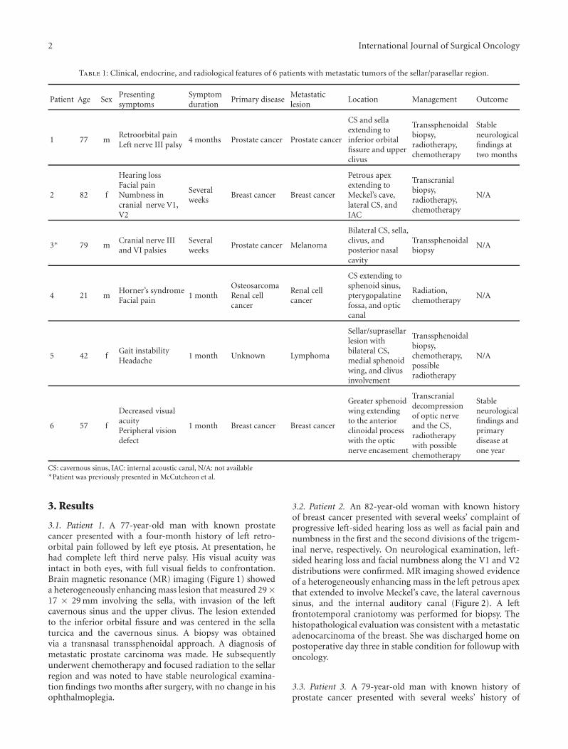
3.1. Patient 1. A 77-year-old man with known prostatecancer presented with a four-month history of left retro-orbital pain followed by left eye ptosis. At presentation, hehad complete left third nerve palsy. His visual acuity wasintact in both eyes, with full visual fields to confrontation.Brain magnetic resonance (MR) imaging (Figure 1) showeda heterogeneously enhancing mass lesion that measured 29×17 × 29 mm involving the sella, with invasion of the leftcavernous sinus and the upper clivus. The lesion extendedto the inferior orbital fissure and was centered in the sellaturcica and the cavernous sinus. A biopsy was obtainedvia a transnasal transsphenoidal approach. A diagnosis ofmetastatic prostate carcinoma was made. He subsequentlyunderwent chemotherapy and focused radiation to the sellarregion and was noted to have stable neurological examina-tion findings two months after surgery, with no change in hisophthalmoplegia.
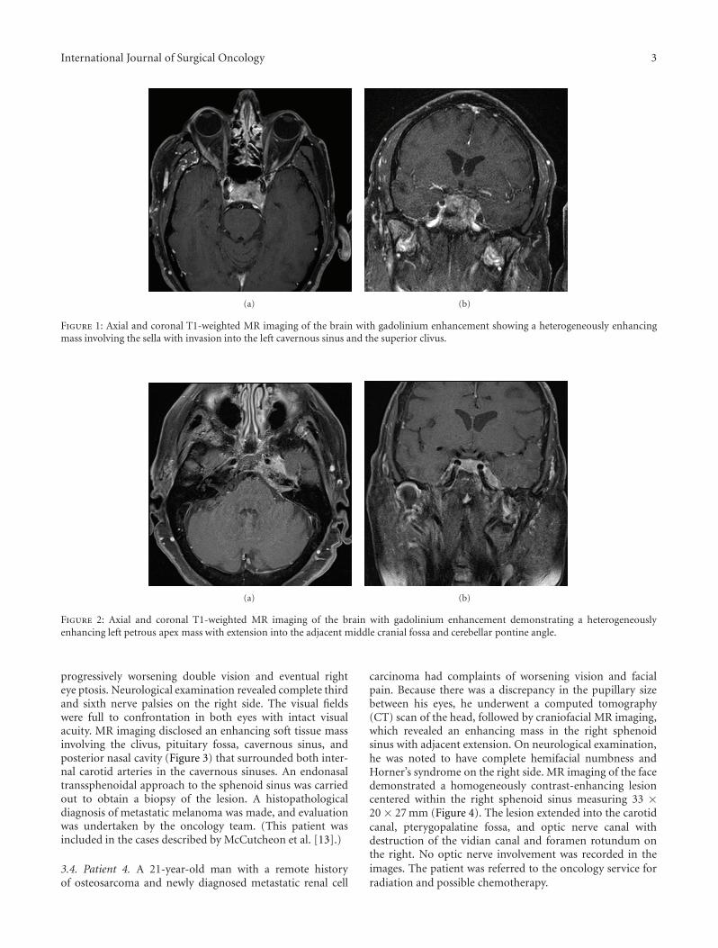
3.2. Patient 2. An 82-year-old woman with known historyof breast cancer presented with several weeks’ complaint ofprogressive left-sided hearing loss as well as facial pain andnumbness in the first and the second divisions of the trigem-inal nerve, respectively. On neurological examination, left-sided hearing loss and facial numbness along the V1 and V2distributions were confirmed. MR imaging showed evidenceof a heterogeneously enhancing mass in the left petrous apexthat extended to involve Meckel’s cave, the lateral cavernoussinus, and the internal auditory canal (Figure 2). A leftfrontotemporal craniotomy was performed for biopsy. Thehistopathological evaluation was consistent with a metastaticadenocarcinoma of the breast. She was discharged home onpostoperative day three in stable condition for followup withoncology.
3.3. Patient 3. A 79-year-old man with known history ofprostate cancer presented with several weeks’ history of
International Journal of Surgical Oncology 3
(a) (b)
Figure 1: Axial and coronal T1-weighted MR imaging of the brain with gadolinium enhancement showing a heterogeneously enhancingmass involving the sella with invasion into the left cavernous sinus and the superior clivus.
(a) (b)
Figure 2: Axial and coronal T1-weighted MR imaging of the brain with gadolinium enhancement demonstrating a heterogeneouslyenhancing left petrous apex mass with extension into the adjacent middle cranial fossa and cerebellar pontine angle.
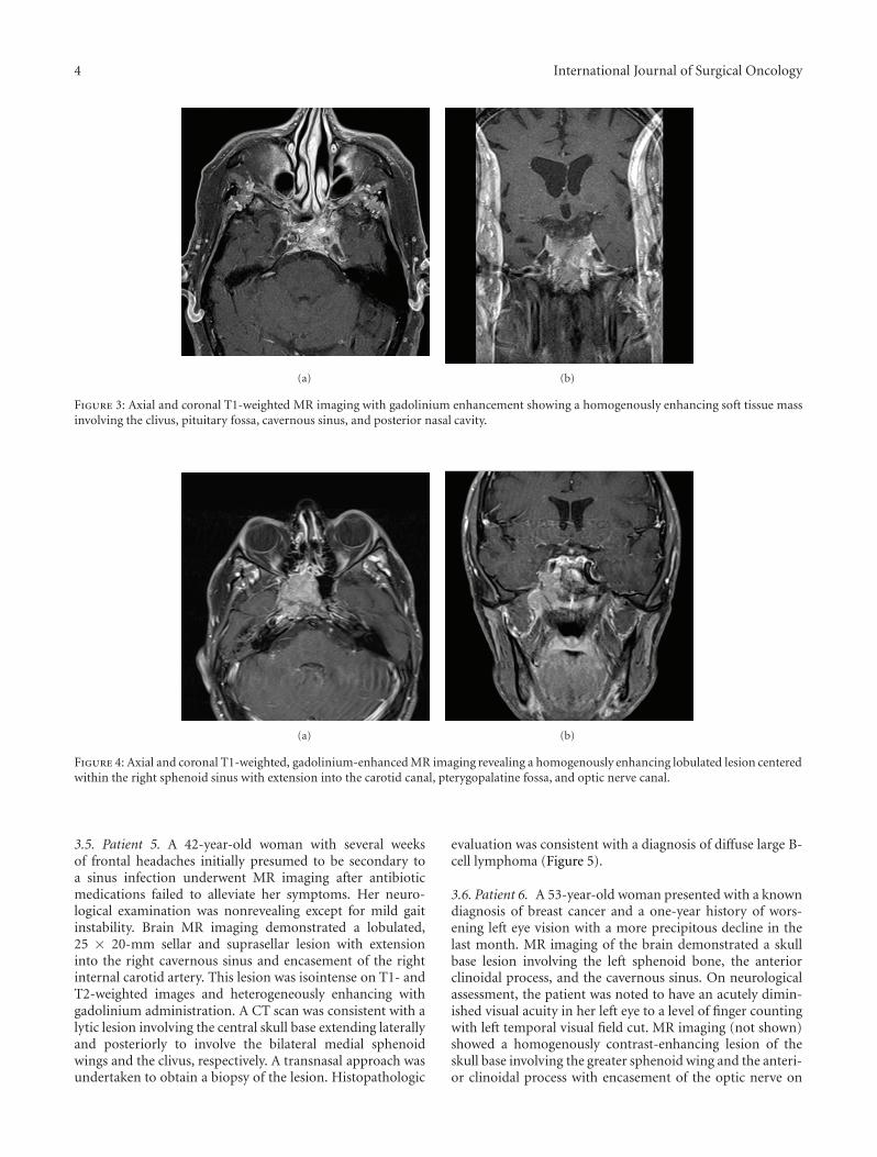
progressively worsening double vision and eventual righteye ptosis. Neurological examination revealed complete thirdand sixth nerve palsies on the right side. The visual fieldswere full to confrontation in both eyes with intact visualacuity. MR imaging disclosed an enhancing soft tissue massinvolving the clivus, pituitary fossa, cavernous sinus, andposterior nasal cavity (Figure 3) that surrounded both inter-nal carotid arteries in the cavernous sinuses. An endonasaltranssphenoidal approach to the sphenoid sinus was carriedout to obtain a biopsy of the lesion. A histopathologicaldiagnosis of metastatic melanoma was made, and evaluationwas undertaken by the oncology team. (This patient wasincluded in the cases described by McCutcheon et al. [13].)
3.4. Patient 4. A 21-year-old man with a remote historyof osteosarcoma and newly diagnosed metastatic renal cell
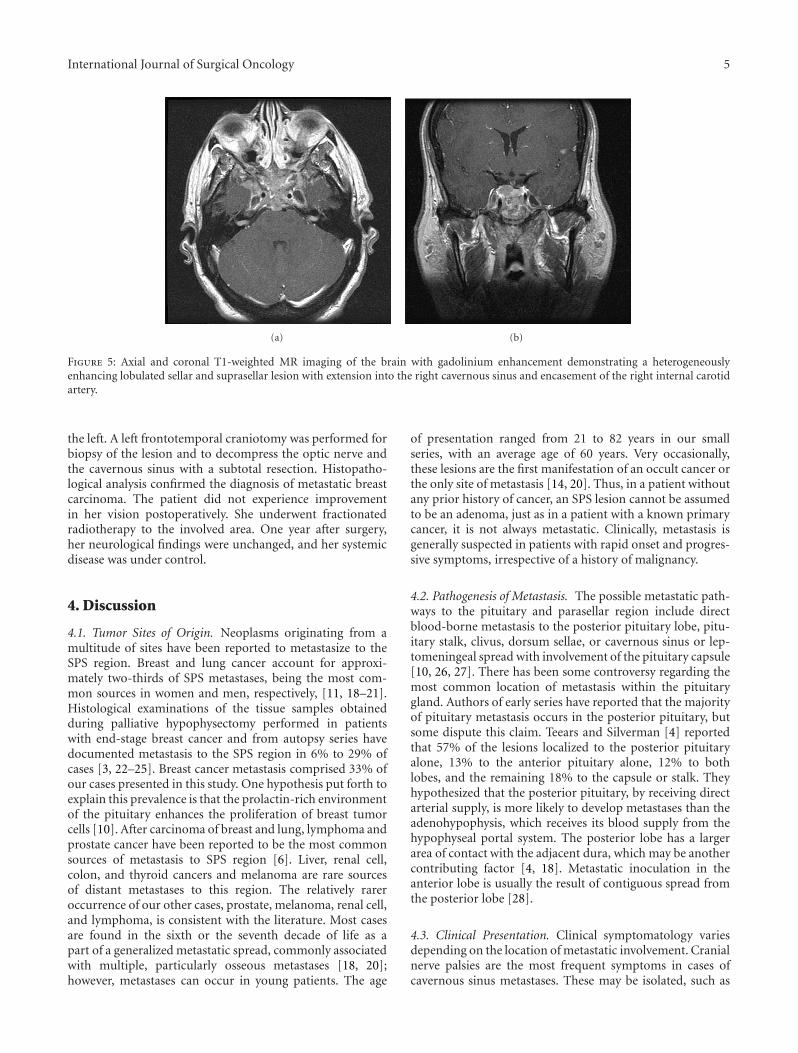
carcinoma had complaints of worsening vision and facialpain. Because there was a discrepancy in the pupillary sizebetween his eyes, he underwent a computed tomography(CT) scan of the head, followed by craniofacial MR imaging,which revealed an enhancing mass in the right sphenoidsinus with adjacent extension. On neurological examination,he was noted to have complete hemifacial numbness andHorner’s syndrome on the right side. MR imaging of the facedemonstrated a homogeneously contrast-enhancing lesioncentered within the right sphenoid sinus measuring 33 ×20 × 27 mm (Figure 4). The lesion extended into the carotidcanal, pterygopalatine fossa, and optic nerve canal withdestruction of the vidian canal and foramen rotundum onthe right. No optic nerve involvement was recorded in theimages. The patient was referred to the oncology service forradiation and possible chemotherapy.
4 International Journal of Surgical Oncology
(a) (b)
Figure 3: Axial and coronal T1-weighted MR imaging with gadolinium enhancement showing a homogenously enhancing soft tissue massinvolving the clivus, pituitary fossa, cavernous sinus, and posterior nasal cavity.
(a) (b)
Figure 4: Axial and coronal T1-weighted, gadolinium-enhanced MR imaging revealing a homogenously enhancing lobulated lesion centeredwithin the right sphenoid sinus with extension into the carotid canal, pterygopalatine fossa, and optic nerve canal.
3.5. Patient 5. A 42-year-old woman with several weeksof frontal headaches initially presumed to be secondary toa sinus infection underwent MR imaging after antibioticmedications failed to alleviate her symptoms. Her neuro-logical examination was nonrevealing except for mild gaitinstability. Brain MR imaging demonstrated a lobulated,25 × 20-mm sellar and suprasellar lesion with extensioninto the right cavernous sinus and encasement of the rightinternal carotid artery. This lesion was isointense on T1- andT2-weighted images and heterogeneously enhancing withgadolinium administration. A CT scan was consistent with alytic lesion involving the central skull base extending laterallyand posteriorly to involve the bilateral medial sphenoidwings and the clivus, respectively. A transnasal approach wasundertaken to obtain a biopsy of the lesion. Histopathologic
evaluation was consistent with a diagnosis of diffuse large B-cell lymphoma (Figure 5).
3.6. Patient 6. A 53-year-old woman presented with a knowndiagnosis of breast cancer and a one-year history of wors-ening left eye vision with a more precipitous decline in thelast month. MR imaging of the brain demonstrated a skullbase lesion involving the left sphenoid bone, the anteriorclinoidal process, and the cavernous sinus. On neurologicalassessment, the patient was noted to have an acutely dimin-ished visual acuity in her left eye to a level of finger countingwith left temporal visual field cut. MR imaging (not shown)showed a homogenously contrast-enhancing lesion of theskull base involving the greater sphenoid wing and the anteri-or clinoidal process with encasement of the optic nerve on
International Journal of Surgical Oncology 5
(a) (b)
Figure 5: Axial and coronal T1-weighted MR imaging of the brain with gadolinium enhancement demonstrating a heterogeneouslyenhancing lobulated sellar and suprasellar lesion with extension into the right cavernous sinus and encasement of the right internal carotidartery.
the left. A left frontotemporal craniotomy was performed forbiopsy of the lesion and to decompress the optic nerve andthe cavernous sinus with a subtotal resection. Histopatho-logical analysis confirmed the diagnosis of metastatic breastcarcinoma. The patient did not experience improvementin her vision postoperatively. She underwent fractionatedradiotherapy to the involved area. One year after surgery,her neurological findings were unchanged, and her systemicdisease was under control.
4. Discussion
4.1. Tumor Sites of Origin. Neoplasms originating from amultitude of sites have been reported to metastasize to theSPS region. Breast and lung cancer account for approxi-mately two-thirds of SPS metastases, being the most com-mon sources in women and men, respectively, [11, 18–21].Histological examinations of the tissue samples obtainedduring palliative hypophysectomy performed in patientswith end-stage breast cancer and from autopsy series havedocumented metastasis to the SPS region in 6% to 29% ofcases [3, 22–25]. Breast cancer metastasis comprised 33% ofour cases presented in this study. One hypothesis put forth toexplain this prevalence is that the prolactin-rich environmentof the pituitary enhances the proliferation of breast tumorcells [10]. After carcinoma of breast and lung, lymphoma andprostate cancer have been reported to be the most commonsources of metastasis to SPS region [6]. Liver, renal cell,colon, and thyroid cancers and melanoma are rare sourcesof distant metastases to this region. The relatively rareroccurrence of our other cases, prostate, melanoma, renal cell,and lymphoma, is consistent with the literature. Most casesare found in the sixth or the seventh decade of life as apart of a generalized metastatic spread, commonly associatedwith multiple, particularly osseous metastases [18, 20];however, metastases can occur in young patients. The age
of presentation ranged from 21 to 82 years in our smallseries, with an average age of 60 years. Very occasionally,these lesions are the first manifestation of an occult cancer orthe only site of metastasis [14, 20]. Thus, in a patient withoutany prior history of cancer, an SPS lesion cannot be assumedto be an adenoma, just as in a patient with a known primarycancer, it is not always metastatic. Clinically, metastasis isgenerally suspected in patients with rapid onset and progres-sive symptoms, irrespective of a history of malignancy.
4.2. Pathogenesis of Metastasis. The possible metastatic path-ways to the pituitary and parasellar region include directblood-borne metastasis to the posterior pituitary lobe, pitu-itary stalk, clivus, dorsum sellae, or cavernous sinus or lep-tomeningeal spread with involvement of the pituitary capsule[10, 26, 27]. There has been some controversy regarding themost common location of metastasis within the pituitarygland. Authors of early series have reported that the majorityof pituitary metastasis occurs in the posterior pituitary, butsome dispute this claim. Teears and Silverman [4] reportedthat 57% of the lesions localized to the posterior pituitaryalone, 13% to the anterior pituitary alone, 12% to bothlobes, and the remaining 18% to the capsule or stalk. Theyhypothesized that the posterior pituitary, by receiving directarterial supply, is more likely to develop metastases than theadenohypophysis, which receives its blood supply from thehypophyseal portal system. The posterior lobe has a largerarea of contact with the adjacent dura, which may be anothercontributing factor [4, 18]. Metastatic inoculation in theanterior lobe is usually the result of contiguous spread fromthe posterior lobe [28].
4.3. Clinical Presentation. Clinical symptomatology variesdepending on the location of metastatic involvement. Cranialnerve palsies are the most frequent symptoms in cases ofcavernous sinus metastases. These may be isolated, such as
6 International Journal of Surgical Oncology
diplopia or ptosis, with the third (oculomotor) and thesixth (abducens) nerves being the most commonly involved,followed by the fourth (trochlear) nerve [14, 29–31], orthey may appear in a constellation of symptoms charac-terized by unilateral, rapidly progressive ophthalmoplegiawith retroorbital pain. This latter presentation is the usualpresentation of the cavernous sinus syndrome, also knownas parasellar syndrome [32]. If the branches of the trigem-inal nerve are affected, alteration in the facial sensation,facial pain, or dysesthesia occurs [33]. Headache has beenreported as a rather common symptom, with an incidenceas high as 70% [14, 34, 35]; however, the majority ofpituitary metastases are clinically silent. In the autopsy studyby Teears and Silverman [4], only 7% of the pituitarymetastases were symptomatic. These metastases are oftenseen in patients with terminal malignancy who presentwith malaise, generalized pain, central nervous systeminvolvement, or treatment-associated symptoms, althoughsymptoms of pituitary insufficiency may be masked. Severalstudies have indicated that diabetes insipidus (DI) was themost common symptom [10, 20, 24, 36, 37]. In the seriesof McCormick et al. [2], DI developed in 70% of patients;however, if the anterior pituitary function is compromised,DI may be concealed by reduced mineralocorticoid func-tion [10]. Once the corticosteroid treatment is instituted,DI becomes clinically evident. In some recent series, DIhas not been reported, which is likely because modernimaging techniques are able to detect abnormalities earlierthan the timeframe required for DI development [35].Other rarer hormonal findings may be hypothyroidismand hypoadrenalism, hypogonadism, or overproduction ofadrenocorticotropic hormone (ACTH), growth hormone(GH), or prolactin [10, 38–40].
Because of their invasiveness, pituitary metastatic lesionshave a high potential to cause visual deficits from suprasellarextension, with an incidence as high as 50% reported byBranch Jr. and Laws Jr. [14] and others [10, 34, 41]. In boththe series by Chiang et al. [18] and that of Sioutos et al.[20], bitemporal hemianopsia was the most common type ofvisual impairment. Cranial nerve palsies involving the thirdand the sixth nerves and facial pain with numbness (trigem-inal origin) were the most common presentation (33%) inour cases with cavernous sinus involvement. Retro-orbitalpain, vision compromise, gait instability, Horner’s syndrome,and hearing loss were infrequent and associated with petro-clival and sphenoorbital extension of the lesions. All thesesymptoms were experienced with a relatively rapid onsetfrom a few weeks to a few months that suggested the aggres-sive character of the lesions.
Symptoms strongly suggesting metastasis in the parasel-lar or sellar space include painful ophthalmoplegia in associ-ation with the sudden onset of DI [10, 26, 34, 42]. The painmay be retro-orbital or may be due to trigeminal dysfunction[43–45]. In our series, 5 of the 6 (83%) patients had a pre-vious history of malignant disease. One of the patientsthat had prior history developed a malignancy (melanoma)other than the original one. Only one patient without aprior history of malignancy first presented with lymphomametastasis to the SPS region.
4.4. Imaging. Because pituitary adenomas also present withinvasion of the sellar floor, cavernous sinus, or clivus, nospecific neuroimaging criteria to define metastatic lesionsin SPS region have been reported. The diagnostic imagingtools for SPS metastasis mainly include high-resolution CTand MR imaging. Although CT is superior to MR imagingin detecting the bone involvement, the latter is preferable todetermine the relationship of the lesion to the surroundingneurovascular structures [46]. Although nonspecific, thecharacteristics of these lesions on MR imaging are an iso-or hypointense mass on T1-weighted imaging with a usuallyhyperintense signal on T2-weighted imaging, and homoge-neously enhancing mass in images obtained after the admin-istration of contrast agent [47]. Invasion of the cavernoussinus, sclerotic changes around the sella turcica and clivus,isointense signal on both T1- and T2-weighted imaging,and loss of high-intensity signal in the posterior pituitaryhave been reported to be helpful in differentiating metastaticlesions from benign ones [15, 30, 48]. Morita et al. [10] andKomninos et al. [40] found that thickening or enhancementof the infundibulum was the most characteristic CT or MRimaging feature [15]. Schubiger and Halter [37] reportedthat the invasion of the infundibular recess by a suprasellarmass is suggestive of metastasis. Because of the rapidgrowth of metastatic lesions, a dumbbell-shaped intra- andsuprasellar tumor with indentation at the diaphragm levelis generally indicative for these cases [20, 37, 49]. Theabove-mentioned imaging characteristics are not specific forsellar or parasellar metastases. The radiodiagnostic findingsthat suggested a malignant/metastatic process in our caseswere the involvement of multiple compartments in theanterior, middle, posterior cranial fossae, extension to theinfratemporal and pterygopalatine fossae, sphenoid sinus,and nasal cavity with bony destruction in the cranial base andasymmetric or bilateral invasion into the cavernous sinus.
4.5. Clinical Management. The management of SPS metas-tases is multimodal, including surgical resection, radiationtherapy, and chemotherapy [50]. Treatment is mainly pallia-tive and depends on the symptoms and the extent of systemicdisease [10, 51]. Because of the invasiveness and the highvascularity of the tumor, total surgical resection is generallynot undertaken [1, 20]. Therefore, surgical treatment shouldaim for symptomatic relief and the preservation of visualfunction, even in patients with widespread primary disease,and should be followed by local radiation treatment and/orchemotherapy [10, 51]. The body of evidence is inconclusiveon the effect of the latter two modalities on survival [14, 52].Morita et al. [10] and Branch Jr. and Laws Jr. [14] reportedimprovement in symptoms, especially in pain and visualfield defects, with no difference in survival after completeresection compared with subtotal or partial resection [10,14]. On the other hand, others have supported the conceptof improvement in survival after surgical resection of thelesion [53–55]. Surgical exploration is also essential if tissuediagnosis is likely to affect therapy in patients with no knownprimary malignancy. Resection is most commonly donevia transsphenoidal route, although subfrontal or pterionalapproaches are also options depending on the location and
International Journal of Surgical Oncology 7
the extension of the lesion. Four out of six patients in ourseries underwent surgical biopsy through either transsphe-noidal or transcranial route and then underwent subse-quent radiation/chemotherapy. One patient who had visualcompromise had subtotal resection for palliation followedby radiation/chemotherapy. One patient who had renal cellcarcinoma was directly referred to radiation oncology forimmediate radiation and subsequent chemotherapy.
Besides its role as an adjunct after surgery [14, 42, 56],radiosurgery or conventional radiation is recommended asthe initial course of treatment in patients with systemic dis-ease out of control, recurrence in the systemic disease withconcomitant SPS metastasis, or medical comorbidities thatput the patient at risk for a surgical intervention [2, 7,18, 41, 57–62]. Conventional radiation therapy can achievesymptom relief as high as 78% [62]. Radiosurgery, whichis considered less invasive than conventional radiation, hasbeen reported to achieve good tumor control [63, 64]. In aseries of 23 patients by Iwai et al. [17], the rates of tumorcontrol and symptom improvement were 67% and 53%,respectively; however, radiosurgery to the SPS region islimited by its potential to cause radiation injury to thesurrounding neurovascular structures such as optic appara-tus, pituitary gland, or cranial nerves coursing in cavernoussinus. Doses reported in the literature for these structuresrange from 8 to 40 Gy [63, 65–68], and the optimal dose maybe quite variable depending on the proximity of the lesion.Furthermore, the debate about whether the radiation shouldbe directed to the SPS region alone or to the entire braincontinues.
Chemotherapy is commonly used alone or along withradiation therapy mostly for palliation in the treatment ofmetastatic disease in SPS region [69]. Its value has not beenadequately studied and reported in the literature.
4.6. Prognosis. The prognosis of patients with metastases tothe SPS region is grim in a majority of cases because of theaggressive character of the primary disease [58]. Even inpatients with no other metastasis at the initial evaluation,the prognosis remains poor because of radiologically unde-tectable microscopic metastases; however, it has been sug-gested that the extent of systemic disease affects survivalin these patients [14]. Patients with a single SPS regionmetastasis may have a better outcome [10, 20]. Mediansurvival is less than 2 years independent of the managementstrategy [10, 14, 70]. The records on the long-term follow-up of the majority of our patients are lacking because thepatients were not monitored for surgical outcome, or thefollow-up periods were too short. The only patient that hadpalliative surgery for vision compromise was seen at one yearafter the surgery with stable neurological findings.
5. Conclusion
Sellar and parasellar metastatic lesions are relatively rare.Breast and lung have been reported to be the most commonsources in both sexes. Suggestive symptoms include rapidonset of progressive ophthalmoplegia with retro-orbital or
facial pain, visual impairment, and/or DI. Management va-ries depending on whether a primary source is identified, thesymptomatology, the location and extent of the lesion, thestage of the primary disease, and the medical comorbidities.Subtotal or partial surgical resection is aimed mainly forsymptom relief. A multimodal approach involving subto-tal resection of the lesion followed by radiation and/orchemotherapy is widely accepted, especially in symptomaticpatients whose primary disease is under control. Radiationwith or without chemotherapy is generally recommendedas first-line treatment in patients with advanced primarydisease or in those with high-risk medical comorbidities.A biopsy usually precedes radiation therapy if the primarysource of the metastasis is unknown. The prognosis forpatients is generally poor, independent of the therapeuticmodality, and the overall survival is less than two years.
References
[1] J. Gsponer, N. De Tribolet, J. P. Deruaz et al., “Diagnosis,treatment, and outcome of pituitary tumors and other abnor-mal intrasellar masses: retrospective analysis of 353 patients,”Medicine, vol. 78, no. 4, pp. 236–269, 1999.
[2] P. C. McCormick, K. D. Post, A. D. Kandji, and A. P. Hays,“Metastatic carcinoma to the pituitary gland,” British Journalof Neurosurgery, vol. 3, no. 1, pp. 71–79, 1989.
[3] U. Roessmann, B. Kaufman, and R. L. Friede, “Metastaticlesions in the sella turcica and pituitary gland,” Cancer, vol.25, no. 2, pp. 478–480, 1970.
[4] R. J. Teears and E. M. Silverman, “Clinicopathologic reviewof 88 cases of carcinoma metastatic to the pituitary gland,”Cancer, vol. 36, no. 1, pp. 216–220, 1975.
[5] W. T. Couldwell, P. T. Chandrasoma, and M. H. Weiss, “Pitu-itary gland metastasis from adenocarcinoma of the prostate:case report,” Journal of Neurosurgery, vol. 71, no. 1, pp. 138–140, 1989.
[6] D. W. Spell, D. S. Gervais Jr., J. K. Ellis, and R. H. Vial, “Cav-ernous sinus syndrome due to metastatic renal cell carci-noma,” Southern Medical Journal, vol. 91, no. 6, pp. 576–579,1998.
[7] B. Onec, B. Oksuzoglu, H. G. Hatipoglu, K. Onec, A. Azak, andN. Zengin, “Cavernous sinus syndrome caused by metastaticcolon carcinoma,” Clinical Colorectal Cancer, vol. 6, no. 8, pp.593–596, 2007.
[8] C. D. Bell, K. Kovacs, E. Horvath, H. Smythe, and S. Asa,“Papillary carcinoma of thyroid metastatic to the pituitarygland,” Archives of Pathology and Laboratory Medicine, vol. 125,no. 7, pp. 935–938, 2001.
[9] A. Chrisoulidou, K. Pazaitou-Panayiotou, N. Flaris et al.,“Pituitary metastasis of follicular thyroid carcinoma,” Hor-mone Research, vol. 61, no. 4, pp. 190–192, 2004.
[10] A. Morita, F. B. Meyer, and E. R. Laws Jr., “Symptomaticpituitary metastases,” Journal of Neurosurgery, vol. 89, no. 1,pp. 69–73, 1998.
[11] C. M. Ogilvie, S. Payne, J. Evanson, T. A. Lister, and A. B.Grossman, “Lymphoma metastasizing to the pituitary: an un-usual presentation of a treatable disease,” Pituitary, vol. 8, no.2, pp. 139–146, 2005.
[12] S. R. Masse, R. W. Wolk, and R. H. Conklin, “Peripituitarygland involvement in acute leukemia in adults,” Archives ofPathology and Laboratory Medicine, vol. 96, no. 2, pp. 141–142,1973.
8 International Journal of Surgical Oncology
[13] I. E. McCutcheon, S. G. Waguespack, G. N. Fuller, and W.T. Couldwell, “Metastatic melanoma to the pituitary gland,”Canadian Journal of Neurological Sciences, vol. 34, no. 3, pp.322–327, 2007.
[14] C. L. Branch Jr. and E. R. Laws Jr., “Metastatic tumors ofthe sella turcica masquerading as primary pituitary tumors,”Journal of Clinical Endocrinology and Metabolism, vol. 65, no.3, pp. 469–474, 1987.
[15] N. A. Mayr, W. T. C. Yuh, M. G. Muhonen et al., “Pituitarymetastases: MR findings,” Journal of Computer Assisted Tomog-raphy, vol. 17, no. 3, pp. 432–437, 1993.
[16] D. Brasnu, O. Laccourreye, V. Bassot, L. Laccourreye, P.Naudo, and F. X. Roux, “Cisplatin-based neoadjuvant chemo-therapy and combined resection for ethmoid sinus adenocar-cinoma reaching and/or invading the skull base,” Archives ofOtolaryngology—Head and Neck Surgery, vol. 122, no. 7, pp.765–768, 1996.
[17] Y. Iwai, K. Yamanaka, Y. Honda, and Y. Matsusaka, “Radio-surgery for pituitary metastases,” Neurologia Medico-Chirurgi-ca, vol. 44, no. 3, pp. 112–116, 2004.
[18] M. F. Chiang, M. Brock, and S. Patt, “Pituitary metastases,”Neurochirurgia, vol. 33, no. 4, pp. 127–131, 1990.
[19] A. Sepehrnia, M. Samii, and M. Tatagiba, “Management ofintracavernous tumours: an 11-year experience,” Acta Neu-rochirurgica, Supplement, vol. 53, pp. 122–126, 1991.
[20] P. Sioutos, V. Yen, and E. Arbit, “Pituitary gland metastases,”Annals of Surgical Oncology, vol. 3, no. 1, pp. 94–99, 1996.
[21] J. E. Thomas and R. E. Yoss, “The parasellar syndrome:problems in determining etiology,” Mayo Clinic Proceedings,vol. 45, no. 9, pp. 617–623, 1970.
[22] H. L. Abrams, R. Spiro, and N. Goldstein, “Metastases in car-cinoma; analysis of 1000 autopsied cases,” Cancer, vol. 3, no.1, pp. 74–85, 1950.
[23] K. J. Gurling, G. B. Scott, and D. N. Baron, “Metastases inpituitary tissue removed at hypophysectomy in women withmammary carcinoma,” British Journal of Cancer, vol. 11, pp.519–522, 1957.
[24] W. A. Houck, K. B. Olson, and J. Horton, “Clinical features oftumor metastasis to the pituitary,” Cancer, vol. 26, no. 3, pp.656–659, 1970.
[25] F. Marin, K. T. Kovacs, B. W. Scheithauer, and W. F. Young,“The pituitary gland in patients with breast carcinoma: ahistologic and immunocytochemical study of 125 cases,” MayoClinic Proceedings, vol. 67, no. 10, pp. 949–956, 1992.
[26] M. B. Max, M. D. Deck, and D. A. Rottenberg, “Pituitarymetastasis: incidence in cancer patients and clinical differen-tiation from pituitary adenoma,” Neurology, vol. 31, no. 8, pp.998–1002, 1981.
[27] K. Takakura, K. Sano, and S. Hojo, Metastatic Tumors of TheCentral Nervous System, Igaku-Shoin, Tokyo, Japan, 1982.
[28] O. B. Leramo, J. D. Booth, and B. Zinman, “Hyperpro-lactinemia, hypopituitarism, and chiasmal compression dueto carcinoma metastatic to the pituitary,” Neurosurgery, vol. 8,no. 4, pp. 477–480, 1981.
[29] J. R. Keane, “Cavernous sinus syndrome: analysis of 151 cases,”Archives of Neurology, vol. 53, no. 10, pp. 967–971, 1996.
[30] M. Kistler and H. W. Pribram, “Metastatic disease of the sellaturcica,” American Journal of Roentgenology, Radium Therapy,and Nuclear Medicine, vol. 123, no. 1, pp. 13–21, 1975.
[31] C. C. K. Lin and J. J. Tsai, “Relationship between the numberof involved cranial nerves and the percentage of lesions locatedin the cavernous sinus,” European Neurology, vol. 49, no. 2, pp.98–102, 2003.
[32] M. L. Supler, W. A. Friedman, C. E. Geist, and B. R. Younge,“Acute bilateral ophthalmoplegia secondary to cavernoussinus metastasis: a case report,” Neurosurgery, vol. 31, no. 4,pp. 783–786, 1992.
[33] J. M. Bumpous, M. D. Maves, S. M. Gomez, B. K. Levy, andF. Johnson, “Cavernous sinus involvement in head and neckcancer,” Head and Neck, vol. 15, no. 1, pp. 62–66, 1993.
[34] J. C. Kattah, R. M. Silgals, H. Manz, J. G. Toro, A. Dritschilo,and F. P. Smith, “Presentation and management of parasellarand suprasellar metastatic mass lesions,” Journal of NeurologyNeurosurgery and Psychiatry, vol. 48, no. 1, pp. 44–49, 1985.
[35] C. Weilbaecher, R. V. Patwardhan, M. Fowler, B. K. Willis,and A. Nanda, “Metastatic lesions involving the sella: reportof three cases and review of the literature,” Neurology India,vol. 52, no. 3, pp. 365–368, 2004.
[36] D. W. Kimmel and B. P. O’Neill, “Systemic cancer presentingas diabetes insipidus: clinical and radiographic features of11 patients with a review of metastatic-induced diabetesinsipidus,” Cancer, vol. 52, no. 12, pp. 2355–2358, 1983.
[37] O. Schubiger and D. Haller, “Metastases to the pituitary-hypothalamic axis: an MR study of 7 symptomatic patients,”Neuroradiology, vol. 34, no. 2, pp. 131–134, 1992.
[38] T. Abe, K. Matsumoto, M. Iida, M. Hayashi, N. Sanno, and R.Yoshiyuki Osamura, “Malignant carcinoid tumor of the ante-rior mediastinum metastasis to a prolactin-secreting pituitaryadenoma: a case report,” Surgical Neurology, vol. 48, no. 4, pp.389–394, 1997.
[39] F. W. F. Hanna, O. M. Williams, J. S. Davies, T. Dawson, J. Neal,and M. F. Scanlon, “Pituitary apoplexy following metastasisof bronchogenic adenocarcinoma to a prolactinoma,” ClinicalEndocrinology, vol. 51, no. 3, pp. 377–381, 1999.
[40] J. Komninos, V. Vlassopoulou, D. Protopapa et al., “Tumorsmetastatic to the pituitary gland: case report and literaturereview,” Journal of Clinical Endocrinology and Metabolism, vol.89, no. 2, pp. 574–580, 2004.
[41] P. B. Nelson, A. G. Robinson, and A. J. Martinez, “Metastatictumor of the pituitary gland,” Neurosurgery, vol. 21, no. 6, pp.941–944, 1987.
[42] P. Juneau, W. C. Schoene, and P. Black, “Malignant tumors inthe pituitary gland,” Archives of Neurology, vol. 49, no. 5, pp.555–558, 1992.
[43] S. Fernandez, O. Godino, S. Martınez-Yelamos et al., “Cav-ernous sinus syndrome: a series of 126 patients,” Medicine, vol.86, no. 5, pp. 278–281, 2007.
[44] E. Sharkawi, K. Tumuluri, and J. M. Olver, “Metastasticchoriocarcinoma causing cavernous sinus syndrome,” BritishJournal of Ophthalmology, vol. 90, no. 5, pp. 654–655, 2006.
[45] Y. C. Yap, V. Sharma, J. Rees, and A. Kosmin, “Cavernous sinussyndrome secondary to metastasis from small cell lung carci-noma,” Annals of Ophthalmology, vol. 39, no. 2, pp. 166–169,2007.
[46] W. L. Hirsch Jr., F. G. Hryshko, L. N. Sekhar et al., “Compari-son of MR imaging, CT, and angiography in the evaluation ofthe enlarged cavernous sinus,” American Journal of Roentgenol-ogy, vol. 151, no. 5, pp. 1015–1023, 1988.
[47] L. E. Ginsberg, “Neoplastic diseases affecting the central skullbase: CT and MR imaging,” American Journal of Roentgenol-ogy, vol. 159, no. 3, pp. 581–589, 1992.
[48] R. Chaudhuri, C. Twelves, T. C. S. Cox, and J. B. Bingham,“MRI in diabetes insipidus due to metastatic breast carci-noma,” Clinical Radiology, vol. 46, no. 3, pp. 184–188, 1992.
[49] P. U. Freda and K. D. Post, “Differential diagnosis of sellarmasses,” Endocrinology and Metabolism Clinics of North Amer-ica, vol. 28, no. 1, pp. 81–117, 1999.
International Journal of Surgical Oncology 9
[50] M. Losa, M. Grasso, E. Giugni, P. Mortini, S. Acerno, and M.Giovanelli, “Metastatic prostatic adenocarcinoma presentingas a pituitary mass: shrinkage of the lesion and clinical im-provement with medical treatment,” Prostate, vol. 32, no. 4,pp. 241–245, 1997.
[51] A. Ruelle, M. Palladino, and G. C. Andrioli, “Pituitary metas-tases as presenting lesions of malignancy,” Journal of Neurosur-gical Sciences, vol. 36, no. 1, pp. 51–54, 1992.
[52] K. D. Post, P. C. McCormick, A. P. Hays, and A. G. Kandji,“Metastatic carcinoma to pituitary adenoma: report of twocases,” Surgical Neurology, vol. 30, no. 4, pp. 286–292, 1988.
[53] W. L. Lynes, D. G. Bostwick, F. S. Freiha, and T. A. Stamey, “Pa-renchymal brain metastases from adenocarcinoma of pro-state,” Urology, vol. 28, no. 4, pp. 280–287, 1986.
[54] K. G. Rao, “Carcinoma of prostate presenting as intracranialtumor with multiple cranial nerve palsies,” Urology, vol. 19,no. 4, pp. 433–435, 1982.
[55] V. C. Smith, D. L. Kasdon, and R. C. Hardy, “Metastatic braintumor from the prostate: two unusual cases,” Surgical Neurol-ogy, vol. 14, no. 3, pp. 189–191, 1980.
[56] S. Bitoh, H. Hasegawa, J. Obashi, and M. Maruno, “Secondarymalignancies involving parasellar region: clinical manifesta-tions, diagnosis and management in 16 patients,” MedicalJournal of Osaka University, vol. 36, no. 1-2, pp. 17–27, 1985.
[57] Y. Ahn, J. Yang, H. Kim et al., “Cavernous sinus metastasisof non-small cell lung cancer,” Tuberculosis and RespiratoryDiseases, vol. 69, pp. 381–384, 2010.
[58] J. Y. Delattre, C. Castelain, L. Davila, B. Schadeck, and M.Poisson, “Pituitary stalk metastasis from breast cancer,” RevueNeurologique, vol. 146, no. 6-7, pp. 455–456, 1990.
[59] H. S. Greenberg, M. D. F. Deck, and B. Vikram, “Metastasis tothe base of the skull: clinical findings in 43 patients,” Neurol-ogy, vol. 31, no. 5, pp. 530–537, 1981.
[60] Y. Iwai, K. Yamanaka, and M. Yoshimura, “Gamma knife radi-osurgery for cavernous sinus metastases and invasion,” Surgi-cal Neurology, vol. 64, no. 5, pp. 406–410, 2005.
[61] Y. Mori, T. Kobayashi, and Y. Shibamoto, “Stereotactic radio-surgery for metastatic tumors in the pituitary gland and thecavernous sinus,” Journal of Neurosurgery, vol. 105, pp. 37–42,2006.
[62] B. Vikram and F. C. H. Chu, “Radiation therapy for metastasesto the base of the skull,” Radiology, vol. 130, no. 2, pp. 465–468,1979.
[63] A. J. Cmelak, R. S. Cox, J. R. Adler, W. E. Fee, and D. R. Gof-finet, “Radiosurgery for skull base malignancies and nasopha-ryngeal carcinoma,” International Journal of Radiation Oncol-ogy Biology Physics, vol. 37, no. 5, pp. 997–1003, 1997.
[64] R. C. Miller, R. L. Foote, R. J. Coffey et al., “The role of stereo-tactic radiosurgery in the treatment of malignant skull basetumors,” International Journal of Radiation Oncology BiologyPhysics, vol. 39, no. 5, pp. 977–981, 1997.
[65] Y. Iwai and K. Yamanaka, “Gamma Knife radiosurgery forskull base metastasis and invasion,” Stereotactic and FunctionalNeurosurgery, vol. 72, supplement 1, pp. 81–87, 1999.
[66] M. Kocher, J. Voges, S. Staar, H. Treuer, V. Sturm, and R. P.Mueller, “Linear accelerator radiosurgery for recurrent malig-nant tumors of the skull base,” American Journal of ClinicalOncology, vol. 21, no. 1, pp. 18–22, 1998.
[67] K. A. Leber, J. Bergloff, and G. Pendl, “Dose-response toler-ance of the visual pathways and cranial nerves of the cavernoussinus to stereotactic radiosurgery,” Journal of Neurosurgery,vol. 88, no. 1, pp. 43–50, 1998.
[68] R. B. Tishler, J. S. Loeffler, L. D. Lunsford et al., “Tolerance ofcranial nerves of the cavernous sinus to radiosurgery,” Inter-national Journal of Radiation Oncology Biology Physics, vol. 27,no. 2, pp. 215–221, 1993.
[69] R. G. Garcıa, J. S. Perez, L. N. Gıas, F. J. R. Campo, and F. J.D. Gonzalez, “Cavernous sinus metastasis from oropharyngealsquamous cell carcinoma,” Medicina Oral, Patologıa Oral yCirugıa Bucal, vol. 12, no. 2, pp. E166–E170, 2007.
[70] F. Laigle-Donadey, S. Taillibert, N. Martin-Duverneuil, J.Hildebrand, and J. Y. Delattre, “Skull-base metastases,” Journalof Neuro-Oncology, vol. 75, no. 1, pp. 63–69, 2005.