Embed Size (px)
Citation preview
at SciVerse ScienceDirect
Biochimie 94 (2012) 628e636
Contents lists available
Biochimie
journal homepage: www.elsevier .com/locate/b iochi
Research paper
Self assembly of human septin 2 into amyloid filaments
Julio Cesar Pissuti Damalioa, Wanius Garciab, Joci Neuby Alves Macêdoa, Ivo de Almeida Marquesa,José M. Andreuc, Rafael Giraldoc, Richard Charles Garratta, Ana Paula Ulian Araújoa,*aCentro de Biotecnologia Molecular Estrutural, Instituto de Física de São Carlos (IFSC), Universidade de São Paulo (USP), Av. Trabalhador Sãocarlense,400, 13560-970 São Carlos, SP, BrazilbCentro de Ciências Naturais e Humanas (CCNH), Universidade Federal do ABC (UFABC), Santa Adélia, 148, Santo André, SP, BrazilcCentro de Investigaciones Biológicas-CSIC, Madrid, Spain
a r t i c l e i n f o
Article history:Received 18 May 2011Accepted 15 September 2011Available online 28 September 2011
Keywords:GTPase domainAggregatesAmyloidNeurodegenerative diseasesSeptin
* Corresponding author. Tel.: þ55 16 33739875; faxE-mail address: [email protected] (A.P. Ulian A
0300-9084/$ e see front matter � 2011 Elsevier Masdoi:10.1016/j.biochi.2011.09.014
a b s t r a c t
Septins are a conserved group of GTP-binding proteins that form hetero-oligomeric complexes whichassemble into filaments. These are essential for septin function, including their role in cytokinesis, celldivision, exocytosis and membrane trafficking. Septin 2 (SEPT2) is a member of the septin family and hasbeen associated with neurofibrillary tangles and other pathological features of senile plaques in Alz-heimer’s disease. An in silico analysis of the amino acid sequence of SEPT2 identified regions witha significant tendency to aggregate and/or form amyloid. These were all observed within the GTP-bindingdomain. This was consistent with the experimental identification of a structure rich in b-sheet duringtemperature induced unfolding transitions observed for both the full length protein and the GTP-bindingdomain alone. This intermediate state is characterized by irreversible aggregation and has the ability tobind Thioflavin-T, suggesting its amyloid nature. Under electron microscopy, fibers extending for severalmicrometers in length could be visualized. The results shown in this study support the hypothesis thatsingle septins, when present in excess or with unbalanced stoichiometries, may be unstable andassemble into amyloid-like structures.
� 2011 Elsevier Masson SAS. All rights reserved.
1. Introduction
The septins are members of a conserved group of GTP-bindingproteins, originally discovered in yeast as being required for thecompletion of the cell cycle [1]. Septins have been identified in allanimals and fungi [2]. In mammals they are involved in a variety ofcellular phenomena, such as microtubule regulation [3,4], vesicletrafficking [5], the assembly of scaffolding platforms [6], actindynamics [7], exocytosis [8], apoptosis [9], DNA repair [10] andmechanical stability [11e13].
All members of the septin family can be divided into threedomains: a variable N-terminal domain, a GTP-binding domain anda C-terminal region, which generally includes sequences with thepotential to assemble as a coiled-coil [14]. In addition, some septinshave a polybasic region between the N-terminal and GTP-bindingdomains, which is responsible for lipid interaction [15]. Both thebinding of GTP and its hydrolysis have been experimentallydemonstrated for several septins in vitro [2,16e18].
: þ55 1633715381.raújo).
son SAS. All rights reserved.
Septins can assemble into high-order hetero-filaments,including two or more members of the family, in vivo and in vitro.These hetero-filaments were isolated from Drosophila melanogaster[19] and fungi [20], and shown to have a width of about 7e9 nmand a variable length. Hetero-filaments were also identified in thecase of the Caenorhabditis elegans septins UNC-59 and UNC-61 [21].Moreover, a complex of recombinant Saccharomyces cerevisiaeseptins, Cdc3, Cdc10, Cdc11 and Cdc12, forms an elongated linearoctamer composed of two copies of each individual septin whichsubsequently polymerize to form filaments [22]. Recently, Sir-ajuddin et al. solved the crystal structures of both the human SEPT2GTP-binding domain alone and the hetero-trimeric complex ofSEPT2eSEPT6eSEPT7 [23]. In both cases the crystal lattice leads tothe formation of filaments. In addiction, members of the samegroup can replace each other in the specific position along theheterofilament [24].
Although the formation of hetero-filaments by septins and theirassociated functions is relatively well established, informationconcerning the existence and physiological role of homo-filamentsis puzzling. The first report of filaments formed by a single septinwas that for Xenopus laevis SEPT2. The authors described homo-filaments of 20 nm in width when the component monomerswere bound to GTP but not to GDP [25]. This observation has been
J.C. Pissuti Damalio et al. / Biochimie 94 (2012) 628e636 629
subsequently questioned, leaving some doubt about the relevanceof homo-filaments in the case of Xenopus septins [26]. However,20e40 nm wide homo-filaments have also been reported forhuman SEPT2 in both the GTP- and GDP-bound states [18]. Thequestion of the existence of homo-filaments is therefore unsettledand has been complicated by the observation of homo-filaments ofhuman SEPT4, which are believed to be amyloid fibers with nophysiological relevance [27].
SEPT2 is essential for cytokinesis, being located near thecontractile ring from anaphase to telophase and finally condensinginto themidbody [7]. It has awide tissue distribution and specificallyin the brain has been observed to co-localizewithGLAST, an astrocyteglutamate transporter [28]. Based on this data SEPT2 has been sug-gested to play a role in signal transmission in the cerebellum [28].Besides its physiological roles, SEPT2 has also been implicated inseveral pathologies including leukemia and lymphoma [29], renalcell carcinoma [30], brain tumors [31,32] and systemic lupus eryth-ematosus [33]. Moreover, SEPT2 together with SEPT1 and SEPT4(all of which are acidic proteins) have been seen to accumulate inneurofibrillary tangles (NFTs) in Alzheimer’s disease [34] where theyco-localize with the basic microtubule-associated protein tau. So far,however, the role of septins in NFT formation and the possiblemechanisms of self-aggregation of septins in neurodegenerativedisorders still remain unclear.
A wide range of human pathologies are associated with uncon-trolled protein misfolding, leading to the conversion of polypeptidechains from their soluble globular states into well-organized fibrillaraggregates rich in b-sheet structure [35,36]. Amyloid formation is thehallmark of medically related disorders, such as Alzheimer disease,andmore than 40 human related diseases have been described, eachhaving a distinct clinical profile and each associated with theaggregation of a single dominant protein [37].
With the aim of shedding some light upon this issue, the presentstudy uses a biophysical approach to investigate the stability andaggregation of SEPT2. Our results show that both SEPT2 and itsGTP-binding domain (SEPT2G) are dimeric in solution, but theyhave the tendency to rapidly aggregate at physiological tempera-tures. These aggregates have the ability to bind Thioflavin-T, sug-gesting that they are amyloids and providing the first insight on themechanisms that cause the formation of the aggregates associatedwith neurodegenerative diseases.
2. Material and methods
2.1. Materials and reagents
The bacterial expression vector pET28a(þ) and Ni-NTA resinwere purchased from Novagen. Restriction endonucleases, iso-propyl-b-D-thiogalactopyranoside, kanamycin, T4 DNA ligase andTaq Polymerase were obtained from Invitrogen. The Superdex-200column 10/30 and native gels for electrophoresis [8e25% (w/v)gradient polyacrylamide] were purchased from Amersham Phar-macia Biotech (GE-Healthcare). Guanosine-50-diphosphate (GDP),protein standard markers and ThT (Thioflavin-T) were purchasedfrom Sigma. All other chemical products used were obtained fromSigma and/or GE-Healthcare.
2.2. In silico analysis
Essentially, protein aggregation is a self-assembly of identicalmolecules [38], and these aggregates can be classified as eitherordered or disordered, on the basis of their intimate structuralfeatures [39]. There is evidence that local unfolding events can causethe aggregation of normally globular proteins into well-organizedfibrils [40]. In order to identify possible regions presenting a high
aggregation probability in SEPT2, we used the in silico analysisprograms TANGO, WALTZ and ZYGGREGATOR. TANGO is a statisticalmechanics algorithm which predicts protein aggregation, based onthe physico-chemical principles of b-sheet formation [41,42]. WALTZand ZYGGREGATOR are algorithms including additional thermody-namic information in thepredictionof regionswith ahighprobabilityto form amyloid sequences [43,44]. The amino acid sequence ofSEPT2 was submitted to these prediction programs using serversbased at http://tango.crg.es/ and http://waltz.switchlab.org/, usingdefault parameters. Additionally, the major isoforms of all 13 humanseptin sequenceswere alsoanalyzedwithWALTZ inorder to comparetheir predicted amyloidogenic regions.
2.3. Plasmid construction and proteins expression
Based on the results of the sequence analysis, which suggestedthe presence of aggregation prone sequences within the GTP-binding domain, a construct was designed corresponding toa truncated SEPT2 protein covering this region of the molecule(SEPT2G, residues 34e308). The cDNA corresponding to SEPT2(residues 1e361) and its GTP-binding domain (SEPT2G) wereamplified using the polymerase chain reaction performed ina Mastercycler thermocycler (Eppendorf), using a fetal brain cDNAlibrary (Gibco BRL) as template. Amplification products were puri-fied and cloned into the pET28a(þ) expression vector, using NdeIand XhoI restriction sites. The recombinant plasmids were trans-formed into Escherichia coli DH5a for propagation and plasmidextraction purposes. These were named pSEPT2 and pSEPT2G andproduced their respective products fused to an N-terminal His-tag.All plasmids were sequenced by the dideoxy chain method [45]using an ABI Prism 377 automated DNA sequencer (PerkineElmer)following the protocol of the manufacturer.
The expression plasmids were used to transform the E. coli hoststrain BL21(DE3). A total of 500 mL of an overnight culture of E. coliBL21(DE3) harboring the pSEPT2 plasmid were inoculated into500 mL of fresh LB medium containing kanamycin (50 mg/mL). Theculture was grown whilst shaking at 37 �C to mid log phase(O.D.600nm ¼ 0.6) and subsequently induced with IPTG at a finalconcentration of 0.4 mM followed by incubation for 10 h at 18 �C.
After centrifugation, the pelleted cells were re-suspended in25 mM TriseHCl pH 7.8 buffer, containing 100 mM GDP and 10%glycerol. The cells were disrupted by the addition of 0.1 mg/mLlysozyme for 30min at 4 �C, followed by sonication. The suspensionwas then centrifuged at 18,000 g for 20 min at 4 �C, and the pelletand supernatant were analyzed by SDS-PAGE to check the solubilityof the recombinant protein. The supernatant containing therecombinant SEPT2 was loaded onto a nickel-affinity columnequilibrated with the same buffer. After the unbound proteins wereeliminated by exhaustive washing, SEPT2 was eluted by increasingconcentrations of imidazole up to 300 mM.
The resulting SEPT2 was then loaded onto a Superdex-20010/300 GL column (GE-Healthcare) pre-equilibrated with 25 mMTriseHCl pH7.8 buffer, containing 10% glycerol and driven by anÅktapurifier. The elution was carried out in the same buffer, at 4 �C andfractions analyzed by means of 15% SDS-PAGE. Protein elution wasmonitored byabsorbance at 280nm. The elution volumes of standardproteins were used to calculate the Kav values [Kav ¼ (elutionvolume� columnvoid volume)/(total columnvolume� columnvoidvolume)]. The standard proteins of known molecular weight werecarbonic anhydrase (29 kDa), ovoalbumin (43 kDa), conalbumin(75 kDa), ferritin (440 kDa) and lactoglobulin (669 kDa). An identicalprotocol was used for the purification of SEPT2G. The proteinconcentration, in all cases, was determined from its absorbance at280 nm, based on its amino acid composition [46], employinga U-2001 Hitachi UVevisible spectrophotometer.
J.C. Pissuti Damalio et al. / Biochimie 94 (2012) 628e636630
2.4. Native gel electrophoresis
SEPT2 (20 mM) and SEPT2G (20 mM) were centrifuged at13,000 � g for 5 min at 4 �C. Subsequently the samples were sub-jected to electrophoresis on an 8e25% (w/v) gradient poly-crylamide gel at pH 8.8 using the Phast System (AmershamBioscience), at 4 �C, and stained following standard protocols.Protein standards of known hydrodynamic radii (thyroglobulin,8.5 nm; ferritin, 6.1 nm; catalase, 5.22 nm; lactate dehydrogenase,4.4 nm; and bovine serum albumin, 3.55 nm) were subjected toelectrophoresis under identical conditions. The mobilities of theindividual bands of the protein standards were plotted as theretardation factors (Rf) versus the Stokes radii (Rh). The linearequation obtained from this calibration was employed to calculatethe Rh of SEPT2 and SEPT2G.
2.5. Circular dichroism spectroscopy (CD)
The thermal unfolding of the recombinant proteins, SEPT2 andSEPT2G was monitored by far-UV CD spectroscopy over a wave-length range of 195e250 nm, using a J-715 Jasco spectropolarimeterequipped with a temperature controller. CD spectra weremeasuredfrom samples in 0.1 cm quartz cuvettes and were the average of 16accumulations, using a scanning speed of 100 nm min�1, a spectralbandwidth of 1 nm, and a response time of 0.5 s. The proteinconcentration, in all cases, was approximately 10 mM in 25 mMTriseHCl, pH 7.8 buffer containing 10% glycerol. Thermal denatur-ationmeasurements were performed by incubating the samples for30 min at temperatures of 15 �C, 30 �C, 37 �C, 45 �C and 60 �C. CDspectra were obtained on a degree ellipticity scale and the buffercontribution was subtracted in all of the experiments. Data pointswere analyzed with the software Origin 7.0 and deconvolution ofthe spectrumwas performed using the K2d algorithm (http://www.embl-heidelberg.de/wandrade/k2d/).
2.6. Right-angle light scattering
SEPT2 and SEPT2G (5 mM) in 25 mM TriseHCl, pH 7.8 buffercontaining 10% glycerol were centrifuged (16,000 � g for 10 minat 4 �C) and each sample placed in a 1 cm path length quartzcuvette in a spectrofluorimeter, model K2 ISS, equipped witha refrigerated circulator. The samples were illuminated with350 nm light, and the scattering at the same wavelength wascollected at an angle of 90�. Measurements were made at 15 �C,30 �C, 37 �C, 45 �C and 60 �C. All intensity measurements werenormalized to values between 0 and 1 (where the latter corre-sponds to the maximum intensity obtained at 60 �C) aftersubtraction of the light scattering by the buffer. Data points wereanalyzed with the software Origin 7.0.
2.7. Thioflavin-T fluorescence assay
Thioflavin T (ThT) is the most commonly used dye for thedetection of amyloid aggregates [47,48], and it is speculated thatThT may bind in between the b-sheets of the fibril [49]. Proteins at5 mM (in 25 mM TriseHCl, 10% glycerol, pH 7.8 buffer) were used inthis experiment to analyze the ThT binding to SEPT2 and SEPT2G.The proteins were incubatedwith 80 mMThT, excited at 450 nm andthe emission measured at 482 nm for a period of 90 min.Measurements weremade at 15 �C, 30 �C, 37 �C, 45 �C and 60 �C. Allintensity measurements were normalized to values between 0 and1, after subtraction of the light scattering by the buffer. Data pointswere analyzed with the software Origin 7.0.
2.8. Electron microscopy
In order to verify the morphology of the SEPT2 aggregates, thesample (5 mM) was maintained at 37 �C for 30 min in buffer con-taining 25 mM TriseHCl, 10% glycerol, pH 7.8. Samples incubated at4 �C for 5 days were also prepared in order to decrease the rate ofamyloid filament formation. Negative staining was performed asfollows. Initially samples were applied to glow-discharged carbon-coated grids for 1 min. These were stained with filtered 1% uranylacetate for 1 min, subsequently washed with a drop of the samebuffer and blotted dry. Images were acquired with a transmissionelectron microscope Philips CM 120 or a JEOL JEM 1230, working at80 or 100 kV.
3. Results and discussion
3.1. SEPT2 presents regions with high probability of aggregationand/or amyloid formation
It is accepted that two mechanisms appear to control theassembly of amyloid oligomers, one sequence-dependant and theother sequence-independent [50]. Obviously, the sequence-dependant mechanism is more predictable and the programsTANGO and WALTZ are very sensitive for the detection of b-sheetaggregation and amyloidogenic regions, respectively [41,43]. TheTANGO results indicated four regions of SEPT2 presenting a highprobability of aggregation through b-sheets, all of them within theGTP-binding domain. The sequences identified included thosebetween residues 38 and 43 (FTLMVV), 52 and 59 (TLINSLFL), 118and 122 (IISYI) and 146 and 153 (VHCCFYFI) (Fig. 1A). AlthoughTANGO aims to predict b-sheet aggregation but not specificallyamyloid formation, there is a good correlation between the two [41].
The program WALTZ aims to minimize the over prediction ofamorphous b-aggregation compared to the regular cross-b structurecharacteristic of amyloid fibrils, making it more specific for the latter[51]. WALTZ also identified four regions within SEPT2 which pre-sented a high amyloidogenic potential: the sequences betweenresidues 118 and 123 (IISYID), 147 and 155 (HCCFYFISP), 163 and 168(LDVAFM), 173 and 178 (NKVNIV) (Fig. 1A). Once again these regionswere all restricted to the GTP-binding domain and two of theWALTZpredictions were effectively identical to those identified by TANGO,being highly suggestive that these regions have a tendency towardsself-assembly and amyloid formation. The region from 118 to 123corresponds to a helical region on the surface of themonomerwhilstthat from147 to 155maps to a buriedb-strand. They are shown in redandblue in Fig.1B andC. Furthermore, bothof these regionswere alsoidentified by the independent algorithm Zyggregator. In both casesconsiderable structural rearrangement of the monomer wouldtherefore be necessary in order for these particular sequences tobecome solvent exposed and directly participate in the cross-b spine,eventually leading to aggregation. In order to investigate this further,SEPT2 and SEPT2G were over-expressed for structural analysis.
3.2. SEPT2 and SEPTG are dimers in solution
The definition of the GTP-binding domain for SEPT2 was basedon that used in a previous study of the molecular dissection ofSEPT4 [52]. DNA amplifications produced fragments of 1083 and822 bp corresponding to the coding regions of SEPT2 and SEPT2G,respectively. After induction with IPTG, E. coli cells harboring theappropriate expression vector produced additional bands of around44 and 34 kDa corresponding to SEPT2 and SEPT2G, respectively. Inboth cases, the majority of the expressed product was soluble aftercell lysis and final yields were typically 3 and 10 mg of recombinantprotein/L of culture medium for SEPT2 and SEPT2G, respectively.
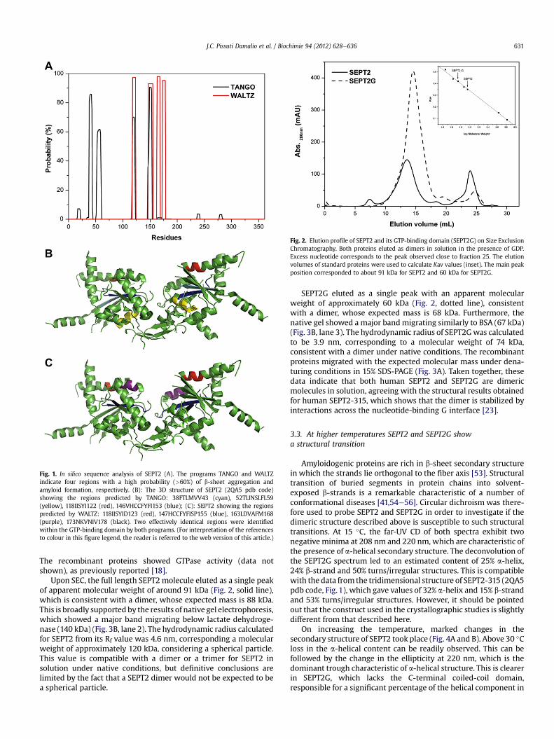
Fig. 2. Elution profile of SEPT2 and its GTP-binding domain (SEPT2G) on Size ExclusionChromatography. Both proteins eluted as dimers in solution in the presence of GDP.Excess nucleotide corresponds to the peak observed close to fraction 25. The elutionvolumes of standard proteins were used to calculate Kav values (inset). The main peakposition corresponded to about 91 kDa for SEPT2 and 60 kDa for SEPT2G.
Fig. 1. In silico sequence analysis of SEPT2 (A). The programs TANGO and WALTZindicate four regions with a high probability (>60%) of b-sheet aggregation andamyloid formation, respectively. (B): The 3D structure of SEPT2 (2QA5 pdb code)showing the regions predicted by TANGO: 38FTLMVV43 (cyan), 52TLINSLFL59(yellow), 118IISYI122 (red), 146VHCCFYFI153 (blue); (C): SEPT2 showing the regionspredicted by WALTZ: 118IISYID123 (red), 147HCCFYFISP155 (blue), 163LDVAFM168(purple), 173NKVNIV178 (black). Two effectively identical regions were identifiedwithin the GTP-binding domain by both programs. (For interpretation of the referencesto colour in this figure legend, the reader is referred to the web version of this article.)
J.C. Pissuti Damalio et al. / Biochimie 94 (2012) 628e636 631
The recombinant proteins showed GTPase activity (data notshown), as previously reported [18].
Upon SEC, the full length SEPT2 molecule eluted as a single peakof apparent molecular weight of around 91 kDa (Fig. 2, solid line),which is consistent with a dimer, whose expected mass is 88 kDa.This is broadly supported by the results of native gel electrophoresis,which showed a major band migrating below lactate dehydroge-nase (140 kDa) (Fig. 3B, lane 2). The hydrodynamic radius calculatedfor SEPT2 from its Rf value was 4.6 nm, corresponding a molecularweight of approximately 120 kDa, considering a spherical particle.This value is compatible with a dimer or a trimer for SEPT2 insolution under native conditions, but definitive conclusions arelimited by the fact that a SEPT2 dimer would not be expected to bea spherical particle.
SEPT2G eluted as a single peak with an apparent molecularweight of approximately 60 kDa (Fig. 2, dotted line), consistentwith a dimer, whose expected mass is 68 kDa. Furthermore, thenative gel showed a major bandmigrating similarly to BSA (67 kDa)(Fig. 3B, lane 3). The hydrodynamic radius of SEPT2Gwas calculatedto be 3.9 nm, corresponding to a molecular weight of 74 kDa,consistent with a dimer under native conditions. The recombinantproteins migrated with the expected molecular mass under dena-turing conditions in 15% SDS-PAGE (Fig. 3A). Taken together, thesedata indicate that both human SEPT2 and SEPT2G are dimericmolecules in solution, agreeing with the structural results obtainedfor human SEPT2-315, which shows that the dimer is stabilized byinteractions across the nucleotide-binding G interface [23].
3.3. At higher temperatures SEPT2 and SEPT2G showa structural transition
Amyloidogenic proteins are rich in b-sheet secondary structurein which the strands lie orthogonal to the fiber axis [53]. Structuraltransition of buried segments in protein chains into solvent-exposed b-strands is a remarkable characteristic of a number ofconformational diseases [41,54e56]. Circular dichroism was there-fore used to probe SEPT2 and SEPT2G in order to investigate if thedimeric structure described above is susceptible to such structuraltransitions. At 15 �C, the far-UV CD of both spectra exhibit twonegative minima at 208 nm and 220 nm, which are characteristic ofthe presence of a-helical secondary structure. The deconvolution ofthe SEPT2G spectrum led to an estimated content of 25% a-helix,24% b-strand and 50% turns/irregular structures. This is compatiblewith the data from the tridimensional structure of SEPT2-315 (2QA5pdb code, Fig. 1), which gave values of 32% a-helix and 15% b-strandand 53% turns/irregular structures. However, it should be pointedout that the construct used in the crystallographic studies is slightlydifferent from that described here.
On increasing the temperature, marked changes in thesecondary structure of SEPT2 took place (Fig. 4A and B). Above 30 �Closs in the a-helical content can be readily observed. This can befollowed by the change in the ellipticity at 220 nm, which is thedominant trough characteristic of a-helical structure. This is clearerin SEPT2G, which lacks the C-terminal coiled-coil domain,responsible for a significant percentage of the helical component in
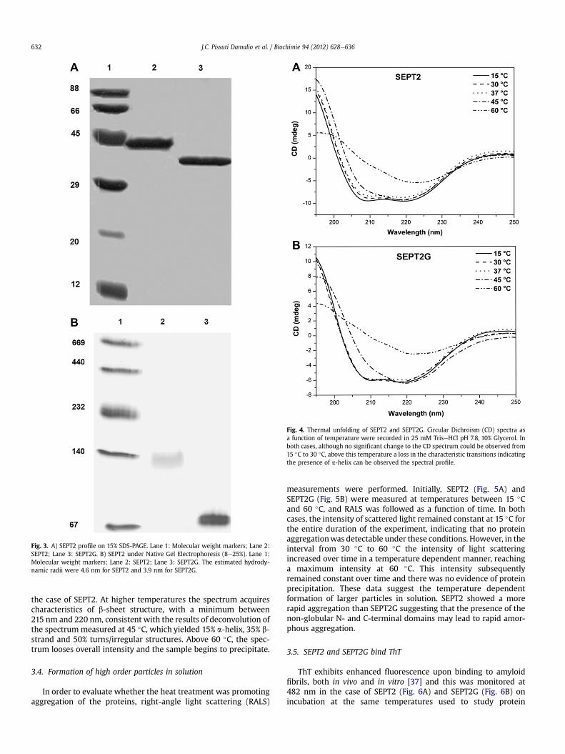
Fig. 4. Thermal unfolding of SEPT2 and SEPT2G. Circular Dichroism (CD) spectra asa function of temperature were recorded in 25 mM TriseHCl pH 7.8, 10% Glycerol. Inboth cases, although no significant change to the CD spectrum could be observed from15 �C to 30 �C, above this temperature a loss in the characteristic transitions indicatingthe presence of a-helix can be observed the spectral profile.
Fig. 3. A) SEPT2 profile on 15% SDS-PAGE. Lane 1: Molecular weight markers; Lane 2:SEPT2; Lane 3: SEPT2G. B) SEPT2 under Native Gel Electrophoresis (8e25%). Lane 1:Molecular weight markers; Lane 2: SEPT2; Lane 3: SEPT2G. The estimated hydrody-namic radii were 4.6 nm for SEPT2 and 3.9 nm for SEPT2G.
J.C. Pissuti Damalio et al. / Biochimie 94 (2012) 628e636632
the case of SEPT2. At higher temperatures the spectrum acquirescharacteristics of b-sheet structure, with a minimum between215 nm and 220 nm, consistent with the results of deconvolution ofthe spectrummeasured at 45 �C, which yielded 15% a-helix, 35% b-strand and 50% turns/irregular structures. Above 60 �C, the spec-trum looses overall intensity and the sample begins to precipitate.
3.4. Formation of high order particles in solution
In order to evaluate whether the heat treatment was promotingaggregation of the proteins, right-angle light scattering (RALS)
measurements were performed. Initially, SEPT2 (Fig. 5A) andSEPT2G (Fig. 5B) were measured at temperatures between 15 �Cand 60 �C, and RALS was followed as a function of time. In bothcases, the intensity of scattered light remained constant at 15 �C forthe entire duration of the experiment, indicating that no proteinaggregationwas detectable under these conditions. However, in theinterval from 30 �C to 60 �C the intensity of light scatteringincreased over time in a temperature dependent manner, reachinga maximum intensity at 60 �C. This intensity subsequentlyremained constant over time and there was no evidence of proteinprecipitation. These data suggest the temperature dependentformation of larger particles in solution. SEPT2 showed a morerapid aggregation than SEPT2G suggesting that the presence of thenon-globular N- and C-terminal domains may lead to rapid amor-phous aggregation.
3.5. SEPT2 and SEPT2G bind ThT
ThT exhibits enhanced fluorescence upon binding to amyloidfibrils, both in vivo and in vitro [37] and this was monitored at482 nm in the case of SEPT2 (Fig. 6A) and SEPT2G (Fig. 6B) onincubation at the same temperatures used to study protein
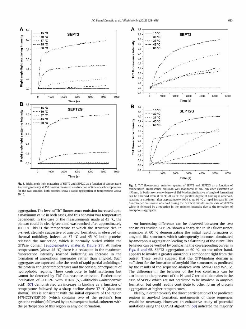
Fig. 6. ThT fluorescence emission spectra of SEPT2 and SEPT2G as a function oftemperature. Fluorescence emission was monitored at 482 nm after excitation at450 nm. In both cases, some degree of ThT binding (indicative of amyloid formation)can be observed even at 30 �C. At 45 �C the greatest degree of binding is observed,reaching a maximum after approximately 1000 s. At 60 �C a rapid increase in thefluorescence emission is observed during the first few minutes in the case of SEPT2Gwhich is followed by a reduction in the emission intensity due to the formation ofamorphous aggregates.
Fig. 5. Right angle light scattering of SEPT2 and SEPT2G as a function of temperature.Scattering intensity at 350 nmwas measured as a function of time at each temperaturefor the two samples. Both proteins show a rapid aggregation at temperatures above30 �C.
J.C. Pissuti Damalio et al. / Biochimie 94 (2012) 628e636 633
aggregation. The level of ThT fluorescence emission increased up toa maximumvalue in both cases, and this behavior was temperaturedependent. In the case of the measurements made at 45 �C, theplateau could be clearly seen and was reached after approximately1000 s. This is the temperature at which the structure rich inb-sheet, strongly suggestive of amyloid formation, is observed onthermal unfolding. Indeed, at 37 �C and 45 �C both proteinsreleased the nucleotide, which is normally buried within theGTPase domain (Supplementary material, Figure S1). At highertemperatures (above 45 �C) there is a reduction in the maximumfluorescence intensity reached indicating an increase in theformation of amorphous aggregates rather than amyloid. Suchaggregates are expected to be the result of rapid partial unfolding ofthe protein at higher temperatures and the consequent exposure ofhydrophobic regions. These contribute to light scattering butcannot be detected by ThT fluorescence emission. Furthermore,incubation of SEPT2G with DTNB (5,50-dithiobis,2-nitrobenzoicacid) [57] demonstrated an increase in binding as a function oftemperature followed by a sharp decline above 37 �C (data notshown). This is consistent with the initial exposure of the region147HCCFYFISP155, (which contains two of the protein’s fourcysteine residues) followed by its subsequent burial, coherent withthe participation of this region in amyloid formation.
An interesting difference can be observed between the twoconstructs studied. SEPT2G shows a sharp rise in ThT fluorescenceemission at 60 �C demonstrating the initial rapid formation ofamyloid-like structures which subsequently becomes dominatedby amorphous aggregation leading to a flattening of the curve. Thisbehavior can be verified by comparing the corresponding curves inFigs. 5 and 6B. SEPT2 aggregation at 60 �C, on the other hand,appears to involve a greater amorphous component right from theoutset. These results suggest that the GTP-binding domain issufficient for the formation of amyloid-like structures as predictedby the results of the sequence analyses with TANGO and WALTZ.The difference in the behavior of the two constructs can beattributed to the presence of the N- and C-terminal domains in thecase of SEPT2 which are not predicted to be involved in amyloidformation but could readily contribute to other forms of proteinaggregation at higher temperatures.
Ideally, in order to verify the direct participation of the predictedregions in amyloid formation, mutagenesis of these sequenceswould be necessary. However, an exhaustive study of potentialmutations using the CUPSAT algorithm [58] indicated the majority
J.C. Pissuti Damalio et al. / Biochimie 94 (2012) 628e636634
of these to be destabilizing. Furthermore, given the possibleinvolvement of more than one amyloidogenic region, a series ofsingle and double mutants would be necessary in order to properlytest this hypothesis. Unfortunately, these limitations would lead toconsiderable ambiguity when interpreting the outcome.
3.6. Amyloid like filaments are visualized by electron microscopy
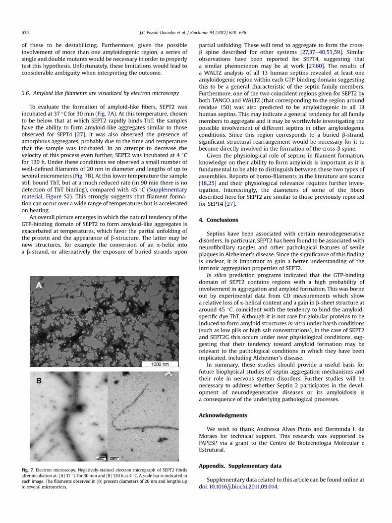
To evaluate the formation of amyloid-like fibers, SEPT2 wasincubated at 37 �C for 30 min (Fig. 7A). At this temperature, chosento be below that at which SEPT2 rapidly binds ThT, the sampleshave the ability to form amyloid-like aggregates similar to thoseobserved for SEPT4 [27]. It was also observed the presence ofamorphous aggregates, probably due to the time and temperaturethat the sample was incubated. In an attempt to decrease thevelocity of this process even further, SEPT2 was incubated at 4 �Cfor 120 h. Under these conditions we observed a small number ofwell-defined filaments of 20 nm in diameter and lengths of up toseveral micrometers (Fig. 7B). At this lower temperature the samplestill bound ThT, but at a much reduced rate (in 90 min there is nodetection of ThT binding), compared with 45 �C (Supplementarymaterial, Figure S2). This strongly suggests that filament forma-tion can occur over a wide range of temperatures but is acceleratedon heating.
An overall picture emerges in which the natural tendency of theGTP-binding domain of SEPT2 to form amyloid-like aggregates isexacerbated at temperatures, which favor the partial unfolding ofthe protein and the appearance of b-structure. The latter may benew structures, for example the conversion of an a-helix intoa b-strand, or alternatively the exposure of buried strands upon
Fig. 7. Electron microscopy. Negatively-stained electron micrograph of SEPT2 fibrilsafter incubation at: (A) 37 �C for 30 min and (B) 120 h at 4 �C. A scale bar is indicated ineach image. The filaments observed in (B) present diameters of 20 nm and lengths upto several micrometers.
partial unfolding. These will tend to aggregate to form the cross-b spine described for other systems [27,37e40,53,59]. Similarobservations have been reported for SEPT4, suggesting thata similar phenomenon may be at work [27,60]. The results ofa WALTZ analysis of all 13 human septins revealed at least oneamyloidogenic region within each GTP-binding domain suggestingthis to be a general characteristic of the septin family members.Furthermore, one of the two coincident regions given for SEPT2 byboth TANGO and WALTZ (that corresponding to the region aroundresidue 150) was also predicted to be amyloidogenic in all 13human septins. This may indicate a general tendency for all familymembers to aggregate and it may be worthwhile investigating thepossible involvement of different septins in other amyloidogenicconditions. Since this region corresponds to a buried b-strand,significant structural rearrangement would be necessary for it tobecome directly involved in the formation of the cross-b spine.
Given the physiological role of septins in filament formation,knowledge on their ability to form amyloids is important as it isfundamental to be able to distinguish between these two types ofassemblies. Reports of homo-filaments in the literature are scarce[18,25] and their physiological relevance requires further inves-tigation. Interestingly, the diameters of some of the fibersdescribed here for SEPT2 are similar to those previously reportedfor SEPT4 [27].
4. Conclusions
Septins have been associated with certain neurodegenerativedisorders. In particular, SEPT2 has been found to be associated withneurofibrillary tangles and other pathological features of senileplaques in Alzheimer’s disease. Since the significance of this findingis unclear, it is important to gain a better understanding of theintrinsic aggregation properties of SEPT2.
In silico prediction programs indicated that the GTP-bindingdomain of SEPT2 contains regions with a high probability ofinvolvement in aggregation and amyloid formation. This was borneout by experimental data from CD measurements which showa relative loss of a-helical content and a gain in b-sheet structure ataround 45 �C, coincident with the tendency to bind the amyloid-specific dye ThT. Although it is not rare for globular proteins to beinduced to form amyloid structures in vitro under harsh conditions(such as low pHs or high salt concentrations), in the case of SEPT2and SEPT2G this occurs under near physiological conditions, sug-gesting that their tendency toward amyloid formation may berelevant to the pathological conditions in which they have beenimplicated, including Alzheimer’s disease.
In summary, these studies should provide a useful basis forfuture biophysical studies of septin aggregation mechanisms andtheir role in nervous system disorders. Further studies will benecessary to address whether Septin 2 participates in the devel-opment of neurodegenerative diseases or its amyloidosis isa consequence of the underlying pathological processes.
Acknowledgments
We wish to thank Andressa Alves Pinto and Derminda I. deMoraes for technical support. This research was supported byFAPESP via a grant to the Centro de Biotecnologia Molecular eEstrutural.
Appendix. Supplementary data
Supplementary data related to this article can be found online atdoi:10.1016/j.biochi.2011.09.014.
J.C. Pissuti Damalio et al. / Biochimie 94 (2012) 628e636 635
References
[1] L.H. Hartwell, Genetic control of the cell division cycle in yeast. IV. Genescontrolling bud emergence and cytokinesis, Exp. Cell Res. 69 (1971) 265e276.
[2] M. Kinoshita, The septins, Genome Biol. 4 (2003) 236.[3] M.C. Surka, C.W. Tsang, W.S. Trimble, The mammalian septin MSF localizes
with microtubules and is required for completion of cytokinesis, Mol. Biol. Cell13 (2002) 3532e3545.
[4] K. Nagata, A. Kawajiri, S. Matsui, M. Takagishi, T. Shiromizu, N. Saitoh, I. Izawa,T. Kiyono, T.J. Itoh, H. Hotani, M. Inagaki, Filament formation of MSF-A,a mammalian septin, in human mammary epithelial cells depends on inter-actions with microtubules, J. Biol. Chem. 278 (2003) 18538e18543.
[5] S.C. Hsu, C.D. Hazuka, R. Roth, D.L. Foletti, J. Heuser, R.H. Scheller, Subunitcomposition, protein interactions, and structures of the mammalian brainsec6/8 complex and septin filaments, Neuron 20 (1998) 1111e1122.
[6] E.T. Spiliotis, M. Kinoshita, W.J. Nelson, A mitotic septin scaffold required formammalian chromosome congression and segregation, Science 307 (2005)1781e1785.
[7] M. Kinoshita, S. Kumar, A. Mizoguchi, C. Ide, A. Kinoshita, T. Haraguchi,Y. Hiraoka, M. Noda, Nedd5, a mammalian septin, is a novel cytoskeletalcomponent interacting with actin-based structures, Gene Dev. 11 (1997)1535e1547.
[8] C.L. Beites, H. Xie, R. Bowser, W.S. Trimble, The septin CDCrel-1 binds syntaxinand inhibits exocytosis, Nat. Neurosci. 2 (1999) 434e439.
[9] S. Larisch, Y.S. Yi, R. Lotan, H. Kerner, S. Eimerl, W.T. Parks, Y. Gottfried,S.B. Reffey, M.P. de Caestecker, D. Danielpour, N. Book-Melamed, R. Timberg,C.S. Duckett, R.J. Lechleider, H. Steller, J. Orly, S.J. Kim, A.B. Roberts, A novelmitochondrial septin-like protein, ARTS, mediates apoptosis dependent on itsP-loop motif, Nat. Cell Biol. 2 (2000) 915e921.
[10] B.E. Kremer, L.A. Adang, I.G. Macara, Septins regulate actin organization andcell-cycle arrest through nuclear accumulation of NCK mediated by SOCS7,Cell 130 (2007) 837e850.
[11] M. Ihara, A. Kinoshita, S. Yamada, H. Tanaka, A. Tanigaki, A. Kitano, M. Goto,K. Okubo, H. Nishiyama, O. Ogawa, C. Takahashi, O. Ogawa, C. Takahashi,S. Itohara, Y. Nishimune, M. Noda, M. Kinoshita, Cortical organization by theseptin cytoskeleton is essential for structural and mechanical integrity ofmammalian spermatozoa, Dev. Cell 8 (2005) 343e352.
[12] J.D. Steels, M.R. Estey, C.D. Froese, D. Reynaud, C. Pace-Asciak, W.S. Trimble,Sept12 is a component of the mammalian sperm tail annulus, Cell Motil.Cytoskel 64 (2007) 794e807.
[13] H. Kissel, M.M. Georgescu, S. Larisch, K. Manova, G.R. Hunnicutt, H. Steller, TheSept4 septin locus is required for sperm terminal differentiation in mice, Dev.Cell 8 (2005) 353e364.
[14] B. Kartmann, D. Roth, Novel roles for mammalian septins: from vesicle traf-ficking to oncogenesis, J. Cell Sci. 114 (2001) 839e844.
[15] J. Zhang, C. Kong, H. Xie, P.S. McPherson, S. Grinstein, W.S. Trimble, Phos-phatidylinositol polyphosphate binding to the mammalian septin H5 ismodulated by GTP, Curr. Biol. 9 (1999) 1458e1467.
[16] M. Kinoshita, Assembly of mammalian septins, J. Biochem. 134 (2003)491e496.
[17] S. Hillebrand, W. Garcia, M.D. Cantu, A.P.U. de Araujo, M. Tanaka, T. Tanaka,R.C. Garratt, E. Carrilho, In vitro monitoring of GTPase activity and enzymekinetics studies using capillary electrophoresis, Anal. Bioanal. Chem. 383(2005) 92e97.
[18] Y.W. Huang, M.C. Surka, D. Reynaud, C. Pace-Asciak, W.S. Trimble, GTPbinding and hydrolysis kinetics of human septin 2, Febs J. 273 (2006)3248e3260.
[19] C.M. Field, O. AlAwar, J. Rosenblatt, M.L. Wong, B. Alberts, T.J. Mitchison,A purified Drosophila septin complex forms filaments and exhibits GTPaseactivity, J. Cell Biol. 133 (1996) 605e616.
[20] J.A. Frazier, M.L. Wong, M.S. Longtine, J.R. Pringle, M. Mann, T.J. Mitchison,C. Field, Polymerization of purified yeast septins: evidence that organizedfilament arrays may not be required for septin function, J. Cell Biol. 143 (1998)737e749.
[21] C.M. John, R.K. Hite, C.S. Weirich, D.J. Fitzgerald, H. Jawhari, M. Faty,D. Schlapfer, R. Kroschewski, F.K. Winkler, T. Walz, Y. Barral, M.O. Steinmetz,The Caenorhabditis elegans septin complex is nonpolar, Embo J. 26 (2007)3296e3307.
[22] A. Bertin, M.A. McMurray, P. Grob, S.S. Park, G. Garcia, I. Patanwala, H.L. Ng,T. Alber, J. Thorner, E. Nogales, Saccharomyces cerevisiae septins: supramo-lecular organization of heterooligomers and the mechanism of filamentassembly, P. Natl. Acad. Sci. USA 105 (2008) 8274e8279.
[23] M. Sirajuddin, M. Farkasovsky, F. Hauer, D. Kuhlmann, I.G. Macara,M. Weyand, H. Stark, A. Wittinghofer, Structural insight into filamentformation by mammalian septins, Nature 449 (2007) 311e315.
[24] M. Nakahira, J.N.A. Macedo, T.V. Seraphim, N. Cavalcante, T.A.C.B. Souza,J.C.P. Damalio, L.F. Reyes, E.M. Assmann, M.R. Alborghetti, R.C. Garratt,A.P.U. Araujo, N.I.T. Zanchin, J.A.R.G. Barbosa, J. Kobarg, A draft of the humanseptin Interactome, Plos One 5 (2010) e0013799.
[25] M. Mendoza, A.A. Hyman, M. Glotzer, GTP binding induces filament assemblyof a recombinant septin, Curr. Biol. 12 (2002) 1858e1863.
[26] A.M. Vrabioiu, S.A. Gerber, S.P. Gygi, C.M. Field, T.J. Mitchison, The majority ofthe Saccharomyces cerevisiae septin complexes do not exchange guaninenucleotides, J. Biol. Chem. 279 (2004) 3111e3118.
[27] W. Garcia, A.P.U. de Araujo, F. Lara, D. Foguel, M. Tanaka, T. Tanaka,R.C. Garratt, An intermediate structure in the thermal unfolding of the GTPasedomain of human septin 4 (SEPT4/Bradeion-beta) forms amyloid-like fila-ments in vitro, Biochemistry-US 46 (2007) 11101e11109.
[28] N. Kinoshita, K. Kimura, N. Matsumoto, M. Watanabe, M. Fukaya, C. Ide,Mammalian septin Sept2 modulates the activity of GLAST, a glutamatetransporter in astrocytes, Genes Cells 9 (2004) 1e14.
[29] N. Cerveira, C. Correia, S. Bizarro, C. Pinto, S. Lisboa, J.M. Mariz, M. Marques,M.R. Teixeira, SEPT2 is a new fusion partner of MLL in acute myeloid leukemiawith t(2; 11)(q37; q23), Oncogene 25 (2006) 6147e6152.
[30] R.A. Craven, S. Hanrahan, N. Totty, P. Harnden, A.J. Stanley, E.R. Maher,A.L. Harris, W.S. Trimble, P.J. Selby, R.E. Banks, Proteomic identification ofa role for the von Hippel Lindau tumour suppressor in changes in theexpression of mitochondrial proteins and septin 2 in renal cell carcinoma,Proteomics 6 (2006) 3880e3893.
[31] K. Sakai, M. Kurimoto, A. Tsugu, S.L. Hubbard, W.S. Trimble, J.T. Rutka,Expression of Nedd5, a mammalian septin, in human brain tumors, J. Neuro-Oncol. 57 (2002) 169e177.
[32] A.A. Khalil, P. James, Biomarker discovery: a proteomic approach for braincancer profiling, Cancer Sci. 98 (2007) 201e213.
[33] P. Margutti, M. Sorice, F. Conti, F. Delunardo, M. Racaniello, C. Alessandri,A. Siracusano, R. Rigano, E. Profumo, G. Valesini, E. Ortona, Screening of anendothelial cDNA library identifies the C-terminal region of Nedd5 as a novelautoantigen in systemic lupus erythematosus with psychiatric manifestations,Arthritis Res. Ther. 7 (2005) R896eR903.
[34] A. Kinoshita, M. Kinoshita, H. Akiyama, H. Tomimoto, I. Akiguchi, S. Kumar,M. Noda, J. Kimura, Identification of septins in neurofibrillary tangles in Alz-heimer’s disease, Am. J. Pathol. 153 (1998) 1551e1560.
[35] D.J. Selkoe, Folding proteins in fatal ways, Nature 426 (2003) 900e904.[36] P. Westermark, M.D. Benson, J.N. Buxbaum, A.S. Cohen, B. Frangione, S.I. Ikeda,
C.L. Masters, G. Merlini, M.J. Saraiva, J.D. Sipe, Amyloid: toward terminologyclarification e report from the nomenclature committee of the Internationalsociety of amyloidosis, Amyloid 12 (2005) 1e4.
[37] F. Chiti, C.M. Dobson, Protein misfolding, functional amyloid, and humandisease, Annu. Rev. Biochem. 75 (2006) 333e366.
[38] F. Rousseau, J. Schymkowitz, L. Serrano, Protein aggregation and amyloidosis:confusion of the kinds? Curr. Opin. Struct. Biol. 16 (2006) 118e126.
[39] C.M. Dobson, Principles of protein folding, misfolding and aggregation, Semin.Cell Dev. Biol. 15 (2004) 3e16.
[40] F. Chiti, C.M. Dobson, Amyloid formation by globular proteins under nativeconditions, Nat. Chem. Biol. 5 (2009) 15e22.
[41] A.M. Fernandez-Escamilla, F. Rousseau, J. Schymkowitz, L. Serrano, Predictionof sequence-dependent and mutational effects on the aggregation of peptidesand proteins, Nat. Biotechnol. 22 (2004) 1302e1306.
[42] R. Giraldo, Defined DNA sequences promote the assembly of a bacterialprotein into distinct amyloid nanostructures, P. Natl. Acad. Sci. USA 104(2007) 17388e17393.
[43] S. Maurer-Stroh, M. Debulpaep, N. Kuemmerer, M.L. de la Paz, I.C. Martins,J. Reumers, K.L. Morris, A. Copland, L. Serpell, L. Serrano,J.W.H. Schymkowitz, F. Rousseau, Exploring the sequence determinants ofamyloid structure using position-specific scoring matrices, Nat. Methods 7(2010) 237eU109.
[44] G.G. Tartaglia, M. Vendruscolo, The Zyggregator method for predicting proteinaggregation propensities, Chem. Soc. Rev. 37 (2008) 1395e1401.
[45] F. Sanger, S. Nicklen, A.R. Coulson, DNA sequencing with chain-TerminatingInhibitors, P. Natl. Acad. Sci. USA 74 (1977) 5463e5467.
[46] S.C. Gill, P.H. Vonhippel, Calculation of protein Extinction Coefficients fromamino-acid sequence data, Anal. Biochem. 182 (1989) 319e326.
[47] H. LeVine 3rd, Thioflavine T interaction with synthetic Alzheimer’s diseasebeta-amyloid peptides: detection of amyloid aggregation in solution, ProteinSci. 2 (1993) 404e410.
[48] H. Naiki, K. Higuchi, M. Hosokawa, T. Takeda, Fluorometric determination ofamyloid fibrils in vitro using the fluorescent dye, thioflavin T1, Anal. Biochem.177 (1989) 244e249.
[49] L.A. Munishkina, A.L. Fink, Fluorescence as a method to reveal structures andmembrane-interactions of amyloidogenic proteins, Bba-Biomembranes 1768(2007) 1862e1885.
[50] M. Lindgren, P. Hammarstrom, Amyloid oligomers: spectroscopic character-ization of amyloidogenic protein states, Febs J. 277 (2010) 1380e1388.
[51] J. Reumers, L. Conde, I. Medina, S. Maurer-Stroh, J. Van Durme, J. Dopazo,F. Rousseau, J. Schymkowitz, Joint annotation of coding and non-coding singlenucleotide polymorphisms and mutations in the SNPeffect and PupaSuitedatabases, Nucleic Acids Res. 36 (2008) D825eD829.
[52] W. Garcia, A.P.U. de Araujo, M.D. Neto, M.R.M. Ballestero, I. Polikarpov,M. Tanaka, T. Tanaka, R.C. Garratt, Dissection of a human septin: definitionand characterization of distinct domains within human SEPT4, Biochemistry-US 45 (2006) 13918e13931.
[53] R. Nelson, D. Eisenberg, Structural models of amyloid-like fibrils, Adv. ProteinChem. 73 (2006) 235e282.
[54] A.P. Pawar, K.F. Dubay, J. Zurdo, F. Chiti, M. Vendruscolo, C.M. Dobson,Prediction of “aggregation-prone” and “aggregation-susceptible” regions inproteins associated with neurodegenerative diseases, J. Mol. Biol. 350 (2005)379e392.
[55] R. Linding, J. Schymkowitz, F. Rousseau, F. Diella, L. Serrano, A comparativestudy of the relationship between protein structure and beta-aggregation in

J.C. Pissuti Damalio et al. / Biochimie 94 (2012) 628e636636
globular and intrinsically disordered proteins, J. Mol. Biol. 342 (2004)345e353.
[56] Y. Kallberg, M. Gustafsson, B. Persson, J. Thyberg, J. Johansson, Prediction ofamyloid fibril-forming proteins, J. Biol. Chem. 276 (2001) 12945e12950.
[57] G.L. Ellman, A colorimetric method for determining low concentrations ofMercaptans, Arch. Biochem. Biophys. 74 (1958) 443e450.
[58] D. Schomburg, V. Parthiban, M.M. Gromiha, CUPSAT: prediction of proteinstability upon point mutations, Nucleic Acids Res. 34 (2006) W239eW242.
[59] J.S. Elam, A.B. Taylor, R. Strange, S. Antonyuk, P.A. Doucette, J.A. Rodriguez,S.S. Hasnain, L.J. Hayward, J.S. Valentine, T.O. Yeates, P.J. Hart, Amyloid-likefilaments and water-filled nanotubes formed by SOD1 mutant proteins linkedto familial ALS, Nat. Struct. Biol. 10 (2003) 461e467.
[60] W. Garcia, N.C. Rodrigues, M.D. Neto, A.P.U. de Araujo, I. Polikarpov,M. Tanaka, T. Tanaka, R.C. Garratt, The stability and aggregation properties ofthe GTPase domain from human SEPT4, Bba-Proteins Proteom. 1784 (2008)1720e1727.

![Colloid-amyloid Bodies in PUVA-treated Human Psoriatic ...Amyloid of primary cutaneous amyloidoses such as lichen amyloidosus [5, 17], macular amyloidosis [6] and amyloid dep- osition](https://img.pdfslide.us/doc/110x75/5e62f6a65098527daa05e73b/colloid-amyloid-bodies-in-puva-treated-human-psoriatic-amyloid-of-primary-cutaneous.jpg)