Embed Size (px)
Citation preview
Self-Assembly of Colloidal Zeolite Precursors intoExtended Hierarchically Ordered SolidsC. Shane Carr,† Stefan Kaskel,‡,§ and Daniel F. Shantz*,†
Max-Planck-Institut for Coal Research, Muelheim, Germany, and Department ofChemical Engineering, Texas A&M University, College Station, Texas 77843
Received December 7, 2003. Revised Manuscript Received April 14, 2004
A new porous material containing both micropores and mesopores has been synthesizedby the self-assembly of silicalite-1 colloidal precursors at low temperatures and thoroughlyinvestigated by diffraction, electron microscopy, porosimetry, and spectroscopy. Our “bottom-up” approach yields mesoporous materials that contain a microporosity different from thatof SBA-15. For the samples where the silicalite-1 mixture is aged at room temperature, wedo not have conclusive evidence that silicalite-1 is responsible for the microporosity in oursamples, as all analytical techniques are inconclusive. By contrast, samples where thesilicalite-1 mixture is heated until Bragg reflections are observed appear by TEM to beheterogeneous materials containing both mesopores and domains of silicalite-1. Nitrogenand argon adsorption show that both the micropore size distribution and the total microporevolume of our samples are different from those of SBA-15. The conclusions from this studyare fourfold: (1) we have created a material containing both micropores and mesopores thatis very well ordered on the mesoscale, (2) our material has a larger micropore volume anddifferent micropore size distribution than SBA-15 made under the same conditions, (3) wehave achieved this high degree of structural ordering and uniformity without the need forhigh-temperature syntheses, and (4) it does not appear possible to use larger (∼50 nm)nanoparticles of silicalite-1 to fabricate homogeneous materials. The ability to synthesizethese materials at low temperatures makes them (and the synthetic concept) ideal forextension into areas such as thin-film syntheses.
Introduction
Zeolites are a technologically important class ofmicroporous oxides that have found widespread use incatalysis, separations, and ion-exchange operations.1,2
This is due to their highly crystalline structures, theuniformity of their pores, and the resulting shapeselectivity. However, zeolites have some limitations inthat their pore sizes are small (<2 nm), inhibiting theirability to process larger molecules. The development ofmesoporous silicas and other oxides (MCM-41,3,4 SBA-155-7) was met with great excitement in the hope that
these materials would exhibit the strong acidity ofzeolites, allowing one to perform catalytic transforma-tions on large substrates. However, these materials didnot exhibit the desired catalytic activity, for the mostpart because of their low hydrothermal stability andweak acidity.8 As such, it would be of great interest ifone could fabricate mesoporous materials that exhibitthe acidity and stability of zeolitic materials.
The synthesis of materials containing both microporos-ity and mesoporosity is an area that has garnered muchinterest recently. Starting with the work of Kloetstraet al.,9 several groups have looked at the synthesis ofmaterials containing a bimodal MCM-41/MFI structure.Many of these have employed a “top-down” approach,taking a mesoporous material and converting its amor-phous pore walls into a zeolitic-type material. Forinstance, Huang et al.10 examined a two-step crystal-lization process that involved the transformation of theamorphous wall of MCM-41 directed by the presence oftetrapropylammonium ions. Karlsson et al.11 reportedaggregates of an MCM-41/MFI material synthesizedthrough the embedment of MFI crystals into the MCM-41 material. Poladi et al.12 examined a two-step process
* Corresponding author: Daniel F. Shantz, Department of ChemicalEngineering, Texas A&M University, Mail Stop 3122, College Station,TX 77843-3122. Phone: (979) 845-3492. Fax: (979) 845-6446. E-mail:[email protected].
† Texas A&M University.‡ Max-Planck-Institut for Coal Research.§ Current address: Institute for Inorganic Chemistry, Technical
University Dresden, Helmholtzstrasse 10, Dresden, Germany 01069.(1) Barrer, R. M. Hydrothermal Chemistry of Zeolites; Academic
Press: London, 1982.(2) Breck, D. W. Zeolite Molecular Sieves; Wiley: New York,
1974.(3) Kresge, C. T.; Leonowicz, M. E.; Roth, W. J.; Vartuli, J. C.; Beck,
J. S. Nature 1992, 359, 710.(4) Beck, J. S.; Vartuli, J. C.; Roth, W. H.; Leonowicz, M. E.; Kresge,
C. T.; Schmitt, K. D.; Chu, C. T.; Olson, D. H.; Sheppard, E. W.;McCullem, S. B.; Higgins, J. B.; Schenker, J. L. J. Am. Chem. Soc.1992, 114, 10834.
(5) Zhao, D.; Huo, Q.; Feng, J.; Chmelka, B. F.; Stucky, G. D. J.Am. Chem. Soc. 1998, 120, 6024.
(6) Zhao, D.; Feng, J.; Huo, Q.; Melosh, N.; Fredrickson, G. H.;Chmelka, B. F.; Stucky, G. D. Science 1998, 279, 548.
(7) Yang, P.; Zhao, D.; Margolese, D. I.; Chmelka, B. F.; Stucky, G.D. Nature 1998, 396, 152.
(8) Corma, A. Chem. Rev. 1997, 97, 2373.(9) Kloetstra, K. R.; van Bekkum, H.; Jansen, J. C. Chem. Commun.
1997, 23, 2281.(10) Huang, L.; Guo, W.; Deng, P.; Xue, Z.; Li, Q. J. Phys. Chem. B
2000, 104, 2817.(11) Karlsson, A.; Stocker, M.; Schmidt, R. Microporous Mesoporous
Mater. 1999, 27, 181.(12) Poladi, R. H. P. R.; Landry, C. C. J. Solid State Chem. 2002,
167, 363.
3139Chem. Mater. 2004, 16, 3139-3146
10.1021/cm035283j CCC: $27.50 © 2004 American Chemical SocietyPublished on Web 07/10/2004

that manipulated the growth of the MCM-41 materialby introducing the tetrapropylammonium ion during thegrowth phase of the mesomaterial. Naik et al.13 studieda dual templating approach that involved the formationof the MFI precursor followed by the addition of asurfactant solution to induce formation of a mesoporousmaterial. Y. Liu et al. examined the utilization ofdifferent zeolite seeds reacted with various mesoporoustemplates including cetyltrimethylammonium bromide,CTAB (MCM-41 template),14,15 and Pluronic P123 (SBA-15 template)16 at high temperatures. J. Liu17 used aTS-1 zeolite precursor with a Pluronic P123 surfactanttemplate at high temperature to form these materials.Xiao and colleagues18-21 reported the use of clear zeoliticsolutions reacted with mesoporous templates at hightemperatures to form various ordered mesoporous alu-minosilicates, titanosilicates, and a pure silica material,MPS-9,21 that is used for comparison in this paper.Zhang et al.22,23 reported the synthesis of a mesoporousaluminosilicate with zeolite-like properties using a dualtemplating approach. On et al.24,25 examined synthesesof bimodal materials by the formation of the amorphousmesoporous precursor followed by the crystallization ofthe amorphous walls with zeolitic precursor. Kremer etal.26 recently performed an HRTEM study on a dualtemplating approach in which colloidal silicalite-1 wasreacted with either CTAB or Pluronic P123 at relativelyhigh temperatures. It is also of note that there havebeen numerous investigations of the synthesis andcharacterization of bimodal micro-/macroscopic materi-als, starting with the work of Holland et al.27
Characterizing these materials poses two primarychallenges. The first is that, if one is truly successful inmaking such micro-/mesoporous materials, the zeolitedomains might be sufficiently small to preclude theirobservation by X-ray diffraction. In this case, demon-strating the existence of the zeolite phase can beachieved, at best, only by using indirect means such ascatalytic testing, porosimetry, or spectroscopy. Thesecond case, observation of the zeolite diffraction peaks,demonstrates the presence of the zeolite phase butnecessitates exhaustive material investigation by TEM
to rule out the possibility that the material is a physicalmixture of bulk zeolite and mesoporous solid. In thecurrent work, we observe both cases. Considering theformer situation, an IR band at approximately 570 cm-1
has been used as a positive indicator that MFI ispresent;28 however, many high-silica zeolites (e.g., ZSM-12) exhibit similar bands in the IR region, and studiesof as-made SBA-15 performed in our laboratories alsoshow this band at 570 cm-1 (vide infra). In both cases,porosimetry can also be employed, but as we discussbelow, given the high external surface area/volumeratios of small zeolite particles, this also has complica-tions. Further complications occur because it has beenshown that SBA-15 also contains micropores.29,30 Dif-ferentiating between the micropores formed by thepolyalkylene oxide block copolymer and any microporescreated by a zeolitic phase poses an interesting chal-lenge that must be examined on a more intensive levelthan routine adsorption analyses such as BET.
We have also decided to examine the synthesis of amicro-/mesoporous MFI/SBA-15 material using a dualtemplating approach. Our approach has two facets thatmake it attractive. The first is that we have chosen the“bottom-up” approach for assembling these materials.The second is that we are forming the materials at lowtemperatures (below 323 K). We have chosen thisapproach for two reasons. First, it appears to be moreamenable to implementation at low temperatures, con-ditions under which self-assembly is easy to utilize andunderstand. A major challenge in synthesizing suchbimodal materials is preventing the formation of largedomains of zeolite (i.e., physically inhomogeneous ma-terials). Also, the ability to form such materials at lowtemperatures will allow us to extend this syntheticapproach beyond powders, for instance, to synthesizehierarchically ordered thin films. This approach iscomparable to the synthesis of the MPS-9 materials, butour synthesis does not involve a high-temperaturehydrothermal treatment after the initial reaction.
As outlined below we have performed a battery ofcharacterization methods to probe the physicochemicalnature of our materials in detail and compared them toMPS-9 and SBA-15 made under the same conditions.We have also studied how preheating the colloidalsilicalite-1 mixture impacts the material formed as thesilicalite-1 precursor or particle size varies.
Experimental Section
Material Synthesis. The synthesis of the colloidal sili-calite-1 mixture was performed as reported earlier (1 TEOS:0.36 TPAOH:18 H2O).31 Nine grams of tetraethoxysilane(>99%, Aldrich) was added to 7.9 g of tetrapropylammoniumhydroxide (40 wt %, Alfa Aesar), and the mixture was stirredat room temperature for 30 min. Deionized water (9 mL) wasadded, and the mixture was stirred at room temperature for60 min. After this time, the mixture appears to be a singlephase. In a separate vessel, 4.2 g of Pluronic P123 wasdissolved in 4 M HCl and water at room temperature. TwoPluronic mixtures (low acid, high acid) were used. The low-
(13) Naik, S. P.; Chiang, A. S. T.; Thompson, R. W.; Huang, F. C.;Kao, H.-M. Microporous Mesoporous Mater. 2003, 60, 213.
(14) Liu, Y.; Zhang, W.; Pinnavaia, T. J. Angew. Chem., Int. Ed.2001, 40, 1255.
(15) Liu, Y.; Zhang, W.; Pinnavaia, T. J. J. Am. Chem. Soc. 2000,122, 8791.
(16) Liu, Y.; Pinnavaia, T. J. Chem. Mater. 2002, 14, 3.(17) Liu, J.; Zhang, X.; Han, Y.; Xiao, F.-S. Chem. Mater. 2002, 14,
2536.(18) Han, Y.; Wu, S.; Sun, Y.; Li, D.; Xiao, F.-S.; Liu, J.; Zhang, X.
Chem. Mater. 2002, 14, 1144.(19) Han, Y.; Xiao, F.-S.; Wu, S.; Sun, Y.; Meng, X.; Li, D.; Lin, S.;
Deng, F.; Ai, X. J. Phys. Chem. B 2001, 105, 7963.(20) Xiao, F.-S.; Han, Y.; Yu, Y.; Meng, X.; Yang, M.; Wu, S. J. Am.
Chem. Soc. 2002, 124, 888.(21) Han, Y.; Li, N.; Zhao, L.; Li, D.; Xu, X.; Wu, S.; Di, Y.; Li, C.;
Zou, Y.; Yu, Y.; Xiao, F.-S. J. Phys. Chem. B 2003, 107, 7551.(22) Zhang, Z.; Han, Y.; Xiao, F.-S.; Qiu, S.; Zhu, L.; Wang, R.; Yu,
Y.; Zhang, Z.; Zou, B.; Wang, Y.; Sun, H.; Zhao, D.; Wei, Y. J. Am.Chem. Soc. 2001, 123, 5014.
(23) Zhang, Z.; Han, Y.; Zhu, L.; Wang, R.; Yu, Y.; Qiu, S.; Zhao,D.; Xiao, F.-S. Angew. Chem., Int. Ed. 2001, 40, 1258.
(24) On, D. T.; Kaliaguine, S. Angew. Chem., Int. Ed. 2001, 40, 3248.(25) On, D. T.; Kaliaguine, S. Angew. Chem., Int. Ed. 2002, 41, 1036.(26) Kremer, S. P. B.; Kirschhock, C. E. A.; Aerts, A.; Villani, K.;
Martens, J. A.; Lebedev, O. I.; Tendeloo, G. V. Adv. Mater. 2003, 15,1705.
(27) Holland, B. T.; Abrams, L.; Stein, A. J. Am. Chem. Soc. 1999,121, 4308.
(28) Ravishankar, R.; Kirschhock, C. E. A.; Verspeurt, F.; Grobet,P. J.; Jacobs, P. A.; Martens, J. A. J. Phys. Chem. B 1999, 103, 4965.
(29) Imperor-Clerc, M.; Davidson, P.; Davidson, A. J. Am. Chem.Soc. 2000, 122, 11925.
(30) Ravikovitch, P. I.; Neimark A. V. J. Phys. Chem. B 2001, 105,6817.
(31) Schoeman, B. J.; Regev, O. Zeolites 1996, 17, 447.
3140 Chem. Mater., Vol. 16, No. 16, 2004 Carr et al.

acid synthesis (LA) employed 30 mL of 4 M HCl in 105 mL ofwater, and the high-acid synthesis (HA) employed 60 mL of 4M HCl in 75 mL of water. The MFI mixture was added to thepolymer solution and allowed to react at various temperatures(23-50 °C) for 24 h. The resulting solids were filtered, washedwith deionized water, and calcined at 550 °C for 8 h. For clarityhereafter, the materials are identified as MM-acidity-temper-ature, so MM-LA-35 represents a low-acid synthesis at 35 °C.To assess materials that should contain different silicalite-1domain sizes, we also prepared samples where the colloidalsilicalite-1 mixture was heated for different periods of time at95 °C (6 and 18 h), cooled to room temperature, and added tothe Pluronic mixture and aged for 24 h at 35 °C. These samplesare referred to below as MM-LA-35-H6 and MM-LA-35-H18,respectively, where Hx refers to the fact that the silicalite-1solutions were heated for x hours at 95 °C.
The SBA-15 materials were made by dissolving 4.2 g ofPluronic P123 in 60 mL of 4 M HCl and 85 mL of distilledwater. TEOS (8.5 g) was added, and the mixture was stirredat various temperatures (23-50 °C) for 24 h. The resultingsolids were filtered, washed with deionized water, and calcinedat 550 °C for 8 h. MPS-9 was synthesized using the procedurereported previously.21
Characterization. Transmission electron microscopy wasperformed on a JEOL 2010 microscope with a lanthanumhexaboride filament and an excitation voltage of 200 kV. Thesamples were ground with a mortar and pestle and thendispersed in ethanol (100%, Aldrich) and placed on a 400-meshcopper grid. Numerous images were taken for each sample atvarious locations to ensure that the images were representa-tive of the bulk materials.
Powder X-ray diffraction (XRD) was performed on a Bruker-AXS D8 powder diffractometer using Cu KR radiation. Sampleswere analyzed over a range of 0.8-30° 2θ using a step scanmode with a step size of 0.01° and a step rate of 5 s/step. Peakintensities and 2θ values were determined using the Brukerprogram EVA.
Infrared spectroscopy was performed on a Thermo NicoletNexus 670 FTIR instrument. Background spectra were col-lected after 30 min of evacuation. A powder mixture of massratio 0.01 sample:0.99 potassium bromide (Aldrich) was pel-letized and analyzed after 30 min of evacuation. One hundredtwenty-eight scans were acquired per spectrum. 29Si solid-stateNMR spectra were measured on a Bruker MSL 300 spectrom-eter at 59.63 MHz. Chemical shifts are referenced to tetra-methylsilane. One-pulse 29Si MAS NMR spectra were acquiredusing a 7-mm probe with ZrO2 rotors, a spinning rate of 3 kHz,a 4-µs 60° pulse, high-power proton decoupling, and a 120-srecycle delay to avoid relaxation effects in the signal intensi-ties.
Nitrogen adsorption experiments were performed on aMicromeritics ASAP 2010 micropore analyzer with a tur-bopump capable of obtaining relative pressures of less than10-6. About 0.1-0.2 g of sample was degassed under vacuumat 100 °C for 4 h and then at 300 °C for 20 h before analysiswas performed. The experiments were conducted in a liquidnitrogen bath at 77 K. The analysis was performed at relativepressures ranging from 10-6 to 0.988. Surface area and porevolumes were determined by the Rs method.32,33 Micropore sizedistributions were determined from nitrogen and argon poro-simetry at 77 K using the Horvath-Kawazoe method using acylindrical pore geometry.34-37 The mesopore size distributionwas calculated from the adsorption branch of the isothermusing the BJH method with a modified equation for thestatistical film thickness.38,39
rs Analysis. The first step required in the Rs analysis wasto obtain the isotherm for a reference nonporous material withchemical surface properties similar to those of the experi-mental sample.29,30 A nonporous silica material, LiChrospherSi-1000,40 was used as the reference material for the Rs
analysis. This isotherm can be converted to an Rs plot bydividing each respective volume adsorbed by the volumeadsorbed at a relative pressure of 0.4. This dimensionlessvolume is defined as Rs. The measured Rs values were plottedversus their respective relative pressures, and Rs(p/po) wasobtained through curve fitting. This relationship was then usedfor all of the experimental work to convert relative pressureto Rs values. This conversion was made for each of the samples,and the adsorbed volume was then plotted versus Rs. Informa-tion such as surface areas and pore volumes could be deter-mined by analysis of the Rs plot. Analysis of the Rs plots hasbeen described previously.32,33
Results and DiscussionIn the current work, we studied how numerous
parameters impact the final properties of the material.In the interest of space, some of the findings will besummarized here and not expounded below. In general,we observed that the final material properties wereinsensitive to the acid content of the synthesis mixture.We also found that aging the colloidal silicalite-1 solu-tion mixture at room temperature for more than 1 h hadlittle effect. We performed aging studies up to 2 weeksin duration with no apparent effect on the materialobtained according to XRD, TEM and absorption analy-ses. We also found that the material properties of theMM materials were not strongly affected by synthesistemperature between the range of 293-343 K. Withthese points in mind, the results discussed below focuson two points: (i) a comparison of how MM-LA-35 differsfrom SBA-15 made at 308 K and from MPS-9 and (ii)an investigation of how heating the colloidal silicalite-1mixture at 368 K for various periods (6, 18 h) beforeadding it to the Pluronic mixture impacts the finalmaterial properties. This second item was studiedbecause we do not observe silicalite-1 by X-ray diffrac-tion in the materials where the silicalite-1 mixture isaged only at room temperature. There are contradictoryresults in the literature pertaining to the exact natureof the colloidal particles in TEOS/TPAOH/H2O solutionsand when they form crystalline silicalite-1.28,31,41-43 Wedo not attempt to resolve this issue here, but rather notethat the TEOS/TPAOH/H2O mixtures we heated in thiswork before synthesizing our micro-/mesoporous mate-rial were heated long enough that crystalline silicalite-1should be present according to a consensus of theexisting literature.28,31,41,43
X-ray Diffraction. The low-angle X-ray diffractionpatterns for calcined MM-LA-35, SBA-15-35, and MPS-9are shown in Figure 1 (top). The pattern shows boththe SBA-15 and our material to be of hexagonal sym-metry with three distinct peaks that can be indexed asthe (100), (110), and (200) reflections. The MPS-9
(32) Gregg, S. J.; Sing, K. S. W. Adsorption, Surface Area, andPorosity; Academic Press: London, 1982.
(33) Rouquerol, F.; Rouquerol, J.; Sing, K. Adsorption by Powdersand Porous Solids; Academic Press: London, 1999.
(34) Saito, A.; Foley, H. C. Microporous Mater. 1995, 3, 531.(35) Saito, A.; Foley, H. C. Microporous Mater. 1995, 3, 543.(36) Saito, A.; Foley, H. C. AIChE J. 1991, 37, 429.(37) Horvath, G.; Kawazoe, K. J. Chem. Eng. Jpn. 1983, 16, 470.(38) Barrett, E. P.; Joyner, L. G.; Halenda, P. P. J. Am. Chem. Soc.
1951, 73, 373.
(39) Kruk, M.; Jaroniec, M.; Sayari, A. Langmuir 1997, 13, 6267.(40) Jaroniec, M.; Kruk, M.; Olivier, J. P. Langmuir 1999, 15, 5410.(41) Kragten, D. D.; Fedeyko, J. M.; Sawant, K. R.; Rimer, J. D.;
Vlachos, D. G.; Lobo, R. F.; Tsapatsis, M. J. Phys. Chem. B 2003, 107,10006.
(42) Ravishankar, R.; Kirschhock, C. E. A.; Knops-Gerrits, P.-P.;Feijen, E. J. P.; Grobet, P. J.; Vanoppen, P.; De Schryver, F. C.; Miehe,G.; Feuss, H.; Schoeman, B. J.; Jacobs, P. A.; Martens, J. A. J. Phys.Chem. B 1999, 103, 4960.
(43) Mintova, S.; Olson, N. H.; Senker, J.; Bein, T. Angew. Chem.,Int. Ed. 2002, 41, 2558.
Self-Assembly of Colloidal Zeolite Precursors Chem. Mater., Vol. 16, No. 16, 2004 3141

sample appears to be much less ordered, which isconsistent with the findings of Han et al.21 The (100)peak of MM-LA-35 is at a d spacing of 71.7 Å, corre-sponding to a unit cell parameter of 8.3 nm. All of thesamples made using our experimental procedure exhibitthree peaks in the diffraction pattern, consistent witha hexagonal mesostructure. We do not observe diffrac-tion peaks between 7 and 10° 2θ for silicalite-1 insample MM-LA-35 (Supporting Information). Figure 1(bottom) shows the low-angle XRD data MM-LA-35,MM-LA-35-H6, and MM-LA-35-H18. Although there arenot appreciable differences in the location of the (100)peak, diffraction peaks at 8.3° and 9.2° 2θ can beobserved for the MM-LA-35-H18 sample, but not for theMM-LA-H6 sample. The appearance of these peakssupports the presence of crystalline silicalite-1 in thissample. Small-angle X-ray scattering (SAXS) measure-ments in our laboratory44 indicate that, after 6 and 18h at 368 K, the particles in the silicalite-1 mixtures areapproximately 6 and 45 nm in diameter, respectively.A key point we will revisit later is the comparison ofthe particle size by SAXS to the wall thickness of thematerials by TEM (see below). The high-angle XRD data
(Supporting Information) for these three samples alongwith Cabosil (Cabot) show that a broad peak at 22° 2θof amorphous silica (Cabosil) is essentially absent in theMM-LA-35 sample and has shifted substantially in thetwo heated samples, consistent with previous work.43
We also observe a very weak diffraction peak at 23.4°2θ for the sample heated 18 h, which is likely the (501)reflection of silicalite-1.
The primary mesopore size is calculated using eq 1,where c is a constant equal to 1.213 for circular poresand 1.155 for hexagonal pores; F is the pore wall density,which is assumed to be 1.6 g/cm3 for these materials(2.2 g/cm3 for amorphous silica); d is the d spacingdetermined by XRD, and Vp is the mesopore volume.39
Assuming that the pores are cylindrical, the primarymesopore size for MM-LA-35 is 5.6 nm, which, usingthe unit cell parameter, yields a wall thickness of 2.7nm. The SBA-15-35 material has a unit cell parameterof 6.8 nm, a calculated mesopore size of 4.4 nm, and awall thickness of 2.4 nm. The unit cell parameters,calculated mesopore sizes, and wall thicknesses for (i)MM-LA-35-H6 and (ii) MM-LA-35-H18 are (i) 7.0, 5.2,and 1.8 nm and (ii) 8.0, 5.5, and 2.5 nm, respectively.The wall thickness is lower for the MM-LA-H6 sampleas compared to the SAXS results, although the valuesare comparable. However, the wall thickness for MM-LA-35-H18 is 5% of the size of the silicalite-1 particlesobserved by SAXS after heating for 18 h. Given thatwe observe Bragg crystallinity in the final material forMM-LA-35-H18, this result would suggest that thismaterial is a physical mixture of silicalite-1 and meso-porous material.
Transmission Electron Microscopy. Hexagonallyordered mesopores are clearly observed by TEM (Figure2). Analysis of the MM-LA-35 image shows the porediameter to be approximately 6 nm and the wall
(44) Cheng, C.-H.; Shantz, D. F. Texas A&M University, CollegeStation, TX. Unpublished data.
Figure 1. Powder X-ray diffraction patterns of (top) (a) MM-LA-35, (b) SBA-15-35, and (c) MPS-9 and (bottom) (d) MM-LA-35-H6 and (e) MM-LA-35-H18.
Figure 2. TEM images of (a) MM-LA-35, (b) SBA-15-35, (c)MPS-9, (d) MM-LA-35-H6, and (e) MM-LA-35-H18.
wd ) cd( FVp
1 + FVp)(1/2)
(1)
3142 Chem. Mater., Vol. 16, No. 16, 2004 Carr et al.

thickness to be approximately 3 nm. These values,within the error of the measurements, are the same asthose calculated from X-ray diffraction. On the basis ofrepeated analyses of multiple domains in multiplesamples, we conclude that these materials are homo-geneous by TEM and not physical mixtures of mesopo-rous silica and other phases. TEM also shows the SBA-15-35 material to contain well-ordered mesopores witha pore diameter of 4.5 nm, in good agreement with theXRD findings. The TEM results for MPS-9 show thematerials to be mainly amorphous with localized areasof disordered hexagonal structure. This is consistentwith findings in previous work.21 The TEM and XRDresults are in good agreement in that our materialscontain a well-ordered mesopore system. MM-LA-35-H6appears to show a well-ordered mesostructure with amesopore size of approximately 6 nm. MM-LA-35-H18also shows areas that contain domains of ordered 6-nmmesopores, but this sample appears to be far lesshomogeneous than the other samples. This is consistentwith the interpretation of the XRD results for thissample, which indicate that it is a physical mixture ofmesoporous silica and silicalite-1. It seems unlikely thatthe domains lacking mesopores are amorphous giventhat the high-angle XRD data appear very different fromthe corresponding data for amorphous silica. This isconsistent with the results from the XRD and small-angle X-ray scattering analyses. The estimated wallthicknesses for the two heated samples are 1.7 nm forMM-LA-35-H6 and 2.5 nm for MM-LA-35-H18. Bothvalues are in good agreement with the findings fromXRD.
Adsorption Analysis. Nitrogen adsorption/desorp-tion isotherms of MM-LA-35, SBA-15, and MPS-9 areshown in Figure 3. The shape of the isotherms is thatof a typical type IV isotherm, except in the low-relative-pressure region where the curve exhibits type I proper-ties.32,33 This indicates the presence of both micro- andmesopores. In the very low relative pressure region (p/po < 0.0001), the slope of the isotherm is very steep,which is indicative of micropore filling (Figure 4). Thesharp adsorption and desorption branches of the hys-teresis loop indicate the presence of uniformly sized
mesopores. These hysteresis loops are located in therelative pressure range of 0.4-0.7. Nitrogen adsorptionwas used for the determination of the mesopore sizedistribution, the surface area, and the mesopore volume.The total surface areas and pore volumes for (i) MM-LA-35, (ii) SBA-15-35, and (iii) MPS-9 determined bythe Rs method are (i) 658 m2/g and 0.51 cm3/g, (ii) 505
Figure 3. Nitrogen adsorption isotherms for (a) MM-LA-35,(b) SBA-15-35, and (c) MPS-9. Open and closed symbolscorrespond to the adsorption and desorption branches, respec-tively. a and c are offset 100 and 200 cm3/g (STP), respec-tively.
Figure 4. (Top) Micropore size distribution of MM-LA-35 (b)and SBA-15-35 (O). Inset is for silicalite-1. (Middle) Microporesize distributions of MM-LA-35, MM-LA-35-H6 and MM-LA-35-H18. (Bottom) Nitrogen adsorption isotherms of silicalite-1(solid line), SBA-15-35 (O), MM-LA-35 (4), MM-LA-35-H6 (+),and MM-LA-35-H18 (× ).
Figure 5. Rs plot of MM-LA-35 nitrogen adsorption data.
Self-Assembly of Colloidal Zeolite Precursors Chem. Mater., Vol. 16, No. 16, 2004 3143

m2/g and 0.44 cm3/g, and (iii) 742 m2/g and 0.53 cm3/g.(See Figure 5 for the Rs plot for sample MM-LA-35.) Oninspection, the surface area for SBA-15-35 is lower thanexpected; however, this is because the synthesis tem-perature (35 °C) employed here is lower than what istypically used (80 °C), and it has been shown thatincreased synthesis temperatures lead to higher porevolumes and surface areas.5 The nitrogen adsorptionisotherms for (iv) MM-LA-35-H6 and (v) MM-LA-35-H18are comparable to that of the MM-LA-35 sample (Sup-porting Information). The materials have total surfaceareas and pore volumes of (iv) 710 m2/g and 0.53 cm3/gand (v) 667 m2/g and 0.46 cm3/g. The mesopore sizedistributions (PSDs) were determined using the BJHformalism,38 assuming cylindrical pores and using theparameters developed by Jaroniec et al.39 for mesopo-rous silicas
where γ is the liquid adsorbate surface tension and VLis the molar volume of the liquid adsorbate (0.00888N/m and 34.68 cm3/mol respectively). The Harkins-Jura representation (eq 2) is valid only for the relative-pressure region between 0.1 and 0.95, so a differentframework must be used to estimate the micropore sizedistribution (vide infra). In the mesopore region, thePSDs typically show one strong maximum for pore sizesbetween 5.5 and 6.3 nm (Supporting Information). Asthe synthesis temperature was increased, the observedtrend was for larger pores to form, as was evidenced bythe PSD data. This trend is also observed in thesynthesis of SBA-155 and is a result of the decreasedsolubility of the PEO block with increasing tempera-ture.45 For MM-LA-35, the primary mesopore size wasdetermined to be 5.8 nm as compared to a mesopore sizeof 5.7 nm for SBA-15-35; these are the same within theerror of the method. The MM-LA-35 values agree wellwith the XRD and TEM results, but the SBA-15-35values do not for unknown reasons. MPS-9 showed abroad range of mesopores between approximately 9-11nm. Both MM-LA-35-H6 and MM-LA-35-H18 exhibit amesopore size of 5.9 nm. That all samples made at 35°C have the same mesopore size is consistent withprevious work that shows that the synthesis tempera-ture strongly determines the mesopore size through thetemperature-dependent solubility of the PEO and PPOblocks.5
Nitrogen and argon adsorption were used to deter-mine the micropore size distributions and micropore
volumes of the samples (Table 1). The cylindrical poremodel (eq 4) derived by Everett and Powel’s cylindricalpotential model46 was used
The constants necessary for the implementation of themodel were found in reported works by Saito andFoley34-36 and Horvath and Kawazoe.37 The differentialvolume divided by the differential effective diameter foreach pressure step was calculated and plotted versusthe effective pore diameter. The micropore size distribu-tions from nitrogen adsorption are shown in Figure 4.MM-LA-35 and MM-LA-35-H18 each show two peaksbetween 0.5 and 0.6 nm. The silicalite-1 peak is signifi-cantly stronger than all of the observed samples and isat approximately 0.55 nm, in good agreement withprevious results.34-36 The SBA-15-35 material shows abroader PSD and lower pore volumes in the pore sizerange of 0.5-0.7 nm, comparatively similar to theapproximated 5-Å micropores found by HRTEM inprevious work.17 MPS-9 does not appear to exhibitappreciable microporosity and therefore is not shown.MM-LA-35-H18 and MM-LA-H6 shows micropore sizemaxima closer to 0.6 nm and have slightly lowermicropore volumes than MM-LA-35 but higher mi-cropore volumes than SBA-15-35.
Because of the presence of micropores in the nativeSBA-15 material, it is impossible to comment conclu-sively on the micropore structure relying exclusively onporosimetry. However, the different pore size distribu-tions between the materials suggest that the microporesin our materials are not the same as those in SBA-15.Also, there is a substantial difference in the microporevolumes of the two samples: 0.07 cm3/g for MM-LA-35and 0.04 cm3/g for SBA-15-35. This difference, althoughnot large in absolute terms, is large in relative terms(∼75% increase in micropore volume) and is completelyreproducible between samples. We also measured argonisotherms at 77 K, and those results are consistent withthe nitrogen adsorption. One complication for the argonadsorption results is that the experiments were run at77 K (triple point of nitrogen) rather than at 87 K (triplepoint of argon). This might have led to the freezing ofthe argon within the sample tube and, therefore, skew-ing of the analysis. As a result, we feel that the nitrogenadsorption data are more reliable. The argon adsorptionresults are consistent with low-pressure nitrogen ad-sorption results showing the MM-LA-35 and SBA-15-35 to have micropore volumes of 0.09 and 0.06 cm3/g,respectively. The two heated samples have the following
(45) Zana, R. Colloids Surf. A 1999, 123-124, 27. (46) Everett, D. H.; Powl, J. C. J. Chem. Soc. 1976, 72, 619.
Table 1. Results from Nitrogen and Argon Adsorption Analysis
characteristic units MM-LA-35 SBA-15-35 MM-LA-35-H6 MM-LA-35-H18 MPS-9
BET surface area m2/g 658 505 710 667 730total surface area m2/g 594 451 646 623 742total pore volume cm3/g 0.51 0.44 0.53 0.46 0.53micropore volume cm3/g 0.07 0.04 0.05 0.04 ∼0mesopore volume cm3/g 0.44 0.40 0.48 0.42 ∼0.53mesopore size nm 5.8 5.7 5.9 5.9 9.0
t(p/po) ) 0.1[ 60.650.03071 - log (p/po)] 0.3968
(2)
r(p/po) )γVL
RT ln(po/p)+ t(p/po) + 0.3 (3)
ln(p/po) )
εo/ ∑
k)0
∞ { 1
k + 1 (1 -do
r )2k[21
32Rk(do
r )10
- âk(do
r )4]} (4)
3144 Chem. Mater., Vol. 16, No. 16, 2004 Carr et al.
micropore volumes according to the nitrogen and argonadsorption results, respectively: (i) H6, 0.05 and 0.07cm3/g; (ii) H18, 0.04 and 0.07 cm3/g. Although we believesome caution is in order regarding the pore size values,the important result here is that our materials and theSBA material exhibit differences in the terms of boththe micropore volumes and the relative size of themicropores.
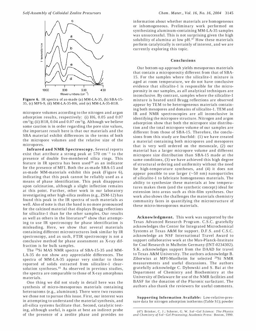
Infrared and NMR Spectroscopy. Several reportsexist that attribute a strong peak at 570 cm-1 to thepresence of double five-membered silica rings. Thisfeature in IR spectra has been used28 as an indicatorfor the presence of MFI. Both the as-made SBA-15 andas-made MM-materials exhibit this peak (Figure 6),indicating that this peak cannot be reliably used as ameans of phase identification. This peak disappearsupon calcination, although a slight inflection remainsat this point. Further, other work in our laboratoryinvestigating other high-silica zeolites (e.g., ZSM-12) hasfound this peak in the IR spectra of such materials aswell. Also of note is that the band is no more pronouncedfor the calcined material that displays Bragg reflectionsfor silicalite-1 than for the other samples. Our resultsas well as others in the literature41 show that attempt-ing to use IR spectroscopy for phase identification ismisleading. Here, we show that several materialscontaining different microstructures look similar by IRspectroscopy, and as such, FTIR spectroscopy is not aconclusive method for phase assessment as X-ray dif-fraction is for bulk samples.
The 29Si MAS NMR spectra of SBA-15-35 and MM-LA-35 do not show any appreciable differences. Thespectra of MM-LA-35 appear very similar to thosereported of solids extracted from silicalite-1 clear-solution syntheses.41 As observed in previous studies,the spectra are comparable to those of X-ray amorphousmaterials.
One thing we did not study in detail here was thesynthesis of micro-/mesoporous materials containingheteroatoms (e.g., aluminum). There were two reasonswe chose not to pursue this issue. First, our interest wasin attempting to understand the material synthesis, andall-silica systems facilitate that. Second, catalytic test-ing, although useful, is again at best an indirect probeof the presence of a zeolite phase and provides no
information about whether materials are homogeneousor inhomogeneous. Preliminary work performed onsynthesizing aluminum-containing MM-LA-35 sampleswas unsuccessful. This is not surprising given the highsolubility of alumina at low pH.47 How these materialsperform catalytically is certainly of interest, and we arecurrently exploring this topic.
Conclusions
Our bottom-up approach yields mesoporous materialsthat contain a microporosity different from that of SBA-15. For the samples where the silicalite-1 mixture isaged at room temperature, we do not have conclusiveevidence that silicalite-1 is responsible for the micro-porosity in our samples, as all analytical techniques areinconclusive. By contrast, samples where the silicalite-1mixture is heated until Bragg reflections are observedappear by TEM to be heterogeneous materials contain-ing both mesopores and domains of silicalite-1. TEM andIR and NMR spectroscopies are all inconclusive inidentifying the micropore structure. Nitrogen and argonadsorption show that both the micropore size distribu-tion and the total micropore volume of our samples aredifferent from those of SBA-15. Therefore, the conclu-sions from this study are fourfold: (1) we have createda material containing both micropores and mesoporesthat is very well ordered on the mesoscale, (2) ourmaterial has a larger micropore volume and differentmicropore size distribution than SBA-15 made at thesame conditions, (3) we have achieved this high degreeof structural ordering and uniformity without the needfor high-temperature syntheses, and (4) it does notappear possible to use larger (∼50 nm) nanoparticlesof silicalite-1 to fabricate homogeneous materials. Theability to synthesize these materials at low tempera-tures makes them (and the synthetic concept) ideal forextension into areas such as thin-film syntheses. Ourwork also shows the challenges the materials chemistrycommunity faces in quantifying the microstructure ofthese micro-/mesoporous materials.
Acknowledgment. This work was supported by theTexas Advanced Research Program. C.S.C. gratefullyacknowledges the Center for Integrated MicrochemicalSystems at Texas A&M for support. D.F.S. and C.S.C.acknowledge an NSF International Travel Award tosupport collaborative work at the Max-Planck-Institutefor Coal Research in Mulheim Germany (INT-0234302).S.K. acknowledges support from the DAAD for travelto Texas A&M University. The authors acknowledge B.Ziborwius at MPI-Muelheim for selected 29Si NMRmeasurements and useful discussions. The authorsgratefully acknowledge C. Dybowski and S. Bai at theDepartment of Chemistry and Biochemistry at theUniversity of Delaware for use of the NMR facilities andBASF for the donation of the Pluronic surfactant. Theauthors also thank the reviewers for useful comments.
Supporting Information Available: Low-relative-pres-sure data for nitrogen adsorption isotherms (Table S1); powder
(47) Brinker, C. J.; Scherer, G. W. Sol-Gel Science: The Physicsand Chemistry of Sol-Gel Processing; Academic Press: Boston, 1990.
Figure 6. IR spectra of as-made (a) MM-LA-35, (b) SBA-15-35, (c) MPS-9, (d) MM-LA-35-H6, and (e) MM-LA-35-H18.
Self-Assembly of Colloidal Zeolite Precursors Chem. Mater., Vol. 16, No. 16, 2004 3145

XRD patterns of MM-LA-35, SBA-15-35, and MPS-9 (FigureS1); high-angle XRD data for MM-LA-35, MM-LA-35-H6, MM-LA-35-H18, and Cabosil (Figure S2); nitrogen adsorptionisotherms for MM-LA-35-H6 and MM-LA-35-H18 (Figure S3);mesopore size distributions for MM-LA-35, SBA-15-35, MM-
LA-35-H6, MM-LA-35-H18, and MPS-9 (Figure S4). Thismaterial is available free of charge via the Internet athttp://pubs.acs.org.
CM035283J
3146 Chem. Mater., Vol. 16, No. 16, 2004 Carr et al.