Embed Size (px)
Citation preview
RESEARCH ARTICLE
Self-assembly of Chloro-Bridged Arene–Ruthenium BasedRectangle: Synthesis, Structural Characterization andSensing Study
Sankarasekaran Shanmugaraju • Harshal Jadhav •
Partha Sarathi Mukherjee
Received: 30 October 2013 / Revised: 22 January 2014 / Accepted: 28 January 2014 / Published online: 4 March 2014
� The National Academy of Sciences, India 2014
Abstract Self-assembly of a chloro-bridged half-sand-
wich p-cymene ruthenium(II) complex [Ru2(l-Cl2)(g6-p-
cymene)2Cl2] 1 with linear ditopic donor [L; trans-1,2-
bis(4-pyridyl)ethylene] in presence of 2 eq. AgNO3 in
CH3CN yielded a chloro-bridged molecular rectangle 2.
The rectangle 2 was isolated as nitrate salt in high yield
(90 %) and characterized by infra-red, 1H NMR spectros-
copy including ESI–MS analyses. Molecular structure of 2
was determined by single crystal X-ray diffraction study
The diffraction analysis shows that 2 adopts a tetranu-
clear rectangular geometry with the dimensions of
5.51 A 9 13.29 A and forming an infinite supramolecular
chain with large internal porosity arising through multiple
p–p and CH–p interactions between the adjacent rectan-
gles. Furthermore, rectangle 2 is used as selective receptor
for phenolic-nitroaromatic compounds such as picric acid,
dinitrophenol and nitrophenol.
Keywords Supramolecular chemistry � Self-assembly �Arene ligands � Ruthenium � X-ray crystallography
Introduction
Discrete molecular architectures of defined shapes and sizes
have attracted a great deal of attention because of their
intriguing structural features and potential applications [1].
A plethora of multifaceted supramolecular complexes with
interesting functional properties have been synthesized by
this elegant synthetic strategy employing various transition
metal acceptors and multidentate organic donors [2]. Plati-
num(II) or Palladium(II) based molecular building units
have been widely used as acceptors due to their rigid square-
planar coordination geometry [3]. Recently, several shape-
persistent macrocycles/cages have been constructed using
arene–ruthenium based organometallic building blocks
because of their structural stability and interesting properties
[4, 5]. The very first metal–ligand directed self-assembly of
molecular rectangle incorporating arene–ruthenium build-
ing units was reported by Suss-Fink et al. in 1997 [6]. Since
then a vast number of arene–ruthenium based molecular
rectangles have been synthesized which are mostly bridged
by O,O- or N,N-chelating organic ligands and connected by
linear bidentate N-donors [7]. But, chloro-bridged metalla-
rectangles employing arene–ruthenium building units are
not yet known because of the difficulty in pre-designing
chloro-bridged arene–ruthenium acceptor having *0� bite
angle between their acceptor sites. Herein we report the
synthesis and structural characterization of a chloro-bridged
tetranuclear cationic molecular rectangle 2 incorporating p-
cymene Ru(II) building block with 1,2-bis(4-pyridyl)ethyl-
ene (L) dipyridyl linker. Rectangle 2 shows a moderate
emission characteristic in CH3OH solution and used as a
macrocyclic receptor to check the binding affinity with ni-
troaromatic compounds. The binding affinity is described
using fluorescence spectroscopic titration study. The fluo-
rescence titration study demonstrated that rectangle 2
selectively binds with phenolic-nitroaromatic compounds
such as picric acid (PA), 2,4-dinitrophenol (DNP) and
4-nitrophenol (NP) by subsequent discernable changes in the
emission intensities. Notably, no such perturbation in
emission intensity was observed upon titration of 2 with
Electronic supplementary material The online version of thisarticle (doi:10.1007/s40010-014-0128-6) contains supplementarymaterial, which is available to authorized users.
S. Shanmugaraju � H. Jadhav � P. S. Mukherjee (&)
Department of Inorganic and Physical Chemistry, Indian
Institute of Science, Bangalore 560 012, India
e-mail: [email protected]
123
Proc. Natl. Acad. Sci., India, Sect. A Phys. Sci. (April–June 2014) 84(2):197–203
DOI 10.1007/s40010-014-0128-6
other non-phenolic nitroaromatics [1,3,5-trinitrotoluene
(TNT), 2,4-dinitrotoluene (DNT), nitrobenzene (NB),
nitrotoluene (NT), nitromethane (NM)]; thereby demon-
strating its potential use as a selective receptor for phenolic
nitroaromatics over the other nitroaromatics.
Experimental
Materials and Methods
The acceptor clip [Ru2(l-Cl2)(g6-p-cymene)2Cl2] (1) was
synthesized following the reported procedure [8]. trans-
1,2-bis(4-pyridyl)ethylene (L) was commercially pur-
chased and used without further purification. 1H NMR was
studied on a Bruker 400 MHz spectrometer. The chemical
shifts (d) are reported in ppm relative to tetramethylsilane
(Me4Si) as internal standard (0.0 ppm) or proton resonance
resulting from incomplete deuteration of the NMR solvent
CD3OD (3.33). IR spectra were recorded on a Bruker
ALPHA FT-IR spectrometer. Electronic absorption spec-
tral measurement was done using Perkin Elmer LAMBDA
750 UV/visible spectrophotometer. Fluorescence titration
studies were carried out on a HORIBA JOBIN–YVON
Fluoromax-4-spectrometer.
Synthesis of Rectangle (2)
A mixture of AgNO3 (50.1 mg, 0.30 mmol) and [Ru2(l-
Cl2)(g6-p-cymene)2Cl2] (1; 91.8 mg, 0.15 mmol) in CH3CN
(10 mL) was stirred at room temperature for 3 h, then filtered
to remove AgCl. Trans-1,2-bis(4-pyridyl)ethylene (L;
27.3 mg, 0.15 mmol) was added to the filtrate and the solution
was stirred at room temperature for 24 h. The solvent was
removed and the residue was extracted with acetonitrile. The
filtrate was concentrated to 2 ml and cold-diethyl ether was
added to precipitate out rectangle 2 as a brownish-orange
powder. Isolated yield: 90 %. 1H NMR (CD3OD, 400 MHz):
d (ppm) 8.34 (d, 8H, Ha-pyridyl, J = 5.6 Hz), 7.55 (d, 8H,
Hb-pyridyl, J = 5.6 Hz), 7.51 (s, 4H, H1-ethylene), 6.00 (d,
8H, H-cymene, J = 6.0 Hz), 5.86 (d, 8H, H-cymene,
J = 6.0 Hz), 2.93 - 2.86 (septet, 4H, H4-CH(CH3)2), 2.17
(s, 12H, H2-CH3), 1.39 (d, 24H, H3-(CH3)2CH, J = 6.8 Hz).
IR: t = 1,607 cm-1 corresponding to C=C double bonds.
UV/Vis (1.0 9 10-5 M, CH3OH) kmax (e) = 315 nm
(24 9 105 M-1 cm-1).
X-ray Data Collection and Structure Refinements
Crystal data of 2 were collected on a Bruker SMART
APEX CCD diffractometer using the SMART/SAINT
software [9]. X-ray quality crystal was mounted on a glass
fiber with traces of viscous oil. Intensity data were col-
lected using graphite-monochromatized Mo-Ka radiation
(0.7107 A) at 150 K. The structures were solved by direct
methods using the SHELX-97 [10–12] incorporated in
WinGX [13–15]. Empirical absorption corrections were
applied with SADABS [16]. All non-hydrogen atoms were
refined with anisotropic displacement coefficients. Hydro-
gen atoms were assigned isotropic displacement coeffi-
cients, U(H) = 1.2U(C) or 1.5U (C-methyl), and their
coordinates were allowed to ride on their respective
carbons.
Fluorescence Sensing Study
A 2 mL stock solution (9.0 9 10-5 M) of rectangle 2 in
CH3OH was placed in a quartz cell of 1 cm width and ni-
troaromatic stock solution (1.0 9 10-3 M) in CHCl3 was
added into it in an incremental fashion. The whole titration
experiment was carried out at 298 K. For all measurements
rectangle 2 was excited at kex = 310 nm and their corre-
sponding emission wavelengths were monitored from
kem = 310 nm onwards. For all the measurements both
excitation and emission slit widths were 5 nm. Analysis of
the normalized fluorescence emission intensity (I0/I) as a
function of increasing quencher concentration [Q] was well
described by the Stern–Volmer equation I0/I = 1
? KSV[Q]. The Stern–Volmer binding constant (KSV) was
calculated from the slope of the Stern–Volmer plot.
Results and Discussion
Synthesis and Characterization of Rectangle (2)
As shown in Scheme 1, the synthesis of chloro-bridged
cationic molecular rectangle 2 was accomplished in a
Scheme 1 Schematic
representation of the formation
of chloro-bridged molecular
rectangle 2
198 Shanmugaraju et al.
123
single step by the reaction of a chloro-bridged Ru2II-
acceptor (1) with dipyridyl donor (L) in presence of 2 eq.
AgNO3.
Upon addition of the solid dipyridyl donor L to a clear
yellow filtrate, which was obtained from the reaction of 1
and AgNO3 in a 1:2 molar ratio in CH3CN at room tem-
perature, and subsequent stirring at room temperature for
24 h resulted in the formation [2 ? 2] self-assembled
molecular rectangle 2 in good yield (90 %). The resulted
macrocycle is highly soluble in common organic solvents
like CH3OH, CH3CN, CH3NO2, CHCl3 and CH2Cl2. For-
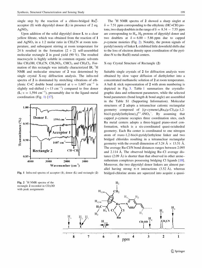
mation of this rectangle was initially characterized IR, 1H
NMR and molecular structure of 2 was determined by
single crystal X-ray diffraction analysis. The infra-red
spectra of 2 is dominated by stretching vibrations of eth-
ylenic C=C double bond centered at t = 1,607 cm-1 is
slightly red-shifted (*13 cm-1) compared to free donor
(L; t = 1,594 cm-1), presumably due to the ligand–metal
coordination (Fig. 1) [17].
The 1H NMR spectra of 2 showed a sharp singlet at
d = 7.51 ppm corresponding to the ethylenic (HC=CH) pro-
tons, two sharp doublets in the range ofd = 8.34 - 7.55 ppm
are corresponding to Ha, Hb protons of dipyridyl donor and
two doublets at d = 6.00 - 5.86 ppm due to capped
p-cymene moieties (Fig. 2). Notably, the proton signals of
pyridyl moiety of linker L exhibited little downfield shifts due
to the loss of electron density upon coordination of the pyri-
dine-N to the Ru(II) metal centers.
X-ray Crystal Structure of Rectangle (2)
Suitable single crystals of 2 for diffraction analysis were
obtained by slow vapor diffusion of diethylether into a
concentrated methanolic solution of 2 at room temperature.
A ball & stick representation of 2 with atom numbering is
depicted in Fig. 3. Table 1 summarizes the crystallo-
graphic data and refinement parameters, while the selected
bond parameters (bond length & bond angle) are assembled
in the Table S1 (Supporting Information). Molecular
structures of 2 adopts a tetranuclear cationic rectangular
geometry composed of [(p-cymene)4Ru4(l-Cl)4(l-1,2-
bis(4-pyridyl)ethylene)2]4?.4NO3-. By assuming that
capped p-cymene occupies three coordination sites, each
Ru metal centers adopts a three-legged piano-stool con-
formation, which is a six-coordinated quasi-octahedral
geometry. Each Ru center is coordinated to one nitrogen
atom of trans-1,2-bis(4-pyridyl)ethylene linker and two
bridged chlorides resulting in a tetranuclear rectangular
geometry with the overall dimension of 3.24 A 9 13.51 A.
The average Ru-Cl/N bond distances ranges between 2.095
and 2.114 A. The observed bridging Ru–Cl average dis-
tance (2.09 A) is shorter than that observed in other arene–
ruthenium complexes possessing bridging Cl ligands [18].
Moreover, the two dipyridyl donor linkers are almost par-
allel having strong p–p interactions (3.52 A), whereas
bridged-chlorine atoms are squeezed into acquire a quasi-Fig. 1 Infra-red spectra of acceptor (1), donor (L) and rectangle (2)
Fig. 2 1H NMR spectra of the
rectangle 2 recorded in CD3OD
with peak assignments
Synthesis, Structural Characterization and Sensing Study 199
123
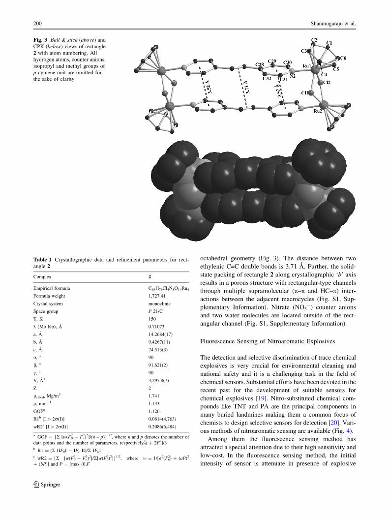
octahedral geometry (Fig. 3). The distance between two
ethylenic C=C double bonds is 3.71 A. Further, the solid-
state packing of rectangle 2 along crystallographic ‘b’ axis
results in a porous structure with rectangular-type channels
through multiple supramolecular (p–p and HC–p) inter-
actions between the adjacent macrocycles (Fig. S1, Sup-
plementary Information). Nitrate (NO3-) counter anions
and two water molecules are located outside of the rect-
angular channel (Fig. S1, Supplementary Information).
Fluorescence Sensing of Nitroaromatic Explosives
The detection and selective discrimination of trace chemical
explosives is very crucial for environmental cleaning and
national safety and it is a challenging task in the field of
chemical sensors. Substantial efforts have been devoted in the
recent past for the development of suitable sensors for
chemical explosives [19]. Nitro-substituted chemical com-
pounds like TNT and PA are the principal components in
many buried landmines making them a common focus of
chemists to design selective sensors for detection [20]. Vari-
ous methods of nitroaromatic sensing are available (Fig. 4).
Among them the fluorescence sensing method has
attracted a special attention due to their high sensitivity and
low-cost. In the fluorescence sensing method, the initial
intensity of sensor is attenuate in presence of explosive
Fig. 3 Ball & stick (above) and
CPK (below) views of rectangle
2 with atom numbering. All
hydrogen atoms, counter anions,
isopropyl and methyl groups of
p-cymene unit are omitted for
the sake of clarity
Table 1 Crystallographic data and refinement parameters for rect-
angle 2
Complex 2
Empirical formula C64H76Cl4N8O14Ru4
Formula weight 1,727.41
Crystal system monoclinic
Space group P 21/C
T, K 150
k (Mo Ka), A 0.71073
a, A 14.2684(17)
b, A 9.4267(11)
c, A 24.513(3)
a, � 90
b, � 91.621(2)
c, � 90
V, A3 3,295.8(7)
Z 2
qcalcd, Mg/m3 1.741
l, mm-1 1.133
GOFa 1.126
R1b [I [ 2r(I)] 0.0814(4,763)
wR2c [I [ 2r(I)] 0.2086(6,484)
a GOF = {R [w(F02 - Fc
2)2]/(n – p)}1/2, where n and p denotes the number of
data points and the number of parameters, respectively02) ? 2Fc
2]/3b R1 = (R IIF0I - IFc II)/R IF0Ic wR2 = {R [w(F0
2 - Fc2)2]/R[w(F0
2)2]}1/2, where w = 1/[r2(F02) ? (aP)2
? (bP)] and P = [max (0,F
200 Shanmugaraju et al.
123
residues [21]. A vast number of organic/inorganic poly-
meric and small molecule chemosensors have been
designed over the past two decades and demonstrated their
sensing ability [22, 23]. However, fluorescence sensor to
selectively discriminate the most common explosives such
as PA and TNT are known to a lesser extent. Despite of
their electron-deficient nature, TNT and PA are differing
much in their structural point of view. TNT is a toluene
based system whereas PA is a phenolic system. Therefore,
one can design a chemosensor with electron-donating
functional groups (preferentially electronegative atoms)
that binds selectively with –OH group of PA; thereby it is
easy to discriminate PA from TNT. Rectangle 2 is com-
posed of trans-1,2-(bis-4-pyridyl)ethylene (L) linkers and
chloro-bridged arene–ruthenium building units. Based on
the aforesaid consideration, it is expected that 2 can
selectively discriminate phenolic-nitroaromatics such as
PA, DNP and NP from other nitroaromatics (TNT, DNT
etc.) possibly via hydrogen bonding interactions. The
electronic absorption spectrum (Fig. 5) of 2 in CH3OH
(10 lM) shows a peak at k = 315 nm (e = 24 9 105 M-1
cm-1), which is tentatively assigned to intra/intermolecular
p–p* transitions/metal-to-ligand charge transfer (MLCT).
Rectangle 2 shows moderate emission characteristics in
CH3OH (10 lM) solution (Fig. 5).
To verify sensing propensity of rectangle 2 towards ni-
troaromatics, we performed fluorescence titration study of
2 with various nitroaromatics (Fig. 4) in CH3OH-CHCl3solution. Upon gradual addition of PA in CHCl3 (1.0 mM)
to a methanolic solution (10 lM) of 2 at room temperature,
the initial fluorescence intensity was quenched rapidly
(Fig. 6). Along with the efficient quenching of emission
intensity, a new low-energy band at k = *437 nm
appeared upon increasing concentration of PA. Similar
titration of 2 with DNP also attenuated the initial emission
intensity and two new low-energy bands at k = 419 nm
and 442 nm were observed upon gradual increasing the
concentration (Fig. S2, Supplementary Information). Sim-
ilar fluorescence titration of 2 with NP caused only a sig-
nificant quenching of emission intensity without any new
low-energy band even at high concentration (Fig. 6). In all
the cases, an upward curvature (nonlinear) type of Stern–
Volmer plot was obtained from the fluorescence quenching
titration profile. According to the Stern–Volmer equation
(see experimental section), for the static or dynamic
quenching a linear Stern–Volmer plot is expected from the
plot of I0/I and quencher concentration [Q]. However, the
obtained nonlinear plot suggest that the fluorescence
quenching of rectangle upon the gradual addition of ni-
troaromatics follows both static as well as dynamic
quenching pathways through charge-transfer complex for-
mation. A significant increase in the absorption intensity of
2 was noticed upon the gradual mixing of NP in CHCl3(1.0 9 10-3 M) to a methanolic solution of 2 (9.0 9 10-5
M) at room temperature (Fig. S3, Supporting Information).
Further, a marked visual color change (light brown to
intense yellow) was observed upon mixing of rectangle 2 in
CH3OH with chloroform solution of DNP and PA (Fig. 7).
The considerable changes of initial absorption intensity
including a sharp visual color change are indicative of the
formation of ground state charge transfer (CT) complex
and supported that the observed fluorescence quenching of
2 mainly follows the static quenching mechanism via
ground-state charge transfer complex formation. Therefore,
Fig. 4 Tested nitroaromatics
Fig. 5 UV–Visible absorption
(left) and emission (right)
spectra of rectangle 2 in CH3OH
(10 lM) at room temperature
Synthesis, Structural Characterization and Sensing Study 201
123
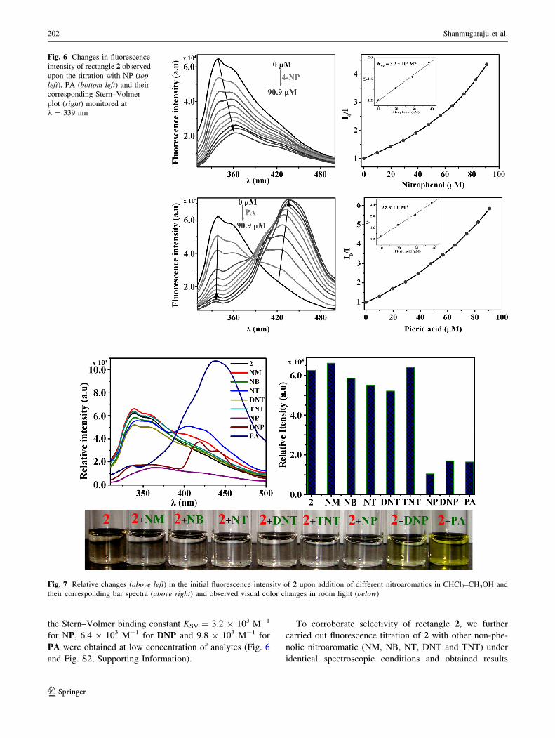
the Stern–Volmer binding constant KSV = 3.2 9 103 M-1
for NP, 6.4 9 103 M-1 for DNP and 9.8 9 103 M-1 for
PA were obtained at low concentration of analytes (Fig. 6
and Fig. S2, Supporting Information).
To corroborate selectivity of rectangle 2, we further
carried out fluorescence titration of 2 with other non-phe-
nolic nitroaromatic (NM, NB, NT, DNT and TNT) under
identical spectroscopic conditions and obtained results
Fig. 6 Changes in fluorescence
intensity of rectangle 2 observed
upon the titration with NP (top
left), PA (bottom left) and their
corresponding Stern–Volmer
plot (right) monitored at
k = 339 nm
Fig. 7 Relative changes (above left) in the initial fluorescence intensity of 2 upon addition of different nitroaromatics in CHCl3–CH3OH and
their corresponding bar spectra (above right) and observed visual color changes in room light (below)
202 Shanmugaraju et al.
123
were compared with phenolic nitroaromatics (PA, DNP
and NP). As shown in Fig. 7, the tested non-phenolic ni-
troaromatics elicited almost no fluorescence quenching
responses whereas the phenolic nitroaromatic exhibited a
significant fluorescence quenching. The possible reason for
the high selectivity of 2 towards phenolic-analytes is pre-
sumably ascribed to the hydrogen bonding interactions
between –OH and bridged Cl atom.
Conclusion
In conclusion, we have shown the synthesis of a chloro-
bridged tetranuclear cationic molecular rectangle 2 via the
self-assembly of arene–ruthenium acceptor 1 with a sym-
metrical dipyridyl donor (L) in quantitative yield. Rect-
angle 2 is fully characterized by 1H NMR, IR and single-
crystal diffraction analysis. Further we have demonstrated
the fluorescence quenching based selective sensing of
phenolic nitroaromatics from other interfering nitroaro-
matics. Thus, rectangle 2 can be viewed as fluorescence
sensor to discriminate PA and TNT.
Acknowledgments P.S.M thanks the Department of Science and
Technology (DST), India, for the financial support.
References
1. Chakrabarty R, Mukherjee PS, Stang PJ (2011) Supramolecular
coordination: self-assembly of finite two- and three-dimensional
ensembles. Chem Rev 111:6810–6918
2. Ruben M, Lehn JM, Muller P (2006) Addressing metal centres in
supramolecular assemblies. Chem Soc Rev 35:1056–1067
3. Shanmugaraju S, Bar AK, Chi KW, Mukherjee PS (2010)
Coordination-driven self-assembly of metallamacrocycles via a
new Pt2II organometallic building block with 90� geometry and
optical sensing of anions. Organometallics 29:2971–2980
4. Shanmugaraju S, Bar AK, Mukherjee PS (2010) Ruthenium–
oxygen coordination-driven self-assembly of a Ru8II incomplete
prism: synthesis, structure, and shape-selective molecular rec-
ognition study. Inorg Chem 49:10235–10237
5. Shanmugaraju S, Joshi SA, Mukherjee PS (2011) Self-assembly
of metallamacrocycles using a dinuclear organometallic acceptor:
synthesis, characterization, and sensing study. Inorg Chem
50:11736–11745
6. Yan H, Suss-Fink G, Neels A, Stoeckli-Evans H (1997) Mono-,
di- and tetra-nuclear p-cymeneruthenium complexes containing
oxalato ligands. J Chem Soc Dalton Trans 22:4345–4350
7. Therrien B (2009) Arene ruthenium cages: boxes full of surprises.
Eur J Inorg Chem 17:2445–2453
8. Zelonka RA, Baird MC (1972) Benzene complexes of ruthe-
nium(II). Can J Chem 50:3063–3072
9. SMART/SAINT (2004) Bruker AXS, Inc., Madison, WI
10. Sheldrick GM (1998) SHELX-97, program for the solution and
refinement of crystal structures. University of Gottingen,
Gottingen
11. Farrugia LJ (2003) WinGX: an integrated system of windows
programs for the solution, refinement and analysis for single
crystal X-ray diffraction data, version 1.65.04. Department of
Chemistry, University of Glasgow, Glasgow
12. Farrugia LJ (1999) WinGX suite for small-molecule single-
crystal crystallography. J Appl Crystallogr 32:837–838
13. Sheldrick GM (1999) SADABS, Bruker nonius area detector
scaling and absorption correction, version 205. University of
Gottingen, Gottingen
14. Farrugia LJ (1997) ORTEP-3 for Windows, version 1.08. J Appl
Crystallogr 30:565
15. Spek AL (1990) PLATON, An integrated tool for the analysis of
the results of a single crystal structure determination. Acta
Crystallogr A 46:C34
16. Govindaswamy P, Suss-Fink G, Therrien B (2007) Self-assem-
bled chloro-bridged (arene)ruthenium metallo-prisms: synthesis
and molecular structure of cationic complexes of the type
[Ru6(g6-arene)6(l3-tpt-jN)2(l-Cl)6]6?(tpt = 2,4,6-tris(pyridi-
nyl)-1,3,5-triazine). Organometallics 26:915–924
17. Therrien B, Ward TR, Pilkington M, Hoffmann C, Gilardoni F,
Weber J (1998) Synthesis and reactivity of tethered g1:g6-
(phosphinoarene)ruthenium dichlorides. Organometallics
17:330–337
18. Germain ME, Knapp MJ (2009) Optical explosives detection:
from color changes to fluorescence turn-on. Chem Soc Rev
38:2543–2555
19. Toal SJ, Trogler WC (2006) Polymer sensors for nitroaromatic
explosives detection. J Mater Chem 16:2871–2883
20. Moore DS (2004) Instrumentation for trace detection of high
explosives. Rev Sci Instrum 75:2499–2512
21. John H, Sailor MJ, Magde D, Trogler WC (2003) Detection of
nitroaromatic explosives based on photoluminescent polymers
containing metalloles. J Am Chem Soc 125:3821–3830
22. Shanmugaraju S, Joshi SA, Mukherjee PS (2011) Fluorescence
and visual sensing of nitroaromatic explosives using electron rich
discrete fluorophores. J Mater Chem 21:9130–9138
23. Shanmugaraju S, Jadhav H, Karthik R, Mukherjee PS (2013)
Electron rich supramolecular polymers as fluorescent sensors for
nitroaromatics. RSC Adv 3:4940–4950
Synthesis, Structural Characterization and Sensing Study 203
123


![Unexpected Cleavage of Thiacalix[4]arene Sulfoxides · Unexpected Cleavage of Thiacalix[4]arene Sulfoxides Jiří Mikšátko†, Václav Eigner,§ and Pavel Lhoták†* †Department](https://img.pdfslide.us/doc/110x75/5ebb58fb58a7af2056069c00/unexpected-cleavage-of-thiacalix4arene-unexpected-cleavage-of-thiacalix4arene.jpg)


![Fluorescent phenanthridine-based calix[4]arene derivatives](https://img.pdfslide.us/doc/110x75/61c21d84d2e79c7793206f73/fluorescent-phenanthridine-based-calix4arene-derivatives-.jpg)
![Development of Calix[4]arene-Functionalized](https://img.pdfslide.us/doc/110x75/61ab1bfbbc68120d180622ab/development-of-calix4arene-functionalized-.jpg)

![SYNTHESIS AND APPLICATIONS OF NOVEL RESORCIN[4]ARENE](https://img.pdfslide.us/doc/110x75/625e5bfc00cedb0726701d72/synthesis-and-applications-of-novel-resorcin4arene-.jpg)