Embed Size (px)
Citation preview
Research ArticleSelenium Polysaccharide SPMP-2a from Pleurotus geesteranusAlleviates H2O2-Induced Oxidative Damage in HaCaT Cells
Yujun Sun,1,2 Cheng Zhou,2 Shoucheng Huang,2 and Changjun Jiang3
1College of Life Sciences, Anhui Agricultural University, Hefei 230036, China2School of Life Science, Anhui Science and Technology University, Fengyang 233100, China3State Key Laboratory of Tea Plant Biology and Utilization, Anhui Agricultural University, Hefei 230036, China
Correspondence should be addressed to Yujun Sun; [email protected] and Changjun Jiang; [email protected]
Received 14 October 2016; Revised 22 December 2016; Accepted 15 January 2017; Published 15 February 2017
Academic Editor: Stanley Brul
Copyright © 2017 Yujun Sun et al. This is an open access article distributed under the Creative Commons Attribution License,which permits unrestricted use, distribution, and reproduction in any medium, provided the original work is properly cited.
Selenium- (Se-) enriched polysaccharide SPMP-2a was extracted and purified from Pleurotus geesteranus. SPMP-2a is a whiteflocculent polysaccharide and soluble in water, with a molecular weight of 3.32 × 10⁴ Da. Fourier transform infrared spectroscopyspectral analysis indicated that it belongs to an acid Se polysaccharide with 𝛼-D-glucopyranoside bond. The effects of Sepolysaccharide SPMP-2a in P. geesteranus against hydrogen peroxide- (H
2O2-) induced oxidative damage in human keratinocytes
(HaCaT) cells were evaluated further. Reduced cell viability and elevated apoptotic rates inH2O2-treatedHaCaT cellswere proven by
MTT and flow cytometry assays. Hoechst 33342 staining revealed chromatin condensations in the nuclei of HaCaT cells. However,with the addition of SPMP-2a, cell viability improved, nuclear condensation declined, and cell apoptotic rates dropped significantly.Ultrastructural observation consistently revealed that treatments with SPMP-2a reduced the number of swollen and vacuolarmitochondria in the H
2O2-treated cells compared with the controls. Furthermore, SPMP-2a increased the superoxide dismutase
(SOD) and catalase (CAT) activities and reduced reactive oxygen species (ROS) content. Western blot analysis showed that SPMP-2a treatment effectively increased B-cell lymphoma 2 (Bcl-2) protein expression. Therefore, SPMP-2a could improve cellularantioxidant enzyme activities, reduce ROS levels, and increase Bcl-2 protein expression levels, thereby reducing cell apoptosis andprotecting HaCaT cells from H
2O2-induced oxidative damage.
1. Introduction
In living organisms, reactive oxygen species (ROS) such assuperoxide anion free radical (O2−∙), hydroxyl free radical(∙OH), and hydrogen peroxide (H
2O2) can be generated [1].
Excessive ROS production exceeds its ROS-scavenging abil-ity, which causes inflammation, aging, and even cancer [2].ROS-mediated oxidative stress has been shown to be corre-lated with the occurrence of various diseases (cardiovasculardiseases, e.g., hypertension, dyslipidemia, and obesity) andcell apoptosis [3]. Previous studies have indicated that H
2O2
can lead to direct oxidation of membrane lipid and proteins[4]. Furthermore, it can pass through the cell membrane andproduce strong activity of free radicals such as ∙OH, therebyinducing cell apoptosis through various metabolic pathways[5].
Increasing evidence has indicated that fungal polysaccha-rides can scavenge free radicals, inhibit lipid peroxidation,and delay aging [6, 7]. Edible fungi polysaccharides areknown as biological response modifiers [8]. Wang et al.[9] reported that polysaccharides from Cordyceps sinensisenhance the activities of glutathione peroxidase (GSH-Px)and superoxide dismutase (SOD), thereby eliminating theaccumulation of ROS, includingO2−∙ and ∙OH.Gao et al. [10]showed that exopolysaccharides from Russula vinosa havestrong in vitro antioxidant activity to scavenge DPPH freeradical, O2−∙, and ∙OH. Moreover, selenium (Se) is one ofthe essential trace elements for animals and humans and isan important component of GSH-Px, which is an antioxidantin red blood cells [11]. Se constitutes the active centers ofseveral oxidases [12], promotes peroxide decomposition, andprotects cell membrane structures [13]. Inorganic Se is the
HindawiBioMed Research InternationalVolume 2017, Article ID 4940384, 9 pageshttps://doi.org/10.1155/2017/4940384
2 BioMed Research International
primary form of Se in nature but is difficult for animals andhumans to absorb [14]. In addition, inorganic Se has greatertoxicity than organic Se, and excessive intake is detrimentalto the animal body [15]. Studies found that ediblemushroomsare capable of accumulating Se [16]. Edible mushrooms linkinorganic Se with polysaccharides, which convert inorganicSe into organic Se polysaccharide. Organic Se polysaccharidehas both polysaccharides and Se, which the human body caneasily absorb [17].
The mushroom Pleurotus geesteranus belongs to Dikaryasubkingdom, Basidiomycota phylum, Agaricomycotina sub-phylum,Agaricomycetes class, Agaricales order, Pleurotaceaefamily, and Pleurotus genus [18]. It is a popular edible mush-room with a unique flavor and smooth taste. Polysaccharidesfrom P. geesteranus have strong antioxidant [19], bloodlipid lowering [20], and antitumor properties [21]. However,information about the Se-combining polysaccharide of P.geesteranus is scarce. In our previous study, polysaccharidesextracted from P. geesteranus exhibited higher superoxideradical- and hydroxyl radical-scavenging activities in a dose-dependent manner [22]. The present study uses MTT assayto examine cell viability, Hoechst 33342 fluorescence stainingto show apoptotic cell morphology, flow cytometry to detectapoptotic rates of HaCaT cells, and Western blot analy-sis to investigate its protective effects and the underlyingmechanisms of SPMP-2a onH
2O2-induced oxidative damage
in human keratinocytes (HaCaT). Results showed that Se-combining polysaccharide from P. geesteranus (SPMP-2a)reduced oxidative stress-induced cell death. This study pro-vided important evidence that SPMP-2a has great potentialto alleviate oxidative stress and cell damage.
2. Materials and Methods
2.1. Bacterial Strains and Cell Lines. Pleurotus geesteranus(GIM5.217) was purchased from the Institute of MicrobialCulture Collection in Guangdong and identified by rDNA-ITS sequence analysis (GenBank number KY417089). HaCaTcell lines were obtained from Shengbo BiopharmaceuticalCo. (Shanghai, China). DEAE-Sepharose Fast Flow andSuperdex-200 were purchased from Amersham BiosciencesCo. (Uppsala, Sweden). Sodium selenite was acquired fromSigma Chemical Co. (St. Louis, MO, USA).
2.2. Preparation of P. geesteranus Se Polysaccharide. P. geester-anus was inoculated on potato dextrose agar (PDA) culturemedium for activation (long-time preserved strain beforeculturing). After two rounds of culture, the seed culturesolution, approximately 10% (v/v) of the final culture solution,was added to a fermentation tank (FUS-50L, GuoqiangBiochemical Engineering Equipment Co., Ltd., Shanghai,China) with the addition of 20 ug/mL selenite sodium. Liquidculture was performed for 7 d at 180 rpm and 25∘C with0.9 vvm (air volume/culture volume/min), and centrifugalseparation was used to collect the mycelia.
Se-enriched mycelium was made into powder and subse-quently added to distilled water. After being treated at 70∘Cfor 3 hours (h), the extract was concentrated and precipitated
by adding threefold volume of 95% ethanol (v/v) and keepingit at 4∘C for 24 h. After centrifugation at 4800 rpm for10min, the supernatant was collected and freeze-dried. TheSe-enriched polysaccharide from mycelia of P. geesteranus(SPMP) was obtained.
SPMP was dissolved in distilled water and then fraction-ated by DEAE-Sepharose Fast Flow (2.6 cm × 50 cm) with adiscontinuous gradient elution of distilled water and 1mol/LNaCl at 1.0mL/min [23]. The elution profile was made bythe phenol-sulfuric acid assay, and two elution peaks, SPMP-1 and SPMP-2, were visualized. SPMP-2 was further appliedto Superdex-200 (1.6 cm × 60 cm) with the AKTA� Purifier10 system and eluted with distilled water at 1.0mL/min flowrate. The elution profile showed three elution peaks, namely,SPMP-2a, SPMP-2b, and SPMP-2c.
2.3. Determination of Purity and Molecular Weight of SPMP-2a. Thepurity andmolecular weight (MW) of SPMP-2aweredetermined by high performance liquid chromatography(HPLC) on an Agilent 1200 system (Agilent Technologies,Palo Alto, CA, USA). A total of 20𝜇L polysaccharide solution(10mg/mL) was injected and eluted with double distilledwater at 35∘C at 1.0mL/min flow rate [26]. A series ofconcentration gradients of dextran was used to draw thestandard curve and make the regression equation, which wasused to calculate the MW of SPMP-2a.
2.4. FTIR Spectral Analysis of SPMP-2a. The functionalgroups of SPMP-2a were detected by Fourier transforminfrared (FTIR) spectrometer. SPMP-2a (1mg) was groundinto powder andmixedwith 100mgKBr powder.Themixturewas then pressed into pellets for FTIR spectra measurementin the 400–4000 cm−1 frequency range [24].
2.5. Effects of SPMP-2a and H2O2 on HaCaT Cell Viability.The HaCaT cells at their logarithmic growth phases wereseeded at densities of 1 × 10⁵ cells/mL into a 96-well cellculture plate. A 100 𝜇L medium containing various concen-trations of SPMP-2a (0, 100, 200, 300, 400, or 600𝜇g/mL) orH2O2(0, 50, 100, 200, 400, 600, or 800𝜇mol/L) was added to
the cell plate after 24 h. After a 20-hour incubation period,the cell medium was aspirated and 20 𝜇L 5mg/mL MTTsolution was added and cultured for 4 h. The supernatantwas discarded, and 150 𝜇L DMSO was added to dissolve theprecipitate. The optical density (OD) value was measuredat 490 nm, and the cell survival rates were calculated. Eachtreatment was repeated five times. The cell survival rateswere calculated by the following formula: (%) = (OD treatedgroup/OD blank) × 100%.
2.6. Effects of SPMP-2a against H2O2-Induced Cell Deathin HaCaT Cells. The experiments were divided into thefollowing groups: the control group, the model group, andthe SPMP-2a treatment groups (100, 200, and 300 𝜇g/mL),plus five replicates for each group. The HaCaT cells at theirlogarithmic growth phases were seeded at densities of 1 ×10⁵ cells/mL into a 96-well cell culture plate with a 100𝜇Lmedium. After a 24-hour incubation period, the 100 𝜇L
BioMed Research International 3
medium containing various concentrations of SPMP-2a wasadded and incubated for 20 h. Except for the control group,100 𝜇L of 200𝜇mol/L H
2O2was added in the other four
groups and incubated for another 6 h before cell viability wascalculated.
2.7. Cell Staining and Cell Apoptosis Detection. The HaCaTcells were seeded into culture dishes at densities of 1 ×10⁵ cells/mL.The cell mediumwas aspirated and washed withphosphate buffer saline (PBS) after SPMP-2a treatment. TheHoechst 33342 dye was diluted to 10 𝜇g/mL in PBS. Cells werestained with Hoechst 33342 at room temperature for 30minand rinsed with PBS to remove the dye. Cell morphologywas examined using confocal laser scanning microscopy(FV1000, Olympus, Japan).
TheHaCaTwas seeded in 6-well plates at 2× 10⁵ cells/welldensity. Cell damage was induced by H
2O2and alleviated
by polysaccharides. The cells were digested with trypsin andcollected by centrifuging at 2000 rpm for 5min.The cellswerewashed with PBS followed by centrifuging at 2000 rpm for5min. A total number of 1–5 × 10⁵ cells were collected andresuspended in a 100 𝜇L 1x binding buffer. A total of 5 𝜇LAnnexin V-FITC and 5 𝜇L PI of staining solution were addedto the cell suspension, mixed gently, and incubated in thedark at room temperature for 10min.Then, 400𝜇L 1x bindingbuffer was added to the mixed cells. Apoptosis was detectedby flow cytometry (FACSAria, American BD Company).
2.8. Ultrastructural Observation. Cell samples were collectedand fixed in 2.5% glutaraldehyde at room temperature for 2 h.Then, the samples were rinsed three times with PBS (0.1M,pH7.2) andfixed in 1.0%osmium tetroxide for 1 h, followed byrinsingwith PBS. Subsequently, the samples were dehydrated,embedded, and cut into thin sections (approximately 70 nm).Ultrathin sections were observed using transmission electronmicroscopy. At least 10 individual cells were observed. Eachsample was sectioned three times, and representative pho-tographs were selected.
2.9. SOD and CAT Activity and ROS Content Measure-ment. Cells were digested, collected, and lysed with a RIPAbuffer. Cell lysate was centrifuged at 12000 rpm/min at 4∘Cfor 5 minutes. The supernatants were collected and testedaccording to the SOD and CAT kit instructions. DCFH-DAwas used as fluorescent probe according to DCFH-DA kitinstructions. Flow cytometry was used to detect ROS contentin the samples, with 488 nm excitation wavelength of 530 nmemission wavelength.
2.10. Western Blot Analysis. All proteins were extractedand subjected to SDS-PAGE. After being transferred to amembrane and blotted with 5% skim milk, the primaryantibodies (anti-Bcl-2, 1 : 1000, and 𝛼-tubulin 1 : 500) wereincubated with membrane overnight at 4∘C. A horseradishperoxidase conjugate antibody (1 : 10000) washed with PBSwas incubated thereafter with the membrane at room tem-perature for 1 h. The enhanced chemiluminescence methodwas used for X-ray film exposure, development, and fixing.
UV1 280 nmUV2 215 nmUV3 260 nm
SPMP-2a
SPMP-2c
SPMP-2b
20 40 60 80 100 120 1400
(mL)
0
200
400
600
800
1000
1200
1400
(mAU
)
1600
1800
2000
2200
2400
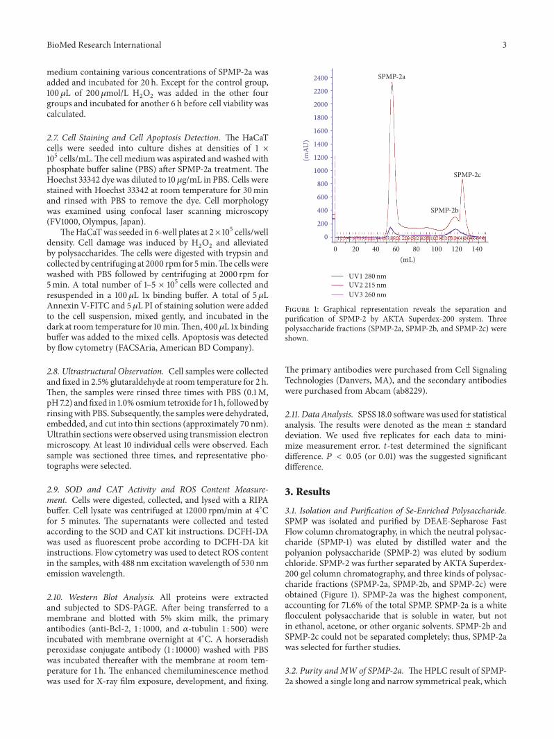
Figure 1: Graphical representation reveals the separation andpurification of SPMP-2 by AKTA Superdex-200 system. Threepolysaccharide fractions (SPMP-2a, SPMP-2b, and SPMP-2c) wereshown.
The primary antibodies were purchased from Cell SignalingTechnologies (Danvers, MA), and the secondary antibodieswere purchased from Abcam (ab8229).
2.11. Data Analysis. SPSS 18.0 software was used for statisticalanalysis. The results were denoted as the mean ± standarddeviation. We used five replicates for each data to mini-mize measurement error. 𝑡-test determined the significantdifference. 𝑃 < 0.05 (or 0.01) was the suggested significantdifference.
3. Results
3.1. Isolation and Purification of Se-Enriched Polysaccharide.SPMP was isolated and purified by DEAE-Sepharose FastFlow column chromatography, in which the neutral polysac-charide (SPMP-1) was eluted by distilled water and thepolyanion polysaccharide (SPMP-2) was eluted by sodiumchloride. SPMP-2 was further separated by AKTA Superdex-200 gel column chromatography, and three kinds of polysac-charide fractions (SPMP-2a, SPMP-2b, and SPMP-2c) wereobtained (Figure 1). SPMP-2a was the highest component,accounting for 71.6% of the total SPMP. SPMP-2a is a whiteflocculent polysaccharide that is soluble in water, but notin ethanol, acetone, or other organic solvents. SPMP-2b andSPMP-2c could not be separated completely; thus, SPMP-2awas selected for further studies.
3.2. Purity and MW of SPMP-2a. TheHPLC result of SPMP-2a showed a single long and narrow symmetrical peak, which
4 BioMed Research International
20
40
60
80
100
120
(mV
)
8.78
2
2 4 6 8 10 120
t (min)
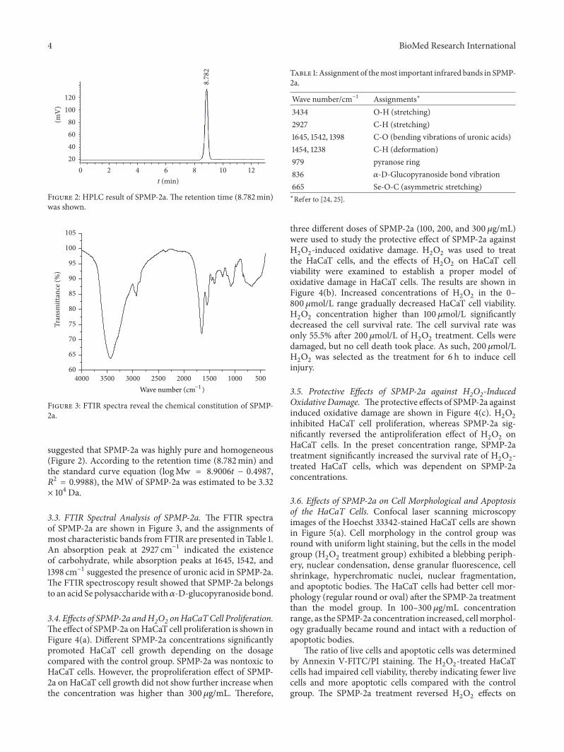
Figure 2: HPLC result of SPMP-2a. The retention time (8.782min)was shown.
3500 3000 2500 2000 1500 1000 5004000Wave number (cm−1
60
)
65
70
75
80
85
90
95
100
105
Tran
smitt
ance
(%)
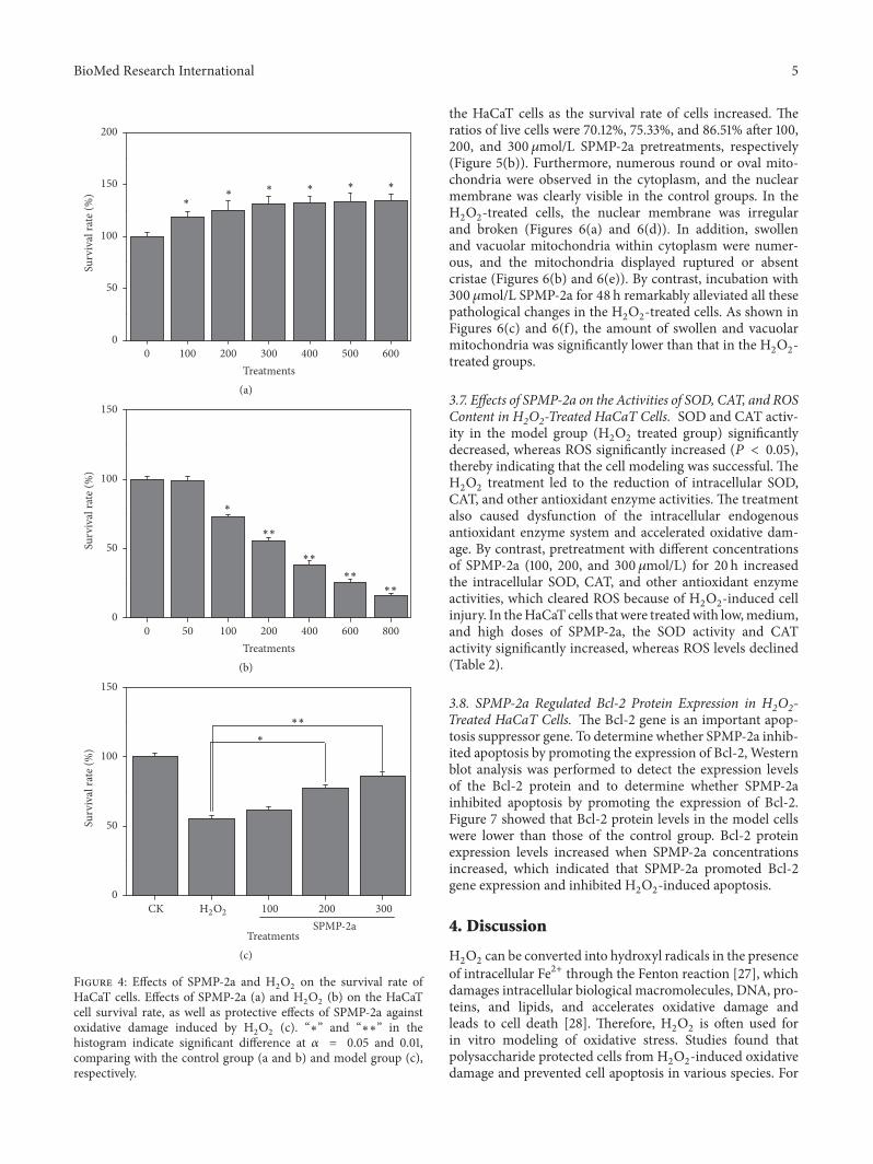
Figure 3: FTIR spectra reveal the chemical constitution of SPMP-2a.
suggested that SPMP-2a was highly pure and homogeneous(Figure 2). According to the retention time (8.782min) andthe standard curve equation (logMw = 8.9006𝑡 − 0.4987,𝑅2 = 0.9988), the MW of SPMP-2a was estimated to be 3.32× 10⁴Da.
3.3. FTIR Spectral Analysis of SPMP-2a. The FTIR spectraof SPMP-2a are shown in Figure 3, and the assignments ofmost characteristic bands from FTIR are presented in Table 1.An absorption peak at 2927 cm−1 indicated the existenceof carbohydrate, while absorption peaks at 1645, 1542, and1398 cm−1 suggested the presence of uronic acid in SPMP-2a.The FTIR spectroscopy result showed that SPMP-2a belongsto an acid Se polysaccharidewith𝛼-D-glucopyranoside bond.
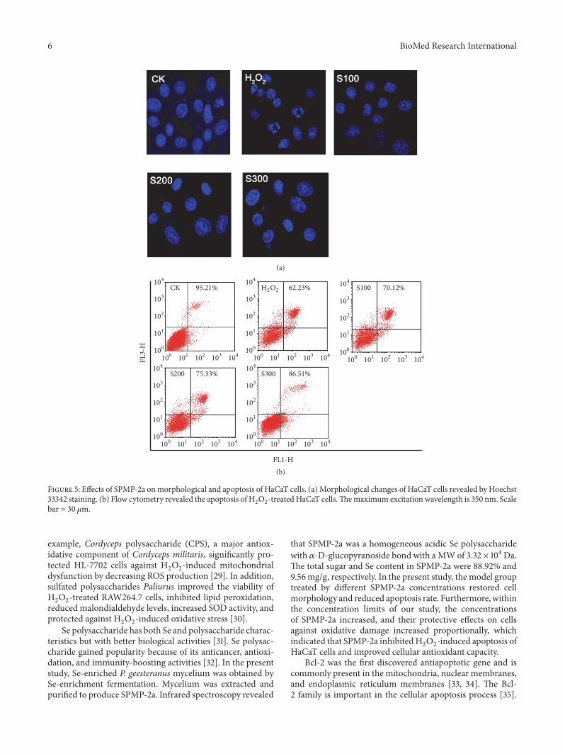
3.4. Effects of SPMP-2a andH2O2 onHaCaTCell Proliferation.Theeffect of SPMP-2a onHaCaT cell proliferation is shown inFigure 4(a). Different SPMP-2a concentrations significantlypromoted HaCaT cell growth depending on the dosagecompared with the control group. SPMP-2a was nontoxic toHaCaT cells. However, the proproliferation effect of SPMP-2a on HaCaT cell growth did not show further increase whenthe concentration was higher than 300 𝜇g/mL. Therefore,
Table 1: Assignment of themost important infrared bands in SPMP-2a.
Wave number/cm−1 Assignments∗
3434 O-H (stretching)2927 C-H (stretching)1645, 1542, 1398 C-O (bending vibrations of uronic acids)1454, 1238 C-H (deformation)979 pyranose ring836 𝛼-D-Glucopyranoside bond vibration665 Se-O-C (asymmetric stretching)∗Refer to [24, 25].
three different doses of SPMP-2a (100, 200, and 300 𝜇g/mL)were used to study the protective effect of SPMP-2a againstH2O2-induced oxidative damage. H
2O2was used to treat
the HaCaT cells, and the effects of H2O2on HaCaT cell
viability were examined to establish a proper model ofoxidative damage in HaCaT cells. The results are shown inFigure 4(b). Increased concentrations of H
2O2in the 0–
800 𝜇mol/L range gradually decreased HaCaT cell viability.H2O2concentration higher than 100𝜇mol/L significantly
decreased the cell survival rate. The cell survival rate wasonly 55.5% after 200𝜇mol/L of H
2O2treatment. Cells were
damaged, but no cell death took place. As such, 200𝜇mol/LH2O2was selected as the treatment for 6 h to induce cell
injury.
3.5. Protective Effects of SPMP-2a against H2O2-InducedOxidative Damage. Theprotective effects of SPMP-2a againstinduced oxidative damage are shown in Figure 4(c). H
2O2
inhibited HaCaT cell proliferation, whereas SPMP-2a sig-nificantly reversed the antiproliferation effect of H
2O2on
HaCaT cells. In the preset concentration range, SPMP-2atreatment significantly increased the survival rate of H
2O2-
treated HaCaT cells, which was dependent on SPMP-2aconcentrations.
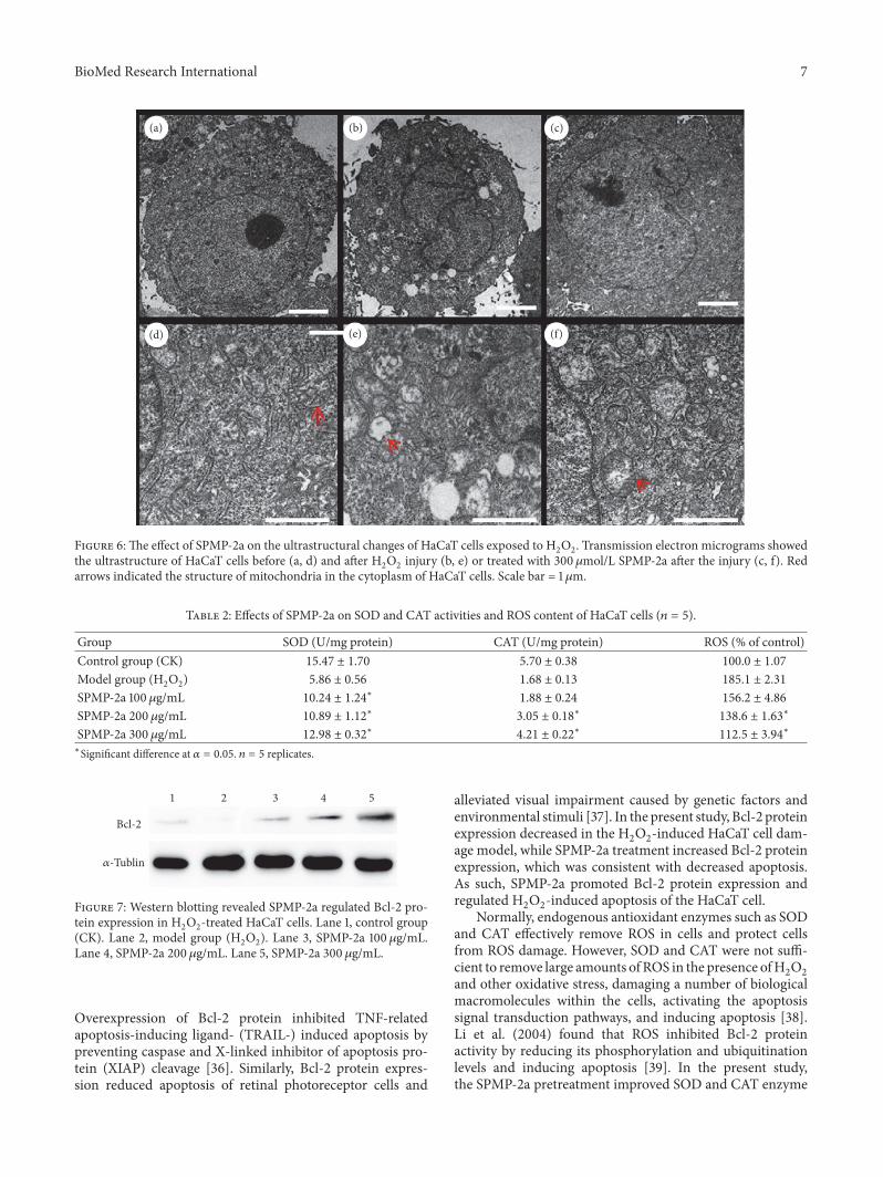
3.6. Effects of SPMP-2a on Cell Morphological and Apoptosisof the HaCaT Cells. Confocal laser scanning microscopyimages of the Hoechst 33342-stained HaCaT cells are shownin Figure 5(a). Cell morphology in the control group wasround with uniform light staining, but the cells in the modelgroup (H
2O2treatment group) exhibited a blebbing periph-
ery, nuclear condensation, dense granular fluorescence, cellshrinkage, hyperchromatic nuclei, nuclear fragmentation,and apoptotic bodies. The HaCaT cells had better cell mor-phology (regular round or oval) after the SPMP-2a treatmentthan the model group. In 100–300𝜇g/mL concentrationrange, as the SPMP-2a concentration increased, cellmorphol-ogy gradually became round and intact with a reduction ofapoptotic bodies.
The ratio of live cells and apoptotic cells was determinedby Annexin V-FITC/PI staining. The H
2O2-treated HaCaT
cells had impaired cell viability, thereby indicating fewer livecells and more apoptotic cells compared with the controlgroup. The SPMP-2a treatment reversed H
2O2effects on
BioMed Research International 5
∗∗∗∗∗
∗
0
50
100
150
200
Surv
ival
rate
(%)
100 200 300 400 500 6000Treatments(a)
∗∗
∗∗
∗∗
∗∗
∗
0
50
100
150
Surv
ival
rate
(%)
50 100 200 400 600 8000Treatments(b)
CK 100 200 300
TreatmentsSPMP-2a
∗∗
∗
0
50
100
150
Surv
ival
rate
(%)
H2O2
(c)
Figure 4: Effects of SPMP-2a and H2O2on the survival rate of
HaCaT cells. Effects of SPMP-2a (a) and H2O2(b) on the HaCaT
cell survival rate, as well as protective effects of SPMP-2a againstoxidative damage induced by H
2O2(c). “∗” and “∗∗” in the
histogram indicate significant difference at 𝛼 = 0.05 and 0.01,comparing with the control group (a and b) and model group (c),respectively.
the HaCaT cells as the survival rate of cells increased. Theratios of live cells were 70.12%, 75.33%, and 86.51% after 100,200, and 300𝜇mol/L SPMP-2a pretreatments, respectively(Figure 5(b)). Furthermore, numerous round or oval mito-chondria were observed in the cytoplasm, and the nuclearmembrane was clearly visible in the control groups. In theH2O2-treated cells, the nuclear membrane was irregular
and broken (Figures 6(a) and 6(d)). In addition, swollenand vacuolar mitochondria within cytoplasm were numer-ous, and the mitochondria displayed ruptured or absentcristae (Figures 6(b) and 6(e)). By contrast, incubation with300 𝜇mol/L SPMP-2a for 48 h remarkably alleviated all thesepathological changes in the H
2O2-treated cells. As shown in
Figures 6(c) and 6(f), the amount of swollen and vacuolarmitochondria was significantly lower than that in the H
2O2-
treated groups.
3.7. Effects of SPMP-2a on the Activities of SOD, CAT, and ROSContent in H2O2-Treated HaCaT Cells. SOD and CAT activ-ity in the model group (H
2O2treated group) significantly
decreased, whereas ROS significantly increased (𝑃 < 0.05),thereby indicating that the cell modeling was successful. TheH2O2treatment led to the reduction of intracellular SOD,
CAT, and other antioxidant enzyme activities. The treatmentalso caused dysfunction of the intracellular endogenousantioxidant enzyme system and accelerated oxidative dam-age. By contrast, pretreatment with different concentrationsof SPMP-2a (100, 200, and 300 𝜇mol/L) for 20 h increasedthe intracellular SOD, CAT, and other antioxidant enzymeactivities, which cleared ROS because of H
2O2-induced cell
injury. In theHaCaT cells that were treatedwith low,medium,and high doses of SPMP-2a, the SOD activity and CATactivity significantly increased, whereas ROS levels declined(Table 2).
3.8. SPMP-2a Regulated Bcl-2 Protein Expression in H2O2-Treated HaCaT Cells. The Bcl-2 gene is an important apop-tosis suppressor gene. To determine whether SPMP-2a inhib-ited apoptosis by promoting the expression of Bcl-2, Westernblot analysis was performed to detect the expression levelsof the Bcl-2 protein and to determine whether SPMP-2ainhibited apoptosis by promoting the expression of Bcl-2.Figure 7 showed that Bcl-2 protein levels in the model cellswere lower than those of the control group. Bcl-2 proteinexpression levels increased when SPMP-2a concentrationsincreased, which indicated that SPMP-2a promoted Bcl-2gene expression and inhibited H
2O2-induced apoptosis.
4. Discussion
H2O2can be converted into hydroxyl radicals in the presence
of intracellular Fe2+ through the Fenton reaction [27], whichdamages intracellular biological macromolecules, DNA, pro-teins, and lipids, and accelerates oxidative damage andleads to cell death [28]. Therefore, H
2O2is often used for
in vitro modeling of oxidative stress. Studies found thatpolysaccharide protected cells from H
2O2-induced oxidative
damage and prevented cell apoptosis in various species. For
6 BioMed Research International
(a)
H S100
S200 S300
CK
FL3-
H
FL1-H
2O 62.23%
86.51%75.33%
70.12%95.21% 2
104
104
103
103
102
102
101
101
100
100
104
104
103
103
102
102
101
101
100
100
104
104
103
103
102
102
101
101
100
100
104
104
103
103
102
102
101
101
100
100
104
104
103
103
102
102
101
101
100
100
(b)
Figure 5: Effects of SPMP-2a onmorphological and apoptosis of HaCaT cells. (a) Morphological changes of HaCaT cells revealed by Hoechst33342 staining. (b) Flow cytometry revealed the apoptosis of H
2O2-treatedHaCaT cells.Themaximum excitation wavelength is 350 nm. Scale
bar = 30 𝜇m.
example, Cordyceps polysaccharide (CPS), a major antiox-idative component of Cordyceps militaris, significantly pro-tected HL-7702 cells against H
2O2-induced mitochondrial
dysfunction by decreasing ROS production [29]. In addition,sulfated polysaccharides Paliurus improved the viability ofH2O2-treated RAW264.7 cells, inhibited lipid peroxidation,
reduced malondialdehyde levels, increased SOD activity, andprotected against H
2O2-induced oxidative stress [30].
Se polysaccharide has both Se and polysaccharide charac-teristics but with better biological activities [31]. Se polysac-charide gained popularity because of its anticancer, antioxi-dation, and immunity-boosting activities [32]. In the presentstudy, Se-enriched P. geesteranus mycelium was obtained bySe-enrichment fermentation. Mycelium was extracted andpurified to produce SPMP-2a. Infrared spectroscopy revealed
that SPMP-2a was a homogeneous acidic Se polysaccharidewith 𝛼-D-glucopyranoside bond with aMWof 3.32 × 10⁴Da.The total sugar and Se content in SPMP-2a were 88.92% and9.56mg/g, respectively. In the present study, the model grouptreated by different SPMP-2a concentrations restored cellmorphology and reduced apoptosis rate. Furthermore, withinthe concentration limits of our study, the concentrationsof SPMP-2a increased, and their protective effects on cellsagainst oxidative damage increased proportionally, whichindicated that SPMP-2a inhibitedH
2O2-induced apoptosis of
HaCaT cells and improved cellular antioxidant capacity.Bcl-2 was the first discovered antiapoptotic gene and is
commonly present in the mitochondria, nuclear membranes,and endoplasmic reticulum membranes [33, 34]. The Bcl-2 family is important in the cellular apoptosis process [35].
BioMed Research International 7
(a) (b) (c)
(d) (e) (f)
Figure 6:The effect of SPMP-2a on the ultrastructural changes of HaCaT cells exposed to H2O2. Transmission electron micrograms showed
the ultrastructure of HaCaT cells before (a, d) and after H2O2injury (b, e) or treated with 300𝜇mol/L SPMP-2a after the injury (c, f). Red
arrows indicated the structure of mitochondria in the cytoplasm of HaCaT cells. Scale bar = 1𝜇m.
Table 2: Effects of SPMP-2a on SOD and CAT activities and ROS content of HaCaT cells (𝑛 = 5).
Group SOD (U/mg protein) CAT (U/mg protein) ROS (% of control)Control group (CK) 15.47 ± 1.70 5.70 ± 0.38 100.0 ± 1.07
Model group (H2O2) 5.86 ± 0.56 1.68 ± 0.13 185.1 ± 2.31
SPMP-2a 100𝜇g/mL 10.24 ± 1.24∗ 1.88 ± 0.24 156.2 ± 4.86
SPMP-2a 200 𝜇g/mL 10.89 ± 1.12∗ 3.05 ± 0.18∗ 138.6 ± 1.63∗
SPMP-2a 300 𝜇g/mL 12.98 ± 0.32∗ 4.21 ± 0.22∗ 112.5 ± 3.94∗
∗Significant difference at 𝛼 = 0.05. 𝑛 = 5 replicates.
𝛼-Tublin
Bcl-2
1 2 3 4 5
Figure 7: Western blotting revealed SPMP-2a regulated Bcl-2 pro-tein expression in H
2O2-treated HaCaT cells. Lane 1, control group
(CK). Lane 2, model group (H2O2). Lane 3, SPMP-2a 100 𝜇g/mL.
Lane 4, SPMP-2a 200 𝜇g/mL. Lane 5, SPMP-2a 300 𝜇g/mL.
Overexpression of Bcl-2 protein inhibited TNF-relatedapoptosis-inducing ligand- (TRAIL-) induced apoptosis bypreventing caspase and X-linked inhibitor of apoptosis pro-tein (XIAP) cleavage [36]. Similarly, Bcl-2 protein expres-sion reduced apoptosis of retinal photoreceptor cells and
alleviated visual impairment caused by genetic factors andenvironmental stimuli [37]. In the present study, Bcl-2 proteinexpression decreased in the H
2O2-induced HaCaT cell dam-
age model, while SPMP-2a treatment increased Bcl-2 proteinexpression, which was consistent with decreased apoptosis.As such, SPMP-2a promoted Bcl-2 protein expression andregulated H
2O2-induced apoptosis of the HaCaT cell.
Normally, endogenous antioxidant enzymes such as SODand CAT effectively remove ROS in cells and protect cellsfrom ROS damage. However, SOD and CAT were not suffi-cient to remove large amounts of ROS in the presence ofH
2O2
and other oxidative stress, damaging a number of biologicalmacromolecules within the cells, activating the apoptosissignal transduction pathways, and inducing apoptosis [38].Li et al. (2004) found that ROS inhibited Bcl-2 proteinactivity by reducing its phosphorylation and ubiquitinationlevels and inducing apoptosis [39]. In the present study,the SPMP-2a pretreatment improved SOD and CAT enzyme
8 BioMed Research International
activity in the damaged HaCaT cells, reduced ROS content,prevented cellular oxidative stress, and significantly increasedthe survival rates of damaged cells.We speculated that SPMP-2a suppressedHaCaT cell apoptosis by promoting antioxidantenzymes SODandCAT activities and decreasing intracellularROS content to reverse Bcl-2 protein level reduction.
5. Conclusions
Thepresent study prepared the selenium-combining polysac-charide (SPMP-2a) from P. geesteranus. Results show thatSPMP-2a is a white flocculent polysaccharide and soluble inwater, with a molecular weight of 3.32 × 10⁴Da and belongsto an acid Se polysaccharide with 𝛼-D-glucopyranosidebond. Reduced cell viability and elevated apoptotic rates inH2O2-treated HaCaT cells were observed. However, with
the addition of SPMP-2a, cell viability improved, nuclearcondensation declined, and cell apoptotic rates dropped sig-nificantly. Treatments with SPMP-2a reduced the number ofswollen and vacuolar mitochondria in the H
2O2-treated cells
compared with the controls. In addition, SPMP-2a increasedthe SOD and CAT activities and reduced ROS content.Furthermore, SPMP-2a treatment effectively increased Bcl-2 gene expression. Therefore, we concluded that SPMP-2acould improve cellular antioxidant enzyme activities, reduceROS levels, and increase Bcl-2 gene expression levels, therebyreducing cell apoptosis and protecting HaCaT cells fromH2O2-induced oxidative damage.
Competing Interests
All authors declare that there is no conflict of interestsregarding the publication of this paper.
Acknowledgments
This work was supported by the grants from the NaturalScience Foundation of Anhui Provincial Department ofEducation (KJ2014A052, KJ2016A172) and Key projects ofAnhui Province University outstanding youth talent supportprogram, China (gxyqZD2016221).
References
[1] K. Apel and H. Hirt, “Reactive oxygen species: metabolism,oxidative stress, and signal transduction,” Annual Review ofPlant Biology, vol. 55, pp. 373–399, 2004.
[2] M. P. Lisanti, U. E. Martinez-Outschoorn, S. Pavlides et al.,“Accelerated aging in the tumormicroenvironment: connectingaging, inflammation and cancer metabolism with personalizedmedicine,” Cell Cycle, vol. 10, no. 13, pp. 2059–2063, 2011.
[3] A. Szuster-Ciesielska, A. Stachura, M. Słotwinska et al., “Theinhibitory effect of zinc on cadmium-induced cell apoptosisand reactive oxygen species (ROS) production in cell cultures,”Toxicology, vol. 145, no. 2-3, pp. 159–171, 2000.
[4] B. Tokur and K. Korkmaz, “The effects of fenton type(Fe+2/H
2O2) oxidation system on lipid and protein oxidation of
grey mullet (Mugil cephalus),” Journal of FisheriesSciences.com,vol. 1, no. 1, pp. 41–47, 2007.
[5] G. C. Chuang, H. Xia, S. E. Mahne, and K. J. Varner, “Envi-ronmentally persistent free radicals cause apoptosis in HL-1cardiomyocytes,” Cardiovascular Toxicology, pp. 1–10, 2016.
[6] C.-H. Chiu, C.-C. Chyau, C.-C. Chen, C.-H. Lin, C.-H. Cheng,and M.-C. Mong, “Polysaccharide extract of Cordyceps sobo-lifera attenuates renal injury in endotoxemic rats,” Food &Chemical Toxicology, vol. 69, pp. 281–288, 2014.
[7] X.-B. Peng, Q. Li, L.-N. Ou, L.-F. Jiang, and K. Zeng, “GC-MS, FT-IR analysis of black fungus polysaccharides and itsinhibition against skin aging in mice,” International Journal ofBiological Macromolecules, vol. 47, no. 2, pp. 304–307, 2010.
[8] H. Kitazawa, T. Itoh, Y. Tomioka, M. Mizugaki, and T.Yamaguchi, “Induction of IFN-𝛾 and IL-1𝛼 production inmacrophages stimulated with phosphopolysaccharide pro-duced by Lactococcus lactis ssp. cremoris,” International Journalof Food Microbiology, vol. 31, no. 1–3, pp. 99–106, 1996.
[9] L.Wang, G.Wang, J. Zhang et al., “Extraction optimization andantioxidant activity of intracellular selenium polysaccharide byCordyceps sinensis SU-02,” Carbohydrate Polymers, vol. 86, no.4, pp. 1745–1750, 2011.
[10] C. Gao, Z. Wang, T. Su, J. Zhang, and X. Yang, “Optimisationof exopolysaccharide production by Gomphidius rutilus and itsantioxidant activities in vitro,” Carbohydrate Polymers, vol. 87,no. 3, pp. 2299–2305, 2012.
[11] J. Turło, B. Gutkowska, and F. Herold, “Effect of seleniumenrichment on antioxidant activities and chemical compositionof Lentinula edodes (Berk.) Pegl. mycelial extracts,” Food andChemical Toxicology, vol. 48, no. 4, pp. 1085–1091, 2010.
[12] S. A. R. Sakr, H. A.-H. Mahran, and A. E. Nofal, “Effectof selenium on carbimazole-induced testicular damage andoxidative stress in albino rats,” Journal of Trace Elements inMedicine & Biology, vol. 25, no. 1, pp. 59–66, 2011.
[13] C. E.Hostetler andR. L. Kincaid, “Maternal seleniumdeficiencyincreases hydrogenperoxide and total lipid peroxides in porcinefetal liver,” Biological Trace Element Research, vol. 97, no. 1, pp.43–56, 2004.
[14] W. R. Butt, S. Leeson, W. Robinson, and A. Shirley, “Seleniumstatus of exclusively breast-fed infants as influenced bymaternalorganic or inorganic selenium supplementation,” AmericanJournal of Clinical Nutrition, vol. 42, no. 5, pp. 829–835, 1985.
[15] D. C. Mahan, T. R. Cline, and B. Richert, “Effects of dietarylevels of selenium-enriched yeast and sodium selenite as sele-nium sources fed to growing-finishing pigs on performance,tissue selenium, serum glutathione peroxidase activity, carcasscharacteristics, and loin quality,” Journal of Animal Science, vol.77, no. 8, pp. 2172–2179, 1999.
[16] M. J. Melgar, J. Alonso, and M. A. Garcıa, “Selenium accumu-lation in wild edible mushrooms: uptake and toxicity,” CyTA—Journal of Food, vol. 7, no. 3, pp. 217–223, 2009.
[17] M. Staaf, Z. Yang, E. Huttunen, and G. Widmalm, “Structuralelucidation of the viscous exopolysaccharide produced byLactobacillus helveticus Lb161,”Carbohydrate Research, vol. 326,no. 2, pp. 113–119, 2000.
[18] D. S. Hibbett, M. Binder, J. F. Bischoff et al., “A higher-levelphylogenetic classification of the Fungi,” Mycological Research,vol. 111, no. 5, pp. 509–547, 2007.
[19] Q. Wang, H. Li, T. T. Chen, and J. R. Han, “Yield, polysaccha-rides content and antioxidant properties of Pleurotus abalonusand Pleurotus geesteranus produced on asparagus straw assubstrate,” Scientia Horticulturae, vol. 134, pp. 222–226, 2012.
[20] D. Mao, Y. Ma, L. Geng, A. Zhao, J. Zheng, and C.-P.Xu, “Fermentation characteristics in stirred-tank reactor of
BioMed Research International 9
exopolysaccharides with hypolipidemic activity produced byPleurotus geesteranus 5#,” Anais da Academia Brasileira deCiencias, vol. 85, no. 4, pp. 1473–1481, 2013.
[21] M. Zhang, L. Zhu, S. W. Cui, Q. Wang, T. Zhou, and H.Shen, “Fractionation, partial characterization and bioactivityof water-soluble polysaccharides and polysaccharide-proteincomplexes from Pleurotus geesteranus,” International Journal ofBiological Macromolecules, vol. 48, no. 1, pp. 5–12, 2011.
[22] Y.-J. Sun, C.-J. Jiang, C.-W. Zhu, and S.-H. Dai, “Physico-chemical characteristics and in vitro antioxidant activity ofpolysaccharide PMP-2a from Pleurotus geesteranus,” ModernFood Science & Technology, vol. 30, no. 12, pp. 79–84, 2014.
[23] T. Hu, D. Liu, Y. Chen, J. Wu, and S. Wang, “Antioxidantactivity of sulfated polysaccharide fractions extracted fromUndaria pinnitafida in vitro,” International Journal of BiologicalMacromolecules, vol. 46, no. 2, pp. 193–198, 2010.
[24] J. Jiang, F.-Y. Meng, Z. He et al., “Sulfated modification oflongan polysaccharide and its immunomodulatory and anti-tumor activity in vitro,” International Journal of BiologicalMacromolecules, vol. 67, pp. 323–329, 2014.
[25] X. Li, R. Hou, C. Yue et al., “The selenylation modification ofEpimedium polysaccharide and Isatis root polysaccharide andthe immune-enhancing activity comparison of their modifiers,”Biological Trace Element Research, vol. 171, no. 1, pp. 224–234,2016.
[26] B. Yang, Y. Jiang, M. Zhao et al., “Structural characterisationof polysaccharides purified from longan (Dimocarpus longanLour.) fruit pericarp,” Food Chemistry, vol. 115, no. 2, pp. 609–614, 2009.
[27] R. G. Zepp, B. C. Faust, and J. Holgne, “Hydroxyl radicalformation in aqueous reactions (pH 3-8) of iron(II) withhydrogen peroxide: the photo-fenton reaction,” EnvironmentalScience and Technology, vol. 26, no. 2, pp. 313–319, 1992.
[28] M. Martchenko, A.-M. Alarco, D. Harcus, and M. Whiteway,“Superoxide dismutases in candida albicans: transcriptionalregulation and functional characterization of the hyphal-induced SOD5 gene,”Molecular Biology of the Cell, vol. 15, no. 2,pp. 456–467, 2004.
[29] W. Liu, E. Qiukai, J. Zuo, Y. Tao, and W. Liu, “Protectiveeffect of Cordyceps polysaccharide on hydrogen peroxide-induced mitochondrial dysfunction in HL-7702 cells,” Molec-ular Medicine Reports, vol. 7, no. 3, pp. 747–754, 2012.
[30] Z.-J. Wang, J.-H. Xie, L.-J. Kan et al., “Sulfated polysaccharidesfromCyclocarya paliurus reduce H
2O2-induced oxidative stress
in RAW264.7 cells,” International Journal of Biological Macro-molecules, vol. 80, pp. 410–417, 2015.
[31] D. Shang, Y. Li, C. Wang, X. Wang, Z. Yu, and X. Fu, “A novelpolysaccharide from Se-enriched Ganoderma lucidum inducesapoptosis of human breast cancer cells,” Oncology Reports, vol.25, no. 1, pp. 267–272, 2011.
[32] R. Zhao, B. M. Yu, G. C. Zhang et al., “Effect of seleniumon immunity and anti-oxidative functions in patients withcolorectal carcinoma,”World Chinese Journal of Digestology, vol.8, no. 9, pp. 1013–1016, 2000.
[33] J.-G. Lin, C.-X. Zhang, and S. Suzuki, “An anti-apoptosis gene ofthe Bcl-2 family frommarine birnavirus inhibiting apoptosis ofinsect cells infected with baculovirus,” Virus Genes, vol. 31, no.2, pp. 185–193, 2005.
[34] M. D. Jacobson, J. F. Burne, M. P. King, T. Miyashrta, J. C.Reed, and M. C. Raff, “Bcl-2 blocks apoptosis in cells lackingmitochondrial DNA,” Nature, vol. 361, no. 6410, pp. 365–369,1993.
[35] J. Misao, Y. Hayakawa, M. Ohno, S. Kato, T. Fujiwara, and H.Fujiwara, “Expression of bcl-2 protein, an inhibitor of apoptosis,and Bax, an accelerator of apoptosis, in ventricular myocytes ofhuman hearts with myocardial infarction,” Circulation, vol. 94,no. 7, pp. 1506–1512, 1996.
[36] S. Fulda, E. Meyer, and K.-M. Debatin, “Inhibition of TRAIL-induced apoptosis by Bcl-2 overexpression,” Oncogene, vol. 21,no. 15, pp. 2283–2294, 2002.
[37] J. Chen, J. G. Flannery, M. M. Lavail, R. H. Steinberg, J. Xu,and M. I. Simon, “bcl-2 overexpression reduces apoptotic pho-toreceptor cell death in three different retinal degenerations,”Proceedings of the National Academy of Sciences of the UnitedStates of America, vol. 93, no. 14, pp. 7042–7047, 1996.
[38] H.-U. Simon, A. Haj-Yehia, and F. Levi-Schaffer, “Role of reac-tive oxygen species (ROS) in apoptosis induction,” Apoptosis,vol. 5, no. 5, pp. 415–418, 2000.
[39] D. Li, E. Ueta, T. Kimura, T. Yamamoto, and T. Osaki, “Reactiveoxygen species (ROS) control the expression of Bcl-2 familyproteins by regulating their phosphorylation and ubiquitina-tion,” Cancer Science, vol. 95, no. 8, pp. 644–650, 2004.

Submit your manuscripts athttps://www.hindawi.com
Hindawi Publishing Corporationhttp://www.hindawi.com Volume 2014
Anatomy Research International
PeptidesInternational Journal of
Hindawi Publishing Corporationhttp://www.hindawi.com Volume 2014
Hindawi Publishing Corporation http://www.hindawi.com
International Journal of
Volume 2014
Zoology
Hindawi Publishing Corporationhttp://www.hindawi.com Volume 2014
Molecular Biology International
GenomicsInternational Journal of
Hindawi Publishing Corporationhttp://www.hindawi.com Volume 2014
The Scientific World JournalHindawi Publishing Corporation http://www.hindawi.com Volume 2014
Hindawi Publishing Corporationhttp://www.hindawi.com Volume 2014
BioinformaticsAdvances in
Marine BiologyJournal of
Hindawi Publishing Corporationhttp://www.hindawi.com Volume 2014
Hindawi Publishing Corporationhttp://www.hindawi.com Volume 2014
Signal TransductionJournal of
Hindawi Publishing Corporationhttp://www.hindawi.com Volume 2014
BioMed Research International
Evolutionary BiologyInternational Journal of
Hindawi Publishing Corporationhttp://www.hindawi.com Volume 2014
Hindawi Publishing Corporationhttp://www.hindawi.com Volume 2014
Biochemistry Research International
ArchaeaHindawi Publishing Corporationhttp://www.hindawi.com Volume 2014
Hindawi Publishing Corporationhttp://www.hindawi.com Volume 2014
Genetics Research International
Hindawi Publishing Corporationhttp://www.hindawi.com Volume 2014
Advances in
Virolog y
Hindawi Publishing Corporationhttp://www.hindawi.com
Nucleic AcidsJournal of
Volume 2014
Stem CellsInternational
Hindawi Publishing Corporationhttp://www.hindawi.com Volume 2014
Hindawi Publishing Corporationhttp://www.hindawi.com Volume 2014
Enzyme Research
Hindawi Publishing Corporationhttp://www.hindawi.com Volume 2014
International Journal of
Microbiology