Embed Size (px)
Citation preview
Biochemistry 1991, 30, 10769-10777 10769
Selectivity of the @-Adrenergic Receptor among G,, Gi’s, and Go: Assay Using Recombinant a! Subunits in Reconstituted Phospholipid Vesicles?
Ronald C. Rubenstein, Maurine E. Linder, and Elliott M. ROSS* Department of Pharmacology and Cell Regulation Graduate Program, Southwestern Graduate School of Biomedical Sciences,
University of Texas Southwestern Medical Center, 5323 Harry Hines Boulevard, Dallas, Texas 75235-9041 Received May 29, 1991; Revised Manuscript Received August 14, 1991
ABSTRACT: The selective regulation of G, (long and short forms), Gi’s (1,2, and 3), and Go by the @adrenergic receptor was assessed quantitatively after coreconstitution of purified receptor, purified G-protein fly subunits, and individual recombinant G-protein a subunits that were expressed in and purified from Escherichia coli. Receptor and fly subunits were incorporated into phospholipid vesicles, and the a subunits bound to the vesicles stoichiometrically with respect to by. Efficient regulation of a subunit by receptor required the presence of @y. Regulation of G proteins was measured according to the stimulation of the initial rate of GTPyS binding, steady-state GTPase activity, and equilibrium GDP/GDP exchange. The assays yielded qualitatively similar results. GDP/GDP exchange was a first-order reaction for each subunit. The rate constant increased linearly with the concentration of agonist-liganded receptor, and the dependence of the rate constant on receptor concentration was a reproducible measurement of the efficiency with which receptor regulated each G protein. Reconstituted a, (long or short form) was stimulated by receptor to approximately the extent described previously for natural G,. Both cql and were regulated with 25-33’36 of that efficiency. Stimulation of a, and ai,2 was weak, and stimulation of a, was barely detectable over its high basal exchange rate. Reduction of the receptor with dithiothreitol increased the exchange rates for all G proteins but did not alter the relative selectivity of the receptor.
GTP-b ind ing regulatory proteins (G proteins) convey in- formation from cell-surface receptors to effector proteins that synthesize or release cytoplasmic second messengers. Selective interactions between receptors and G proteins and between G proteins and effectors are therefore central to reliable signal transduction. Receptors were first thought to be highly se- lective for individual G proteins. Prototypes for such selectivity are the P-adrenergic and glucagon receptors, which stimulate adenylyl cyclase via Gs.’ However, it is now apparent that a signal can be routed from one receptor to multiple G proteins or from one G protein to several effectors [see Ross (1989) for a review]. The M1 muscarinic cholinergic receptor can stimulate phospholipase C activity through both pertussis toxin sensitive and pertussis toxin insensitive G proteins (Ashkenazi et al., 1989). Receptors for bradykinin and neuropeptide Y regulate a CaZ+ channel through a complex combination of Go and several Gi’s (Ewald et al., 1989). The &adrenergic receptor, which usually regulates G,, can also interact with Gi (Abramson et al., 1985, 1987; Asano et al., 1984) and, perhaps, other G proteins (Barber et al., 1989).
G proteins are heterotrimers. The a subunit is activated by binding GTP and, when activated, regulates the activity of effector proteins. The a subunits are a family of homolo- gous proteins that display strong conservation of amino acid sequence, particularly around the GTP-binding site [see Gilman (1987) and Bourne et al. (1991) for reviews]. A unique a subunit defines each G protein and is assumed to determine selectivity for both receptors and effectors. Re- ceptors promote the activation of G proteins by accelerating the binding of GTP to the a subunit, which is otherwise a slow
These studies were supported by US. Public Health Service Grants T32GM07062 (R.C.R.), ROlGM30355 (E.M.R.), and ROlGM34497 (A. G. Gilman), by R. A. Welch Foundation Grant 1-982 (E.M.R.), and by a grant from the Lucille P. Markey Foundation (A.G.G.). A pre- liminary report of some of these findings was presented (Rubenstein et al., 1988).
process. It is thus likely that the receptor-binding domains on a subunits are similar and conserved but that each is ad- equately distinctive to provide the appropriate level of selec- tivity among receptors.
It is a major challenge to understand the physiological se- lectivity of a receptor among a cell’s complement of G proteins. A receptor’s selectivity can be defined according to its acti- vation of effector proteins: channels, adenylyl cyclase, or phospholipase C. Such definition depends on knowing which G proteins are present in the cell, which can couple to each effector protein, and the efficiency with which the effector is stimulated. More direct assays of a receptor’s ability to reg- ulate a specific G protein, such as guanine nucleotide binding or GTPase activity, are not usually feasible in a native mem- brane because it is difficult or impossible to assign the binding or hydrolytic reaction to any one G protein. Measuring the regulation of purified G proteins by receptors in reconstituted systems provides an alternative test of selectivity (Asano et al., 1984; Cerione et al., 1986; Senogles et al., 1987, 1990). Assays are straightforward and can be both quantitative and reproducible. Selectivity determined in vitro gives information about which G proteins are potential targets of a receptor rather than which G protein conveys a signal in a specific cell. Such information can be combined with information on the cell’s spectrum of G proteins and receptors to suggest likely signal transduction pathways.
Purifying individual G proteins to study selectivity in vitro is difficult because these homologous proteins tend to co- fractionate. Even if the preparation is “homogeneous” with respect to the G-protein a subunit, the identity of & subunits among different preparations cannot be assumed. Recently,
~~ ~
I Abbreviations: GTP-yS, guanosine 5’-0-(3-thiotriphosphate); DTT, dithiothreitol; INE, isoproterenol; G and a, each followed by a subscript, denote a specific GTP-binding regulatory protein or its distinctive a subunit.
0006-2960/91/0430-10769$02.50/0 0 199 1 American Chemical Society
10770 Biochemistry, Vol. 30, No. 44, 1991
the two forms of as, the three a{s, and a, have been produced in Escherichia coli (Graziano et al., 1989; Linder et al., 1990). Preparations of each are not contaminated by other a subunits and can be recombined with a uniform, although heteroge- neous, preparations of @y subunits to form a trimeric G protein (Parker et al., 1991; Kurose et al., 1991). To address the biochemical basis of receptor-G-protein selectivity, we re- constituted the @-adrenergic receptor in phospholipid vesicles with different recombinant a subunits and By subunits. This system has allowed us to establish a quantitative assay of receptor-G-protein regulation and determine the receptors' selectivity among the various a subunits.
EXPERIMENTAL PROCEDURES Materials. (-)-Propranolol, (-)-alprenolol, (*)-
cyanopindolol, and Lubrol 12A9 were gifts from Ayerst Laboratories, Hassle Pharmaceuticals, Dr. G. Engel of Sandoz Pharmaceuticals, and ICI, Ltd. GDP and GTP were from Sigma. GTPyS from Boehringer Mannheim was purified as described by Asano et al. (1984). Pertussis toxin was pur- chased from List Biologicals. [3SS]GTPyS, NalZSI, [32P]NAD, [3H]dihydroalprenolo1, and [y-3zP]GTP were from Du- pont/NEN. [ ~ Y - ~ ~ P ] G T P was from Dupont/NEN or Am- ersham. 1 -Palmitoyl-2-oleoylphosphatidylcholine, phospha- tidylserine, and phosphatidylethanolamine were from Avanti Polar Lipids. Dimyristoylphosphatidylcholine and phospha- tidylglycerol prepared from egg lecithin were from Sigma. The sources of other reagents have been listed previously (Asano et al., 1984; Brandt & Ross, 1986).
Protein Purification. @-Adrenergic receptor was purified from turkey erythrocytes either exactly as described by Brandt and Ross (1 986) or using a modification of that protocol in which membranes were prepared according to Cooper et al. (1989) and receptors were solubilized using a mixture of di- gitonin and deoxycholate (101). Soluble receptor was assayed using [3H]dihydr~alpren~l~l (Fleming & Ross, 1980).
Recombinant bovine G, a subunits (long and short forms) were purified from E . coli according to the procedure of Graziano et al. (1989). Recombinant a subunits of rat Gi,l, Gi,2, Gi,3, and Go were purified according to the procedure of Linder et al. (1990). A preparation of a 41-kDa a subunit, presumably ai., or ai,3, was purified from bovine brain ac- cording to Mumby et al. (1988) and was a gift from Dr. L H . Pang of this department. Bovine cy,, purified according to Sternweis and Robishaw (1984), was a gift from Dr. P. J. Casey, also of this department.
Bovine brain By subunits, partially purified by the method of Casey et al. ( 1989) through heptylamine-Sepharose chro- matography, were a gift of Dr. P. J. Casey. To remove con- taminating a subunits, &containing fractions were further chromatographed on FPLC Mono Q (Casey et al., 1989) (preparation 1) or on hydroxyapatite (preparation 2). Hy- droxyapatite chromatography was performed in 20 mM Na- Hepes (pH 8.0), 50 mM NaCl, 1% cholate; Py subunits were eluted with a 0-80 mM linear gradient of KPi in the same buffer. The major peak of By from either preparation con- tained less than 1% contaminating a subunit according to the binding of [3SS]GTPyS. The By subunits were assayed ac- cording to their ability to support the ADP-ribosylation of a, by pertussis toxin (Casey et al., 1989). The presence of a, does not interfere with this determination (not shown).
Reconstitution. &Adrenergic receptor and G-protein sub- units were reconstituted into phospholipid vesicles using modifications of the method of Brandt and Ross (1986). 1-Palmitoyl-2-oleoylphosphatidylcholine and phosphatidyl- glycerol (2:1, w/w) were dispersed by sonication in a solution
Rubenstein et al.
of 0.3% deoxycholate and 0.06% cholate in 20 mM NaHepes (pH 8.0), 2 mM MgClz, 1 mM EDTA, and 100 mM NaCl at a final total concentration of 1 mg/mL. Receptor and G-protein subunits were added to the lipid dispersion to give a final lipid concentration of 0.5 mg/mL in an initial recon- stitution mixture of 50-pL total volume. The mixture was then chromatographed to remove detergent on Ultrogel AcA34 (3 X 150 mm) in 20 mM NaHepes (pH 8.0), 1 mM EDTA, and 100 mM NaCl with either 2 or 3 mM MgCl2. Ultrogel AcA 34 was used for gel filtration because its larger pore size allows the separation of reconstituted vesicles from unreconstituted proteins, detergent micelles, and small molecules (nucleotides, detergents). Vesicles eluted at the void volume of the column in a total volume of about 250 pL, and unreconstituted proteins eluted later with the detergent. Typically, only 5-10'36 of the &adrenergic receptor in the original mixture was recovered in the vesicles. This yield of receptor is considerably lower than that previously reported (Brandt & Ross, 1986) and appears to reflect the change from Sephadex G-50 to UltroGel AcA34 as the matrix used for removal of detergent. The recovery of G-protein subunits is discussed under Results. Between 15 and 50% (usually -30%) of reconstituted a subunit was sensitive to regulation by receptor in the vesicles. Less than quantitative coupling has been a constant observa- tion. It probably reflects incorrect orientation of receptor and/or G protein in the vesicles and the distribution of small numbers of receptors among vesicles.
In other experiments (not shown), similar results were ob- tained using a phospholipid mixture of phosphatidylethanol- amine and phosphatidylserine (3:2 w/w). The addition of 10% cholesteryl hemisuccinate to either phospholipid mixture was also without significant effect.
In some experiments, [CI-~~P]GTP was bound to a subunits prior to reconstitution by incubation in 50 mM NaHepes (pH 8.0), 1 mM EDTA, 1 mM DTT, and 10 mM MgClZ with a 5-10-fold molar excess of [CY-~~P]GTP (5-200 cpm/fmol). Incubations were 10 min at room temperature for a,, 30 min at room temperature for a,, and 90 min at 30 OC for the a{s. Incubations were terminated by chilling to 0 "C and addition of EDTA to 1 mM in excess over total Mg2+. The [ ( Y - ~ ~ P ] - GDP-liganded a subunits were then reconstituted with receptor and G,, as described above. The amount of GDP bound to a was determined by diluting an aliquot of vesicles to 50 pL with the reconstitution buffer, adding 100 pL of buffer con- taining 20 mM NaHepes (pH 8.0), 100 mM NaCl, 0.1% Lubrol 12A9, 1 mM GTP, 0.1% 2-mercaptoethanol, 0.1 mM (-)-propranolol, 30 pM AlC13, 10 mM MgClZ, and 10 NaF at 0 OC, and filtering the sample on Schleicher & Schuell BA85 nitrocellulose filters exactly as described for the assay of GTPyS binding [see below and Asano et al. (1984)l. To confirm the identity of the 32P-labeled nucleotide as GDP, reconstituted vesicles were denatured by incubation in 70% methanol, and an aliquot of the supernatant was subjected to thin-layer chromatography on poly(ethylenimine)-cellulose in 0.75 M Tris-OH/0.45 M HC1/0.4 M LiCl (Bochner & Ames, 1982). All radioactivity migrated with the GDP standard.
The specific activity of [c~-~~P]GDP-liganded G, was de- termined by assays of bound 3zP and total GTPyS-binding activity (see below). Comparison of the specific activities of 32P-labeled nucleotide before and after loading the G:s in- dicated that 60-90% of the total G, was labeled during the loading protocol. Reconstituted, [~~-~~P]GDP-l iganded a subunits retained over 90% of their ligand after 2 days at 0 "C.
G-Protein Selectivity of 0-Adrenergic Receptors
Assays of Reconstituted Vesicles. Reconstituted receptor was assayed by the binding of the &adrenergic antagonist [~251]iodocyanopindolol using filtration on glass fiber filters to remove unbound ligand as previously described (Fleming & Ross, 1980). Because detergent-solubilized receptor flows through these filters, unreconstituted receptor was not detected by this assay.
The total amount of a subunit was assayed according to the binding of [%]GTPyS in the presence of Lubrol 12A9 and 30 mM MgC12 exactly as described by Asano et al. (1984), except that the concentration of GTPyS in the assay was 2 pM and the incubation at 30 "C was for 1 h. Experiments using unlabeled GTPyS confirmed that all of the [32P]GDP that had been bound to G, was released during this assay.
Receptor-stimulated [35S]GTPyS binding (Asano et al., 1984; Brandt & Ross, 1986) and receptor-stimulated GTPase activity (Brandt et al., 1983) were assayed essentially as de- scribed previously. The assay buffer contained 20 mM Na- Hepes (pH KO), 1 mM EDTA, 100 mM NaCI, 0.1 mM as- corbate, 200 nM nucleotide, and either 1.1 mM MgCl, for ai,, and a, or 3 mM MgC12 for all other a subunits.
Reconstituted vesicles that contained [a-32P]GDP-liganded a subunits were assayed for receptor-stimulated GDP release under conditions similar to those used for the assays of re- ceptor-stimulated GTPyS binding and GTPase activity, except that the buffer contained 3 mM MgCl, and the unlabeled guanine nucleotide shown in the figure legends (usually GDP). GDP release assays were quenched at 0 "C in the buffer described for the GDP-binding assay. Bound GDP was stable after quenching for at least 3 h. Unless otherwise noted, all assays of receptor-a subunit interaction were performed at 30 "C.
To measure initial rates of GDP release at different con- centrations of receptor (see Table IV), vesicles that contained [32P]GDP-liganded G protein and varying concentrations of 0-adrenergic receptor were incubated in the presence of 1 pM GDP and either 0.1 pM (-)-propranolol (basal) or 10 pM (-)-isoproterenol. Bound [cY-~,]GDP was assayed over 30-60 s, as appropriate to the rates. Data from duplicate four point time courses were fit to a first-order dissociation model (see text, eq 2 and 3):
In ([GDP bound]/[GDP bound at t = 011 = -kobst
The values of kobs increased linearly with increasing concen- trations of receptor. The slopes of plots of kobs vs receptor concentration (dk,,/d[R]) were determined by linear least- squares fits.
Data Analysis. The data shown are generally means of duplicate determinations. First-order rate constants were derived using a weighted nonlinear curve-fitting program (KINETIC, Elsevier-BIosoFr), and linear regressions were performed using an unweighted least-squares algorithm.
RESULTS Role of G-Protein By Subunits in the Reconstitution of
Receptor with Recombinant a Subunits. G-Protein Py sub- units are required for the association of isolated a subunits with lipid bilayers (Sternweis et al., 1986). This requirement also holds for recombinant a subunits expressed in E. coli. In the absence of Py, the association of as,long with the vesicles was negligible. Essentially all of the a, was recovered within the included volume of the column, while reconstituted vesicles were found in the void volume (not shown). The total recovery of a, activity was also low, probably because Py subunits stabilize a subunits against denaturation (Northup et al., 1983). When f ly subunits were included in the mixture to be
Biochemistry, Vol. 30, No. 44, 1991 10771
=. 10.0, ,1.0
z I ,g I . 10.2
I h wc0"ered p7 applied
0 0 . 0 1 1:' : , : . : v : 0.0 0 10 20 30 40 50 60 70 80
a in Reconstitution (pmol)
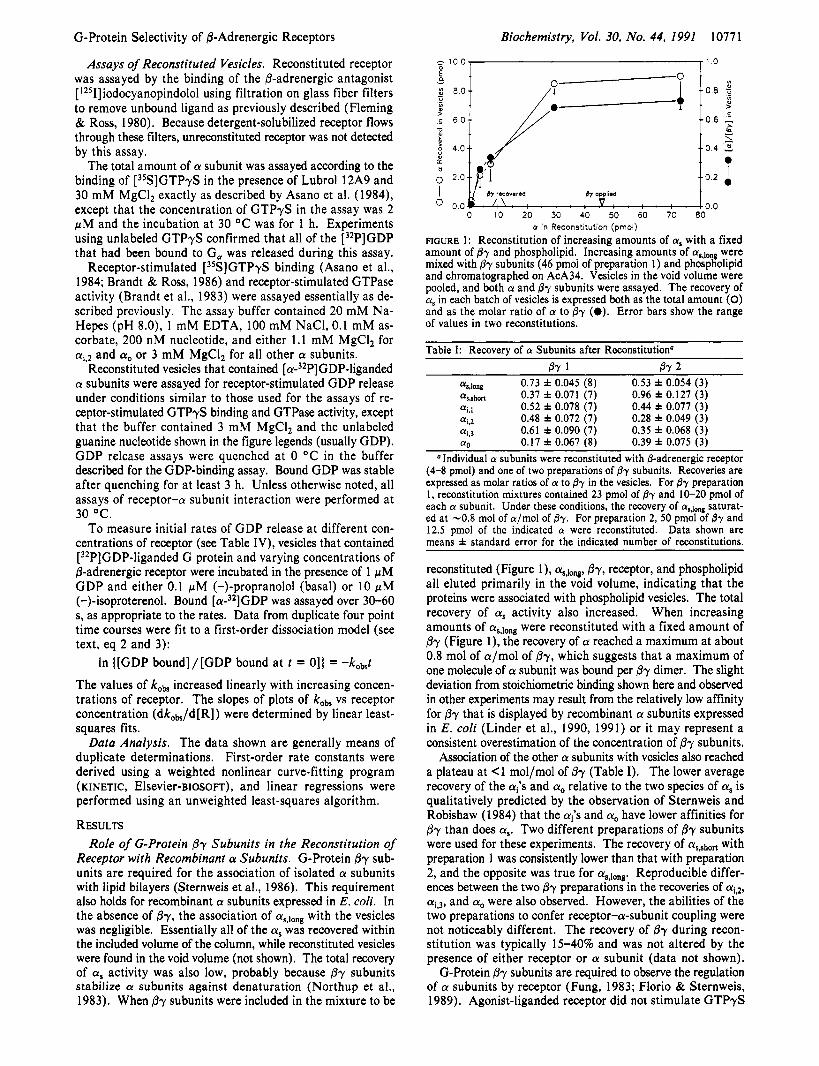
FIGURE 1 : Reconstitution of increasing amounts of a, with a fixed amount of @r and phospholipid. Increasing amounts of as,long were mixed with fir subunits (46 pmol of preparation 1) and phospholipid and chromatographed on AcA34. Vesicles in the void volume were pooled, and both a and @r subunits were assayed. The recovery of CY, in each batch of vesicles is expressed both as the total amount (0) and as the molar ratio of a to @r (e). Error bars show the range of values in two reconstitutions.
Table I: Recovery of a Subunits after Reconstitution' Br 1 Br 2
ffssong 0.73 f 0.045 (8) 0.53 f 0.054 (3) ~ r , , h ~ d 0.37 f 0.071 (7) 0.96 k 0.127 (3) ffi.1 0.52 f 0.078 (7) 0.44 f 0.077 (3) ffi.2 0.48 f 0.072 (7) 0.28 f 0.049 (3) ai,3 0.61 f 0.090 (7) 0.35 f 0.068 (3) a0 0.17 f 0.067 (8) 0.39 f 0.075 (3)
,,Individual CY subunits were reconstituted with 6-adrenergic receptor (4-8 pmol) and one of two preparations of fir subunits. Recoveries are expressed as molar ratios of CY to @y in the vesicles. For j3r preparation 1, reconstitution mixtures contained 23 pmol of @y and 10-20 pmol of each CY subunit. Under these conditions, the recovery of CY^,^^^^ saturat- ed at -0.8 mol of a/mol of By. For preparation 2, 50 pmol of @-y and 12.5 pmol of the indicated a! were reconstituted. Data shown are means f standard error for the indicated number of reconstitutions.
reconstituted (Figure l), asJong, by, receptor, and phospholipid all eluted primarily in the void volume, indicating that the proteins were associated with phospholipid vesicles. The total recovery of a, activity also increased. When increasing amounts of as,long were reconstituted with a fixed amount of Py (Figure l), the recovery of a reached a maximum at about 0.8 mol of a/mol of @y, which suggests that a maximum of one molecule of a subunit was bound per Py dimer. The slight deviation from stoichiometric binding shown here and observed in other experiments may result from the relatively low affinity for By that is displayed by recombinant a subunits expressed in E . coli (Linder et al., 1990, 1991) or it may represent a consistent overestimation of the concentration of Py subunits.
Association of the other a subunits with vesicles also reached a plateau at <1 mol/mol of 0y (Table I). The lower average recovery of the ai's and a, relative to the two species of a, is qualitatively predicted by the observation of Sternweis and Robishaw (1984) that the a;s and a, have lower affinities for @y than does a,. Two different preparations of Py subunits were used for these experiments. The recovery of asrhofi with preparation 1 was consistently lower than that with preparation 2, and the opposite was true for a,,long. Reproducible differ- ences between the two Py preparations in the recoveries of ai,,, ai,3, and a, were also observed. However, the abilities of the two preparations to confer receptor-a-subunit coupling were not noticeably different. The recovery of f ly during recon- stitution was typically 1540% and was not altered by the presence of either receptor or a subunit (data not shown).
G-Protein by subunits are required to observe the regulation of a subunits by receptor (Fung, 1983; Florio & Sternweis, 1989). Agonist-liganded receptor did not stimulate GTPyS

10772 Biochemistry, Vol. 30, No. 44, 1991 Rubenstein et al.
I * I
Time (min)
200 C
Time (min)
200 B 1
Time (niin)
100,
_ * +... .- .. 0 2 3 4
0
T i m (mn)
FIGURE 2: Regulation of GTPyS binding by receptor depends on association of as with vesicle-bound 87. Recombinant a s , l o n g . ~ a ~ combined with &adrenergic receptor containing vesicles using four different protocols described below. GTPyS binding was measured In the presence of 10 pM isoproterenol (0) or 100 nM propranolol (0). (A) 8-Adrenergic receptor, aalang, and fly (preparation 1) were reconstituted as described under Experimental Procedures. Each assay contained 2.0 fmol of receptor, 180 fmol of total a,,io , and 236 fmol of By. (B) Receptor alone was reconstituted as described, and soluble a,,lon was added to the vesicles. Each assay containA 2.9 fmol of receptor and 200 fmol of as. (C) Receptor and by were reconstituted as descrhed, and soluble a,Jong was added to the vesicles. Assays contained 2.3 fmol of receptor, 200 fmol of as,long, and 192 fmol of 87. (D) Receptor alone was reconstituted, and both as,long (200 fmol/assay) and fly (230 fmol/assay) were added to vesicles that contained 2.9 fmol of receptor per assay. For each system (A-D), Lubrol 12A9 was added to give a final concentration in the assays of 10 ppm to account for Lubrol that was added with the By subunits.
binding if recombinant a, was added to preformed receptor- containing vesicles in the absence of /3y (Figure 2B). If soluble a was added to vesicles that contained both receptor and By subunits or if soluble a and Lubrol-solubilized By were added to preformed vesicles that contained receptor alone (Figure 2C,D), slight stimulation of GTPyS binding was consistently observed during multiple exploratory experiments. However, the basal rate of GTPyS binding was much higher than when all three protein components were coreconstituted by gel filtration and accurate quantitation of coupling was not feasible. Free a subunits are known to display a relatively high nucleotide exchange rate that is suppressed by by (Northup et al., 1982; Higashijima et al., 1987). As predicted by this explanation, the basal rate of GTPyS binding to a mixture of free as,long and receptorlpy vesicles was markedly reduced by chromatography on Ultrogel AcA34 although the agon- ist-stimulated binding rate was essentially unchanged (not shown).
Regulation of Different a Subunits by the &Adrenergic Receptor. To study the selectivity of the 8-adrenergic receptors among different G proteins, purified recombinant LY subunits were mixed with By subunits and @adrenergic receptor and reconstituted into vesicles as described above. Measurements of agonist-stimulated GTPyS binding and agonist-stimulated GTPase yielded roughly similar patterns of selectivity. As shown in Figure 3, isoproterenol accelerated GTPyS binding to both forms of a, and to all three ai)s. Stimulation of GTPyS binding to recombinant or cq3 from E. coli was about equal to the stimulation of a preparation of bovine brain ai that was similarly reconstituted. These data are consistent with the finding of Asano et al. (1 984) that the @-adrenergic receptor can regulate rabbit hepatic Gi. The 8-adrenergic receptor also stimulated the steady-state GTPase activities of the as's and ai's in an agonist-dependent manner (Figure 4, Table 11).
Table 11: Receptor-Stimulated GTPase Activities of Receptor-a-& Vesiclesu
GTPase (min-I) [Y subunit INE DTT/INE %,long 0.088 0.220 [Ys.horl 0.121 0.363 ai,I 0.014 0.092 ai,2 0.008 0.039
a0 0.002 0.013 aI.3 0.018 0.12s
bovine ai 0.008 0.00s uVesicles that contained receptor, By, and each of the recombinant
a subunits or the bovine 41-kDa a subunit (prepared as described for Figure 3) were assayed for GTPase activity as described in the legend to Figure 4. Activities in a 10-min assay (1 S min for the bovine ai) are expressed as moles of GTP hydrolyzed per minute per mole of total a subunit. DTT, treated with dithiothreitol (see Figure 4); INE, 10 rM isoproterenol. Activities measured in the presence of propranolol alone have been subtracted from all of the values shown.
Again, receptor stimulated the ais more than the a/s. Slight stimulation of a, was sometimes detected.
This general rank order of selectivity among the different a subunits (s > i,,3 > i, I o ) was maintained throughout a large number of GTPase and GTPyS-binding experiments over several months, although quantitative variation in selectivity ratios was frequently greater than 2-fold. The variability did not appear to result from the choice of /3y preparation used in a specific experiment. A more reproducible assay is in- troduced below.
Treatment of the reconstituted vesicles with pertussis toxin inhibited isoproterenol-stimulated GTPyS binding to the ai)s by 80-90% in several experiments (not shown). Inhibition was NAD-dependent. Such inhibition is characteristic of regula- tion of Gi by receptors that are generally considered to be Gi-coupled. There is no ADP-ribosylation site for pertussis

G-Protein Selectivity of @-Adrenergic Receptors
- 1.0 j: E 2.0 1 0.0
P t -/ I
I 0 1 2 3 1 1
T i m ( m n )
FIGURE 3: Stimulation of GTPyS binding to different a subunits by the 8-adrenergic receptor. @-Adrenergic receptor (4-8 pmol) and & subunits (preparation I , 46 pmol) were reconstituted with 10-20 pmol of the indicated a subunit as described under Experimental Procedures. The resulting vesicles were incubated for 2 h at 0 O C
with 0.1 mM ascorbate and 0.1 pM propranolol in either the presence ( 0 , O ) or the absence (A, A) of 5 mM DTT. Propranolol was added to stabilize thiol-reduced receptors (Pedersen & Ross, 1986). Vesicles were diluted 12-fold, and GTPrS binding was assayed at the indicated times in the presence of 10 pM (-)-isoproterenol (0, A) or 0.1 pM (-)-propranolol (0, A). The assays contained total concentrations of (A) 1.05 fmol of receptor and 1 1 1 fmol of a8,10ng, (B) 0.81 fmol of receptor and 33 fmol of as,short. (C) 1.2 fmol of receptor and 78 fmol of q,,, (D) 1.02 fmol of receptor and 27 fmol of cq2, (E) 1.8 fmol of receptor and 38 fmol of ai 3, (F) 2.25 fmol of receptor and 22 fmol of ao, and (G) 0.54 fmol of receptor and 72 fmol of 41-kDa ai from bovine brain.
toxin on as, and treatment of a,-containing vesicles was without effect.
Selectivity of the &Adrenergic Receptor among G-Protein a Subunits. We routinely monitored the efficiency with which the @-adrenergic receptor regulates individual a subunits by assaying receptor-catalyzed exchange of bound and free GDP at equilibrium. This assay yielded more reproducible data than did the GTPase or GTPyS-binding assay (Table 111). Because GDP/GDP exchange and the binding of radiolabeled GTPyS monitor the same rate-limiting reaction, the results of both assays should be comparable [see Ross (1989)], and this prediction was confirmed by the data shown in Figure 5 . The release of prebound [c~-~*P]GDP mirrored both the rate and the extent of GTPyS binding to similar vesicles that had been prepared with unlabeled a subunit. Similar initial rates of receptor-catalyzed nucleotide exchange and a similarly sized pool of receptor-coupled G protein were observed using either protocol. The release of [ d 2 P ] G D P did not depend on the concentration of the reconstituted a subunit over a 20-fold range (Rubenstein, 1990). Neither the rate nor the extent of
Biochemistry, Vol. 30, No. 44, 1991 10773
' B 240-
0 10 20 Time (min)
FIGURE 4: Regulation of the steady-state GTPase activity of recon- stituted as,long by the P-adrenergic receptor. The same vesicles used in the experiment shown in Figure 3A were similarly incubated in the presence or absence of DTT. GTPase activity was assayed in the presence of either 10 pM isoproterenol or 0.1 pM propranolol. Each assay contained 0.7 fmol of receptor, 74 fmol of aqlong, and 104 fmol of Pr.
Table 111: @-Adrenergic Receptor Stimulated GDP Release from Reconstituted a Subunits'
dkObld[RI basal kob dkob/d[R] + DTT (mi+) (min-I nM-') r (min-1 nh4-l) r
%,long 0.39 1.19 0.96 2.15 0.99 %hart 0.32 1.44 0.98 ndb Ni.1 0.18 0.39 0.91 0.69 0.99 ai.2 0.26 0.13 0.94 nd ai,3 0.19 0.34 0.93 nd a" 0.84 0.13 0.77 0.25 0.96
'Vesicles that contained each a subunit, @r (preparation 2) , and increasing concentrations of @-adrenergic receptor were prepared and assayed for equilibrium GDP exchange in the presence of isoproterenol and propranolol as described under Experimental Procedures. Each value of the first-order exchange rate constant, kob, was derived from a nonlinear, one-component fit of duplicate four-point dissociation time courses, with the zero time value allowed to float during the fit. Each value of dkob/d[R], the slope of the plot of kob vs the concentration of agonist-liganded receptor, was derived from a linear fit for six to eight values of kob. each obtained with a separate batch of vesicles that contained varying amounts of receptor ( r , correlation coefficient). Separate batches of vesicles that contained a,,long. ai,,, and a, were in- cubated with DTT ("+DTT" in table) as described in the legend to Figure 3, and values of kobs were obtained from duplicate four-point time courses at 20 OC. For the DTT-treated vesicles, values of dkobs/d[R] were determined from four values of kob. bnd, not deter- mined.
[CY-~*P]GDP release depended on whether GTPyS or GDP was used as the unlabeled exchanging nucleotide or on the con- centration of unlabeled GDP (0.1-10 pM).
Guanine nucleotide exchange reactions in reconstituted systems indicate the existence of two pools of G protein, one that is regulated by receptor ("coupled") and another that is apparently inaccessible to receptor ("uncoupled"). The ratio of the two pools varies among preparations (Asano et al., 1984b; Brandt & Ross, 1986). Such behavior predicts biex- ponential kinetics in the presence of agonist: a slow rate displayed by the uncoupled G proteins and a faster rate dis- played by the coupled G proteins. Coupled G proteins are predicted to display single-component, first-order kinetics regardless of the extent of stimulation (e.g., at different agonist concentrations) with the extent of stimulation reflected only in the magnitude of the single rate constant [see Chau et al. (1981)l. The receptor-G-protein vesicles used in this study also displayed a small amount of initial rapid GDP/GDP exchange in the presence or absence of agonist. This burst was observed for both equilibrium GDP/GDP exchange and GTPyS binding (Figure 5 and 6A). I ts interference in the

A 100..
'i; 40.' =
120. 80- C D
20..
01 : : 07 0 1 2 3 4 5 0 1 2 3 4 5
Tim (min) h e (min)
FIGURE 5 : Comparison of receptor-stimulated ["PIGDP release and [35S]GTPyS binding. Either aa,lo (A and C; 30 pmol) or (B and D; 17.5 pmol) was incubated with either [(Y-~~PIGTP (C and D) or equimolar unlabeled GTP (A a n d l ) as described under Experimental Procedures. The (Y subunits were then reconstituted with 8-adrenergic receptor (8.6 pmol) and By subunits (preparation 2, 80 pmol), and the vesicles were incubated either with ( 0 , O ) or without (A, A) DTT as described in the legend to Figure 3. The vesicles were assayed either for [35S]GTPyS binding (A and B) or for [32P]GDP release (C and D) in the presence of 10 rM isoproterenol (0, A) or 0.1 rM (-)-propranolol (0, A) after 10-fold dilution. The concentration of GTPyS, either 3SS-labeled (A and B) or unlabeled (C and D), was 200 nM. Each assay contained (A) 3.2 fmol of receptor and 116 fmol of aS,long, (B) 3.7 fmol of receptor and 64 fmol of q3, (C) 3.2 fmol of receptor and 124 fmol of and (D) 4.4 fmol of receptor and 80 fmol of ( ~ i , ~ .
B
assay could be avoided by measuring exchange after the first 2 min of incubation. After the initial rapid release of GDP, exchange was monophasic in the absence of agonist and pro- ceeded with a rate similar to that previously reported for hepatic G, (Asano & Ross, 1984; Brandt & Ross, 1986). Agonist-stimulated exchange was biphasic, with a stimulated component (primarily coupled G protein) followed by a slower component (primarily uncoupled G protein). The slow com- ponent displayed a rate constant equal to that observed in the absence of agonist. Both basal and receptor-stimulated ex- change could be fit to a two-component first-order model?
where a.D* is [32P]GDP-liganded CY subunit, k, is the apparent first-order rate constant for receptor-stimulated exchange, kb is the rate constant for basal exchange, and F is the fraction of CY subunit that is accessible to receptor.
When GDP exchange was measured in vesicles that con- tained increasing amounts of @-adrenergic receptor, it was found that k,, the rate constant for receptor-stimulated ex- change, increased linearly with the concentration of receptor (Figure 6B). The values of kb that were determined from a one-component fit of exchange in the absence of agonist or from a two-component fit in the presence of agonist were equal and did not vary with the concentration of receptors in the vesicles (Figure 6B). The fraction of a subunit that was accessible to receptor (F) was not dependent on the concen- tration of receptor over the range used in these experiments (Figure 6C). This suggests that the dependence of k, on the concentration of receptor does not in some way reflect a
Derivation of eq 1-3 and supporting data for this analysis are given by Rubenstein (1990).
variation in the number of G proteins that have access to receptors. The coupled fraction also did not differ by more than 2-fold among vesicles prepared with different CY subunits.
Although the approach to measuring exchange shown in Figure 6 is analytically valid, it depends on initiating recep- tor-catalyzed nucleotide exchange after significant dissociation of labeled GDP has occurred. It also depends on a multi- parameter fit in which errors in estimates of single parameters may be large and heavily dependent on the fits of other pa- rameters. Therefore, we routinely compared the regulation of different CY subunits by receptor by analyzing only the initial rates of dissociation according to a single-component model. The initial rate of GDP/GDP exchange is described by a single rate contant, k&, that is a weighted combination of k, (defined as described above), k{ (a composite description of all non- receptor-promoted exchange), and F (still defined as the fraction of G protein that undergoes receptor-stimulated ex- change). If k,,' and F remain constant within an experiment, then values of kobs obtained at an arbitrary early time can be related to k, by the equations (see Discussion):
The values of kobs that were determined by analysis of initial rates of GDP exchange increased linearly with the concen- tration of @-adrenergic receptor, as was found using the more complex fitting protocol described above (Figure 6B). There was a negligible effect of receptor on the rate in the absence of agonist, as would be expected if only the agonist-liganded receptor acts as a catalyst of nucleotide exchange.
The linear dependence of kobs on the concentration of re- ceptor (dk,/d[R]; as in Figure 6B) is an index of the intrinsic efficiency with which the receptor regulates different CY sub-

G-Protein Selectivity of @-Adrenergic Receptors 1
0 e e c
0
e
0
A
0 0.1 . : : . . : . : ’ . ; . : : 0 2 4 6 8 10 12
Time (min) . , 2.0
h , b ‘ l a B 0.0 !?h I
0.0 0 1 02 0 3 0 4 0 5 ._ [@-Adrenergic Receptor] (nM in ossoy)
0.0 ~
0.0 0 1 0 2 0 3 0 4 0 5 [@-Adrenergic Receptor] (nM in ossoy)
FIGURE 6: Dissociation of [32P]GDP from and ai,]. (A) Dis- sociation time courses for a,. Receptor-stimulated dissociation of [CX-~~PIGDP from cyak.~ was measured as described under Experimental Procedures (0). Isoproterenol (10 fiM) was added after 2 min of incubation in the absence of @-adrenergic ligand. The basal rate of dissociation (0) was also assayed in medium that contained 100 nM (-)-propranolol. (B) Dissociation of [(Y-’~P]GDP from vesicles that contained subunits, varying amounts of receptor, and either aSJm (open symbols) or q l (closed symbols). Dissociation was measured after 2 min as described for panel A. Data were fit to the two- component model described in the text (eq 1). The dissociation rate constants k, (receptor-stimulated; 0 , O ) and kb (basal; A, A) are shown for each batch of vesicles. Values of kb were also obtained from one-component fits of dissociation in the presence of propranolol (0, m). The slopes of the plots of k, vs receptor concentration were 4.2 m i d nM-l for and 2.7 min-l nM-’ for q l . (C) The fractions of as,lons (0) and cqI2 (0 ) that were stimulated by receptor (F in eq 1) in the experiments described in panel B were also obtained from two-component fits. Error bars indicate standard errors.
units. To determine the efficiency of coupling of the @-ad- renergic receptor with different G-protein a subunits, we as- sayed the initial rates of [32P]GDP release from multiple batches of vesicles that contained different concentrations of @-adrenergic receptor and constant amounts of each of the various [32P]GDP-liganded a subunits. For each G protein, dk,&/d[R] was determined using values of kob obtained for six to eight preparations of vesicles that contained different concentrations of receptor. The results of several such titra- tions are summarized in Table 111. For the @-adrenergic receptor, dk,/d[R] was about 3-4-fold higher for either form of a, than for either or ai,3, which is consistent with our previous studies of hepatic Gi (Asano et al., 1984). @-Adre- nergic stimulation of and ai,3 was observed in all experi- ments. The receptor regulated both ai,* and a, much less
Biochemistry, Vol. 30, No. 44, 1991 10775
efficiently than the as’s; dk,,,/d[R] was about 10-fold lower for both Gi,2 and Go. This level of regulation of was a consistent observation and might be physiologically important. The stimulation of a,, which was undetectable in assays of GTPyS binding and steady-state GTPase, is probably not meaningful. The nonstimulated rate of GDP exchange for a. was already quite high, and our ability to quantitate stimu- lation over this basal rate was in fact marginal. We do not believe that the differences in dkob/d[R] between the long and short forms of a, or between and ( ~ i , ~ are meaningful; they are within the range of experimental variability.
Effects of Thiols on the Selectivity of the @-Adrenergic Receptor. Reduction of disulfide bridges in the @-adrenergic and muscarinic cholinergic receptors potentiates the ability of the receptors to stimulate G proteins (Pedersen & Ross, 1985; Florio & Sternweis, 1989; Moxham et al., 1988; Parker et al., 1991). Treatment of the reconstituted @-adrenergic receptor with dithiothreitol potentiated its ability to regulate all of the recombinant a subunits when assayed in the presence of isoproterenol (Figures 3-5; Tables I1 and 111). In the absence of agonist or in the presence of antagonist, however, the DTT-treated receptor stimulated the ai’s slightly, if at all. Values of dkob/d[R] for DTT-treated, agonist-liganded re- ceptor were compared for as,long, ai,l, and a, (Table 111). Incubation with dithiothreitol did not alter the selectivity of the agonist-liganded receptor among different a subunits.
DISCUSSION Extensive sequence similarity among the G-protein a sub-
units and the G-protein-coupled receptors suggests that a single receptor is able to regulate more than one G protein or that a single G protein could be regulated by more than one re- ceptor. For some receptors, such divergent coupling has been relatively easy to demonstrate. For example, the M1 mus- carinic cholinergic receptor expressed in CHO cells stimulates inositol triphosphate release through the action of two G proteins, only one of which is sensitive to inhibition by pertussis toxin (Ashkenazi et al., 1989). The number and identity of the G proteins that convey a receptor’s signal are usually more difficult to determine.
The initial observation that the @-adrenergic receptor can stimulate Gi (Asano et al., 1984a) was puzzling because 0- adrenergic stimulation has been consistently associated only with the G,-mediated stimulation of adenylyl cyclase and the only known activity of Gi was to inhibit the cyclase. Because G, and Gi are now known to regulate several effector proteins, the &adrenergic stimulation of Gi,l and Gi,3 can be interpreted as a mechanism for generating multiple second messengers from a single P-adrenergic signal.
The data reported here suggest that signaling from the @-adrenergic receptor is primarily mediated by G, but that it is also significantly routed through Gi,l and Gi,3. Regulation of these Gi’s predicts the Occurrence of @-adrenergic stimu- lation of a phospholipase A2, a K+ channel, or another physiological target of Gi, and we are attempting to measure such regulation. From the present data, it seems less likely that the @-adrenergic receptor regulates Gi,2 to a significant extent, and significant regulation of Go seems improbable. Gw which is more widespread among animal tissues than Gi*l or Gi,3 (Mumby et al., 1988), may respond to a more restricted or different range of receptors than do GL, and G,. The range of targets of the @-adrenergic receptor may actually be broader than G,, Gi,l, and Gi,3. We have not observed significant @-adrenergic regulation of G, (Parker et al., 1991; S. K.-F. Wong, unpublished data), but there is some evidence that the receptor can modulate an Na/H exchange activity through

10776 Biochemistry, Vol. 30, No. 44, 1991
a G protein that is neither G, nor Gi (Barber et al., 1989). These data and those of Asano et al. (1984a) indicate that
the @-adrenergic receptor requlates Gi more efficiently than was observed by Cerione et al. (1985), who also used a re- constituted assay system. Neither group determined which ai’s were in their Gi preparations. Asano et al. (1984a) used Gi prepared from rabbit liver, which can contain variable ratios of to ai,, plus ai,3. Cerione et al. may have used a prep- aration that contained relatively more Gi,Z, although Gi,2 does not appear to be the major ai in purified human erythrocyte Gi (Carty & iyengar, 1990). Reduction of disulfides in the receptor, which probably occurred in the experiments of Asano et al. (1984a), is not a determinant of the receptor’s selectivity (Table 11) and therefore does not account for receptor-Gi coupling. Treatment of the receptor with DTT simply makes the less efficient regulation of Gi easier to observe.
The parameter that was used to compare the efficiency of regulation of different G proteins, dko,/d[R], was reproducible among multiple sets of data and intuitively provides a good measure of coupling. A more concrete comparison and more basic mechanistic information could have been derived from a study of dk,/d[R], the actual efficiency of the receptor as a catalyst of nucleotide exchange. Significant non-receptor- mediated GDP exchange precluded our reliably extracting this constant from the data with acceptable precision. Because kob increased linearly with receptor concentration and because the fraction of each G protein that was regulated by receptor did not vary widely (0.2-0.5), the dependence of kob, on receptor was a valid basis for comparison of coupling.3 We did not observe the interesting saturable dependence of coupling on the concentration of G protein that was reported by Senogles et al. (1990) for the D2 dopaminergic receptor. There is no theoretical reason either to support or to question such kinetic behavior over a given range of G-protein concentrations. The lack of dependence of kobs on the concentration of G protein found in the present study is consistent with the relatively low affinity of the turkey erythrocyte &adrenergic receptor for G, that was predicted by Levitzki and co-workers from kinetic analysis of adenylyl cyclase activation [Tolkovsky & Levitzki, 1978, 1981; Tolkovsky et al., 1982; discussed by Rubenstein ( 1990)].
Reconstituted Systems for Assaying the Selectivity of Receptors and G Proteins. The analysis of receptor4-protein selectivity in a purified and reconstituted system has both strengths and weaknesses. The most obvious advantage is that a reconstituted system can use a clearly identified G protein. Measurements of second-messenger responses in intact cells or membranes often do not indicate which G protein (or proteins) may be conveying the signal from a receptor. The use of purified recombinant a subunits further clarifies matters. Because a subunits are structurally similar, it is difficult to purify either a single a subunit or a trimeric G protein free of contamination by others. It is also difficult to demonstrate such purification conclusively. Reconstitution using different recombinant a subunits and a single preparation of By avoids any selective influence on coupling that may be exerted by the different Py subunits that are found in preparations of trimeric G proteins (see below). Reconstitutive assays also allow a
Rubenstein et al.
The relationship between dk,/d[R] and dk,/d[R] is complex and dependent upon the fraction of G protein that is regulated by receptor (F or F?. k , is predicted to increase linearly with the concentration of receptor (dk,/d[R] is constant). In an incompletely coupled system, dk,,/d[R] will only be constant at low receptor concentrations. Deriv- ations of eq 1-3 and a more complete discussion of their applicability to these data have been presented elsewhere (Rubenstein, 1990).
quantitative comparison of the regulation of different G pro- teins by receptors. Interpreting G-protein activation according to signaling in intact systems is complicated by our ignorance of the efficiency with which G proteins regulate effectors.
The assay system described here is applicable to any G- protein-coupled receptor, including recombinant wild-type and mutant receptors that have been expressed in heterologous cells. The receptor need only be relatively concentrated and free of substantial G-protein contamination. We have used this system to study the G-protein selectivity of muscarinic cholinergic receptors (Parker et al., 1991), to study the se- lectivity of chimeric receptors ( S . K.-F. Wong and E. M. Ross, in preparation), and to study the regulation of site-directed mutants in a,.
The recombinant a subunits from E. coli appear to interact normally with the /3-adrenergic receptor. The behavior of recombinant ai,l and was similar to that of the bovine 41-kDa a subunit (Figure 3 and other data not shown), and the behavior of recombinant a, was similar to that observed previously with purified rabbit hepatic G, [see Brandt and Ross (1985) for example]. However, a subunits expressed in E. coli do differ in some respects from the same proteins in mam- malian cells. Both ai and a, in animal cells are myristoylated at their amino termini, but these subunits are not myristoylated when expressed in E . coli (Linder et al., 1991). Although mammalian a, is not myristoylated, its amino terminus is blocked by an unknown modification, and it is more hydro- phobic than would be predicted from its amino acid sequence (Sternweis, 1986). G-Protein a subunits from E . coli regulate adenylyl cyclase and ion channels with lower potency than do the same subunits from mammalian cells (Graziano et al., 1989; Mattera et al., 1989) and bind to By with lower affinity (Linder et al., 1991) Non-myristoylated a, does not bind tightly to the plasma membrane of mammalian cells (Mumby et al., 1990). Data on protein-protein interactions of recom- binant subunits from E . coli must therefore be interpreted carefully.
Role of the fly Subunits. The G-protein By subunits are required for the association of a subunits with phospholipid bilayers (Sternweis, 1986; this study) and for receptor-a- subunit coupling (Fung, 1983; Florio & Sternweis, 1989; this study). They also regulate nucleotide binding to the a subunit (Higashijima et al., 1987). Non-retinal tissues express at least two species of 0 subunit (Evans et al., 1987) and at least three y subunits (Tamir et al., 1991), and preparations of isolated Py subunits or of trimeric G proteins generally contain mix- tures of p’s and y’s. No previous evidence suggested that non-retinal Py subunits were selective among either receptors or a subunits. However, the two preparations of @y used here differed reproducibly in their ability to efficiently reconstitute (Y,,$hort. A third preparation of Py behaved essentially iden- tically to preparation 2. More recently, we have noticed that different bysubunit preparations also alter the reconstitution of other receptors and a subunits (T. Higashijima, G. Berstein, E. M. Parker, and E. M. Ross, unpublished data). We do not know the basis of these differences among preparations of fly. It is likely that we will need to purify dimers composed of known p and y species, expressed in an appropriate cellular background, in order to sort out these effects. The recon- stituted system described here may provide a good assay medium for probing specific functions of j3 and y when these defined By dimers become available.
ACKNOWLEDGMENTS
project. M.E.L. thanks Alfred G. Gilman for his support during this

G-Protein Selectivity of /+Adrenergic Receptors
REFERENCES Abramson, S . N., & Molinoff, P. B. (1985) J. Biol. Chem.
Abramson, S . N., Shorr, R. G. L., & Molinoff, P. B. (1987)
Asano, T., & Ross, E. M. (1984) Biochemistry 23,5467-5471. Asano, T., Katada, T., Gilman, A. G., & Ross, E. M. (1984a)
Asano, T., Pedersen, S. E., Scott, C. W., & Ross, E. M.
Ashkenazi, A., Peralta, E. G., Winslow, J. W., Ramachandran,
Barber, D., McGuire, M., & Ganz, M. (1989) J. Biol. Chem.
Bochner, B. R., & Ames, B. N. (1982) J. Biol. Chem. 257,
Bourne, H., Sanders, D., & McCormick, F. (1991) Nature
Brandt, D. R., & Ross, E. M. (1986) J. Biol. Chem. 261,
Brandt, D. R., Asano, T., Pedersen, S. E., & Ross, E. M.
Carty, D. J., & Iyengar, R. (1990) FEES Lett. 262, 101-103. Casey, P. J., Graziano, M. P., & Gilman, A. G. (1989) Bio-
chemistry 28, 61 1-616. Cerione, R. A., Staniszewski, C., Benovic, J. L., Lefkowitz, R. J., Caron, M. G., Gierschik, P., Somers, R., Spiegel, A. M., Codina, J., & Birnbaumer, L. (1985) J. Biol. Chem.
Chau, V., Romero, G., & Biltonen, R. L. (1981) J. Biol. Chem. 256, 5591-5596.
Codina, J., Olate, J., Abramowitz, J., Mattera, R., Cook, R. G., & Birnbaumer, L. (1988) J. Biol. Chem. 263,
Cooper, C. L., Morris, A. J., & Harden, T. K. (1 989) J. Biol.
Evans, T., Fawzi, A., Fraser, E. D., Brown, M. L., & Northup,
Ewald, D. A., Pang, I.-H., Sternweis, P. C., & Miller, R. J.
Fleming, J. W., & Ross, E. M. (1980) J. Cyclic Nucleotide
Florio, V. A., & Sternweis, P. C. (1989) J. Biol. Chem. 264,
Fung, B. K.-K. (1983) J. Biol. Chem. 258, 10495-10502. Gilman, A. G. (1987) Annu. Rev. Biochem. 56, 615-649. Graziano, M. P., Freissmuth, M., & Gilman, A. G. (1989)
260, 14580-14588.
Biochem. Pharmacol. 36, 2263-2269.
J . Biol. Chem. 259, 9351-9354.
(1984b) Biochemistry 23, 5460-5467.
J., & Capon, D. J. (1989) Cell 56, 487-493.
264, 21038-21042.
9759-9769.
349, 117-127.
1656-1 664.
(1983) Biochemistry 22, 4357-4362.
260, 1493-1 500.
6746-6750.
Chem. 264, 6202-6206.
J. K . (1987) J . Biol. Chem. 262, 176-181.
(1989) Neuron 2, 1185-1 193.
Res. 6, 407-419.
3909-391 5.
J . Biol. Chem. 264, 409-418.
Biochemistry, Vol. 30, No. 44, 1991 10777
Harris, B. A., Robishaw, J. D., Mumby, S. M., & Gilman, A. G. (1985) Science 229, 1274-1277.
Higashijima, T., Ferguson, K. M., Sternweis, P. C., Smigel, M. D., & Gilman, A. G. (1987) J. Biol. Chem. 262,
Kurose, H., Regan, J. W., Caron, M. G., & Lefkowitz, R. J. (1991) Biochemistry 30, 3335-3341.
Linder, M. E., Ewald, D. A., Miller, R. J., & Gilman, A. G. (1990) J. Biol. Chem. 265, 8243-8251.
Linder, M., Pang, 1.-H., Duronio, R., Gordon, J., Sternweis, P., Gilman, A. G. (1991) J. Biol. Chem. 266,4654-4659.
Mattera, R., Graziano, M. P., Yatani, A,, Zhou, Z., Graf, R., Codina, J., Birnbaumer, L., Gilman, A. G., & Brown, A. M. (1989) Science 243, 804-807.
Moxham, C. P., Ross, E. M., George, S. T., & Malbon, C. C. (1988) Mol. Pharmacol. 33, 486-492.
Mumby, S., Pang, 1.-H., Gilman, A. G., & Sternweis, P. C. (1988) J. Biol. Chem. 263, 2020-2026.
Mumby, S. M., Heukeroth, R. O., Gordon, J. I., & Gilman, A. G. (1990) Proc. Natl. Acad. Sci. U.S.A. 87, 728-732.
Northup, J. K., Smigel, M. D., & Gilman, A. G. (1982) J. Biol. Chem. 257, 11416-1 1423.
Northup, J. K., Sternweis, P. C., & Gilman, A. G. (1983) J. Biol. Chem. 258, 11361-1 1368.
Parker, E. M., Kameyama, K., Higashijima, T., & Ross, E. M. (1991) J. Biol. Chem. 266, 519-527.
Pedersen, S. E., & Ross, E. M. (1985) J. Biol. Chem. 260,
Ross, E. M. (1989) Neuron 3, 141-152. Rubenstein, R. C. (1990) Ph.D. Thesis, University of Texas
Southwestern Graduate School of Biomedical Science. Rubenstein, R., Linder, M., Gilman, A., & Ross, E. (1988)
J . Cell Biol. 107, 708a. Senogles, S. E., Benovic, J. L., Amlaiky, N., Unson, C.,
Milligan, G., Vinitsky, R., Spiegel, A. M., & Caron, M. G. (1 987) J. Biol. Chem. 262, 4860-4867.
Senogles, S . E., Spiegel, A. M., Padrell, E., Iyengar, R., & Caron, M. G. (1990) J. Biol. Chem. 265, 4507-4514.
Sternweis, P. C. (1986) J. Biol. Chem. 261, 631-637. Sternweis, P. C. & Robishaw, J. D. (1 984) J. Biol. Chem. 259,
Tolkovsky, A. M., & Levitzki, A. (1978) Biochemistry 17,
Tolkovsky, A. M., & Levitzki, A. (1981) J. Cyclic Nucleotide
Tolkovsky, A. M., Braun, S., & Levitski, A. (1982) Proc. Nutl.
7 6 2-7 66.
14 1 50- 14 1 57.
1 3806-1 38 1 3.
3795-3810.
Res. 7, 139-150.
Acad. Sci. U.S.A. 79, 213-217.