Embed Size (px)
Citation preview
Selective Blockade of Trypanosomatid Protein Synthesisby a Recombinant Antibody Anti-Trypanosoma cruzi P2bProteinMaximiliano Juri Ayub1,2., Benson Nyambega1,3,4., Leandro Simonetti1, Tomas Duffy1, Silvia A. Longhi1,
Karina A. Gomez1, Johan Hoebeke5, Mariano J. Levin1, Cristian R. Smulski1,5*¤
1 Laboratorio de Biologıa Molecular de la Enfermedad de Chagas, Instituto de Investigaciones en Ingenierıa Genetica y Biologıa Molecular, Consejo Nacional de
Investigaciones Cientıficas y Tecnicas, Buenos Aires, Argentina, 2 Instituto Multidisciplinario de Investigaciones Biologicas de San Luis, Consejo Nacional de Investigaciones
Cientıficas y Tecnicas, Universidad Nacional de San Luis, San Luis, Argentina, 3 Department of Medical Biochemistry, Maseno University, Kisumu, Kenya, 4 Zentrum fur
Molekulare Biologie der Universitat Heidelberg, Heidelberg, Germany, 5 Immunologie et Chimie Therapeutiques (UPR 9021), Institut de Biologie Moleculaire et Cellulaire
(IBMC), Strasbourg, France
Abstract
The ribosomal P proteins are located on the stalk of the ribosomal large subunit and play a critical role during theelongation step of protein synthesis. The single chain recombinant antibody C5 (scFv C5) directed against the C-terminalregion of the Trypanosoma cruzi P2b protein (TcP2b) recognizes the conserved C-terminal end of all T. cruzi ribosomal Pproteins. Although this region is highly conserved among different species, surface plasmon resonance analysis showed thatthe scFv C5 possesses very low affinity for the corresponding mammalian epitope, despite having only one single amino-acid change. Crystallographic analysis, in silico modelization and NMR assays support the analysis, increasing ourunderstanding on the structural basis of epitope specificity. In vitro protein synthesis experiments showed that scFv C5 wasable to specifically block translation by T. cruzi and Crithidia fasciculata ribosomes, but virtually had no effect on Rattusnorvegicus ribosomes. Therefore, we used the scFv C5 coding sequence to make inducible intrabodies in Trypanosomabrucei. Transgenic parasites showed a strong decrease in their growth rate after induction. These results strengthen theimportance of the P protein C terminal regions for ribosomal translation activity and suggest that trypanosomatid ribosomalP proteins could be a possible target for selective therapeutic agents that could be derived from structural analysis of thescFv C5 antibody paratope.
Citation: Juri Ayub M, Nyambega B, Simonetti L, Duffy T, Longhi SA, et al. (2012) Selective Blockade of Trypanosomatid Protein Synthesis by a RecombinantAntibody Anti-Trypanosoma cruzi P2b Protein. PLoS ONE 7(5): e36233. doi:10.1371/journal.pone.0036233
Editor: Ben L. Kelly, Louisiana State University, United States of America
Received November 1, 2011; Accepted March 28, 2012; Published May 3, 2012
Copyright: � 2012 Juri Ayub et al. This is an open-access article distributed under the terms of the Creative Commons Attribution License, which permitsunrestricted use, distribution, and reproduction in any medium, provided the original author and source are credited.
Funding: This research has been initially supported by grants from the World Health Organization/Special Program for Research and Training in TropicalDiseases, Universidad de Buenos Aires, Ministerio de Salud y Accion Social-Beca Ramon Carrillo-Arturo Onativia, and the National Agency of Scientific andTechnological Promotion (FONCYT BID 1201/OC-AR 01–14389). Support of ECOS-SECyT project ‘‘Anticorps anti-proteines ribosomales P de T. cruzi commeinhibiteur specifique de la traduction’’ (France-Argentine, 2005–2008) is also acknowledged. The funders had no role in study design, data collection and analysis,decision to publish, or preparation of the manuscript.
Competing Interests: The authors have declared that no competing interests exist.
* E-mail: [email protected]
. These authors contributed equally to this work.
¤ Current address: Department of Biochemistry, University of Lausanne, Epalinges, Switzerland
Introduction
Trypanosoma cruzi is a protozoan parasite responsible for Chagas’
disease. This is an endemic disease in Latin America that affects
18–20 million people. No vaccines are available at present and
drugs used for treatment show undesirable side effects. The
identification of new targets for chemotherapy is a major challenge
in the control of the disease and the protein synthesis machinery
has been proven to be such a target in other species. Insight into
the mechanism capable of selectively blocking protein synthesis
could thus lead to the discovery of new therapeutic agents.
The large subunit of the eukaryotic ribosome possesses a long
and protruding stalk formed by the ribosomal P proteins. These
proteins include P0, an approximately 34 kDa polypeptide, and
two distinct, but closely related peptides of about 11 kDa, P1 and
P2. All of them share a conserved, highly acidic motif at its C-
terminal end. An additional P protein, named P3, has been
described in plants [1]. The number of P1/P2 subtypes varies
among species. In higher eukaryotes, the P1 and P2 families have
only one member. In Saccharomyces cerevisiae, these families have two
members, P1a/P1b and P2a/P2b [2]. Trypanosoma cruzi also
possesses two different P1 and P2 proteins [3,4]. Interestingly, the
T. cruzi P0 protein has a C-terminal end that deviates from the
eukaryotic P consensus and bears similarity to that of the L10
protein of Archaea [5]. The GTPase activity of the eukaryotic
elongation factor 2 (eEF-2), which catalyses the translocation of
peptidyl-tRNA from the A to the P site of the ribosome, is
dependent on the presence of P proteins on the large ribosomal
subunit [6]. Specifically, the C-terminal region of the ribosomal P
proteins was shown to be essential during this step [7,8]. Thus, the
ribosomal stalk is directly involved in the translocation step of
PLoS ONE | www.plosone.org 1 May 2012 | Volume 7 | Issue 5 | e36233
protein synthesis [9]. It has been previously shown that antibodies
against the C-terminal region of ribosomal P proteins (markers of
systemic lupus erythematosus in humans) and their scFv recom-
binant forms posses the ability to block in vitro translation in a
rabbit reticulocyte lysate system [10,11]. In chronic Chagas’ heart
disease, antibodies against the C-terminal region of T. cruzi
ribosomal P proteins have been also detected [12,13]. However,
fine epitope mapping demonstrated that the specificity of the
antibodies induced in these two pathological disorders is different
[14,15]. The single chain recombinant antibody (scFv) C5 directed
against the C-terminal region of the ribosomal P2b protein of T.
cruzi (R13 epitope), targets the five P proteins that constitute the
stalk [16,17]. Four of them (P1a, P1b, P2a, P2b) contain the same
C-terminal epitope, R13 (Figure 1A); and the fifth, P0, has a
closely related epitope called P015 (Figure 1A) [3,16,18]. This
antibody however, as shown in this work, possesses very low
affinity for the corresponding mammalian epitope (H13) that has
one single non-conservative amino acid change in the third
residue. We found that the scFv C5 was able to specifically block in
vitro protein synthesis by trypanosomatid ribosomes, but had
virtually no effect on translation by mammalian ribosomes. We
expressed for the first time an intrabody (intracellular antibody),
derived from scFv C5, in trypanosomatid cells resulting in growth
arrest. Therefore, we propose the ribosomal stalk as a novel
potential chemotherapeutic target, and the scFv C5 paratope as a
model for peptide mimetics synthesis for selective blocking of the
parasite protein synthesis apparatus.
Materials and Methods
Synthetic peptidesPeptides were prepared by the solid-phase method of Merrifield
as was previously described [19], using a semiautomatic multi-
synthesizer NPS 4000 (NeoMPS SA, Strasbourg, France).
Surface Plasmon ResonanceThe BIACORE 3000 system, sensor chip CM5, surfactant P20,
amine coupling kit containing N-hydroxysuccinimide (NHS) and
N-Ethyl-N9-dimethylaminopropyl carbodiimide (EDC), ethanol-
amine were from BIACORE (Uppsala, Sweden). Biosensor assays
were performed with HBS-EP buffer as running buffer (10 mM
HEPES, 150 mM sodium chloride, 3 mM EDTA, 0.005% (v/v)
surfactant P20, pH 7.4). The low carboxylated dextran matrix (B1)
was activated with 35 ml of a mixture 0.2 M N-ethyl-N-
dimethylaminopropyl carbodiimide and 0.05 M N-hydroxysucci-
nimide at 5 ml/min. TcP2b-GST fusion protein and GST (as
control) were immobilized with the standard BIACORE protocol
at a density of 0.05 pmol/mm2 [4]. The scFv C5 was then pre-
incubated with different concentrations of P015, R13 or H13
peptides for 30 min and then injected on the sensor chip for
2 min, followed by a dissociation phase of 3 min at a flow rate of
30 ml/min. The sensor chip surface was regenerated after each
experiment by injecting 20 mL of 3 M MgCl2. From the decrease
in the initial linear kinetics of the interaction in the presence of
increasing amounts of inhibitor, the IC50 could be determined for
each inhibitor as previously described [16].
ScFv C5 DNA constructs and intrabody expressionExpression of scFv C5 in E. coli and purification was performed
as previously described [16]. For in vivo expression, the DNA
encoding for scFv C5 was amplified by PCR using the following
oligonucleotides carrying HindIII restriction site (C5ForHIND: 59-
GT AAGCTT GCC ATG GCC GAA GTG CAG C-39) and
BamHI restriction site (C5RevBAM: 59-GT GGATCC CCG
TTT TAT TTC CAG CTT GGT CC-39) and inserted into
pHD1700 vector as previously described [20]. The N-terminal
myc-tagged scFv C5 construct was transfected into blood stream
1313-514 T. brucei inducible cell line [21], expressed and detected
by Western blot using peroxidase anti-myc monoclonal antibody
(1:50,000) (Santa cruz biotechnology). Monoclonal antibody to
aldolase [22] (1:4000) was used to detect aldolase, used as the
loading control. For growth rate analysis, parasites were seeded at
26105 cells/ml and counted every 24 hours. The concentration
was adjusted (by diluting appropriately) every 24 hours back to
26105 cells/ml. The cumulative number of parasites corresponds
to the number of parasites per milliliter multiplied by the dilution
factor for each day (day after day).
Molecular modelingAll procedures were performed with Discovery Studio 2.5
software from Accelrys (San Diego, CA, USA). The scFv C5
model derives from the parental mAb 17.2 crystal structure Apo
(3SGD) or in complex with R13 peptide (3SGE). The former
structure was used as template to build a model antibody-H13
interaction. For this purpose, we mutated the aspartic acid 3 of
R13 by serine and optimized the conformation of both the
mutated residues and any surrounding residues that lay within a
cut-off radius of 2 A. Five models thus obtained were scored by the
Discrete Optimized Protein Energy (DOPE). We continued
analysis using the lowest energy model (DOPE = 296583.96 k-
cal/mol). Finally, the complex was subjected to a minimization
step (max 500), RMS gradient 0.1 Kcal/mol by conjugated
gradient.
Gel electrophoresis and Western blot analysisSDS-PAGE analysis was performed as a standard procedure
using 12% acrylamide gels followed by immunoblotting. Proteins
were transferred from gels onto a Hybond ECL nitrocellulose
transfer membrane (Amersham Pharmacia, UK) using a mini
trans-blot system (Bio-Rad, Hercules, CA, USA) in transfer buffer
(25 mM Tris-HCl, 190 mM glycine, 20% (v/v) methanol,
pH 8.3). Membranes were soaked in PBS-T (20 mM Na2HPO4,
1.8 mM K2HPO4, 150 mM NaCl, 2.7 mM KCl, 0.1% Tween 20,
pH 7.4) supplemented with 5% (p/v) non-fat milk powder. This
was followed by incubation with scFv C5 200 nM together with
peroxidase-conjugated anti-His Ab (,350 nM) (Sigma, St. Louis,
MO, USA), for detection of ribosomal P proteins and with
peroxidase-conjugated anti-His Ab (,350 nM) alone for intra-
body detection. The Ab was diluted in the blocking solution PBS-
T 1% (p/v) non-fat milk powder. Proteins on transferred
membranes were revealed with tetramethyl-benzidine (TMB)
(Sigma, St. Louis, MO, USA) after a brief wash with dextran
sulphate 1% (Sigma, St. Louis, MO, USA).
Ribosome purificationPurification of ribosomes from different sources and in vitro
protein synthesis assays were performed as described [23]. Briefly,
mammalian ribosomes were obtained from rat liver (20 g), which
was washed in sucrose 0.25 M and then homogenized in buffer
containing 50 mM Tris–HCl, pH 7.5; 250 mM KCl; 5 mM
magnesium acetate; sucrose 0.25 M and supplemented with 10%
of S150 fraction [23]. The homogenate was treated with 10 U/ml
of a-amylase and 0.1 mM CaCl2 for 15 min at 4uC, and then
centrifuged for 4 min at low speed. The supernatant was again
centrifuged for 20 min at 23,000 g. The pellet was discarded and
the supernatant containing polysomes was supplemented with
Triton X-100 1%, deoxycholate 0.5% and centrifuged for 5 min
at 16,000 g. The new supernatant fluid (around 12 ml) was
Inducible Intrabodies on Trypanosoma brucei
PLoS ONE | www.plosone.org 2 May 2012 | Volume 7 | Issue 5 | e36233
layered onto two layers of 2 M and 1.5 M sucrose made up in
Buffer A (50 mM Tris–HCl, pH 7.5; 5 mM magnesium acetate;
250 mM KCl, dithiothreitol 1 mM and 10% of S150 fraction) and
centrifuged 16 h at 140,000 g. The pellet corresponding to
polysomes was rinsed and resuspended in a buffer containing
10 mM Tris–HCl, pH 7.5; 10 mM KCl and 1.5 mM magnesium
acetate. Ribosome concentration was determined by optical
density at 260 nm.
Polysomes from trypanosomatids were obtained from cultures at
the exponential phase of growing. Cultures were treated with
cycloheximide (50 ug/ml) for 10 min before harvesting by
centrifugation at 4uC. Cells were washed twice with PBS
containing 50 ug/ml cycloheximide. The cells were resuspended
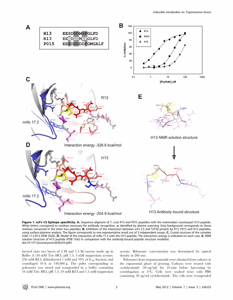
Figure 1. scFv C5 Epitope specificity. A. Sequence aligment of T. cruzi R13 and P015 peptides with the mammalian counterpart H13 peptide.White letters correspond to residues necessary for antibody recognition, as identified by alanine scanning. Grey background corresponds to thoseresidues conserved in the other two peptides. B. Inhibition of the interaction between scFv C5 and TcP2b protein by R13, P015 and H13 peptides,using surface plasmon analysis. The figure corresponds to one representative result out of 3 independent assays. C. Crystal structure of the complexmAb 17.2-R13 (PDB 3SGE). D. Model of the interaction of mAb 17.2 with the H13 peptide. The interaction energy is indicated on each case. E. NMRsolution structure of H13 peptide (PDB 1S4J) in comparison with the antibody-bound peptide structure modeled.doi:10.1371/journal.pone.0036233.g001
Inducible Intrabodies on Trypanosoma brucei
PLoS ONE | www.plosone.org 3 May 2012 | Volume 7 | Issue 5 | e36233

in Lysis Buffer (20 mM Tris–HCl, pH 7.5; 1 mM MgCl2; 5 mM
KCl; 3 mM CaCl2; 5 mM 2-merchaptoethanol and 250 mM
sucrose) and lysed with 0.2–0.4% of Nonidet P40 at 4uC. The
homogenate was centrifuged 2–3 times for 20 min at 1,000 g and
the final supernatant fraction containing ribosomes was layered
onto a discontinuous gradient of 2 and 1.5 M of sucrose made up
in the following buffer: 10 mM Tris–HCl, pH 7.5; 1 mM
magnesium acetate; and 100 mM potasium acetate. The gradient
was centrifuged for 16 h at 140,000 g. The supernatant was
discarded and the pellet carefully rinsed and resuspended as
described above.
In vitro protein synthesis assaysThe reaction mixtures were prepared on ice and contained: 19
amino acids 50 uM each (excepting Met); 2 mM dithiothreitol;
100 mM potassium acetate; 3.5 mM magnesium acetate; 75 ug/
ml wheat germ tRNA; 18 mM Hepes/KOH, pH 7.5; 1 mM
ATP; 0.5 mM GTP; 7.5 mM creatine phosphate; 37.5 ug/ml
creatine phosphokinase; rat liver S150 fraction (24 ug of protein);
0.3 A260U of ribosomes and 2 uCi of [35S] methionine in a final
volume of 30 ul. Reactions were performed at 30uC during
60 min and stopped by adding 150 ul of 1.5 M NaOH; 1 mM
Met, 170 ug/ml BSA. After incubation for 30 min at 37uC,
proteins were precipitated with 1 ml of cold TCA 25%. After
60 min on ice, the samples were filtered and washed with TCA
10% and ethanol using glass fiber filters. Radioactivity retained in
the filters was measured by liquid scintillation counting. For
inhibition assays, reaction mixtures were incubated on ice with the
scFv C5 antibody. For inhibition reversion, the scFv C5 was
preincubated with the H13/R13 peptides for 20 min. After that,
reactions were initiated by incubation at 30uC.
Trypanosome growth and transfectionBloodstream form of Trypanosoma brucei was cultured in HMI-9
medium [24] supplemented with 10% (v/v) fetal calf serum
(Sigma-Aldrich), and transfected as described previously [25].
Transfected cells were then cloned by serial dilution in a 24 well
microtiter plate. For induction, cells were cultured in the medium
above containing 0,1 mg/ml tetracycline and monitored for
phenotypic changes (96 hours) or expression levels (72 hours) post
induction.
Data mining and phylogenetic analysisRibosomal P protein sequences of Trypanosoma cruzi, Saccharomy-
ces cerevisiae and Homo sapiens were used to perform BLAST searches
[26] against NCBI-GenBank and GeneDB databases, using the
tblastn and blastp algorithms. All protein sequences used for the
phylogenetic analysis are listed in Table S1. Sequences were
aligned with MEGA 4.0 software [27] using the Clustal W
algorithm [28] with the PAM protein weight matrix, and all
parameters with default settings. A phylogenetic tree was modeled
using the minimum evolution method with the pairwise deletion
option, and all parameters with default settings. Support for the
branching order was determined by 1,000 bootstrap replicates.
Statistical analysisTranslation inhibition studies were analyzed by two-way
ANOVA with Bonferroni’s post test. Densitometry was analyzed
by one-way ANOVA with Tukey’s Multiple Comparison Test. T.
brucei growth rate was analyzed by two-way ANOVA with
Bonferroni’s post test. All analysis were done using GraphPad
Prism version 5.00 for Windows, GraphPad Software, San Diego
California USA, www.graphpad.com.
Results
Epitope SpecificityAs it was previously reported the scFv C5 (that derives from the
monoclonal antibody 17.2) recognizes the C-terminal region of T.
cruzi ribosomal P2b protein (TcP2b, R13 epitope), and is able to
recognize all five T. cruzi ribosomal P proteins [16,29]. Four of
them (TcP1a, TcP1b, TcP2a and TcP2b) contain the R13 epitope
(Figure 1A); and the fifth, TcP0, contains a homologous C-
terminal region called P015 (Figure 1A). Alanine replacement of
R13 peptide showed as important residues for antibody interaction
the motif 3-EdDDmGF-9 [29] (Figure 1A, white letters). The
mammalian counterpart epitope (H13) possesses one single, non-
conservative amino acid change in the first key glutamic acid
position (Figure 1A). Therefore, using surface plasmon resonance
(SPR) analysis, we tested the ability of the three peptides to inhibit
the interaction between the scFv C5 and TcP2b. Inhibition curves
were performed using recombinant TcP2b protein fixed to a
sensor chip and the scFv C5 in solution together with increasing
concentrations of R13, P015 and H13 peptides (Figure 1B).
Complete inhibition curves could be only obtained with R13
(IC50 = 2,3760.23 mM) (Rsqr = 0.992) and P015 (IC50 = 22,646
2.03 mM) (Rsqr = 0.997) peptides but not with the H13 peptide.
These results demonstrated the low affinity of scFv C5 for the
mammal epitope. Interestingly, both P015 and H13 peptides
possess one amino acid change in this motif but the replacement of
glycine 8 by aspartic acid in P015 peptide seemed to be less
essential than the replacement of glutamic 3 by serine in H13
peptide for scFv C5 interaction.
The crystallographic structure of mAb 17.2 in complex with
R13 peptide has been recently solved (PDB 3SGE) [17]. In silico
replacement of GLU3 by SER showed a strong reduction of the
interaction energies that fall from 2326.6 kcal/mol (R13 complex)
to 2204.9 kcal/mol (H13 complex) (Figure 1C and D).
Particularly, there were three hydrogen bonds established between
GLU3 and CDR-H2 residues (ARG-52, SER-53 and ASN-56)
that were completely lost by the replacement by SER. In addition
H13 peptide, but not R13, was able to acquire a stable
conformation in solution using NMR [30], indicating that a
strong conformational change is needed for the transition from the
free solution form of H13 to the antibody bound conformation
(Figure 1E) been energetically more expensive than binding to
R13 peptide.
Translation inhibitionWe analyzed the ability of scFv C5 to detect the ribosomal P
proteins of T. cruzi, T. brucei, C. fasciculata and R. norvegicus by
western blot. As it was previously shown [16] the scFv C5 strongly
reacted with the T. cruzi P0, P1 and P2 proteins (Figure 2A, lane1). The sequence of the C-terminal peptide of T. brucei shows a
conservative change of Glu3 by Asp in three of the low molecular
weight P proteins (Table S2), and the sequences of C. fasciculata P
proteins are not known. Figure 2A shows that scFv C5 clearly
detected the P proteins on T. brucei and C. fasciculata extracts
(Figure 2A, lanes 2 and 3 respectively). Similar results were
obtained analyzing equal amounts of purified ribosomes from
these different organisms (not shown). In contrast, no protein
bands were detected using rat extracts (Figure 2A lane 4), as was
expected from SPR analysis.
Because scFv C5 reacts with the functional domain of the
ribosomal P proteins, we used an in vitro cell-free translation system
to determine the ability of this antibody to inhibit protein
synthesis. Background values of radioactivity incorporation in
the absence of protein synthesis were obtained in the presence of
Inducible Intrabodies on Trypanosoma brucei
PLoS ONE | www.plosone.org 4 May 2012 | Volume 7 | Issue 5 | e36233
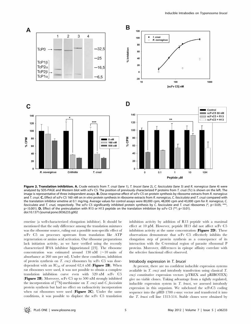
emetine (a well-characterized elongation inhibitor). It should be
mentioned that the only difference among the translation mixtures
was the ribosome source, ruling out a possible non-specific effect of
scFv C5 on processes upstream from translation like ATP
regeneration or amino acid activation. Our ribosome preparations
lack initiation activity, as we have verified using the recently
characterized IF4A inhibitor hippuristanol [23]. The ribosome
concentration was estimated around 130 nM (,10 units of
absorbance at 260 nm per ml). Under these conditions, inhibition
of protein synthesis on T. cruzi ribosomes by scFv C5 was dose-
dependent with an IC50 of around 62,4 nM (Figure 2B). When
rat ribosomes were used, it was not possible to obtain a complete
translation inhibition curve even with 520 nM scFv C5
(Figure 2B). Moreover, scFv C5 up to 500 nM strongly inhibited
the incorporation of [35S]-methionine on T. cruzi and C. fasciculata
protein synthesis but had no effect on radioactivity incorporation
when rat ribosomes were used (Figure 2C). Under the same
conditions, it was possible to displace the scFv C5 translation
inhibition activity by addition of R13 peptide with a maximal
effect at 10 mM. However, peptide H13 did not affect scFv C5
inhibition activity at the same concentration (Figure 2D). These
observations demonstrate that scFv C5 effectively inhibits the
elongation step of protein synthesis as a consequence of its
interaction with the C-terminal region of parasite ribosomal P
proteins. Moreover, differences in epitope affinity correlate with
the selective functional effect observed.
Intrabody expression in T. bruceiAt present, there are no confident inducible expression systems
available in T. cruzi and intrabody transfection using classical T.
cruzi constitutive expression vectors (pTREX and pRIBOTEX)
give no viable clones. Taking advantage from a tightly regulated,
inducible expression system in T. brucei, we assessed intrabody
expression in this organism. We subcloned the scFvC5 coding
sequence into the pHD 1700 c-myc vector and transfected it into
the T. brucei cell line 1313-514. Stable clones were obtained by
Figure 2. Translation inhibition. A. Crude extracts from T. cruzi (lane 1), T. brucei (lane 2), C. fasciculata (lane 3) and R. norvegicus (lane 4) wereanalyzed by SDS-PAGE and Western blot with scFv C5. The position of previously characterized P proteins from T. cruzi (Tc) is shown on the left. Theimage is representative of three independent assays. B. Dose-response effect of scFv C5 on protein synthesis by ribosome extracts from R. norvegicusand T. cruzi. C. Effect of scFv C5 160 nM on in vitro protein synthesis in ribosome extracts from R. norvegicus, C. fasciculata and T. cruzi compared withthe translation inhibitor emetine at 0.1 mg/mg. Average values for control assays were 80,000 cpm, 48,000 cpm and 43,000 cpm for R. norvegicus, C.fasciculata and T. cruzi, respectively. The scFv C5 significantly inhibited protein synthesis by C. fasciculata and T. cruzi ribosomes (*, p,0.05; ***,p,0.001). D. Effect of the preincubation with R13 or H13 peptide on the translation inhibition by scFv C5 (**; p,0.01).doi:10.1371/journal.pone.0036233.g002
Inducible Intrabodies on Trypanosoma brucei
PLoS ONE | www.plosone.org 5 May 2012 | Volume 7 | Issue 5 | e36233
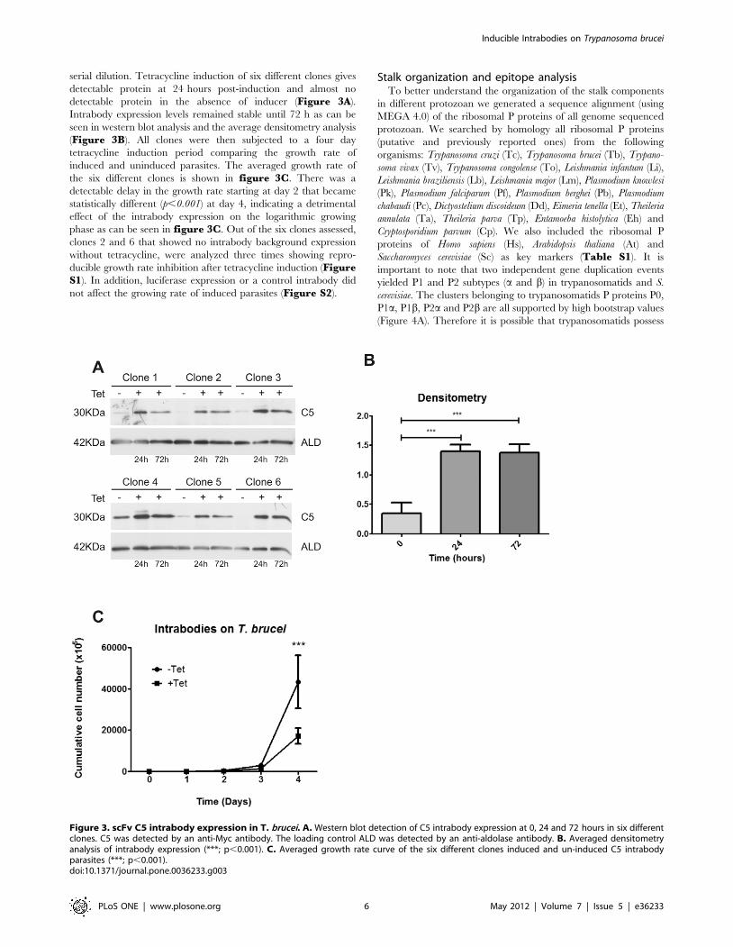
serial dilution. Tetracycline induction of six different clones gives
detectable protein at 24 hours post-induction and almost no
detectable protein in the absence of inducer (Figure 3A).
Intrabody expression levels remained stable until 72 h as can be
seen in western blot analysis and the average densitometry analysis
(Figure 3B). All clones were then subjected to a four day
tetracycline induction period comparing the growth rate of
induced and uninduced parasites. The averaged growth rate of
the six different clones is shown in figure 3C. There was a
detectable delay in the growth rate starting at day 2 that became
statistically different (p,0.001) at day 4, indicating a detrimental
effect of the intrabody expression on the logarithmic growing
phase as can be seen in figure 3C. Out of the six clones assessed,
clones 2 and 6 that showed no intrabody background expression
without tetracycline, were analyzed three times showing repro-
ducible growth rate inhibition after tetracycline induction (FigureS1). In addition, luciferase expression or a control intrabody did
not affect the growing rate of induced parasites (Figure S2).
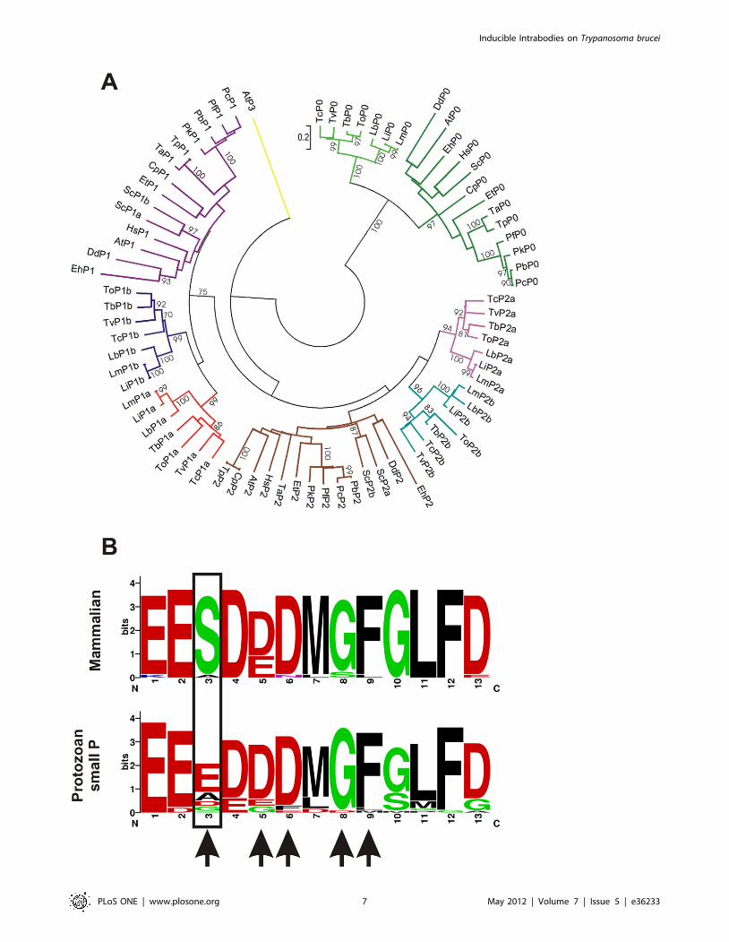
Stalk organization and epitope analysisTo better understand the organization of the stalk components
in different protozoan we generated a sequence alignment (using
MEGA 4.0) of the ribosomal P proteins of all genome sequenced
protozoan. We searched by homology all ribosomal P proteins
(putative and previously reported ones) from the following
organisms: Trypanosoma cruzi (Tc), Trypanosoma brucei (Tb), Trypano-
soma vivax (Tv), Trypanosoma congolense (To), Leishmania infantum (Li),
Leishmania braziliensis (Lb), Leishmania major (Lm), Plasmodium knowlesi
(Pk), Plasmodium falciparum (Pf), Plasmodium berghei (Pb), Plasmodium
chabaudi (Pc), Dictyostelium discoideum (Dd), Eimeria tenella (Et), Theileria
annulata (Ta), Theileria parva (Tp), Entamoeba histolytica (Eh) and
Cryptosporidium parvum (Cp). We also included the ribosomal P
proteins of Homo sapiens (Hs), Arabidopsis thaliana (At) and
Saccharomyces cerevisiae (Sc) as key markers (Table S1). It is
important to note that two independent gene duplication events
yielded P1 and P2 subtypes (a and b) in trypanosomatids and S.
cerevisiae. The clusters belonging to trypanosomatids P proteins P0,
P1a, P1b, P2a and P2b are all supported by high bootstrap values
(Figure 4A). Therefore it is possible that trypanosomatids possess
Figure 3. scFv C5 intrabody expression in T. brucei. A. Western blot detection of C5 intrabody expression at 0, 24 and 72 hours in six differentclones. C5 was detected by an anti-Myc antibody. The loading control ALD was detected by an anti-aldolase antibody. B. Averaged densitometryanalysis of intrabody expression (***; p,0.001). C. Averaged growth rate curve of the six different clones induced and un-induced C5 intrabodyparasites (***; p,0.001).doi:10.1371/journal.pone.0036233.g003
Inducible Intrabodies on Trypanosoma brucei
PLoS ONE | www.plosone.org 6 May 2012 | Volume 7 | Issue 5 | e36233
Inducible Intrabodies on Trypanosoma brucei
PLoS ONE | www.plosone.org 7 May 2012 | Volume 7 | Issue 5 | e36233
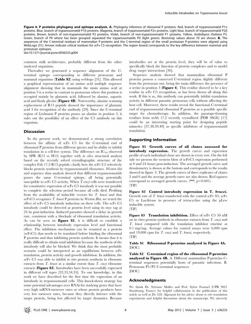
common stalk architecture, probably different from the other
analyzed organisms.
Thereafter we generated a sequence alignment of the C-
terminal epitope corresponding to different protozoan and
mammal organisms (Table S2) using weblogo [31]. This allowed
a graphical representation of an amino acid multiple sequence
alignment showing that in mammals the main amino acid at
position 3 is a serine in contrast to protozoan where this position is
occupied mainly by glutamic acid, followed by alanine, aspartic
acid and finaly glycine (Figure 4B). Noteworthy, alanine scanning
replacement of R13 peptide showed the importance of glutamic
acid 3 for recognition of the antibody [29]. Since the C-terminal
region of Leishmania P proteins posses an alanine in position 3, it
rules out the possibility of an effect of the C5 antibody on this
organism.
Discussion
In the present work, we demonstrated a strong correlation
between the affinity of scFv C5 for the C-terminal end of
ribosomal P proteins from different species and its ability to inhibit
translation in a cell-free system. The difference on IC50 observed
by SPR (R13 vs H13) together with in silico structural analysis
based on the recently solved crystallographic structure of the
complex Fab 17.2-R13 peptide; clarify how the scFv C5 selectively
blocks protein synthesis by trypanosomatid ribosomes. Western blot
and sequence data analysis showed that different trypanosomatids
posses the same C-terminal epitope, all being potentially
susceptible to scFv C5 activity. When T.cruzi cells were transfected
for constitutive expression of scFv C5 intrabody it was not possible
to complete the selection period because all cells died. Profiting
from the availability of inducible vectors for T. brucei and that
scFvC5 recognizes T. brucei P proteins in Western Blot, we tested the
effect of scFv C5 intrabody induction on these cells. The scFv C5
intrabody could be detected at protein level using its myc tag at
24 hs post-induction. Induced parasites showed a delay in growth
rate, consistent with a blockade of ribosomal translation activity.
As can be seen on figure S1, it is difficult to establish a
relationship between intrabody expression level and functional
effect. The inhibition mechanism can be resumed as a protein
(scFvC5) that needs to be translated before binding the ribosomal
P proteins and thus inhibiting protein synthesis. It means that it is
really difficult to obtain total inhibition because the synthesis of the
intrabody will also be blocked. We think that the most probable
scenario could be interpreted as an equilibrium between scFv
translation, protein activity and growth inhibition. In addition, the
scFv C5 was able to inhibit in vitro protein synthesis in ribosome
extracts from T. brucei at a similar extent than T. cruzi ribosome
extracts (Figure S3). Intrabodies have been successfully expressed
in different cell types [32,33,34,35]. To our knowledge, in this
work we have described for the first time the expression of an
intrabody in trypanosomatid cells. This knock-down strategy has
some potential advantages over RNAi for studying genes that have
very high mRNA-turnover rates or whose protein products have
very low turnover rates, because they directly interact with the
target protein, being less affected by target dynamics. Because
intrabodies act at the protein level, they will be of value to
specifically block the function of protein complexes and to model
drug–target interactions [36].
Sequence analysis showed that mammalian ribosomal P
proteins possess a conserved C-terminal region slightly different
from the protozoan one, being the main difference the presence of
a serine in position 3 (Figure 4). This residue showed to be a key
residue in scFv C5 recognition, as has been shown all along this
work. If this is so, this antibody can potentially inhibit translation
activity in different parasitic protozoan cells without affecting the
host cell. Moreover, these results reveal the functional C-terminal
end of trypanosomatid ribosomal P proteins as a possible specific
target for chemotherapy. In addition, the paratope-forming
residues from mAb 17.2 recently crystallized (PDB 3SGE) [17]
could be an interesting starting point for designing peptide
mimetics [37,38,39,40] as specific inhibitors of trypanosomatid
translation.
Supporting Information
Figure S1 Growth curves of all clones assessed forintrabody expression. The growth curves and expression
profile of each individual clone are shown in every row. On the left
side we present the western blots of scFvC5 expression performed
at 0 and 24 hours post-induction. The averaged growth curve and
densitometry is shown at the bottom and corresponds to the results
showed in figure 3. The growth curves of three replicates of clones
2 and 6 and the average growth curve are also shown. Red squares
correspond to averaged growth curves. (***; p,0.001).
(TIF)
Figure S2 Control intrabody expression in T. brucei.Growth rate of T. brucei transfected with the control scFv S3, scFv
C5 or Luciferase in presence of tetracycline using the pLew
inducible system.
(TIF)
Figure S3 Translation inhibition. Effect of scFv C5 50 nM
on in vitro protein synthesis in ribosome extracts from T. cruzi and
T. brucei compared with the translation inhibitor emetine at
0.1 mg/mg. Average values for control assays were 6,000 cpm
and 19,000 cpm for T. cruzi and T. brucei, respectively.
(TIF)
Table S1 Ribosomal P-proteins analyzed in Figure 4A.(DOC)
Table S2 C-terminal region of the ribosomal P-proteinsanalyzed in Figure 4B. A. Different mammalian P-proteins C-
terminal sequences potentially hosts of parasitic infections. B.
Protozoan P1/P2 C-terminal sequences.
(DOC)
Acknowledgments
We thank Dr. Sylviane Muller and Prof. Sylvie Fournel (UPR 9021
Strasbourg, France) for helpful collaboration in the publication of this
article as well as Dr. I.D. Algranati for his advice about in vitro translation
experiments and helpful discussions about the manuscript. We sincerely
Figure 4. P proteins phylogeny and epitope analysis. A. Phylogeny inference of ribosomal P proteins. Red, branch of trypanosomatid P1aproteins. Blue, branch of trypanosomatid P1b proteins. Magenta, branch of trypanosomatid P2a proteins. Light blue, branch of trypanosomatid P2bproteins. Brown, branch of non-trypanosomatid P2 proteins. Violet, branch of non-trypanosomatid P1 proteins. Yellow, Arabidopsis thaliana P3.Green, branch of P0 where has been grouped separated the trypanosomatids P0 (light green). Bootstrap values above 70 are shown. B. Thesequences of the C-terminal residues of mammalian P-proteins and the C-terminal region of the small protozoan P-proteins were aligned usingWebLogo [31]. Arrows indicate critical residues for scFv C5 recognition. The region boxed corresponds to the key difference between mammal andprotozoan epitopes.doi:10.1371/journal.pone.0036233.g004
Inducible Intrabodies on Trypanosoma brucei
PLoS ONE | www.plosone.org 8 May 2012 | Volume 7 | Issue 5 | e36233
thank Prof. Dr. Christine Clayton (ZMBH, Heidelberg, Germany) for
facilitating the expressions in T. brucei. M.J.A., S.A.L. and K.A.G. are
members of scientific career of CONICET. L.S. and T.D. have fellowships
from CONICET.
In memory of Dr. Mariano J. Levin (1951–2010), Director of the Laboratory of
Molecular Biology of Chagas’ disease, INGEBI-CONICET, Buenos Aires, Argentina,
from 1985 to 2010.
Author Contributions
Conceived and designed the experiments: MJL CRS MJA BN. Performed
the experiments: CRS MJA BN LS TD SAL. Analyzed the data: CRS
MJA BN LS TD SAL KAG JH. Contributed reagents/materials/analysis
tools: JH. Wrote the paper: CRS MJA.
References
1. Bailey-Serres J, Vangala S, Szick K, Lee CH (1997) Acidic phosphoprotein
complex of the 60S ribosomal subunit of maize seedling roots. Components andchanges in response to flooding. Plant Physiol 114: 1293–1305.
2. Planta RJ, Mager WH (1998) The list of cytoplasmic ribosomal proteins ofSaccharomyces cerevisiae. Yeast 14: 471–477.
3. Juri Ayub M, Smulski CR, Nyambega B, Bercovich N, Masiga D, et al. (2005)Protein-protein interaction map of the Trypanosoma cruzi ribosomal P protein
complex. Gene 357: 129–136.
4. Smulski CR, Longhi SA, Ayub MJ, Edreira MM, Simonetti L, et al. (2010)Interaction map of the Trypanosoma cruzi ribosomal P protein complex (stalk)
and the elongation factor 2. J Mol Recognit 24: 359–370.5. Levin MJ, Vazquez M, Kaplan D, Schijman AG (1993) The Trypanosoma cruzi
ribosomal P protein family: classification and antigenicity. Parasitol Today 9:
381–384.6. Lavergne JP, Conquet F, Reboud JP, Reboud AM (1987) Role of acidic
phosphoproteins in the partial reconstitution of the active 60 S ribosomalsubunit. FEBS Lett 216: 83–88.
7. Gonzalo P, Reboud JP (2003) The puzzling lateral flexible stalk of the ribosome.Biol Cell 95: 179–193.
8. Bargis-Surgey P, Lavergne JP, Gonzalo P, Vard C, Filhol-Cochet O, et al. (1999)
Interaction of elongation factor eEF-2 with ribosomal P proteins. Eur J Biochem262: 606–611.
9. Ballesta JP, Rodriguez-Gabriel MA, Bou G, Briones E, Zambrano R, et al.(1999) Phosphorylation of the yeast ribosomal stalk. Functional effects and
enzymes involved in the process. FEMS Microbiol Rev 23: 537–550.
10. Zampieri S, Mahler M, Bluthner M, Qiu Z, Malmegrim K, et al. (2003)Recombinant anti-P protein autoantibodies isolated from a human autoimmune
library: reactivity, specificity and epitope recognition. Cell Mol Life Sci 60:588–598.
11. Stacey DW, Skelly S, Watson T, Elkon K, Weissbach H, et al. (1988) Theinhibition of protein synthesis by IgG containing anti-ribosome P autoantibodies
from systemic lupus erythematosus patients. Arch Biochem Biophys 267:
398–403.12. Levin MJ, Mesri E, Benarous R, Levitus G, Schijman A, et al. (1989)
Identification of major Trypanosoma cruzi antigenic determinants in chronicChagas’ heart disease. Am J Trop Med Hyg 41: 530–538.
13. Mesri EA, Levitus G, Hontebeyrie-Joskowicz M, Dighiero G, Van
Regenmortel MH, et al. (1990) Major Trypanosoma cruzi antigenic determinantin Chagas’ heart disease shares homology with the systemic lupus erythematosus
ribosomal P protein epitope. J Clin Microbiol 28: 1219–1224.14. Kaplan D, Ferrari I, Bergami PL, Mahler E, Levitus G, et al. (1997) Antibodies
to ribosomal P proteins of Trypanosoma cruzi in Chagas disease possessfunctional autoreactivity with heart tissue and differ from anti-P autoantibodies
in lupus. Proc Natl Acad Sci U S A 94: 10301–10306.
15. Lopez Bergami P, Mateos P, Hoebeke J, Levin MJ, Baldi A (2003) Sequenceanalysis, expression, and paratope characterization of a single-chain Fv fragment
for the eukaryote ribosomal P proteins. Biochem Biophys Res Commun 301:819–824.
16. Smulski C, Labovsky V, Levy G, Hontebeyrie M, Hoebeke J, et al. (2006)
Structural basis of the cross-reaction between an antibody to the Trypanosomacruzi ribosomal P2beta protein and the human beta1 adrenergic receptor.
FASEB J 20: 1396–1406.17. Pizarro JC, Boulot G, Bentley GA, Gomez KA, Hoebeke J, et al. (2011) Crystal
Structure of the Complex mAb 17.2 and the C-Terminal Region of
Trypanosoma cruzi P2b Protein: Implications in Cross-Reactivity. PLoS NeglTrop Dis 5: e1375.
18. Lopez Bergami P, Scaglione J, Levin MJ (2001) Antibodies against the carboxyl-terminal end of the Trypanosoma cruzi ribosomal P proteins are pathogenic.
FASEB J 15: 2602–2612.19. Muller S, Couppez M, Briand JP, Gordon J, Sautiere P, et al. (1985) Antigenic
structure of histone H2B. Biochim Biophys Acta 827: 235–246.
20. Manful T, Cristodero M, Clayton C (2009) DRBD1 is the Trypanosoma brucei
homologue of the spliceosome-associated protein 49. Mol Biochem Parasitol
166: 186–189.
21. Alibu VP, Storm L, Haile S, Clayton C, Horn D (2005) A doubly inducible
system for RNA interference and rapid RNAi plasmid construction inTrypanosoma brucei. Molecular and Biochemical Parasitology 139: 75–82.
22. Clayton CE (1987) Import of fructose bisphosphate aldolase into the glycosomesof Trypanosoma brucei. J Cell Biol 105: 2649–2654.
23. Juri Ayub M, Ma KW, Shaw PC, Wong KB (2008) Trypanosoma cruzi: highribosomal resistance to trichosanthin inactivation. Exp Parasitol 118: 442–447.
24. Hirumi H, Hirumi K (1989) Continuous cultivation of Trypanosoma bruceiblood stream forms in a medium containing a low concentration of serum
protein without feeder cell layers. J Parasitol 75: 985–989.
25. Biebinger S, Wirtz LE, Lorenz P, Clayton C (1997) Vectors for inducible
expression of toxic gene products in bloodstream and procyclic Trypanosomabrucei. Mol Biochem Parasitol 85: 99–112.
26. Altschul SF, Gish W, Miller W, Myers EW, Lipman DJ (1990) Basic localalignment search tool. J Mol Biol 215: 403–410.
27. Kumar S, Nei M, Dudley J, Tamura K (2008) MEGA: a biologist-centricsoftware for evolutionary analysis of DNA and protein sequences. Brief
Bioinform 9: 299–306.
28. Thompson JD, Higgins DG, Gibson TJ (1994) CLUSTAL W: improving the
sensitivity of progressive multiple sequence alignment through sequence
weighting, position-specific gap penalties and weight matrix choice. NucleicAcids Res 22: 4673–4680.
29. Mahler E, Sepulveda P, Jeannequin O, Liegeard P, Gounon P, et al. (2001) Amonoclonal antibody against the immunodominant epitope of the ribosomal
P2beta protein of Trypanosoma cruzi interacts with the human beta 1-adrenergic receptor. Eur J Immunol 31: 2210–2216.
30. Soares MR, Bisch PM, Campos de Carvalho AC, Valente AP, Almeida FC(2004) Correlation between conformation and antibody binding: NMR structure
of cross-reactive peptides from T. cruzi, human and L. braziliensis. FEBS Lett
560: 134–140.
31. Crooks GE, Hon G, Chandonia JM, Brenner SE (2004) WebLogo: a sequence
logo generator. Genome Res 14: 1188–1190.
32. Boldicke T, Weber H, Mueller PP, Barleon B, Bernal M (2005) Novel highly
efficient intrabody mediates complete inhibition of cell surface expression of thehuman vascular endothelial growth factor receptor-2 (VEGFR-2/KDR).
J Immunol Methods 300: 146–159.
33. Wolfgang WJ, Miller TW, Webster JM, Huston JS, Thompson LM, et al. (2005)
Suppression of Huntington’s disease pathology in Drosophila by human single-chain Fv antibodies. Proc Natl Acad Sci U S A 102: 11563–11568.
34. Paganetti P, Calanca V, Galli C, Stefani M, Molinari M (2005) beta-site specificintrabodies to decrease and prevent generation of Alzheimer’s Abeta peptide.
J Cell Biol 168: 863–868.
35. Kontermann RE (2004) Intrabodies as therapeutic agents. Methods 34:
163–170.
36. Stocks MR (2004) Intrabodies: production and promise. Drug Discov Today 9:
960–966.
37. Saragovi HU, Fitzpatrick D, Raktabutr A, Nakanishi H, Kahn M, et al. (1991)
Design and synthesis of a mimetic from an antibody complementarity-
determining region. Science 253: 792–795.
38. Takasaki W, Kajino Y, Kajino K, Murali R, Greene MI (1997) Structure-based
design and characterization of exocyclic peptidomimetics that inhibit TNF alphabinding to its receptor. Nat Biotechnol 15: 1266–1270.
39. Berezov A, Greene MI, Murali R (2003) Structure-based approaches toinhibition of erbB receptors with peptide mimetics. Immunol Res 27: 303–308.
40. Casset F, Roux F, Mouchet P, Bes C, Chardes T, et al. (2003) A peptide mimeticof an anti-CD4 monoclonal antibody by rational design. Biochem Biophys Res
Commun 307: 198–205.
Inducible Intrabodies on Trypanosoma brucei
PLoS ONE | www.plosone.org 9 May 2012 | Volume 7 | Issue 5 | e36233