Embed Size (px)
Citation preview
Psychiatr Clin N Am 29 (2006) 947–967
PSYCHIATRIC CLINICSOF NORTH AMERICA
Selected Sleep Disorders: Restless LegsSyndrome and Periodic LimbMovement Disorder, Sleep ApneaSyndrome, and Narcolepsy
Milton K. Erman, MDa,b,*aDepartment of Psychiatry, School of Medicine, University of California San Diego, CA, USAbPacific Sleep Medicine, 10052 Mesa Ridge Court, Suite 101, San Diego, CA 92121, USA
Complaints of disturbed sleep are common in psychiatric patients and areassociated with the diagnosis of specific disorders, such as mania andmajor depression. Despite this, clinicians often perceive sleep com-
plaints to be a symptomatic reflection of the therapeutic response to treatment:If the treatment for depression is effective, sleep complaints should resolve.Most practicing psychiatrists have received little formal instruction about sleepdisorders during their training in medical school and residency [1]. This leavesthem incompletely prepared to recognize symptoms of sleep disorders, whichoften are subtle in their presentations, and the symptomatic complaints ofwhich may overlap with various psychiatric syndromes.
The specific sleep disorders discussed in this article all are likely to be seenby practicing psychiatrists and may be misinterpreted by them as reflecting anelement of a comorbid psychiatric syndrome. Additionally, the presence ofthese conditions may interfere with treatment efforts being provided for psychi-atric conditions. For these reasons, awareness of these disorders and their pre-sentations allows psychiatrists to function more effectively in clinical care.
Use of sleep studies to establish the diagnosis of several of these conditions isaddressed later in this article. Polysomnography (PSG) techniques for establish-ing sleep stages have been reviewed elsewhere in this issue and include the elec-troencephalogram, electro-oculogram, and submental electromyogram. Thetypical clinical PSG also includes an electrocardiogram, respiratory effort (usu-ally including measures of chest and abdominal effort), bilateral anterior tibialiselectromyography, measurement of airflow (using a thermistor or a pressuretransducer), and measurement of oxygen saturation (pulse oximetry). Thestudies must be performed in a temperature-controlled, sound-attenuated,
*Pacific Sleep Medicine, 10052 Mesa Ridge Court, Suite 101, San Diego, CA 92121,USA. E-mail address: [email protected]
0193-953X/06/$ – see front matter ª 2006 Elsevier Inc. All rights reserved.doi:10.1016/j.psc.2006.09.007 psych.theclinics.com
948 ERMAN
and light-attenuated environment to ensure that the data obtained would not bedisrupted by outside environmental factors. Guidelines for the use of PSG inthe practice of sleep medicine have been established by a committee of theAmerican Academy of Sleep Medicine [2]. Alternative methods for the diagno-sis of various sleep disorders, including narcolepsy and sleep apnea, have beenproposed, and are discussed in the sections pertaining to these diseases.
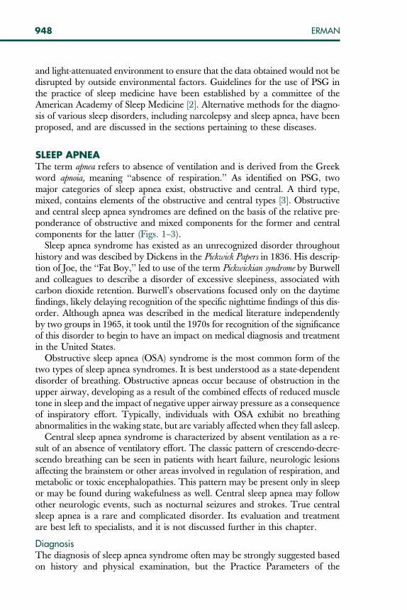
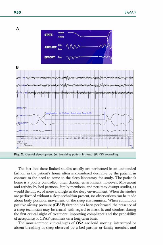
SLEEP APNEAThe term apnea refers to absence of ventilation and is derived from the Greekword apnoia, meaning ‘‘absence of respiration.’’ As identified on PSG, twomajor categories of sleep apnea exist, obstructive and central. A third type,mixed, contains elements of the obstructive and central types [3]. Obstructiveand central sleep apnea syndromes are defined on the basis of the relative pre-ponderance of obstructive and mixed components for the former and centralcomponents for the latter (Figs. 1–3).
Sleep apnea syndrome has existed as an unrecognized disorder throughouthistory and was descibed by Dickens in the Pickwick Papers in 1836. His descrip-tion of Joe, the ‘‘Fat Boy,’’ led to use of the term Pickwickian syndrome by Burwelland colleagues to describe a disorder of excessive sleepiness, associated withcarbon dioxide retention. Burwell’s observations focused only on the daytimefindings, likely delaying recognition of the specific nighttime findings of this dis-order. Although apnea was described in the medical literature independentlyby two groups in 1965, it took until the 1970s for recognition of the significanceof this disorder to begin to have an impact on medical diagnosis and treatmentin the United States.
Obstructive sleep apnea (OSA) syndrome is the most common form of thetwo types of sleep apnea syndromes. It is best understood as a state-dependentdisorder of breathing. Obstructive apneas occur because of obstruction in theupper airway, developing as a result of the combined effects of reduced muscletone in sleep and the impact of negative upper airway pressure as a consequenceof inspiratory effort. Typically, individuals with OSA exhibit no breathingabnormalities in the waking state, but are variably affected when they fall asleep.
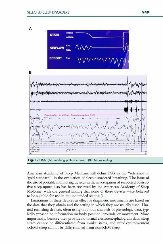
Central sleep apnea syndrome is characterized by absent ventilation as a re-sult of an absence of ventilatory effort. The classic pattern of crescendo-decre-scendo breathing can be seen in patients with heart failure, neurologic lesionsaffecting the brainstem or other areas involved in regulation of respiration, andmetabolic or toxic encephalopathies. This pattern may be present only in sleepor may be found during wakefulness as well. Central sleep apnea may followother neurologic events, such as nocturnal seizures and strokes. True centralsleep apnea is a rare and complicated disorder. Its evaluation and treatmentare best left to specialists, and it is not discussed further in this chapter.
DiagnosisThe diagnosis of sleep apnea syndrome often may be strongly suggested basedon history and physical examination, but the Practice Parameters of the
949SELECTED SLEEP DISORDERS
American Academy of Sleep Medicine still define PSG as the ‘‘reference or‘gold standard’’’ in the evaluation of sleep-disordered breathing. The issue ofthe use of portable monitoring devices in the investigation of suspected obstruc-tive sleep apnea also has been reviewed by the American Academy of SleepMedicine, with the general finding that none of these devices were believedto be suitable for use in an unattended setting [4].
Limitations of these devices as effective diagnostic instruments are based onthe data that they obtain and the setting in which they are usually used. Lim-ited recording devices, often using only four channels of physiologic data, typ-ically provide no information on body position, arousals, or movement. Moreimportantly, because they provide no formal electroencephalogram data, sleepstates cannot be differentiated from awake states, and rapid-eye-movement(REM) sleep cannot be differentiated from non-REM sleep.
Fig. 1. OSA. (A) Breathing pattern in sleep. (B) PSG recording.
950 ERMAN
The fact that these limited studies usually are performed in an unattendedfashion in the patient’s home often is considered desirable by the patient, incontrast to the need to come to the sleep laboratory for study. The patient’shome is a poorly controlled, often chaotic, environment, however. Movementand activity by bed partners, family members, and pets may disrupt studies, aswould the impact of noise and light in the sleep environment. When the studiesare performed without a sleep technician present, no observations can be madeabout body position, movement, or the sleep environment. When continuouspositive airway pressure (CPAP) titration has been performed, the presence ofa sleep technician may be crucial with regard to mask fit and comfort duringthe first critical night of treatment, improving compliance and the probabilityof acceptance of CPAP treatment on a long-term basis.
The most common clinical signs of OSA are loud snoring, interrupted orabsent breathing in sleep observed by a bed partner or family member, and
Fig. 2. Central sleep apnea. (A) Breathing pattern in sleep. (B) PSG recording.
951SELECTED SLEEP DISORDERS
excessive daytime sleepiness. The patient himself or herself is usually unawareof the existence of a problem, often boasting that they have no sleepproblem—‘‘I can fall asleep anywhere.’’ Patients often deny the presence ofthese symptoms, and it is helpful to have a bed partner or family memberpresent when symptoms suggesting sleep apnea are reviewed.
Some patients report awareness of awakening from sleep with a sensation ofchoking, at times associated with dreams of drowning or suffocating. Othercommon symptoms include a history of restless sleep, morning headache,morning sore throat, and daytime fatigue.
Specific abnormalities in the physical examination often are seen in patientswith OSA. Many patients are obese and have a large neck or crowded upper air-way, and a history of weight gain associated with increased severity of snoring orapnea often is reported. It should not be assumed that all patients, however, are
Fig. 3. Mixed sleep apnea. (A) Breathing pattern in sleep. (B) PSG recording.
952 ERMAN
morbidly obese or obese at all because some sleep apnea patients have a normalbody habitus. Common structural abnormalities, such as narrow nasal passage,long soft palate, large tonsils, or a short (retrognathic or micrognathic) mandiblecontribute to airway obstruction.
OSA is associated with an increased risk of cognitive abnormalities and ofaffective disorders, such as depression [5]. Cognitive impairment associatedwith apnea may be misdiagnosed as dementia, with capacity for substantial im-provement in cognition associated with effective treatment. Similarly, untreatedapnea may exacerbate depression severity or limit response to therapy, withsubstantial improvement in mood seen after treatment is initiated [6].
EpidemiologyOSA is a common disorder. The Wisconsin Sleep Cohort Study [7], using re-strictive methodologies in a large population of employed men and women 30to 60 years old, reported a prevalence rate for OSA of approximately 15% inmen and 5% in women based on an Apnea Hypopnea Index (AHI)—numberof apneas or hypopneas, episodes of partial apnea, per hour of sleep—of 10or higher. Other studies of OSA prevalence using laboratory PSG to estimatethe prevalence of severe OSA, defined as an AHI greater than 15, report 7% to14% in men and 2% to 7% in women. If mild disease is considered (AHI >5events per hour), the prevalence is 17% to 26% in men and 9% to 28% inwomen older than 20 years. The presence of mild disease is relevant becausean AHI of 5 events per hour has been shown to be associated with increasedrisks for hypertension and other health consequences.
Some patients may be unaware of or may deny the existence of snoring; con-firmation of snoring may be particularly difficult for patients who sleep alone.Often, bed partners acknowledge that they have been driven from their bedand shared bedroom by their partner’s loud snoring. Although they may beable to comment on snoring from the past, they can say little about thepresence or absence of apneic pauses at the present time.
Another factor that may limit the reliability of information provided bya spouse or bed partner is hearing loss, a problem seen most frequently in olderpatients and their spouses. Some spouses may deny or may be unaware of theseverity of their hearing loss, affecting their ability to provide accurate informa-tion about snoring and apneas.
Pathophysiology and ConsequencesOSA involves repetitive obstructions of the airway in sleep, leading to oxyhe-moglobin desaturation, inspiratory efforts against the occluded airway, and ter-mination of the event by arousal from sleep. The daytime sleepiness andfatigue characteristic of OSA is likely a consequence of the fragmented sleepassociated with this disorder, generated by the need for recurrent arousals toavoid suffocation and death.
Patients with OSA have an increased incidence of hypertension comparedwith individuals without OSA, and OSA seems to be an independent risk factorfor the development of hypertension. OSA seems to be implicated in stroke and
953SELECTED SLEEP DISORDERS
transient ischemic attacks and coronary heart disease, heart failure, and cardiacarrhythmias. These cardiovascular risks are likely due to the release of proin-flammatory and prothrombotic factors that have been identified to beimportant in the development of atherosclerosis as a consequence of apneaevents and by ‘‘downstream effects’’ of arousals that terminate apnea events.
Other consequences of the disorder include excessive daytime sleepiness,which can be relatively mild or may be severe enough to interfere with employ-ment or driving an automobile. Several studies have documented an increasedrisk of automobile crashes in patients with OSA, such that it is considered a dis-order associated with loss of consciousness in some states. Quality of life is de-monstrably impaired in patients with OSA, including complaints of fatigue,memory impairment, reduced concentration, depressed mood, and irritability.Some patients may experience decreased libido or erectile dysfunction. Thesesymptoms virtually always improve, and may resolve completely, with effec-tive treatment.
TreatmentNasal CPAP administered via a nasal mask or interface is the treatment ofchoice for moderate-to-severe OSA. The positive pressure generated byCPAP functions as a pneumatic stent for the upper airway, preventing theairway collapse that otherwise would occur as a consequence of negative inspi-ratory pressure.
Nasal CPAP therapy for sleep apnea was first described in 1981, but it wasnot initially widely accepted in treatment. CPAP is administered by a soft maskheld in place against the face, usually covering only the nose (although somepatients may need a ‘‘full face’’ mask covering the nose and mouth). Sufficientpressure is introduced to eliminate apneas, hypopneas, and snoring. At appro-priate pressures, CPAP is almost always effective in treatment of OSA. Limita-tions have included the bulk and noise of early CPAP units and problems withearly masks including poor fit and the need to use an adhesive to attach themask to the face. Continuing technical improvements in the design of masksand CPAP units has led to dramatically increased acceptance of CPAP therapy.
Surgical therapy for sleep apnea includes a broad range of procedures.Although tracheostomy is curative of this disorder, it is associated with signif-icant medical risks and lifestyle limitations and generally is reserved for patientswith severe sleep apnea unable to use nasal CPAP in treatment.
The most widely used surgery for sleep apnea, uvulopalatopharyngoplasty(UPPP), was described in 1981 [8]. This procedure involves removal of redun-dant or excess tissue in the throat to improve airway patency, reduce snoring,and reduce apnea severity. It was seen initially as a viable and mildly invasivealternative to CPAP, initially perceived as burdensome and ‘‘a treatment, nota cure.’’
Outcome analyses for UPPP procedures suggested that significant improve-ment (defined as a �50% reduction in the AHI) was seen in only about 50% ofpatients. Patients often complained of significant pain associated with the
954 ERMAN
procedure, which required hospitalization for at least several days. In addition,complications such as altered speech and velopalatal incompetence were seen insome patients. These complications seemed to be more common in the earlyyears in which this procedure was performed and in cases in which the surgerymay have been more aggressive. As a consequence of these issues, the numberof UPPP procedures has reduced dramatically in recent years.
A variant of this procedure, laser-assisted uvulopalatoplasty, was developedto provide a less invasive treatment alternative. Although it is often describedas providing results comparable to UPPP, pain associated with the multiple(typically three to five) laser treatments needed to complete this procedure lim-ited patient acceptance. Treatment outcome is not likely to be equal to that ofUPPP because laser-assisted uvulopalatoplasty relies primarily on scarring ofthe airway and retraction of tissue as a consequence rather than removal of softtissue in the airway as occurs with UPPP.
Radiofrequency ablation is another alternative to UPPP and laser-assisteduvulopalatoplasty that works by stiffening the soft palate and shrinking tissuein the airway. An advantage of radiofrequency ablation is that it is minimallyinvasive, with few treatment-associated complications. Although it has beenshown to be a benefit in patients who snore, few data have been published sug-gesting benefit in patients with significant sleep apnea.
Weight reduction surgery must be mentioned among surgical options for thetreatment of sleep apnea. Although many sleep apnea patients may be of nor-mal weight, weight is an exacerbating factor for virtually all apnea patients, anda high proportion of morbidly obese patients have sleep apnea [9]. Patients withsleep apnea who undergo bariatric surgery typically have a progressive reduc-tion in the apnea severity associated with weight loss and usually are able toreduce their CPAP pressure as they lose weight. Not all patients are ‘‘cured’’of apnea as a consequence of weight loss, and continued use of nasal CPAP,use of an oral appliance, or surgical treatment of the upper airway may berequired after maximal weight loss.
Alternative ApproachAn alternative nonsurgical approach for patients who are not candidates for orwho cannot tolerate CPAP is use of an oral appliance, also known as an airwaydilator. These devices, at times similar in appearance to a ‘‘bite plate’’ or nightguard used for bruxism or an athletic mouthpiece, are often beneficial inselected patients [10]. The therapeutic effect of these devices is presumed tobe due to stimulation of muscles in the airway in sleep, reducing the tendencyto collapse that otherwise occurs in association with the muscle flaccidity ofsleep. The fabrication and fitting of these appliances is best performed by adentist knowledgeable about sleep apnea and with significant experience inthe use of these devices.
At present, no pharmacologic treatments have been shown to be effective inthe treatment of sleep apnea. Although some limited treatment trials have sup-ported the hypothesis that tricyclic antidepressants and some selective
955SELECTED SLEEP DISORDERS
serotonin reuptake inhibitors may be beneficial for some patients [11], no con-trolled studies have shown a clear therapeutic benefit associated with treatmentwith these agents, and none are approved for apnea treatment.
Other medical options may be important as adjuncts to these treatment op-tions or as therapeutic interventions alone if the patient cannot accept or toler-ate CPAP or oral appliance therapy as a primary treatment. These includeefforts at weight loss, avoidance of alcohol and other sedating agents at nightif CPAP is not being used, and avoidance of the supine position in sleep.
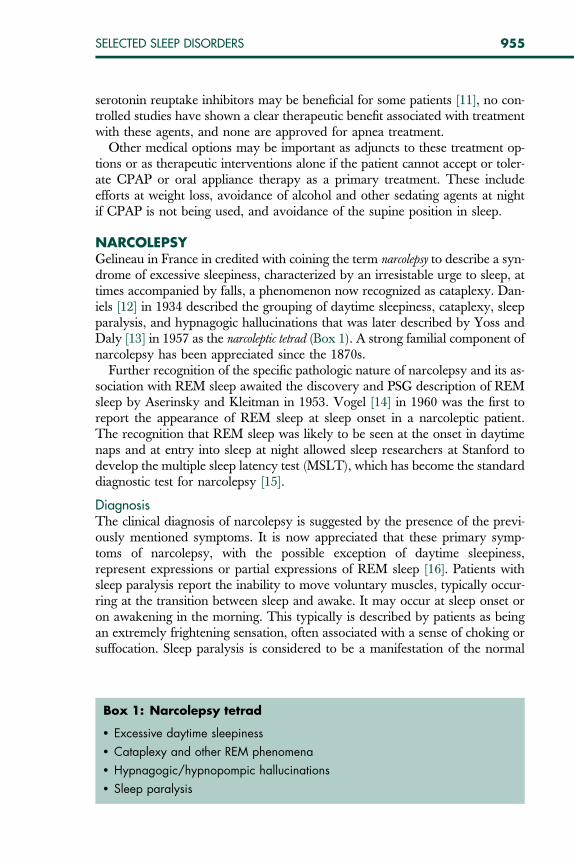
NARCOLEPSYGelineau in France in credited with coining the term narcolepsy to describe a syn-drome of excessive sleepiness, characterized by an irresistable urge to sleep, attimes accompanied by falls, a phenomenon now recognized as cataplexy. Dan-iels [12] in 1934 described the grouping of daytime sleepiness, cataplexy, sleepparalysis, and hypnagogic hallucinations that was later described by Yoss andDaly [13] in 1957 as the narcoleptic tetrad (Box 1). A strong familial component ofnarcolepsy has been appreciated since the 1870s.
Further recognition of the specific pathologic nature of narcolepsy and its as-sociation with REM sleep awaited the discovery and PSG description of REMsleep by Aserinsky and Kleitman in 1953. Vogel [14] in 1960 was the first toreport the appearance of REM sleep at sleep onset in a narcoleptic patient.The recognition that REM sleep was likely to be seen at the onset in daytimenaps and at entry into sleep at night allowed sleep researchers at Stanford todevelop the multiple sleep latency test (MSLT), which has become the standarddiagnostic test for narcolepsy [15].
DiagnosisThe clinical diagnosis of narcolepsy is suggested by the presence of the previ-ously mentioned symptoms. It is now appreciated that these primary symp-toms of narcolepsy, with the possible exception of daytime sleepiness,represent expressions or partial expressions of REM sleep [16]. Patients withsleep paralysis report the inability to move voluntary muscles, typically occur-ring at the transition between sleep and awake. It may occur at sleep onset oron awakening in the morning. This typically is described by patients as beingan extremely frightening sensation, often associated with a sense of choking orsuffocation. Sleep paralysis is considered to be a manifestation of the normal
Box 1: Narcolepsy tetrad
� Excessive daytime sleepiness� Cataplexy and other REM phenomena� Hypnagogic/hypnopompic hallucinations� Sleep paralysis
956 ERMAN
atonia (muscle paralysis) of REM sleep, occurring at an inopportune time. Thepatient suddenly finds himself or herself unable to move for a few minutes,most often on falling asleep or waking up.
During hypnagogic and hypnopompic hallucinations, patients experiencedreamlike auditory or visual hallucinations, usually occurring at the interfacebetween sleep and wakefulness. These experiences, although often bizarreand hard to explain, are perceived by patients to be so real that, in somecircumstances, they have led physicians to a misdiagnosis of schizophrenia. Italso has been argued that the experience of alien abduction reported bysome patients without a clear history of psychiatric illness may be associatedwith symptoms of sleep paralysis and hypnagogic hallucinations.
Cataplexy often is considered to be a pathognomonic feature for narcolepsy.Cataplexy is the sudden loss of muscle tone or strength, usually elicited bystrong emotions, such as elation, anger, or sexual arousal. This symptom seemsto reflect the appearance of normal REM sleep atonia, experienced pathologi-cally in the waking state. The severity of cataplexy varies tremendously amongindividual patients. In very mild cases, the patient’s knees may buckle slightlyor the patient may feel weak when he or she hears a joke, is startled by a noise,or experiences other strong emotions. Other patients may report weakness ofthe neck such that their head may drop to their chest or their jaw may becomeslack. Although there is a perception that cataplexy typically is associated with‘‘drop attacks,’’ only in the most severe cases do patients fall or experience rel-atively complete paralysis for a few seconds to several minutes.
The association with a specific human leukocyte antigen (HLA) pattern wasdescribed in the 1980s and has supported the hypothesis that this disorder maydevelop as a consequence of an autoimmune phenomenon. Recognition thatthis HLA pattern was seen in most narcoleptics led to efforts to use this test as a di-agnostic test, theoretically minimizing the need for PSG evaluation. The notionthat a specific blood test could be diagnostic for this disorder also was appealing.
The specific HLA subtypes that have been discovered to be associated withnarcolepsy are only predisposing factors and are not sufficient by themselves tocause narcolepsy. More than 20% of the general population carries the sameHLA subtypes (HLA-DR2, HLA-DQB1*0602) [17]. Many patients with symp-tomatic narcolepsy but without cataplexy do not have HLA-DQB1*0602.
Although the clinical diagnosis of narcolepsy may be strongly suggested bythe presence of symptoms of the narcolepsy tetrad, formal diagnosis of this con-dition requires PSG evaluation. Historically, many patients have been given thediagnosis of narcolepsy based on a report of sleepiness, without ever havingreceived formal PSG evaluation. This phenomenon was more common beforethe development of the MSLT and publication of specific recommendationswith regard to diagnosis by the American Academy of Sleep Medicine [18].Many patients with OSA have been erroneously diagnosed with narcolepsy,leading to treatment with stimulants that may provide symptomatic relief butthat leave untreated, and can even exacerbate, medical consequences of theOSA, such as arrhythmias and hypertension.
957SELECTED SLEEP DISORDERS
From an administrative perspective, the realization that some patients withnarcolepsy receive lifelong access to stimulant medications supports the benefitsof formal PSG evaluation. Some drug-abusing patients may seek a diagnosis ofnarcolepsy to provide ‘‘legal’’ access to amphetamines and other stimulants;other patients who have received stimulant medications in treatment of theircomplaints of sleepiness, and who perceive this to be of benefit for them,may be treated more appropriately with other therapies when it is appreciatedthat they do not meet the formal diagnostic criteria for narcolepsy.
A distinction is now made between narcolepsy with cataplexy and betweennarcolepsy without cataplexy and has been incorporated in the revision of the.International Classification of Sleep Disorders (Box 2) [19]. A diagnosis of nar-colepsy with cataplexy requires documentation of a definite history of cata-plexy, but can be made without PSG and MSLT evaluation. Narcolepsywithout cataplexy is diagnosed based on appropriate clinical history andPSG evaluation with an MSLT.
The specifics of PSG diagnosis are relatively straightforward. The MSLT isperformed after a full (8-hour) nighttime PSG, performed to ensure that theMSLT data are not contaminated by the effects of sleep deprivation, andthat other sleep disorders that could cause excessive sleepiness are not present.
The MSLT consists of PSG monitoring of sleep parameters for 20 minutes ina quiet, dark, and comfortable bedroom. The first test usually is performedabout 2 hours after the patient has awakened. The patient is given four orfive nap opportunities to fall asleep at 2-hour intervals throughout the day.The time to sleep onset and the presence of a sleep-onset REM period are docu-mented. REM sleep that occurs within 15 minutes of sleep onset is considereda sleep-onset REM period. A mean sleep latency of less than 8 minutes and thepresence of REM sleep in at least two naps are needed to establish a diagnosisof narcolepsy without cataplexy.
Box 2: Diagnosis of narcolepsy
Narcolepsy with cataplexy
Daily excessive sleepiness for at least 3 mo
Definite history of cataplexy, triggered by emotions, is present
Diagnosis should be confirmed, when possible, with PSG and MSLT
Excessive sleepiness not caused by another medical disorder or by medications
Narcolepsy without cataplexy
Daily excessive sleepiness for at least 3 mo
Typical cataplexy not present, although cataplexy-like symptoms may be reported
Diagnosis must be confirmed with PSG and MSLT
Excessive sleepiness not caused by another medical disorder or by medications
958 ERMAN
EpidemiologyThe prevalence of narcolepsy is not known with certainty because it often goesunrecognized or unreported. The prevalence also seems to vary among differ-ent populations throughout the world, perhaps in relation to variability amongdifferent ethnic and racial groups. In European populations, narcolepsy hasbeen reported to occur at a rate of about 3 to 5 per 10,000 with men andwomen equally affected. It has been reported to be seen much less frequentlyin Israel and more frequently in Japan, emphasizing the probable genetic com-ponents associated with predisposition to this disorder [20,21]. Narcolepsy maymanifest at any age, but is diagnosed most often within the first 2 decades oflife. Narcolepsy also may contribute to childhood behavioral problems stem-ming from other disorders, such as attention-deficit disorder.
EtiologyNarcolepsy research has been facilitated by the existence of animal models forthis disorder, including canine narcolepsy. Canine narcolepsy was firstdescribed at Stanford University in the early 1970s. A colony of affectedanimals was established; the breeding of these animals led to the demonstrationthat narcolepsy was transmitted as a single autosomal recessive gene with fullpenetrance in narcoleptic Dobermans.
Striking parallels between canine and human narcolepsy were shown, includ-ing the presence of cataplexy and predisposition to sleep-onset REM episodes,characterized by the onset of REM sleep shortly after falling asleep. Substantialdifferences between human and canine narcolepsy also were discovered,including the predominant canine autosomal dominant hereditary pattern,which has not been described in human narcolepsy.
A research team led by Mignot of Stanford was able to use their colony ofnarcoleptics dogs to identify the gene responsible for canine narcolepsy. Thisresearch group determined that the dogs carry a mutation in the receptor fora neurotransmitter variously called hypocretin or orexin [22]. Virtually simulta-neously, a group working with knockout mice [23] determined that the absenceof a functional version of the hypocretin gene in these mice was associated withthe appearance of REM sleep at sleep onset.
Because human narcolepsy does not seem to follow the same simple geneticpatterns seen in canine narcolepsy, it was not expected that a single gene anomaly ofthe sort described in narcoleptic dogs and knockout mice would explain human nar-colepsy. In 2000, Mignot’s group showed that human narcolepsy is associated withundetectable hypocretin-1 levels in the cerebrospinal fluid [24], implicating the hy-pocretin system in human narcolepsy. Hypocretin/orexin is generated in the brainonly in the hypothalamus; the functions of hypocretin seem to include regulation ofbody weight, control of water balance, and regulation of body temperature.
Although the cause of human narcolepsy is not formally established, the datathat have been generated suggest that destruction of hypocretin-secreting cellsin the hypothalamus best explains the absence of hypocretin-1 in cerebrospinalfluid described by Mignot. That specific HLA subtypes are associated with
959SELECTED SLEEP DISORDERS
narcolepsy suggests that an HLA-related autoimmune response of unknownorigin may trigger damage to these hypocretin-secreting cells leading to thedevelopment of narcolepsy.
ConsequencesThe impact of narcolepsy on social and interpersonal function is dramatic andwell documented. The excessive sleepiness that is a core symptom of the disorderis associated with school performance problems, often leading to limitations in ed-ucational achievement owing to the perception that the patient with narcolepsy islazy or disinterested. The excessive daytime sleepiness of narcolepsy also gener-ates a high automobile accident rate in patients with this disorder, leading to lim-itations on licensure to drive a car in some states. It has been shown to lead toa high socioeconomic burden, including increased risk of unemployment.
Compared with population norms, baseline quality-of-life scores for subjects innarcolepsy studies have shown substantial burdens in vitality, social functioning,and performance of usual activities. Consequences for patients with narcolepsyare dramatic [25], particularly because this condition often is not appropriatelydiagnosed until affected individuals have had their symptoms for many years.Even with treatment, patients may have problems functioning successfully inschool or at work. The sleepiness that is beyond the narcoleptic’s control maybe interpreted as disinterest or laziness by family members or a spouse; cataplexymay be interpreted as behavior suspicious of sedative abuse. These patients maybe referred for evaluation for depression as a consequence of misperceptions onthe part of relatives that these symptoms are signs of depression. The risk of de-veloping depression may be increased as a consequence of the social and interper-sonal stresses these patients experience.
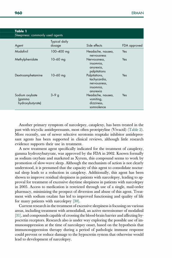
TreatmentThe excessive sleepiness of narcolepsy historically has been treated using am-phetamine and other stimulant agents, such as methylphenidate and pemoline(Table 1) [26]. These compounds all are centrally acting sympathomimeticagents that promote the release of monoamines and block their reuptake.The problems associated with these medications are well recognized and welldescribed, including risks of abuse and diversion; provocation of or exacerba-tion of tendencies to psychosis; and other side effects, such as tremulousness,elevation of blood pressure, agitation, and motor restlessness. Pemoline hasbeen withdrawn from the market because of hepatotoxicity.
Alternative treatments for excessive sleepiness in narcolepsy also have been de-veloped. The wake-promoting compound modafinil was approved by the USFood and Drug Administration (FDA) for the treatment of excessive sleepinessin narcolepsy in 1998 and subsequently has received additional approvals fortreatment of residual sleepiness in treated patients with sleep apnea and for thetreatment of shift work sleep disorder [27,28]. Modafinil has a novel mechanismand is theorized to work in a localized manner, using hypocretin, histamine, epi-nephrine, c-aminobutyric acid, and glutamate [29]. It is a well-tolerated medica-tion with low propensity for abuse.
960 ERMAN
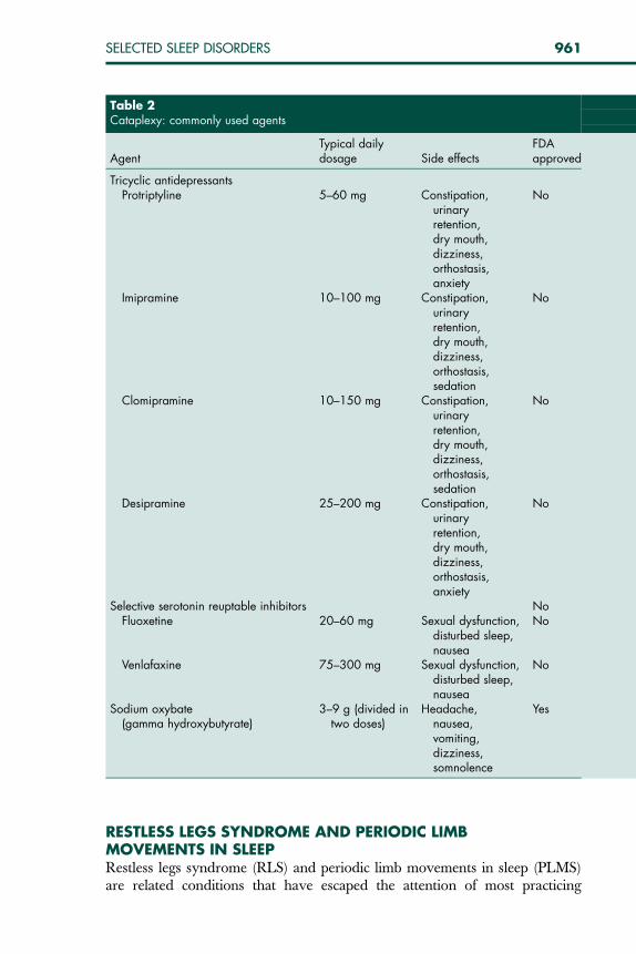
Another primary symptom of narcolepsy, cataplexy, has been treated in thepast with tricyclic antidepressants, most often protriptyline (Vivactil) (Table 2).More recently, use of newer selective serotonin reuptake inhibitor antidepres-sant agents has been supported in clinical reviews, although little researchevidence supports their use in treatment.
A new treatment agent specifically indicated for the treatment of cataplexy,gamma hydroxybutyrate, was approved by the FDA in 2002. Known formallyas sodium oxybate and marketed as Xyrem, this compound seems to work bypromotion of slow-wave sleep. Although the mechanism of action is not clearlyunderstood, it is presumed that the capacity of this agent to consolidate noctur-nal sleep leads to a reduction in cataplexy. Additionally, this agent has beenshown to improve residual sleepiness in patients with narcolepsy, leading to ap-proval for treatment of excessive daytime sleepiness in patients with narcolepsyin 2005. Access to medication is restricted through use of a single, mail-orderpharmacy, minimizing the prospect of diversion and abuse of this agent. Treat-ment with sodium oxalate has led to improved functioning and quality of lifefor many patients with narcolepsy [30].
Current research in the treatment of excessive sleepiness is focusing on variousareas, including treatment with armodafinil, an active stereoisomer of modafinil[31], and compounds capable of crossing the blood-brain barrier and affecting hy-pocretin receptors. Research also is under way exploring the possible use of im-munosuppression at the time of narcolepsy onset, based on the hypothesis thatimmunosuppression therapy during a period of pathologic immune responsecould prevent or reduce damage to the hypocretin system that otherwise wouldlead to development of narcolepsy.
Table 1Sleepiness: commonly used agents
AgentTypical dailydosage Side effects FDA approved
Modafinil 100–400 mg Headache, nausea,nervousness
Yes
Methylphenidate 10–60 mg Nervousness,insomnia,anorexia,palpitations
Yes
Dextroamphetamine 10–60 mg Palpitations,tachycardia,nervousness,insomnia,anorexia
Yes
Sodium oxybate(gammahydroxybutyrate)
3–9 g Headache, nausea,vomiting,dizziness,somnolence
Yes
961SELECTED SLEEP DISORDERS
RESTLESS LEGS SYNDROME AND PERIODIC LIMBMOVEMENTS IN SLEEPRestless legs syndrome (RLS) and periodic limb movements in sleep (PLMS)are related conditions that have escaped the attention of most practicing
Table 2Cataplexy: commonly used agents
AgentTypical dailydosage Side effects
FDAapproved
Tricyclic antidepressantsProtriptyline 5–60 mg Constipation,
urinaryretention,dry mouth,dizziness,orthostasis,anxiety
No
Imipramine 10–100 mg Constipation,urinaryretention,dry mouth,dizziness,orthostasis,sedation
No
Clomipramine 10–150 mg Constipation,urinaryretention,dry mouth,dizziness,orthostasis,sedation
No
Desipramine 25–200 mg Constipation,urinaryretention,dry mouth,dizziness,orthostasis,anxiety
No
Selective serotonin reuptable inhibitors NoFluoxetine 20–60 mg Sexual dysfunction,
disturbed sleep,nausea
No
Venlafaxine 75–300 mg Sexual dysfunction,disturbed sleep,nausea
No
Sodium oxybate(gamma hydroxybutyrate)
3–9 g (divided intwo doses)
Headache,nausea,vomiting,dizziness,somnolence
Yes
962 ERMAN
physicians until recent years. RLS is a diagnosed on the basis of symptomaticcomplaints; PLMS (also known as periodic limb movements disorder) is a sleepdisorder, diagnosed through PSG studies. The two conditions overlap tremen-dously; most RLS patients, if studied in the sleep laboratory, exhibit periodicleg movement activity. When PLMS are noted as an ‘‘incidental finding’’ ina sleep study of a patient not known to have RLS, restless legs symptoms usu-ally are present but may not have been reported.
The complaint of restless legs has been present throughout history, but thefirst description of this complaint as a specific condition was published by theSwedish neurologist Ekbom in 1945. This disorder, also known as Ekbom’ssyndrome, usually is described as a hard-to-define disorder of discomfort inthe legs. A broad range of terms is used by patients to describe their symptoms:pain, discomfort, creeping, crawling, tingling, pulling, twitching, tearing, aching, throbbing,prickling, or grabbing sensation in the calves, legs, or arms. Symptoms typically aremore severe during periods of inactivity or rest or while sitting or lying down.
These symptoms are distinct from complaints of akathisia, a complaint ofmotoric restlessness frequently experienced by psychiatric patients. Akathisiais seen most frequently in patients taking neuroleptic medications, but alsomay be reported as a consequence of use of selective serotonin reuptake inhib-itors and tricyclic antidepressants.
Patients with RLS also are not experiencing symptoms of an anxiety disor-der or attention-deficit/hyperactivity disorder. Although they are disturbed bythe restlessness they experience, their mood state otherwise is usually normal,and they typically do not have the inattention, distractibility, or hyperactivityseen in attention-deficit/hyperactivity disorder.
DiagnosisAs noted previously, RLS is diagnosed on the basis of symptomatic complaints;PLMS must be diagnosed on the basis of PSG evaluation. It should not be as-sumed, however, that a complaint of restless legs and leg movement in sleeprequires a PSG evaluation. RLS does not require PSG, and the finding ofPLMS usually is associated with PSG evaluation of other complaints or condi-tions, including treatment-resistant RLS.
The second edition of the International Classification of Sleep Disorders (AmericanAcademy of Sleep Medicine) requires the presence of four diagnostic criteria, inthe absence of other medical or psychiatric causes, as follows: (1) the urge tomove the legs, accompanied by uncomfortable or unpleasant sensations inthe legs; (2) worsening of symptoms in association with inactivity; (3) partialrelief of symptoms with movement; and (4) the presence of symptoms onlyin the evening hours or worsening of symptoms at night or in the evening.
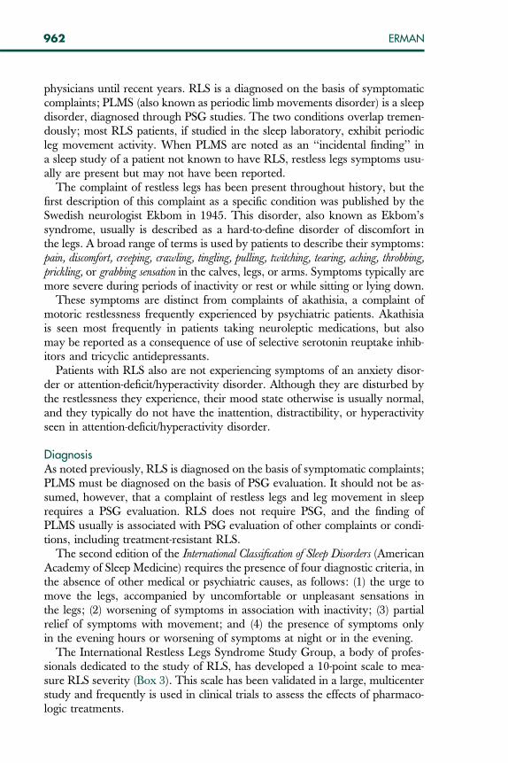
The International Restless Legs Syndrome Study Group, a body of profes-sionals dedicated to the study of RLS, has developed a 10-point scale to mea-sure RLS severity (Box 3). This scale has been validated in a large, multicenterstudy and frequently is used in clinical trials to assess the effects of pharmaco-logic treatments.
963SELECTED SLEEP DISORDERS
EpidemiologyCurrent estimates suggest that 5% to 10% of adults have RLS [32], with initialsymptoms often appearing in the third decade of life. The disorder may appearin childhood, with estimates that 1% to 2% of the general pediatric populationmay be affected. In general, older patients are more likely to report restless legscomplaints than younger ones, but patients historically have been unlikely toseek attention from primary care physicians leading to significant underdiagno-sis of this problem. Patients may report symptoms present for 25 to 30 yearswithout having had a diagnosis established or having received treatment.
EtiologyPrimary and secondary forms of RLS exist. Strong historical evidence suggeststhat there is a genetic predisposition to the development of primary RLS,
Box 3: Restless legs syndrome diagnosis per the InternationalRestless Legs Syndrome Study Group
International Restless Legs Syndrome Study Group rating scale
1. Overall, how would you rate the RLS discomfort in your legs or arms?
2. Overall, how would you rate the need to move around because of your RLSsymptoms?
3. Overall, how much relief of your RLS arm or leg discomfort do you get frommoving around?
4. Overall, how severe is your sleep disturbance from your RLS symptoms?
5. How severe is your tiredness or sleepiness from your RLS symptoms?
6. Overall, how severe is your RLS as a whole?
7. How often do you get RLS symptoms?
8. When you have RLS symptoms, how severe are they on an average day?
9. Overall, how severe is the impact of your RLS symptoms on your ability tocarry out your daily affairs (eg, living a satisfactory family, home, social,school, or work life)?
10. How severe is your mood disturbance from your RLS symptoms (eg, angry,depressed, sad, anxious, or irritable)?
� Each question (except question 3) has the following multiple choices: (4) Verysevere, (3) Severe, (2) Moderate, (1) Mild, (0) None (sometimes with a moreoperational definition in parentheses)
� Question 3: (4) No relief, (3) Slight relief, (2) Moderate relief, (1) Either com-plete or almost complete relief, (0) No RLS symptoms and therefore questiondoes not apply
Adapted from The International Restless Legs Syndrome Study Group: Validation of the In-ternational Restless Legs Syndrome Study Group rating scale for restless legs syndrome.Sleep Med 2003;4:121–32; with permission.
964 ERMAN
although the specific genetic basis for this disorder has not been elucidated. Notall patients report a family history for this disorder. Familial and spontaneoustypes likely exist.
Secondary RLS occurs in medical conditions associated with the symptom ofrestless legs, including end-stage renal disease, iron deficiency, and pregnancy,all of which may be associated with low iron stores. Ekbom first noted a rela-tionship between RLS and impaired iron storage or iron metabolism in 1955,and impaired iron storage or metabolism is now recognized as a reversiblecause of RLS. Evaluation of a patient with RLS complaints always shouldinclude measurement of iron, iron stores, and ferritin levels.
These abnormalities of iron storage and metabolism may play a central rolein the cause of this disorder. Iron seems to play a role in normal dopaminergicfunction in the central nervous system, perhaps related to dopamine transportand to dopamine synthesis. Although a clear-cut link with iron metabolism hasnot been established, it is interesting to consider this relationship in the contextof the role of dopaminergic agonists in the treatment of this disorder.
ConsequencesThe primary impact of RLS on health is mediated through the sleep distur-bance that is characteristic of RLS. As would be expected, the inability to fallasleep, disrupted sleep through the night with inability to resume sleep afterawakening, and insufficient hours of sleep are common among individualswith RLS. Individuals with RLS frequently report disturbance of daytime activ-ities and function, however, and the risk of daytime symptoms is increased inassociation with greater chronicity.
Quality of life in RLS also has been evaluated using the Short Form�36Health Survey (SF-36), an extensively tested and validated tool that assesseseight dimensions of health-related quality of life: physical functioning, physicallimitations on normal role activities, bodily pain, general health, energy andvitality, social functioning, emotional limitations on normal role activities,and mental health. Based on SF-36 evaluation, patients with RLS are signifi-cantly different from the general population, with a quality of life comparableto that seen in patients with chronic medical conditions, such as type 2 diabetesand clinical depression.
TreatmentVarious agents have been used to provide symptomatic relief for RLS andPLMS. In the 1950s, efforts at treatment of RLS with intravenous iron wereundertaken, with significant improvement noted in small patient populations.Formal approval by the FDA of ropinirole in 2005 in treatment of RLS wasaccomplished as a consequence of multiple studies showing efficacy of thisagent [33,34].
At the time of this writing (July 2006), another dopaminergic agonistapproved for the treatment of Parkinson’s disease, pramipexole, is awaitingword from the FDA with regard to possible approval for RLS treatment. Other
965SELECTED SLEEP DISORDERS
dopaminergic agonist compounds, such as carbidopa-levodopa, bromocriptine,and pergolide, may be beneficial for some treatment-resistant patients.
Until the approval of ropinirole in treatment of RLS, all treatment was off-label. For many years, sedating agents such as the benzodiazepine clonazepam(Klonopin) were prescribed in treatment and were recommended in clinicalreviews of the treatment of the complaint of restless legs and in sleep textbooks.Although it was recognized that these sedating agents did not alter the frequencyof leg movement activity and might lead to residual sedation, patients often expe-rienced a sense of relief, presumably on the basis of sleep consolidation. Opioidssuch as codeine, hydrocodone, oxycodone, propoxyphene, and methadone havebeen used in treatment and may be beneficial for patients whose complaintsinclude severe pain, not affected by other treatment modalities.
Anticonvulsants such as carbamazepine and gabapentin are often used whendopamine agonists have failed. They may be useful in patients with coexistingperipheral neuropathy or when RLS discomfort is described as pain.
Iron treatment is indicated for patients with evidence of iron deficiency, typ-ically for patients with serum ferritin levels less than 50 lg. Oral treatment usuallyis given as ferrous sulfate, 325 mg or its equivalent twice per day. Absorption ofiron is improved by coadministration of vitamin C. For patients who have troubleabsorbing iron or may have significant problems with side effects, such as indiges-tion or constipation, intravenous iron supplementation may be used.
Because some patients with RLS may be iron deficient on the basis of dietaryiron intake, it is always necessary to ask patients about foods that they mayavoid or not eat because of dietary restrictions. Patients may not eat redmeat for ‘‘health reasons’’ and may have limited iron intake available fromother sources. Although repletion of iron stores by increased dietary intakemay not always be possible, patients should be educated about iron-rich foodsthat they may be able to eat (ie, beef, turkey, shrimp, sardines, tuna, iron-enriched breakfast cereals, green leafy vegetables) and encouraged to increasetheir intake of these foods.
SUMMARYSleep disorders, including RLS and periodic limb movements disorder, sleepapnea syndrome, and narcolepsy, are prevalent medical conditions, likely tobe seen by practicing psychiatrists. Awareness of these conditions and their pre-sentations, pathophysiology, and treatment allows psychiatrists to treat theseconditions where appropriate, to minimize complications and healthconsequences associated with delayed diagnosis, and to reduce the burden ofdisease that these conditions may pace on patients already experiencing pri-mary psychiatric disorders.
References[1] Rosen R, Mahowald M, Chesson A, et al. The Taskforce 2000 Survey: medical education in
sleep and sleep disorders. Sleep 1998;21:235–8.[2] Kushida CA, Littner MR, Morgenthaler T, et al. Practice parameters for the indications for
polysomnography and related procedures: an update for 2005. Sleep 2005;28:499–521.
966 ERMAN
[3] Flemons WW. Clinical practice: obstructive sleep apnea. N Engl J Med 2002;347:498–504.
[4] Chesson AL Jr, Berry RB, Pack A. Practice parameters for the use of portable monitoring de-vices in the investigation of suspected obstructive sleep apnea in adults. Sleep 2003;26:907–13.
[5] El-Ad B, Lavie P. Effect of sleep apnea on cognition and mood. Int Rev Psychiatry 2005;17:277–82.
[6] Suhner AG, Darko DD, Erman MK, et al. Depressive symptoms in patients with OSA and theimpact of nasal CPAP treatment. Sleep 2003;25:A225.
[7] Young T, Palta M, Dempsey J, et al. The occurrence of sleep-disordered breathing amongmiddle-aged adults. N Engl J Med 1993;328:1230–5.
[8] Fujita S, Conway W, Zorick F, et al. Surgical correction of anatomic abnormalities in obstruc-tive sleep apnea syndrome: uvulopalatopharyngoplasty. Otolaryngol Head Neck Surg1981;89:923–34.
[9] Crookes PF. Surgical treatment of morbid obesity. Annu Rev Med 2006;57:243–64.[10] Walker-Engstrom ML, Tegelberg A, Wilhelmsson B, et al. 4-year follow up of treatment with
dental appliance or uvulopalatopharyngoplasty in patients with obstructive sleep apnea:a randomized study. Chest 2002;121:739–46.
[11] Hanzel DA, Proia NG, Hudgel DW. Response of obstructive sleep apnea to fluoxetine andprotriptyline. Chest 1991;100:416–21.
[12] Daniels L. Narcolepsy. Medicine (Baltimore) 1934;13:1–22.[13] Yoss RE, Daly DD. Criteria for the diagnosis of the narcoleptic syndrome. Mayo Clin Proc
1957;32:320–8.[14] Vogel G. Studies in psychophysiology of dreams: III. the dream of narcolepsy. Arch Gen
Psychiatry 1960;3:421–8.[15] Carskadon MA, Dement WC, Mitler MM, et al. Guidelines for the multiple sleep latency test
(MSLT): a standard measure of sleepiness. Sleep 1986;9:519–24.[16] Scammell TE. The neurobiology, diagnosis, and treatment of narcolepsy. Ann Neurol
2003;53:154–66.[17] Honda Y, Asake A, Tanaka Y, et al. Discrimination of narcolepsy by using genetic markers
and HLA. Sleep Res 1983;12:254.[18] Littner MR, Kushida C, Wise M, et al. Practice parameters for clinical use of the multiple
sleep latency test and the maintenance of wakefulness test. Sleep 2005;28:113–21.[19] American Academy of Sleep Medicine. International classification of sleep disorders:
diagnostic and coding manual. 2nd edition. Westchester (IL): American Academy of SleepMedicine; 2005.
[20] Mignot E. Genetic and familial aspects of narcolepsy. Neurology 1998;50(Suppl 1):S16–22.
[21] Juji T, Matsuki K, Tokunaga K, et al. Narcolepsy and HLA in the Japanese. Ann N YAcad Sci1988;540:106–14.
[22] Lin L, Faraco J, Li R, et al. The sleep disorder canine narcolepsy is caused by a mutation in thehypocretin (orexin) receptor 2 gene. Cell 1999;98:365–76.
[23] Chemelli RM, Sinton CM, Yanagisawa M. Polysomnographic characterization of orexin-2receptor knockout mice. Sleep 2005;23:A296–7.
[24] Nishino S, Ripley B, Overeem S, et al. Hypocretin (orexin) deficiency in human narcolepsy.Lancet 2000;355:39–40.
[25] Goswami M. The influence of clinical symptoms on quality of life in patients with narcolepsy.Neurology 1998;50(Suppl 1):S31–6.
[26] Mitler M, Erman M, Hajdukovic R. The treatment of excessive somnolence with stimulantdrugs. Sleep 1993;16:203–6.
[27] Thorpy M, Black J, Erman M, et al. Tolerability of modafinil in disorders of sleep and wake-fulness. Sleep 2004;27:A130.
967SELECTED SLEEP DISORDERS
[28] Schwartz JR, Hirshkowitz M, Erman MK, et al. Modafinil as adjunct therapy for daytimesleepiness in obstructive sleep apnea: a 12-week, open-label study. Chest 2003;124:2192–9.
[29] Ballon JS, Feifel D. A systematic review of modafinil: potential clinical uses and mechanismsof action. J Clin Psychiatry 2006;67:554–66.
[30] Mamelak M, Scharf MB, Woods M. Treatment of narcolepsy with gamma-hydroxybutyrate:a review of clinical and sleep laboratory findings. Sleep 1986;9:285–9.
[31] Harsh JR, Hayduk R, Rosenberg R, et al. The efficacy and safety of armodafinil as treatmentfor adults with excessive sleepiness associated with narcolepsy. Curr Med Res Opin2006;22:761–74.
[32] Phillips B, Young T, Finn L, et al. Epidemiology of restless legs symptoms in adults. Arch InternMed 2000;160:2137–41.
[33] Trenkwalder C, Garcia-Borreguero D, Montagna P, et al. Ropinirole in the treatment of rest-less legs syndrome: results from the TREAT RLS 1 study, a 12 week, randomised, placebocontrolled study in 10 European countries. J Neurol Neurosurg Psychiatry 2004;75:92–7.
[34] Bogan RK, Fry JM, Schmidt MH, et al. Ropinirole in the treatment of patients with restless legssyndrome: a US-based placebo-controlled clinical trial. Mayo Clin Proc 2006;81:17–27.