Embed Size (px)
Citation preview

Seizures & Stroke
1st international congress on epilepsy in cerebrovascular disease
Gothenburg 20-22 February 2019
PROGRAM

2 3
Seizures & Stroke
WELCOME!Dear Delegate,
We are very pleased to welcome you to Seizures & Stroke, the first international conference on seizures in the context of cerebrovascular disease. The interest in the meeting has by far exceeded our expectations and it seems that more than one hundred participants will travel to Gothenburg for three days of scientific discussions. In addition to plenary lectures by very distinguished faculty, many abstracts will be presented as posters or platform talks.
We are very grateful to Gothenburg university for hosting the conference, the Swedish Research Council for providing travel support for speakers, the city of Gothenburg for providing the welcome reception and our industry sponsors Eisai and UCB.
We hope that you will enjoy the conference and take the opportunity to discuss research and clinical management.
Johan Zelano Francesco Brigo
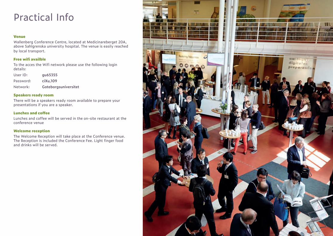
Practical Info
VenueWallenberg Conference Centre, located at Medicinareberget 20A, above Sahlgrenska university hospital. The venue is easily reached by local transport.
Free wifi availbleTo the acces the Wifi network please use the following login details:User ID: gu65355Password: ciXu,109Network: Goteborgsuniversitet
Speakers ready roomThere will be a speakers ready room available to prepare your presentations if you are a speaker.
Lunches and coffeeLunches and coffee will be served in the on-site restaurant at the conference venue
Welcome receptionThe Welcome Reception will take place at the Conference venue. The Reception is included the Conference Fee. Light finger food and drinks will be served.
6 7
Seizures & Stroke
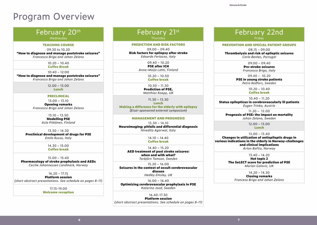
February 20thWednesday
TEACHING COURSE09.30 to 10.20
“How to diagnose and manage poststroke seizures”Francesco Brigo and Johan Zelano
10.20 – 10.40 Coffee Break10:40 – 12:00
“How to diagnose and manage poststroke seizures”Francesco Brigo and Johan Zelano
12.00 – 13.00 Lunch
PRECLINICAL13.00 – 13.10
Opening remarksFrancesco Brigo and Johan Zelano
13.10 – 13.50 Modelling PSE
Asla Pitkänen, Finland
13.50 – 14.30 Preclinical development of drugs for PSE
Emilo Russo, Italy
14.30 – 15.00 Coffee break
15.00 – 15.40 Pharmacology of stroke prophylaxis and AEDs
Cecilie Johannessen Landmark, Norway
16.20 – 17.15 Platform session
(short abstract presentations. See schedule on pages 8-11)
17.15-19.00Welcome reception
February 21stThursday
PREDICTION AND RISK FACTORS09.00 – 09.40
Risk factors for epilepsy after strokeEdoardo Ferlazzo, Italy
09.40 – 10.20 PSE after ICH
Anna-Maija Lahti, Finland
10.20 – 10.50 Coffee break
10.50 – 11.30 Prediction of PSE,Matthias Koepp, UK
11.30 – 13.30 Lunch
Making a difference for the elderly with epilepsy(Eisai-sponsored external symposium)
MANAGEMENT AND PROGNOSIS13.30 – 14.10
Neuroimaging; pitfalls and differential diagnosisNivedita Agarwal, Italy
14.10 – 14.40 Coffee break
14.40 – 15.20 AED treatment of post stroke seizures:
when and with what? Torbjörn Tomson, Sweden
15.20 – 16.00 Seizures in the context of occult cerebrovascular
diseaseHedley Emsley, UK
16.00 – 16.40 Optimizing cerebrovascular prophylaxis in PSE
Katarina Jood, Sweden
16.40-17.30 Platform session
(short abstract presentations. See schedule on pages 8-11)
February 22ndFriday
PREVENTION AND SPECIAL PATIENT GROUPS08.15 – 09.00
Thrombolysis and risk of epileptic seizuresCarla Bentes, Portugal
09.00 – 09.40 Pre-stroke seizuresFrancesco Brigo, Italy
09.40 – 10.20 PSE in young stroke patients
Petra Redfors, Sweden
10.20 – 10.40 Coffee break
10.40 – 11.20 Status epilepticus in cerebrovascularly ill patients
Eugen Trinka, Austria11.20 – 12.00
Prognosis of PSE: the impact on mortalityJohan Zelano, Sweden
12.00 – 13.00 Lunch
13.00 – 13.40 Changes in utilization of antiepileptic drugs in
various indications in the elderly in Norway-challenges and clinical implications
Arton Baftiu, Norway
13.40 – 14.20 Hot topic 2
The SeLECT score for prediction of PSEMarian Galovic, UK
14.20 – 14.30 Closing remarks
Franceso Brigo and Johan Zelano
Program Overview
8 9
Seizures & Stroke
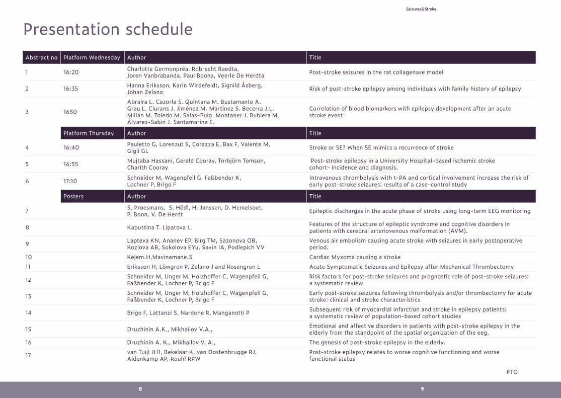
Presentation scheduleAbstract no Platform Wednesday Author Title
1 16:20 Charlotte Germonpréa, Robrecht Raedta, Joren Vanbrabanda, Paul Boona, Veerle De Herdta Post-stroke seizures in the rat collagenase model
2 16:35 Hanna Eriksson, Karin Wirdefeldt, Signild Åsberg, Johan Zelano Risk of post-stroke epilepsy among individuals with family history of epilepsy
3 1650
Abraira L. Cazorla S. Quintana M. Bustamante A. Grau L. Ciurans J. Jiménez M. Martinez S. Becerra J.L. Millán M. Toledo M. Salas-Puig. Montaner J. Rubiera M. Álvarez-Sabin J. Santamarina E.
Correlation of blood biomarkers with epilepsy development after an acute stroke event
Platform Thursday Author Title
4 16:40 Pauletto G, Lorenzut S, Corazza E, Bax F, Valente M, Gigli GL Stroke or SE? When SE mimics a recurrence of stroke
5 16:55 Mujtaba Hassani, Gerald Cooray, Torbjörn Tomson, Charith Cooray
Post-stroke epilepsy in a University Hospital-based ischemic stroke cohort- incidence and diagnosis.
6 17:10 Schneider M, Wagenpfeil G, Faßbender K, Lochner P, Brigo F
Intravenous thrombolysis with t-PA and cortical involvement increase the risk of early post-stroke seizures: results of a case-control study
Posters Author Title
7 S. Proesmans, S. Hödl, H. Janssen, D. Hemelsoet, P. Boon, V. De Herdt Epileptic discharges in the acute phase of stroke using long-term EEG monitoring
8 Kapustina T. Lipatova L. Features of the structure of epileptic syndrome and cognitive disorders in patients with cerebral arteriovenous malformation (AVM).
9 Lapteva KN, Ananev EP, Birg TM, Sazonova OB, Kozlova AB, Sokolova EYu, Savin IA, Podlepich VV
Venous air embolism causing acute stroke with seizures in early postoperative period.
10 Kejem.H,Mavinamane.S Cardiac Myxoma causing a stroke11 Eriksson H, Löwgren P, Zelano J and Rosengren L Acute Symptomatic Seizures and Epilepsy after Mechanical Thrombectomy
12 Schneider M, Unger M, Holzhoffer C, Wagenpfeil G, Faßbender K, Lochner P, Brigo F
Risk factors for post-stroke seizures and prognostic role of post-stroke seizures: a systematic review
13 Schneider M, Unger M, Holzhoffer C, Wagenpfeil G, Faßbender K, Lochner P, Brigo F
Early post-stroke seizures following thrombolysis and/or thrombectomy for acute stroke: clinical and stroke characteristics
14 Brigo F, Lattanzi S, Nardone R, Manganotti P Subsequent risk of myocardial infarction and stroke in epilepsy patients: a systematic review of population-based cohort studies
15 Druzhinin A.K., Mikhailov V.A., Emotional and affective disorders in patients with post-stroke epilepsy in the elderly from the standpoint of the spatial organization of the eeg.
16 Druzhinin A. K., Mikhailov V. A., The genesis of post-stroke epilepsy in the elderly.
17 van Tuijl JH1, Bekelaar K, van Oostenbrugge RJ, Aldenkamp AP, Rouhl RPW
Post-stroke epilepsy relates to worse cognitive functioning and worse functional status
PTO
10 11
Seizures & Stroke
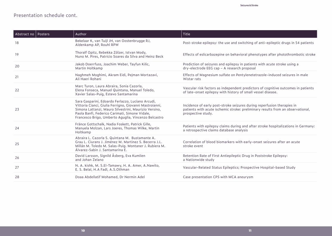
Abstract no Posters Author Title
18 Bekelaar K, van Tuijl JH, van Oostenbrugge RJ, Aldenkamp AP, Rouhl RPW Post-stroke epilepsy: the use and switching of anti-epileptic drugs in 54 patients
19 Thoralf Opitz, Rebekka Zölzer, Istvan Mody, Nuno M. Pires, Patrício Soares da Silva and Heinz Beck Effects of eslicarbazepine on behavioral phenotypes after photothrombotic stroke
20 Jakob Doerrfuss, Joachim Weber, Tayfun Kilic, Martin Holtkamp
Prediction of seizures and epilepsy in patients with acute stroke using a dry-electrode EEG cap – A research proposal
21 Naghmeh Moghimi, Akram Eidi, Pejman Mortazavi, Ali Haeri Rohani
Effects of Magnesium sulfate on Pentylenetetrazole-induced seizures in male Wistar rats
22Marc Turon, Laura Abraira, Sonia Cazorla, Elena Fonseca, Manuel Quintana, Manuel Toledo, Xavier Salas-Puig, Estevo Santamarina
Vascular risk factors as independent predictors of cognitive outcomes in patients of late-onset epilepsy with history of small vessel disease.
23
Sara Gasparini, Edoardo Ferlazzo, Luciano Arcudi, Vittoria Cianci, Giulia Ferrigno, Giovanni Mastroianni, Simona Lattanzi, Mauro Silvestrini, Maurizio Versino, Paola Banfi, Federico Carimati, Simone Vidale, Francesco Brigo, Umberto Aguglia, Vincenzo Belcastro
Incidence of early post-stroke seizures during reperfusion therapies in patients with acute ischemic stroke: preliminary results from an observational, prospective study.
24Fränce Gottschalk, Nadia Foskett, Patrick Gille, Manuela Molzan, Lars Joeres, Thomas Wilke, Martin Holtkamp
Patients with epilepsy claims during and after stroke hospitalizations in Germany: a retrospective claims database analysis
25
Abraira L. Cazorla S. Quintana M. Bustamante A. Grau L. Ciurans J. Jiménez M. Martinez S. Becerra J.L. Millán M. Toledo M. Salas-Puig. Montaner J. Rubiera M. Álvarez-Sabin J. Santamarina E.
Correlation of blood biomarkers with early-onset seizures after an acute stroke event
26 David Larsson, Signild Åsberg, Eva Kumlien and Johan Zelano
Retention Rate of First Antiepileptic Drug in Poststroke Epilepsy: a Nationwide study
27 N. A. kishk, M. S.El-Tamawy, H. A. Amer, A.Nawito, E. S. Belal, H.A Fadl, A.S.Othman Vascular-Related Status Epileptics; Prospective Hospital-based Study
28 Doaa Abdellatif Mohamed, Dr Nermin Adel Case presentation CPS with MCA aneurysm
Presentation schedule cont.
12 13
Seizures & Stroke
MethodsThis study included patients treated for stroke collected from the national Swedish stroke register. Linkage to other databases is possible through a personal identification number. Swedish multi-generation register identified first-degree relatives. The database was established in 1932 why this study only cover patients born after that date. Additional cross-referencing with National Patient Register (NPR) relayed information of diagnoses of index patients and their first-degree relatives. The risk of epilepsy was assessed with survival-analysis and hazard ratio was estimated with cox regression. ResultsPatients with ischemic stroke or intracranial bleeding with no prior epilepsy diagnoses registered in the national stroke register between year 2001 and 2012 were included. Preliminary data indicate that having first-degree relatives with epilepsy is associated with a higher risk of post stroke epilepsy after two and five years. Additionally, the hazard ratio in patients with first-degree relatives with epilepsy is significantly elevated compared to patients without relatives with epilepsy.ConclusionsOur data suggests that patients with first-degree relatives with epilepsy have a greater risk of developing post stroke epilepsy.
ABSTRACT 3
Correlation of blood biomarkers with epilepsy development after an acute stroke eventAuthors: Abraira L.1; Cazorla S.1; Quintana M.1; Bustamante A.2; Grau L.3; Ciurans J.3; Jiménez M.3; Martinez S.3; Becerra J.L.3; Millán M.4; Toledo M.1; Salas-Puig.1; Montaner J.2; Rubiera M.5; Álvarez-Sabin J.2; Santamarina E.1Affiliations: 1. Epilepsy Unit, Neurology Deparment, Vall d’Hebron University Hospital. 2. Neurovascular Research Laboratory, Vall d’Hebron Institut de Recerca (VHIR). 3. Epilepsy Unit, Neurology Department, Germans Trias i Pujol University Hospital. 4. Stroke Unit, Neurology Department, Germans Trias i Pujol University Hospital. 5. Neurovascular Unit, Neurology Department, Vall d’Hebron University HospitalBackground and purpose: We aimed to describe the clinical factors and biomarkers present during the acute stroke process and its association with epilepsy at long-term of follow up. Methods: We studied 1065 stroke patients (ischaemic and haemorrhagic) diagnosed between 2012-2013 (Stroke-Chip study). We excluded those with history of epilepsy, subaranoid haemorrhage, arteriovenous malformation and subdural haematoma. We analysed demographics, stroke and epilepsy related factors (type of stroke and aetiology, timing from stroke to seizure, treatment and functional disability (mRs)), and blood biomarkers (NT-proBNP, IGFBP, TNF-R1, GroA, FasL, IL-6, D-dimer, vWF, VAP-1, Endostatin, S100B, HsC70, Apo CIII, NCAM). We evaluated the association with the development of epilepsy.Results: Mean age was 72.2 ± 13.1, 56.2% men. Median time of follow-up was 4.7 years, 40.5% died. The 85.7% (n=913) had ischaemic stroke with median baseline NIHSS 8 (IQR 3-16). Fifty-five patients (5.6%) had late-onset seizures and median time of appearance was 253 days. The clinical variables that were independently associated with epilepsy were a greater baseline NIHSS (NIHSS>8) (HR 3.919 (CI 2.173–7.071)(p<0.001) and history of early-onset seizures (HR 3.979 (CI 1.781– 8.891) p<0.001).
AbstractsABSTRACT 1
Post-stroke seizures in the rat collagenase modelAuthors: Charlotte Germonpréa, Robrecht Raedta, Joren Vanbrabanda, Paul Boona, Veerle De Herdta Affiliations: (a) Laboratory for Clinical and Experimental Neurophysiology, Neurobiology and Neuropsychology (LCEN3), Ghent University, Belgium PURPOSE Intracerebral hemorrhage (ICH) is a known risk factor for the development of seizures, but little is known about the incidence and consequence of seizures in the acute phase post-ICH. In this study, ICH was induced by injecting different doses of collagenase in dorsal striatum and the occurrence of seizures in each condition was assessed. METHODS Fifteen male Sprague-Dawley rats were implanted with scalp electrodes (4AP +/-2ML and -4.5AP +/-3ML, coordinates in mm and relative to bregma) and injection-site was marked. After minimum one week of baseline video-EEG recording, rats were injected with collagenase (0.2U (n=2), 0.4U (n=3), 0.6U (n=3), 0.8U (n=4) and 1U (n=3), 0.5AP 3.5ML 6DV) and immediately reconnected to the video-EEG setup during 7 days. Afterwards animals were euthanized to visualize the hemorrhage by histology and video-EEG recordings were analyzed. RESULTS ICH was present in all animals. Epileptic seizures occurred in 4/15 animals: 1/3 rats injected with 0.6U (n=25, 47h-96h after ICH induction), 1/4 rats injected with 0.8U (n=12, 4h-25h after ICH induction) and 2/3 rats injected with 1U (n=4, 12h-53h/8h-61h after ICH induction). Two animals died during post-ICH recordings (1/4 rats injected with 0.8U and 1/3 rats injected with 1U). CONCLUSION Injecting rats with 0.6U of collagenase seems to be the most promising condition to study post-stroke seizures in the rat collagenase model as it resulted in hemorrhagic stroke in all animals without causing mortality and with an incidence rate of stroke-induced seizures of 30%. However this needs to be confirmed in a larger study population.
ABSTRACT 2
Risk of post stroke epilepsy among individuals with family history of epilepsy Authors: Hanna Eriksson, Karin Wirdefeldt, Signild Åsberg, Johan ZelanoAffiliations: Sahlgrenska university hospital, Karolinska university hospital, Uppsala university hospital.BackgroundStroke is the most common cause of acquired epilepsy. Some risk factors of developing post stroke epilepsy are known, but to date, the risk in a given individual is relatively unknown. Previous studies have shown that patients with a family history of epilepsy had an increased risk of epilepsy after traumatic brain injury. This study aims to assess the risk of epilepsy after stroke in individuals with and without a family history of epilepsy.
14 15
Seizures & Stroke
For the analysis of blood biomarkers we performed an adjusted multiple regression model and showed that greater levels of FAsL (HR 2.114 (CI 1.069–4.183)(p=0.031) and Endostatin (HR 10.761 (CI 1.480–78.243)(p=0.019) and lower levels of HsC70 (HR 2.007 (CI 1.071-3.760)(p=0.030) were independent predictors for the development of epilepsy.Conclusion: Higher levels of FAsL and Endostatin and lower levels of HsC70 predict epilepsy at long-term follow up.
ABSTRACT 4
Stroke or SE? When SE mimics a recurrence of strokeAuthors: Pauletto G, Lorenzut S, Corazza E, Bax F, Valente M, Gigli GLAffiliation: Neurology Unit, Udine University Hospital, ItalyAims: Status Epilepticus (SE) may arise in patients with previous stroke as breakthrough or complication of post-stroke epilepsy (PSE). Sometimes, SE is misdiagnosed as a new cerebro-vascular event, determining a delayed diagnosis and an inappropriate use of diagnostic and therapeutic procedures. Aim of this study is to evaluate the clinical characteristics of patients with previous stroke (both ischemic and hemorrhagic stroke) who developed SE, with particular regards to stroke-mimicking SE.Methods: We performed a retrospective analysis of patients, who were admitted to our Neurology Unit for acute stroke, between January 2016 and 2018.Results: in the time span considered, 1150 patients were admitted to our Neurology Unit for suspected stroke. Among them, 11 presented with SE mimicking a stroke.They were equally distributed between women and men, with a high mean age (77.7 years) and a clinical history of previous stroke, four intra-intraparenchymal hemorrhages and seven ischemic stroke respectively. All patients had negative symptoms as main clinical manifestation of SE, in particular: six (54%) with complete aphasia, four (36%) with worsening of pre-existing aphasia and hemiparesis and one (10%) with dysarthria.In the majority of cases (64%), SE was the clinical onset of symptomatic epilepsy, in the remaining patients, SE developed in the setting of an already diagnosed PSE (36%). All patients were treated in our Stroke Unit, under EEG, ECG and blood pressure monitoring. The first bolus of anti-epileptic drug was sufficient to stop clinical symptoms and EEG epileptic activity in 90% of patients. Rhythmic focal slow activity could be seen for 24 hours after SE resolution. Levetiracetam was the first choice therapy, followed by Lacosamide, in those patients who were already treated with Levetiracetam for their epilepsy or had a history of irritability and behavior disturbances. Only one patient, with previous large ischemic stroke, developed a super refractory SE and died.Four patients had a relapse of SE within one year. All the relapses had the same clinical features of the first SE and they occurred. Subjects with previous intra-parenchymal hemorrhage or haemorragic infarction of ischemic stroke.Conclusions: SE is not uncommon in patients with previous stroke and it may presents with negative symptoms mimicking a new cerebro-vascular events. Hemorrhagic stroke and large ischemic stroke with neurological sequences are more prone to develop SE, sometimes relapsing. EEG should be considered as a diagnostic tool in the clinical management of acute suspected stroke.
ABSTRACT 5
Post-stroke epilepsy in a University Hospital-based ischemic stroke cohort- incidence and diagnosis.Authors: Mujtaba Hassani, Gerald Cooray, Torbjörn Tomson, Charith CoorayAffiliations: Department of Clinical Neurosciences, Karolinska Institutet, Stockholm and Department of Neurology, Karolinska University Hospital, Stockholm, Sweden Introduction- Post-stroke epilepsy (PSE) is a common cause of adult-onset epilepsy. Despite an increasing awareness of the diagnosis, there is a concern for under-diagnosis of the condition. We aimed to study the diagnosis and incidence of PSE in an ischemic stroke-cohort admitted to a tertiary University Hospital. Methods- We retrospectively investigated the occurrence and diagnosis of unprovoked seizures and PSE in all ischemic stroke patients admitted to Karolinska University Hospital in Stockholm during 2015 and registered in the Swedish Stroke Register. Patient records were scrutinized for the presence of post-stroke seizures/epilepsy.Results- A total of 240 patients fulfilling the inclusion criteria of the study were surveyed. Median follow-up time was 1062 days (IQR 589-1195 days). Thirteen patients were diagnosed with PSE according to the study criteria, the incidence of PSE 23/1000 person-years. Median time to PSE from stroke-onset was 237 days (IQR 33-688). Eleven of 13 PSE patients (85%) received an epilepsy diagnosis, 8 patients after one unprovoked seizure and 3 patients after two. Median age comparing PSE with non-PSE patients was 65 vs 71 years (p-value 0.187), 30.8% vs 45.4% females (p-value 0.303), and median baseline NIHSS score 6.5 vs 3 (p-value 0.062). Conclusion- The incidence of PSE in this ischemic stroke cohort was in line with that described in previous studies. Despite concern for under-diagnosis of this condition, the majority of PSE patients were given an epilepsy diagnosis and treated with adequate anti-convulsants. There is still potential for improvement in the adherence to the latest updated epilepsy definitions.
ABSTRACT 6
Intravenous thrombolysis with t-PA and cortical involvement increase the risk of early post-stroke seizures: results of a case-control studyAuthors: Schneider M1, Wagenpfeil G1, Faßbender K1, Lochner P1, Brigo F2,3Affiliations: 1Saarland University Medical Center, Homburg, Germany; 2Franz Tappeiner Hospital, Merano, Italy; 3University of Verona, ItalyAim of this study was to identify risk factors for early post-stroke seizures (PSS) in patients with acute ischemic stroke. We undertook a case-control study at a single stroke center. Patients with seizure occurring during the first 7 days following ischemic stroke admitted at our stroke center between 2010 and 2016 were retrospectively identified and matched with controls on age and gender. We included 79 cases and 158 controls. Blood sugar levels on admission, stroke localization, NIHSS- and Rankin score, and intravenous (i.v.) thrombolysis with rtPA were statistically associated with early PSS in univariated analysis. Multiple logistic regression after forward and backward variable selection identified cortical stroke localization (OR 2.49; 95% CI 1.35 to 4.59; p= 0.003) and intravenous thrombolysis (OR 2.26; 95% CI 1.16 to 4.43) as being independently associated with occurrence of early PSS. Accordingly, the predicted risk of early PSS in stroke patients with cortical involvement and i.v. thrombolysis is 57%, whereas the risk in subcortical or lacunar stroke not treated with thrombolysis is 19%.
16 17
Seizures & Stroke
Cortical involvement and i.v. thrombolysis are independent risk factors associated with the occurrence of early PSS. This association is not explained by age or gender, concomitant drugs, diabetes or alcoholism, sodium and cholesterol levels, blood pressure on admission, stroke etiology or severity, and hemorrhagic transformation or hemorrhage following i.v. thrombolysis. Our results are consistent with those of a previous case-control study conducted in a smaller population. Further prospective studies are required to fully elucidate the association between i.v. thrombolysis and early PSS.
ABSTRACT 7
Epileptic discharges in the acute phase of stroke using long-term EEG monitoringAuthors: S. Proesmans1,2, S. Hödl1,2, H. Janssen1, D. Hemelsoet1, P. Boon1,2, V. De Herdt1,2Affiliations: 1 Ghent University Hospital, Department of Neurology, Gent, Belgium 2 4Brain Lab, Ghent University, Gent, BelgiumBackgroundStroke is a common cause of seizures, especially in the elderly population.However, the incidence, associated factors and influence on outcome of interictal epileptic discharges and subclinical electrographic seizures in the acute phase of stroke are unknown. MethodsIn this prospective study, 55 patients underwent long-term video-EEG monitoring within 3 days after intracerebral haemorrhage or ischemic stroke. Epileptic activity on the EEG, including spikes, spike-waves, PLEDs and electrographic seizures, was analysed and correlated with clinical and neuroradiological patient characteristics, the occurrence of clinical seizures and functional outcome. ResultsIn a preliminary analysis, data of the first 31 patients was investigated. Analysis of the whole study population is ongoing and will be completed by mid-February 2019. In the preliminary analysis, 7/31 (23%) of patients had epileptic activity on the EEG and 2/31 (6%) of patients had electrographic seizures. No association was found between the occurrence of epileptic discharges and functional outcome or any of the studied patient characteristics. ConclusionIn the preliminary analysis of the first 31 patients, epileptic discharges were frequently found. Analysis of all patient data is ongoing and will be presented during the conference.
ABSTRACT 8
Features of the structure of epileptic syndrome and cognitive disorders in patients with cerebral arteriovenous malformation (AVM)Authors Kapustina T., Lipatova L.Affiliation: St.Petersburg V.M.Bekhterev Psychoneurological Research Institute Objective: to assess the frequency, structure of epileptic syndrome and cognitive impairment in patients with AVM. The analysis of the results of examination of
patients with AVM of different localization. The main group consisted of 78 patients with cerebral AVM:average age of 36.2. The results of the study:1 subgroup- a right-hemisphere localization-41(53%);2 subgroup-a left-hemisphere localization–37(47%). Of these, 33%frontal lobe; 12% the temporal lobe;17% area central convolutions; 28% the parietal and the occipital lobe;10% - subcortical structures. In all cases evaluated, the frequency of epileptic seizures:frequent 11%; moderate50%; rare 28%; seizures were not observed in 11% cases. The structure of epileptic syndrome was presented in the following seizures:partial sensorimotor in 29%; vegetative-visceral -40%;mixed: complex partial; secondary generalized - 31%. Changes in EEG in 14% cases, changes were not observed; moderate diffuse changes at the level of diencephalic structures -23%;paroxysmal activity of malformations with an interest in diencephalic structures of the brain-41%;the formation of a mirror focus-10%, moderate irritative changes in the anterior leads of the hemisphere in combination with remote scattering sharp potentials-7%;the presence of the pattern of epileptiform activity-5%.Were identified with cognitive impairment of varying severity, light cognitive disorders – 21%; mild cognitive impairment– 64%;dementia from mild to severe, severity – 13%. Conclusion:The duration and frequency of the epileptic syndrome of the complex structure aggravate the course of cognitive impairment, thereby reducing quality of life of patients with AVM.A comprehensive assessment of cognitive disorders, structure and frequency of epileptic syndrome can be a diagnostic criterion of the results of the immediate outcomes of endovascular treatment of AVM
ABSTRACT 9
Venous air embolism causing acute stroke with seizures in early postoperative period.Authors: Lapteva KN, Ananev EP, Birg TM, Sazonova OB, Kozlova AB, Sokolova EYu, Savin IA, Podlepich VVAffiliations: NN Burdenko National Medical and Research Center for Neurosurgery, Moscow, Russia Introduction. Venous air embolism (VAE) is a potentially serious complication in neurosurgery. There are only single case studies in literature, describing patients with new neurological disturbance after VAE: hemiparesis, altered level of consciousness, CN failure, seizures.The aim of our study was to analyze postoperative neurological disorders and ictal EEG-patterns in patients with VAE during fossa posterior surgery.Methods. We performed retrospective study of 16 patients from 2014 to 2018. Inclusion criteria were VAE (confirmed clinically by decrease in ETCO2 and hemodynamic disturbances intraoperatively), and NICU LOS > 48 hrs. New neurological deficits developed in 13 of 16 patients (81%). Seizures in ICU developed in 5 (31%) patients. EEG and MRI were performed during the first 24 hours after operation. Results. Ictal epileptic activity developed in 5 patients (“spike-wave”, “sharp-and-slow wave”): generalized tonic-clonic seizures in 2 cases, NCSE in 3 cases.The epileptic activity initiated in occipital lobe in 4 cases. The foci of hyperintense signal in DWI were determined in the cerebral cortex on MRI. At least one of the revealed lesions matched with the EEG-confirmed source of epileptic activity.
18 19
Seizures & Stroke
Conclusion.VAE during neurosurgery can lead to new neurological disorders and seizures. But there are some doubts about the correlation between new neurological symptoms and seizures and the fact of VAE. Nevertheless in some cases seizures are resistant to antiepileptic therapy. Therefore, EEG-monitoring should be performed in patients with altered level of consciousness or clinical seizure to exclude NCSE or to control the efficacy of antiepileptic therapy.
ABSTRACT 10
Cardiac Myxoma causing a strokeAuthors:Kejem.H,Mavinamane.SAffiliations:St Helens and knowsley NHS TrustAtrial Myxoma is a rare primary cardiac tumor . Embolisation occurs in 35% of all atrial myxoma1. Atrial myxoma is responsible for only 0.5% of stroke 1. We report a case of stroke secondary to atrial myxoma.A 67 year old male was admitted with 3 days history of loss of vision,dizziness and 2 week history of difficulty concentrating. His past medical history included hypertension and hyperlipidemia .His neurological examination showed right homonymous hemianopia. CT head was suggestive of infarct in the left occipital lobe. On MRI head there were multiple areas of recent infarcts involving the right occipital cortex , posterior left parietal , left occipital lobe and corpus callosum. Transthoracic echocardiogram showed 4.3cmx3.3cm mobile mass in the left atrium below the anterior mitral leaflet prolapsing back and forth through the mitral valve causing transient mild valve obstruction. Subsequently ,he was transferred to the cardiothoracic unit where he had resection and atrial patch .Histology confirmed atrial myxoma.The incidence of myxoma is 0.5% per million every year,the aetiology of which is currently unknown2.Although most cases are sporadic ,10% have an autosomal dominant genetic pattern2 A high index of suspicion is required to diagnose atrial myxoma in patient with multiple infarcts.ReferencesLam K.Y., Dickens P., Chan A.C. Tumors of the heart. A 20-year experience with a review of 12,485 consecutive autopsies. Arch Pathol Lab Med. 1993;117:1027–1031 2. Pinede L, Duhaut P, Loire R. Clinical presentation of left atrial myxoma. A series of 112 consecutive cases. Medicine (Baltimore) 2001;80(3):159-72.
ABSTRACT 11
Acute Asymptomatic Seizures and Epilepsy after Mechanical ThrombectomyAuthors: Eriksson H, Löwgren P, Zelano J and Rosengren LAffiliations: Department of clinical neuroscience, Sahlgrenska university hospitalRationale Cerebrovascular disease is the most common cause of acquired epilepsy. Large stroke and cortical lesions increase the risk of epilepsy. Since mechanical thrombectomy is typically indicated in large strokes, patients undergoing this procedure constitute a potential high-risk population. We investigated the incidence of acute symptomatic seizures and late seizures (epilepsy) in 88 patients that participated in a randomized controlled trial of mechanical thrombectomy at Sahlgrenska university hospital.
MethodsPatients included in the AnStroke trial of mechanical trombectomy were included and information on seizures extracted through chart review, at least one year after thrombectomy. The study was approved by the relevant ethics comittee (EPN Gothenburg 13-13).ResultsA total of 88 patients were included, with a median age of 72 years and a median NIHSS of 18 prior to thrombectomy. Recanalization was achieved in approximately 90%. During follow-up, four patients had acute symptomatic seizures and four patients had late-onset seizures, resulting in a cumulative incidence of 4.5% for both outcomes. ConclusionsPreliminary analyses of our data do not indicate that mechanical thrombectomy increases the cumulative incidence of epileptic seizures beyond the levels expected. Subsequent analyses will determine if infarction volume or other clinical characteristics can predict epilepsy after thrombectomy.
ABSTRACT 12
Risk factors for post-stroke seizures and prognostic role of post-stroke seizures: a systematic reviewAuthors: Schneider M1, Unger M1, Holzhoffer C1, Wagenpfeil G1, Faßbender K1, Lochner P1, Brigo F2,3Affiliations: 1Department of Neurology, Saarland University Medical Center, Homburg, Germany; 2Franz Tappeiner Hospital, Merano, Italy; 3University of Verona, ItalyWe performed a systematic review of the literature to evaluate: (a) the association between intravenous (i.v.) thrombolysis with rt-PA and the occurrence of post-stroke seizures (PSS); (b) the variables associated with PSS; (c) the effect of PSS on functional outcome and mortality in stroke patients.The database MEDLINE (accessed through PubMed) was searched from inception to April 18, 2017. Thirteen studies were eventually included. Six studies explored the association between i.v. thrombolysis and PSS; 8 studies assessed factors associated with PSS and the prognostic role of PSS. Included studies differed considerably in terms of study design, length of follow-up and definitions of early PSS. Only one retrospective case-control study found an association between i.v. thrombolysis and the occurrence of early PSS, whereas the remaining 5 studies did not find an association with either early or late PSS. Variables most frequently associated with PSS were: stroke severity (5 studies), cortical involvement (6), and hemorrhagic transformation (2). A significant association between PSS and poor functional outcome was found in the 2 studies addressing this issue. Three out of 4 studies showed an association between PSS and increased mortality.In the available literature there is no evidence of an association between i.v. thrombolysis and the occurrence of PSS, but further studies are required to draw definite conclusions. Stroke severity, cortical involvement, and hemorrhagic transformation are associated with an increased risk of PSS. Post-stroke seizures can have detrimental effects on functional outcomes in stroke patients, and can be associated with increased mortality.
20 21
Seizures & Stroke
ABSTRACT 13
Early post-stroke seizures following thrombolysis and/or thrombectomy for acute stroke: clinical and stroke characteristicsAuthors: Schneider M1, Unger M1, Holzhoffer C1, Wagenpfeil G1, Faßbender K1, Lochner P1, Brigo F2,3Affiliations: 1Department of Neurology, Saarland University Medical Center, Homburg, Germany; 2Franz Tappeiner Hospital, Merano, Italy; 3University of Verona, ItalyWe explored the clinical and stroke characteristics of patients treated with thrombolysis and/or mechanical thrombectomy for an acute stroke and experiencing early post-stroke seizures within 7 days of the cerebrovascular accident. Patients with prior epilepsy, primary intracerebral hemorrhage or transient ischemic attacks, and taking antiepileptic drugs were excluded. We retrospectively identified 32 patients admitted between 2010 and 2016 (mean age 75 years; range: 49-90; 14 females and 18 males). Most patients did not have a prior history of stroke (18; 56.3%), alcoholism (30; 93.8%) or diabetes (19; 59.4%); did not take statins (19; 59.4%) or anticoagulants (29; 90.6%); had a diagnosis of hypertension (30; 93.8%). Half of them (16; 50%) was treated with antiplatelet agents. 71.9% of strokes (23) had a cortical involvement, 12.5% (4) were subcortical and 15.6% (5) lacunar. Median NIHSS- and Rankin-score on admission were 12 and 4, respectively. 25 patients were treated with intravenous and 7 with intra-arterial thrombolysis, whereas 16 underwent thrombectomy. An hemorrhagic transformation occurred in 7 (21.9%) patients, and an hemorrhage following thrombolysis in 6 (18.8%). Focal-onset aware seizure (not secondarily generalized) was the most frequent seizure type (46.7%), followed by primarily generalized (43.3%), and focal unaware seizure (6.7%). No case of status epilepticus was observed. The median time between stroke and seizure occurrence was 2 days; in 75.9% of cases seizures occurred within the first 3 days. Early post-stroke seizures are associated with cortical stroke involvement, are usually focal without impairment of awareness, and occur mostly within the first 3 days.
ABSTRACT 14
Subsequent risk of myocardial infarction and stroke in epilepsy patients: a systematic review of population-based cohort studiesAuthors: Brigo F1,2, Lattanzi S3, Nardone R2, Manganotti P4Affiliations: 1University of Verona, Italy; 2”Franz Tappeiner” Hospital, Merano, Italy; 3Marche Polytechnic University, Ancona, Italy; 4University of Trieste, ItalyAim: To review epidemiological studies evaluating the risk of subsequent cardio/cerebrovascular disease in epilepsy patients.Methods: We systematically searched MEDLINE (from inception to 19th October, 2018) to identify population-based cohort studies evaluating the risk of subsequent cardio-cerebrovascular disease in epilepsy patients without prior vascular disease compared with subjects without epilepsy and not receiving antiepileptic drugs (AED). We excluded case-control studies and cohort studies not providing data on vascular risk. Results: 16,641 articles were screened, and 6 were eventually included. 4 studies provided data on risk of subsequent stroke and 2 on risk of myocardial infarction. The adjusted hazard ratio (aHR) of subsequent ischemic stroke for epilepsy patients ranged between 1.6 and 2.89, whereas the aHR of subsequent myocardial infarction ranged between 1.09 and 1.24. Only one study assessed the risk of subsequent haemorrhagic stroke, showing an increased risk (aHR: 3.30; 95% CI: 2.46-4.43) among epilepsy patients. One study showed that high AED dose was associated with increased risk of
ischemic and haemorrhagic stroke (aHR: 5.84; 95% CI: 5.02-6.80). In a further study, carbamazepine was associated with increased risk of MI (HR: 1.17; 95% CI: 1.05–1.30), whereas valproate was associated with decreased risk of MI (HR: 0.80; 95% CI: 0.66–0.95); other AED treatments were significantly associated with increased risk of stroke.Discussion: Epilepsy patients are at high risk of subsequent stroke and MI. Individual AEDs may carry different risk of clinically overt cardio/cerebrovascular disease. Further studies should investigate the cardio/cerebrovascular risk of individual AEDs.
ABSTRACT 15
Emotional and affective disorders in patients with post-stroke epilepsy in the elderly from the standpoint of the spatial organization of the eeg. Authors: Druzhinin A.K. Mikhailov V.A. Affiliations: National Medical Research Center of Psychiatry and Neurology, V.M. Bekhterev Saint –Petersburg, Department of rehabilitation of patients with psychosomatic disorders.Introduction: Coherence analysis of the EEG in patients with post-stroke epilepsy in the elderly allows to assess the degree of impairment of psychopathological disorders. The aim of the study was to identify specific EEG characteristics, according to the analysis associative relations of the EEG in patients with post-stroke epilepsy in the elderly, to identify correlations with the manifestation of emotional-affective disorders.Materials and methods: the study included 121 patients: the 1st control group of 40 patients without gross neurological diseases, 2-nd group (PSE) 41 patients with post-stroke epilepsy. In the 3rd group (PS), 41пациент with the consequences of stroke without paroxysmal state of an epileptic nature. Psychological research was conducted using the BDI, HRDS, SCL-90-R.Results: the minimum number associative relations defined in the 1st group, with a uniform distribution on cortical networks in alpha band. The greatest number of ”new” links in the 2nd group of patients (PSE): in the Delta and theta bands with the formation of hemispheric relations (T6-P3, T4-T3, P4-O1), with a subsequent increase in the number of these relationships (T6, T5, F8, P3). Group 3 - increased activity compared to the 1st group differs from the group of PSE: less associative relations to the theta rhythm, alpha and beta bands, a marked decline in slow wave activity. Indicators of depression (BDI): 34,8±2,7 (PSE) and 28.6±3.1 points (PS); HRDS - 21,8 ±1,5 (PSE) and 13.8±1,4 (PS), respectively. In the study (SCL-90-R): GSI (General index of the severity of the symptoms) was 1.2±0.13 points in the PSE group and 0.70±0,06 in PS. Conclusions: 1. When evaluating EEG method for analyzing the coherence of the observed differences in patients with post-stroke epilepsy, which confirms a pronounced influence of the epileptic process on the state of the neural connections between different brain regions. 2. The results of EEG analysis has allowed to draw a conclusion about a specific pattern of distribution associative links in certain topographical areas of the brain and their correlation with emotional-affective disorders: most of the matches were noted in the frontal and temporal areas (9.7% of total sample), parietal and temporal areas (18.4 per cent and 14.6 per cent of the total sample). 3. The revealed changes of slow-wave activity, with in associative relations of hemisfer, parietal and occipital lobes, the presence of pathological changes in the alpha band in patients with epilepsy (T6, T5, F8, P3) are specific characteristic ”marker” epilepsy.
22 23
Seizures & Stroke
ABSTRACT 16
The genesis of post-stroke epilepsy in the elderly.(neurophysiological and radiological aspects of diagnosis).Authors: Druzhinin A. K., Mikhailov V. A.,Affiliation: “V. M. Bekhterev national medical research center of psychiatry and neurology ” of the Ministry of health of the Russian Federation.Epileptic seizures develop in 10 % of cases after suffering from cancer and are one of the main causes of epilepsy in old age. In this regard, the diagnosis of post-stroke epilepsy in adults and the elderly is of particular importance in the context of the rehabilitation of this age population. Materials and methods: the study included 2 groups of patients with acute cerebral circulation disorders in the number of 81 patients. The 1st group - post-stroke epilepsy) in the number of 41 patients. The 2nd group of comparison was recruited from patients older than 60 years with the consequences of stroke, without epileptic seizures, a total of 40 patients, aged 60,75 to 65 years.Distribution structure of seizure types in group 1 (PE): complex partial seizures occurred in 19 patients (46.34%), complex partial seizures with secondary generalization in 10 patients (24.39%), simple partial seizures in 7 patients (17.07%), generalized seizures-in 5 patients (12.19%).According to MRI study brain 1st comparison group (PE) is characterized by a large number of multiple lesions of the brain, 37 patients (90,24%) compared with the 2nd, 30 patients (75%). The nature of the changes of the morphological structure of the brain in the 1st group were mainly determined cystic glial changes in 30 (73,2%) of patients while in 2nd group were mostly identified glial changes in 20 (50%) patients. Analysis of indicators of brain BEA of the 1st and 2nd groups of comparison showed the following results: diffuse changes of BEA in the 1st group (PE) were revealed in 41 patients (100 %), local changes in combination with diffuse in 37 (88,1%), only local nonspecific changes in activity in 4 (9,5%) patients, paroxysmal changes in the background sample were determined in 31 (73,8 %), with functional samples in 37 (88,1%) patients, specific epileptic patterns occurred in 11 patients (26,83 %). Local changes in BEA were predominantly bilateral in 22 (55%) subjects. The results of the EEG examination of the 1st and 2nd comparison groups were also analyzed using coherent analysis. We have obtained data allowing us to conclude that in the 1st group of comparison (PE) communication were mainly distributed between temporal leads (T3-T5, T4-T5) in the delta range, parietal and occipital leads on the left (P3-O1) in the theta range; frontal and occipital (F3-O2), temporal (F8-T4), central temporal (C3-T4, C3-T6), central parietal leads (C4-T4). P4) in the alpha range of EEG. Summary: For patients with post-stroke epilepsy in the elderly is mainly characterized by brain damage by multiple extensive foci with predominantly bilateral presence of destructive changes in the cortical and subcortical localization, with cystic-glious changes.EEG pattern in patients with post-stroke epilepsy in the elderly is characterized by a combination of diffuse and local changes in the activity of nonspecific nature, with a small number of epileptic paroxysmal changes (10.89%) in temporal and frontal leads.
When epilepsy in the elderly, vascular genesis, changes the synchronization of BEA between the anatomical regions of the brain in the direction of reducing the number of structural relationships. The weakening of the spatial organization of the alpha rhythm leads to an increase in slow-wave (delta and theta rhythm), frequency ranges BEA brain, there by leads to a violation of the functional links between the areas of the cerebral cortex, responsible for the organization of higher mental functions.
ABSTRACT 17
Post-stroke epilepsy relates to worse cognitive functioning and worse functional statusAuthors: van Tuijl JH1,2, Bekelaar K1, van Oostenbrugge RJ1,3,4, Aldenkamp AP1,4,5,6, Rouhl RPW1,4,6.Affiliations: Department of Neurology, Maastricht University Medical Centre+ (MUMC+), Maastricht, NetherlandsDepartment of Neurology, Elisabeth-TweeSteden Hospital, Tilburg, NetherlandsSchool for Cardiovascalar Diseases (CARIM), Maastricht University, Maastricht, NetherlandsSchool for Mental Health and Neuroscience (MHeNS), Maastricht University, Maastricht, NetherlandsDepartment of Electrical Engineering, Technical University Eindhoven, Eindhoven, NetherlandsAcademic Center for Epileptology Kempenhaeghe/MUMC+, Heeze and Maastricht, NetherlandsBACKGROUND – Epilepsy after stroke, or post-stroke epilepsy, serves as a double hit model for cognitive problems. A variety of cognitive problems can occur after stroke, but the occurrence is probably higher when epilepsy develops.METHODS – We performed a case-control study in 36 patients with epilepsy after stroke (PSE) and 36 age, sex, and stroke matched controls. To roughly estimate the extent of (mainly physical) neurological damage caused by the stroke we used the NIHSS, we assessed cognitive functioning with MMSE and a computerized visual search task (CVST), and global and psychological functioning (in combined measures) with the SID-AED and SA-SIP questionnaires.RESULTS – We examined PSE and controls around 6 years after their stroke. In PSE and controls left and right sided strokes were equally represented (in PSE 17 left, 19 right; in controls 16 left, 20 right), in the PSE cohort there were 5 hemorrhages, in the control cohort 6. PSE patients had lower functional status than controls (NIHSS 4.86 vs 1.64; p<0.01 and SA-SIP physical functioning 43.7% vs 25.3%, p<0.01). Cognitive functioning was worse in PSE patients (MMSE 24.6 vs 28.2, p<0.05), and PSE patients were significantly slower in the CVST (12.7 vs 8.0, p<0.01). Furthermore, PSE patients complained more often of cognitive and mood problems.CONCLUSION – We found that stroke followed by epilepsy relates to more cognitive and functional problems than stroke only in this case-control study.
24 25
Seizures & Stroke
ABSTRACT 18
Post-stroke epilepsy: the use and switching of anti-epileptic drugs in 54 patientsAuthors: Bekelaar K1, van Tuijl JH1,2, van Oostenbrugge RJ1,3,4, Aldenkamp AP1,4,5,6, Rouhl RPW1,4,6.Affiliations: Department of Neurology, Maastricht University Medical Centre+ (MUMC+), Maastricht, NetherlandsDepartment of Neurology, Elisabeth Twee Steden Hospital, Tilburg, NetherlandsSchool for Cardiovascalar Diseases (CARIM), Maastricht University, Maastricht, NetherlandsSchool for Mental Health and Neuroscience (MHeNS), Maastricht University, Maastricht, NetherlandsDepartment of Electrical Engineering, Technical University Eindhoven, Eindhoven, NetherlandsAcademic Center for Epileptology Kempenhaeghe/MUMC+, Heeze and Maastricht, NetherlandsObjective: Currently, in post-stroke epilepsy the choice for anti-epileptic drugs (AED) is mainly determined by demographic factors and personal preferences of the treating physician. In the present study, we assessed the decisions and reasons for switching between AED. Method: We gathered a case series of 54 patients with post-stroke epilepsy and assessed these for use of AED and the reason for switches between AED during treatment. We also determined the daily drug dose (DDD) at the switching moment.Results: There was a median follow-up of 62 months in this case series. 24 patients started carbamazepine, 14 levetiracetam, 10 fenytoine and 5 valproic acid. There were 25 switches in 21 patients. 11 switches were because of ineffectivity, whereas 14 switches because of side effects only. The DDD was significantly (p>0.001) higher in case of ineffectivity (median 1.20; IQR 0.33) compared to switching related to side effects (median 0.67; IQR 0.07), which was independent of the AED (previously) used. Conclusion: In our case series, 39% of patients needed to switch their AED, mostly because of side effects in lower dosage ranges, whereas 61% retained their primary prescribed AED.
ABSTRACT 19
Effects of eslicarbazepine on behavioral phenotypes after photothrombotic stroke Authors: Thoralf Opitz1, Rebekka Zölzer1, Istvan Mody1, Nuno M. Pires2, Patrício Soares da Silva2, 3 and Heinz Beck1Affiliations: 1Institute for Experimental Epileptology and Cognition Research, University of Bonn Medical School, Sigmund-Freud-Straße 25, 53105 Bonn, Germany, 2BIAL – Portela& Ca. SA, S. Mamede do Coronado, Portugal; 3MedInUP - Center for Drug Discovery and Innovative Medicines, University of Porto, Porto, Portugal; Cerebrovascular incidents cause neuronal hyperexcitability. Studies aimed at understanding the mechanisms of hyperexcitability and the deleterious effects thereof in models of cerebrovascular trauma or stroke are of crucial importance for the development and preclinical testing of drugs that counteract stroke-associated hyperexcitability.
We have examined the effects of the antiepileptic drug eslicarbazepine acetate (ESL) on stroke induced behavioral and motor deficits. Mice received small photothrombotic stroke lesions in the somatosensory cortex, or sham operations. 48 hours after stroke induction, they were treated with either 75 or 150 mg/kg ESL daily via gavage for 5 weeks. Gridwalking tests did not reveal a significant improvement of motor skill with ESL treatment. Additionally, stroke volumes were unaltered by ESL treament. However, the open field behavior showed a surprising effect of ESL administration in stroke-induced animals. Weekly examination of the open field exploration behavior revealed that sham-stroke animals treated with vehicle or ESL habituate to the environment with respect to exploration, exploring less with successive trials. Animals with experimental stroke do not show this habituation effect, leading to sustained exploration over sessions. Application of ESL counteracts this stroke effect, effectively reversing the behavioral effects in the open field exploration behavior. We hypothesize that the focal stroke may increase neuronal activity, potentially even at remote sites relevant for exploration, learning and memory. We further hypothesize that ESL can improve behavioral phenotypes of focal stroke via normalization of abnormal activity patterns. Further behavioral and electrophysiological studies are under way to test these hypotheses.
ABSTRACT 20
Prediction of seizures and epilepsy in patients with acute stroke using a dry-electrode EEG cap – A research proposal Authors: Jakob Doerrfuss1,2, Joachim Weber1,2, Tayfun Kilic1,2, Martin Holtkamp1Affiliations: Klinik für Neurologie, Charité- Universitätsmedizin Berlin, Germany. Berlin Institute of Health, BIH, Berlin, GermanyBackground: In patients with stroke, the occurrence of seizures and the development of epilepsy is a clinically relevant complication. Recently, it was demonstrated that EEG background asymmetry and interictal epileptiform activity are independently associated with the development of post-stroke epilepsy (PSE) (Bentes et al., 2018). Modern dry-electrode EEG cap systems may facilitate the electrophysiological assessment of stroke patients in the future, however their application as a prediction tool has yet to be studied. Scientific research questionWe hypothesize, that a standardized quantitative assessment with dry-electrode EEG recordings in the acute phase after stroke may be helpful for the individual risk prediction of PSE.Study designThe study is designed as a substudy to the Berlin Longterm Observation of Vascular Events (BeLOVE) study. In the BeLOVE study, patients with an acute cardio- or cerebrovascular event are followed up for up to 5 years in study visits after 3 and 24 months and by annual telephone interviews. A multitude of data regarding stroke-specific risk factors and outcome parameters are evaluated. In our substudy, we will perform an EEG recording, using a 64-channel dry-electrode cap system (actiCap Xpress, BrainProducts ®), in the acute phase after stroke (i.e. within 7 days) and after 3 months. The development of PSE will be assessed after 3, 6 and 12 months and from then annually via telephone interviews using a standardized screening tool (Ottman et al, 2010). We plan to include 500 stroke patients into this substudy.
26 27
Seizures & Stroke
ReferencesBentes C, Martins H, Peralta AR, et al. Early EEG predicts poststroke epilepsy. Epilepsia Open 2018:1-10.Ottman R, Barker-Cummings C, Leibson CL, Vasoli VM, Hauser WA, Buchhalter JR. Validation of a brief screening instrument for the ascertainment of epilepsy. Epilepsia 2010;51:191-7.
ABSTRACT 21
Effects of Magnesium sulfate on Pentylenetetrazole-induced seizures in male Wistar ratsAuthors: Naghmeh Moghimi,1,* Akram Eidi,1 Pejman Mortazavi,2 Ali Haeri Rohani,11. Department of Biology, Science and Research Branch, Islamic Azad University, Tehran, Iran 2. Department of Pathology, Faculty of Veterinary, Science and Research Branch, Islamic Azad University, Tehran, Iran Magnesium (Mg2+) is the second most abundant divalent and ubiquitous intracellular cation in mammalian cells and an essential cofactor that activates ATPases. Magnesium is an important modulator of intracellular free Ca2+ concentration, secretion, motility and proliferation. Mg2+ deficiency causes an increased incidence of cardiovascular diseases, including hypertension, stroke and atherosclerosis, and gastrointestinal disorders, such as loss of appetite, nausea, and vomiting. The present study was designed to assess the effect of magnesium sulfate on antioxidant enzymes alterations in the brain of Pentylenetetrazole (PTZ)-induced epileptic adult rats.The rats were randomly divided into 8 groups: normal control, magnesium sulfate (0.05, 0.1, and 0.2 g/kg intragastrically, daily) alone, epileptic control rats (PTZ, 35 mg/kg, i.p.), magnesium sulfate (0.05, 0.1, and 0.2 g/kg intragastrically, daily) together with PTZ, and treatment was performed accordingly. Magnesium sulfate or distilled water was administrated 90 min before the injection of PTZ and the treatments lasted for 28 consecutive days. The rats were sacrificed on day 29 and parameters of oxidative stress, superoxide dismutase (SOD), catalase (CAT), glutathione peroxidase (GPX) and malondialdehyde (MDA) activity, were measured in brain homogenate. Our results revealed that administration of magnesium sulfate increased the activities of antioxidant enzymes, including SOD, CAT, GPX, while decreased MDA levels. These results indicate that magnesium sulfate could have protective activities against oxidative stress induced by PTZ.
ABSTRACT 22
Vascular risk factors as independent predictors of cognitive outcomes in patients of late-onset epilepsy with history of small vessel disease.Authors: Marc Turon, Laura Abraira, Sonia Cazorla, Elena Fonseca, Manuel Quintana, Manuel Toledo, Xavier Salas-Puig, Estevo SantamarinaAffiliations: Epilepsy Unit, Hospital Vall Hebron, BarcelonaBACKGROUND: New-onset epilepsy in elderly people often has underlying cerebrovascular disease and has been associated with cognitive deficits even dementia. The contribution of various risk factors to the cognitive outcomes in late-onset epilepsy remains unknown.
OBJECTIVE: To explore the contribution of vascular-related and epilepsy-related factors on cognitive outcomes in an elderly sample of new-onset epilepsy with history of small vessel disease.METHODS: In this cross-sectional study, a comprehensive neurocognitive assessment was performed in 25 new-onset epilepsy patients aged >65yr with history of small-vessel disease. Raw scores of cognitive tests were transformed in T scores and were grouped in 6 cognitive domains. Regression models were performed to explore the contribution of vascular risk factors (Diabetes Mellitus, arterial hypertension, dyslipidemia and smoking habit) and epilepsy-related factors (drug-resistance, number of AEDs, age at epilepsy onset and epilepsy localization), controlling for age, gender, years of epilepsy evolution and years of education.RESULTS: DM (B=-5.79; p=0-03) and smoking habit (B=-5.41; p=0.045) were the best independent factors to predict attention performance; DM predicted visual memory function (B=-14.49; p=0.015); gender was related to verbal memory performance (B=7.399; p=0.04) and speed processing (B=11.73; p=0.02). Age at onset predicted executive function (B=-0.371; p=0.048); age (B=-0.86; p=0.008) and gender (B=9.49; p=0.033) were the major contributors to language performance. None of the other epilepsy-related factors were associated with cognitive outcomes. CONCLUSIONS: Vascular risk factors and sociodemographic variables were the best predictors of cognitive outcomes in an elderly sample of new-onset epilepsy with history of small-vessel disease. Epilepsy did not modify these associations. Longitudinal studies are necessary to clarify the role of epilepsy-related variables on progression of cognitive deterioration.
ABSTRACT 23
Incidence of early post-stroke seizures during reperfusion therapies in patients with acute ischemic stroke: preliminary results from an observational, prospective study.Authors: Sara Gasparini,1, Edoardo Ferlazzo,1, Luciano Arcudi,1, Vittoria Cianci,1, Giulia Ferrigno,1, Giovanni Mastroianni,1, Simona Lattanzi,2, Mauro Silvestrini,2 Maurizio Versino,3, Paola Banfi,3, Federico Carimati,3, Simone Vidale,4, Francesco Brigo,5, Umberto Aguglia1, Vincenzo Belcastro, 4.Affiliations: 1. Department of Medical and Surgical Sciences, Magna Graecia University of Catanzaro, Italy; Neurology and Stroke Unit, Bianchi-Melacrino-Morelli Great Metropolitan Hospital, Reggio Calabria, Italy. 2. Department of Experimental and Clinical Medicine, Neurological Clinic, Marche Polytechnic University, Ancona, Italy; 3. Neurology and Stroke Unit, Circolo Hospital and Macchi Foundation, Varese, Italy; 4. Neurology Unit, S. Anna Hospital, Como, Italy; 5. Hospital Franz Tappeiner, Department of Neurology, Merano, Italy Acute stroke treatment includes intravenous recombinant tissue plasminogen activator (rtPA) and mechanical thrombectomy. Data on early post-stroke seizures in acutely treated patients are conflicting. Aim of this real-world prospective study is to investigate the occurrence of early (within 7 days) post-stroke seizures in patients undergoing acute stroke treatment, compared to untreated stroke patients (controls). A total of 136 patients per group will be eventually included. We aim to assess the occurrence and features of early post-stroke seizures and predictive factors. Analyzed variables include: clinical data, stroke type and etiology, stroke treatment, neuroimaging. Herein, we present the preliminary results from the first 108 included patients. Eighty-seven received acute treatment and 21 were controls. Early seizures
28 29
Seizures & Stroke
were recorded in 4/87(4.6%) patients receiving acute treatment and in 2/21(9.5%) of the controls (p=0.33). Treated patients and controls did not differ in terms of demographics (age, sex) and stroke risk factors (hypertension, atrial fibrillation, smoking, diabetes mellitus. NIHSS was higher in treated patients than controls both at admission (median 15, range 3-23 vs. median 6, range 1-18, p<0.001) and after acute treatment or at discharge (median 11, range 0-24 vs. median 6, range 0-15, p=0.003). However, NIHSS improved significantly more in treated patients than in controls (median delta 10, range -7 to 22, vs. median delta 1, range -6 to 12). These preliminary data suggest that early seizures are rare in acutely treated patients and controls, without significant differences. More data will be collected to investigate differences between groups and risk factors for seizures.
ABSTRACT 24
Epileptogenic network reorganization induced by excitatory synaptogenesis – a modeling study.Authors: Jan Pyrzowski1, Michel le Van Quyen2, Grzegorz Kozera1Affiliations: 1 Department of Neurology, Ludwik Rydygier Collegium Medicum in Bydgoszcz, Nicolas Copernicus University in Toruń, Poland 2 Sorbonne University, UPMC Univ. Paris 06, F-75005, Paris, FranceExcitatory synaptogenesis is likely to play an important role in persistent network alterations leading to the emergence of post-stroke epilepsy (PSE). While its relationship to blood-brain barrier breakdown and albumin/TGF-β signaling has been established, the downstream mechanisms leading to epileptogenic network reorganization remain to be elucidated. High-frequency oscillations (HFOs) are a recently emerged biomarker which may be capable to distinguish normal from epileptic tissue. In this work, we have studied a computational model of a network of spiking cortical neurons, interacting through synapses with conduction delays whose weights evolve according to spike-timing-dependent plasticity (STDP). We show that the simulated network markedly changes its pattern of steady state activity after the introduction of new excitatory synapses. In particular, we robustly observe a transition from a state with dominant ~90Hz gamma-like oscillations to spontaneous HFO-like bursts (>250Hz) along with suppression of all other oscillatory behaviour. We also show that this transition results from large-scale STDP-mediated network reorganization rather than just from the net increase of excitation over inhibition. Our results, therefore, suggest a putative mechanistic link between synaptogenesis, network reorganization and epileptogenesis and provide experimental predictions ready to be tested in animal models of PSE. This may, in turn, contribute to the development of future antiepileptogenic treatments.
ABSTRACT 25
Correlation of blood biomarkers with early-onset seizures after an acute stroke eventAuthors: Abraira L.1; Cazorla S.1; Quintana M. 1; Bustamante A.2; Grau L.3; Ciurans J.3; Jiménez M.3; Martinez S.3; Becerra J.L.3; Millán M.4; Toledo M.1; Salas-Puig.1; Montaner J.2; Rubiera M.5; Álvarez-Sabin J.2; Santamarina E.1Affiliations: 1. Epilepsy Unit, Neurology Deparment, Vall d’Hebron University Hospital.
2. Neurovascular Research Laboratory, Vall d’Hebron Institut de Recerca (VHIR). 3. Epilepsy Unit, Neurology Department, Germans Trias i Pujol University Hospital. 4. Stroke Unit, Neurology Department, Germans Trias i Pujol University Hospital. 5. Neurovascular Unit, Neurology Department, Vall d’Hebron University HospitalBackground and purpose: We aimed to describe the clinical factors and biomarkers present during the acute stroke process and its association with the development of early-onset seizures. Methods: We studied 1065 stroke patients (ischaemic and haemorrhagic) diagnosed between 2012-2013 (Stroke-Chip study). We excluded those with history of epilepsy, subaranoid haemorrhage, arteriovenous malformation and subdural haematoma. We analysed demographics, stroke and epilepsy related factors (type of stroke and aetiology, timing from stroke to seizure, treatment and functional disability (mRs)), and blood biomarkers (NT-proBNP, IGFBP, TNF-R1, GroA, FasL, IL-6, D-dimer, vWF, VAP-1, Endostatin, S100B, HsC70, Apo CIII, NCAM). We evaluated the association with early-onset seizures occurrence.Results: Mean age was 72.2 ± 13.1 and 56.2% men. Median time of follow-up was 4.7 years and 40.5% died. The 85.7% (n=913) had ischaemic stroke with median baseline NIHSS 8 (IQR 3-16). Thirty-eight patients (3.6%) had early-onset seizures; median time from stroke to seizure onset was 2.1 days. The clinical variables that were independently associated with higher likelihood of early-onset seizures were haemorrhagic stroke (OR 2.269 (CI 1.070 – 4.811) p=0.033) and greater baseline (NIHSS ≥10) (OR 4.351 (CI 1.946 – 9.732) p<0.001). For the analysis of blood biomarkers, we performed an adjusted multiple regression model and observed that greater levels of NCAM (OR 2.638 (CI 1.278 – 5.445) p=0.009) and lower levels of TNF-R1 (OR 3.354 (IC 1.422 – 7.913) p=0.006) were independent predictors for early-onset seizures.Conclusion: Higher levels of NCAM and lower levels of TNF-R1 predict the occurrence of early-onset seizures.
ABSTRACT 26
Retention Rate of First Antiepileptic Drug in Poststroke Epilepsy: a Nationwide studyAuthors: David Larsson1, Signild Åsberg2, Eva Kumlien3, and Johan Zelano1. Affiliations: 1 Department of Clinical Neuroscience, Sahlgrenska Academy, University of Gothenburg and Sahlgrenska University Hospital, Sweden. 2 Department of Medical sciences, Uppsala University, Uppsala, Sweden. 3 Department of Neuroscience, Uppsala University, Uppsala, Sweden.We performed a nationwide register-based investigation of retention rates of the first antiepileptic drug (AED) in patients with poststroke epilepsy (PSE). Two small, open-label RCTs have demonstrated better short-term tolerability of lamotrigine and levetiracetam compared to slow-release carbamazepine in patients with PSE. Our research question was if other AEDs demonstrated higher long-term retention rates than carbamazepine, as suggested by the RCTs.The Swedish Stroke Register, which has 94% coverage and high-resolution data on stroke, comorbidities, and disability, was cross-referenced to the National Patient Register, Drug Register, and Cause-of-Death Register. A total of 4991 patients with an onset of AED-treated epilepsy after stroke in 2005-2010 were included.
30 31
Seizures & Stroke
An algorithm based on prescription renewal intervals was used to analyze treatment data until the end of 2014. First AEDs analyzed were carbamazepine (n=2373), valproic acid (n=943), levetiracetam (n=555), lamotrigine (n=519), phenytoin (n=176), and oxcarbazepine (n = 89). The five-year retention rate was highest for lamotrigine (75%, 95%CI:70.4-79.4), followed by levetiracetam (69%, 95%CI:62.9-74.3), oxcarbazepine (68%, 95%CI:55.2-79.8), valproic acid (62%, 95%CI:57.8-66.4), carbamazepine (60%, 95%CI:57.6-62.4), and phenytoin (55%, 95%CI:45.2-64.0). In a Cox model adjusted for baseline characteristics, the risk of discontinuation was lower for lamotrigine (HR 0.53, 95%CI:0.43-0.67) and levetiracetam (HR 0.75, 95%CI:0.60-0.94) when compared to carbamazepine.To summarize, lamotrigine and levetiracetam have higher long-term retention rates than carbamazepine in PSE.
ABSTRACT 27
Vascular-Related Status Epileptics; Prospective Hospital-based StudyAuthors: N. A. kishk, M. S.El-Tamawy, H. A. Amer, A.Nawito, E. S. Belal, H.A Fadl, A.S.OthmanAffiliations: Neurology department, Kasr-alainy school of medicine, Cairo University, Egypt.Objective: To study the magnitude of vascular causes of status epilepticus (SE), in relation to the outcome (survival versus death).Method: This is a prospective hospital-based study, involving consecutive SE patients admitted to Cairo university hospitals, along 16 months. SE was defined in concordance with the 2015 ILAE report. Episodes were labelled as acute or remote in relation to the vascular event, with a cut-off of 4 weeks. Results: 144 patients presented with SE were recruited, among which, 51patients (35.4%) were related to vascular etiology, with mean age of 39±30 years. The vascular etiology was acute in 39 (76.5 %) [19 (37.3%) acute arterial infarction,12 (23.5%) cerebral venous sinus thrombosis (CVST) , 4 (7.8%) intracerebral hemorrhage (ICH) and 4(7.8%) Posterior Reversible Encephalopathy Syndrome (PRES)].while 12 (23.5 %) were due to remote arterial infractions. Semiology was mainly focal (N= 47, 92.2%).Outcome was favorable in most episodes, with survival of 34 cases (66.7%), while fatal outcome was noted in the rest 17 cases (33.3%).Mortality was 100 % in cases due to ICH & 50% in cases of PRES, regardless of the semiology. Outcome was best for cases with remote post-stroke SE, with survival of 11 cases (91.7%). Whereas, it was almost similar for cases with acute arterial infarction (68.4%) & CVST (66.7%).Conclusion: Vascular disorders contribute significantly to the etiology of SE, with good outcome in most remote, acute arterial & venous ischemic events, whereas ICH & PRES were associated with poor outcome, which denote the need for aggressive management.
ABSTRACT 28
Case presentation CPS with MCA aneurysmAuthors:Doaa Abdellatif Mohamed, Dr Nermin AdelAffiliations: Kasr Alainy Medical School,A 12 year old female patient student single born and living at Banha, Egypt has
no special habits of medical importance irrelevant perinatal history, Normal developmental milestones irrelevant family historyAt the age of 5 the patient developed speech arrest salivation Nausea retching frequently daily at which she sought medical advice at gastroenterologists for 4 years with no improvementAt the age of 9 years old she developed tonic convulsion Preictal bad smell, Ictal Tonic UL&LL urine incontinence, Cyanosis, Rolling up of eye, No tongue biting, Post ictal sleepiness 3 times a week which was focal with secondary generalization and no history of status epilepticus her neurological assessment was normal ,Neuropsychology: IQ Test 92 (average) and EEG and MRI was doneEEG was NormalMRI Brain MRA Left MCA Branch Aneurysmal dilatation associated with Mural thrombus and calcificationShe was managed with antiepileptic fairly controlled on Controlled on levetiracetam 3000 mg CBZ 800 mg Indovepamide 200 mg Previous Seizure frequency: daily Current Seizure frequency once/month and not to intervene regarding the aneurysm for the possibility of producing neurological deficits for annual follow up 4 vessel angiography

32 33
Seizures & Stroke
Notes

34 35
Seizures & Stroke
Notes

We thank our sponsors
The conference is hosted by
supported by
THANK YOU!