Embed Size (px)
Citation preview
5/24/2016
1
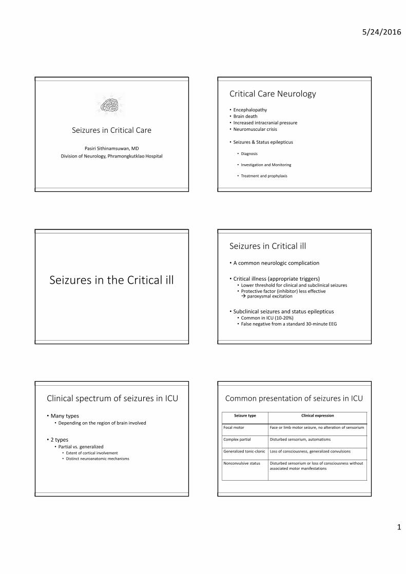
Seizures in Critical Care
Pasiri Sithinamsuwan, MDDivision of Neurology, Phramongkutklao Hospital
Critical Care Neurology
• Encephalopathy• Brain death• Increased intracranial pressure• Neuromuscular crisis
• Seizures & Status epilepticus
• Diagnosis
• Investigation and Monitoring
• Treatment and prophylaxis
Seizures in the Critical ill
Seizures in Critical ill
• A common neurologic complication
• Critical illness (appropriate triggers)• Lower threshold for clinical and subclinical seizures• Protective factor (inhibitor) less effective Æ paroxysmal excitation
• Subclinical seizures and status epilepticus• Common in ICU (10‐20%)• False negative from a standard 30‐minute EEG
Clinical spectrum of seizures in ICU
• Many types• Depending on the region of brain involved
• 2 types• Partial vs. generalized
• Extent of cortical involvement• Distinct neuroanatomic mechanisms
Common presentation of seizures in ICU
Seizure type Clinical expression
Focal motor Face or limb motor seizure, no alteration of sensorium
Complex partial Disturbed sensorium, automatisms
Generalized tonic‐clonic Loss of consciousness, generalized convulsions
Nonconvulsive status Disturbed sensorium or loss of consciousness without associated motor manifestations

5/24/2016
2
Focal seizures
• Structural disease (Local excitatory aberrations)• A portion of the cortex and its corresponding functional systems • Spreading to adjacent cortical regions via local synaptic connections
• Simple partial seizures • Example: the classic ‘‘Jacksonian march
• Complex partial seizures (esp. temporal epilepsies)• More involved: the recruitment of deeper brain elements• Affect conscious behavior: limbic brain, hippocampus or amygdala• Automatisms‐ ictal events, speech or behavioral mannerisms
• Lip smacking, blinking, or repetitive hand movements
• Non‐motor: more difficult to diagnose
Focal epileptic discharges and slow delta from left hemisphere
Kaplan PW, J Clin Neurophysiol 2006
Generalized seizures
• Primary generalized
• No cortical nidus identified
• ? Focal but fast spreading
• Initiation: brainstem/subcortical structure
• Propagation: paroxysmal activity; via cortical networks or cortical‐subcortical circuits
• Bilateral symmetric/synchronous in clinical and EEG
Kaplan PW. J Clin Neurophysiol 2006., Scharfman HE. Curr Neurol Neurosci Rep 2007., Slaght SJ. Epileptic Disord 2002.Timofeev I. Neuroscience 2004., Norden AD. Epilepsy Behav 2002., Huguenard JR. Trends Neurosci 2007.
Generalized tonic‐clonic seizures
J Clin Neurophysiol 2004
Etiologies
• Seizures as a consequence of critical illness
• Primary neurologic causes
Seizures as a consequence of critical illness

5/24/2016
3
Etiology
• Sepsis
• Hemodynamic instability
• Hypoxia/ischemia
• Metabolic abnormalities (30‐35%)
• Hyponatremia• Hypoglycemia, hyperglycemia• Hypophosphatemia• Renal/hepatic dysfunction• Hypomagnesemia
Etiology
• Drug/substance toxicity (15%‐45%)
• Antibiotics• Antidepressants• Antipsychotics• Bronchodilators• Local anesthetics• Immunosuppressive• Cocaine, amphetamines
• Drug substance withdrawal
• Barbiturates• Benzodiazepines• Opioids• Alcohol
Antibiotics
• Decrease central neuro‐inhibitory tone• All antagonize the action of GABA
• Penicillin class:• Well established animal seizure model for focal epilepsy• Primary proconvulsant effect by blocking GABAA Cl channels• Prevent GABA binding to GABAA receptors Æ inhibiting Cl currents
• Renal insufficiency: a predisposing factor for E‐lactam drug toxicity
Carbapenem
• Imipenem/cilastatin• In rat, induce convulsive behavior at lower serum concentrations than other penicillins
• A risk for seizures of 1.8% to 6.0%• More severe than other antibiotics
• Newer agents: meropenem, biapenem• lower convulsant risks than imipenem (risk 0.8%) as weaker affinity for the GABA‐Areceptor
Alldredge BK. The medical treatment of epilepsy. 1992., Wallace KL. Crit Care Clin 1997. Norby SR. 1996.
Psychiatric medications
• Antidepressants• 0.1‐4% risk of seizures, usually in an overdose• Low risk: trazodone, doxepin, monoamine oxidase inhibitors• Medium risk: tricyclics antidepressant, bupropion• Highest risk: maprotiline, amoxapine
• Psychotropic medication• Phenothiazines: lower seizure threshold: clinical & lab• Chlorpromazine: highest risk (3%–5%)
Theophylline
• Theophylline
• Risk 8‐14% in toxic dose (rare if level < 20 mg/mL)
• Clinical can be notoriously difficult to treat
• Mortality 50%
• Rx: benzodiazepines, barbiturates, general anesthesia and hemodialysis

5/24/2016
4
Anesthesia
• Lidocaine
• Na+ channel antagonist
• Topical, IV, intratracheal
• Risk of seizures is well correlated with serum concentration as linearly dose‐dependent
• Low risk: at therapeutic levels (1–5 mg/L) for the treatment of arrhythmias and as an anesthetic supplement
• High risk: level 8–12 mg/L
Primary neurologic causes
Etiology
• Neurovascular: stroke, AVM, etc.
• Tumor: primary, metastatic
• CNS infection: abscess, meningitis, encephalitis
• Inflammatory disease: vasculitis, ADEM, limbic encephalitis
• Traumatic brain injury: contusion, hemorrhage
• Primary epilepsy
Wijdicks EFM. Neurology 1993., Alldredge BK. The medical treatment of epilepsy. 1992.
Primary neurologic causes• Incidence
• Higher seizures after monitoring with continuous‐EEG (up to 22%)
• 52% NCSE• 9% subtle
• 1/3 minimal signs and all died
Vespa PM. J Neurosurg 1999;91:750–60.
Stroke & seizures
• Incidence of seizures (4.4‐13.8%)
• Most common cause of seizures in patient age> 60 years
• Location: • Lobar & subcortical > basal ganglia, thalamus • Posterior fossa (no risk)
Hauser WA. Epilepsia 1975., Bladin CF. Arch Neurol 2000.
Stroke & seizures
• Onset
• Early (24 hours – 4 weeks): risk 1.8‐15%• Excitatory or inhibitory alterations in the penumbral tissue
• Late (>4weeks): risk 2.5‐15%• ? result from gliosis and a meningocerebral scar• almost 3 times higher risk for subsequent stroke Æ epilepsy
Cleary P. Lancet 2004., Labovitz DL. Neurology 2001., Rumbach L. Neurology 2000.
5/24/2016
5
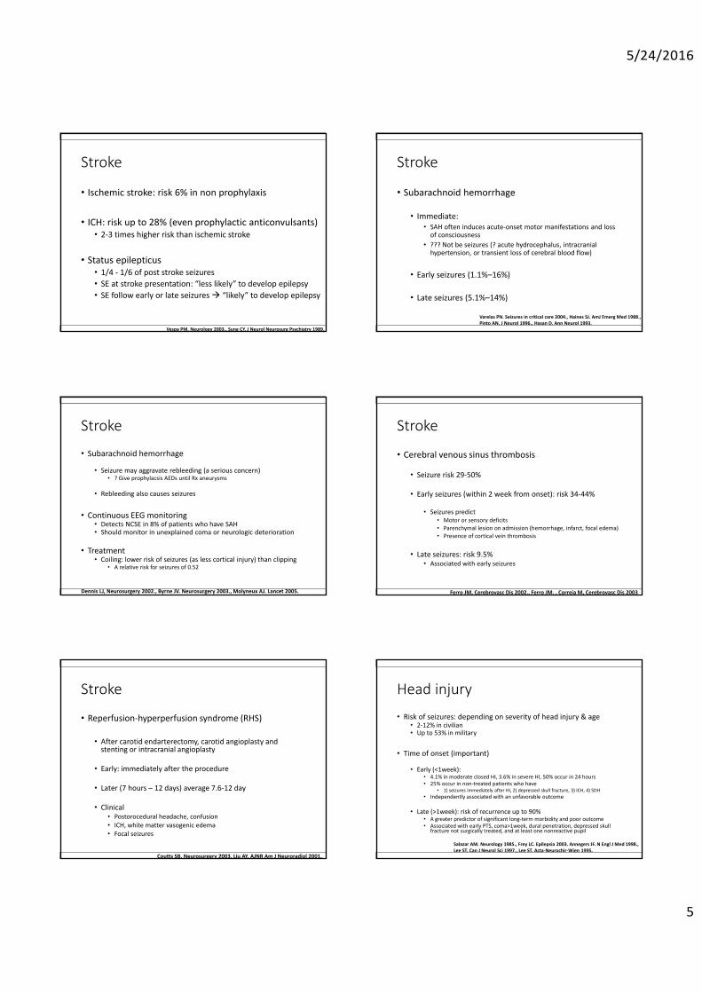
Stroke
• Ischemic stroke: risk 6% in non prophylaxis
• ICH: risk up to 28% (even prophylactic anticonvulsants)• 2‐3 times higher risk than ischemic stroke
• Status epilepticus• 1/4 ‐ 1/6 of post stroke seizures• SE at stroke presentation: “less likely” to develop epilepsy• SE follow early or late seizures Æ “likely” to develop epilepsy
Vespa PM. Neurology 2003., Sung CY. J Neurol Neurosurg Psychiatry 1989.
Stroke
• Subarachnoid hemorrhage
• Immediate: • SAH often induces acute‐onset motor manifestations and loss of consciousness
• ??? Not be seizures (? acute hydrocephalus, intracranial hypertension, or transient loss of cerebral blood flow)
• Early seizures (1.1%–16%)
• Late seizures (5.1%–14%)
Varelas PN. Seizures in critical care 2004., Haines SJ. AmJ Emerg Med 1988., Pinto AN. J Neurol 1996., Hasan D. Ann Neurol 1993.
Stroke
• Subarachnoid hemorrhage
• Seizure may aggravate rebleeding (a serious concern)• ? Give prophylacsis AEDs until Rx aneurysms
• Rebleeding also causes seizures
• Continuous EEG monitoring• Detects NCSE in 8% of patients who have SAH• Should monitor in unexplained coma or neurologic deterioration
• Treatment• Coiling: lower risk of seizures (as less cortical injury) than clipping
• A relative risk for seizures of 0.52
Dennis LJ, Neurosurgery 2002., Byrne JV. Neurosurgery 2003., Molyneux AJ. Lancet 2005.
Stroke
• Cerebral venous sinus thrombosis
• Seizure risk 29‐50%
• Early seizures (within 2 week from onset): risk 34‐44%
• Seizures predict • Motor or sensory deficits• Parenchymal lesion on admission (hemorrhage, infarct, focal edema)• Presence of cortical vein thrombosis
• Late seizures: risk 9.5% • Associated with early seizures
Ferro JM, Cerebrovasc Dis 2002., Ferro JM. , Correia M, Cerebrovasc Dis 2003
Stroke
• Reperfusion‐hyperperfusion syndrome (RHS)
• After carotid endarterectomy, carotid angioplasty and stenting or intracranial angioplasty
• Early: immediately after the procedure
• Later (7 hours – 12 days) average 7.6‐12 day
• Clinical• Postorocedural headache, confusion• ICH, white matter vasogenic edema• Focal seizures
Coutts SB. Neurosurgery 2003. Liu AY. AJNR Am J Neuroradiol 2001.
Head injury
• Risk of seizures: depending on severity of head injury & age• 2‐12% in civilian• Up to 53% in military
• Time of onset (important)
• Early (<1week): • 4.1% in moderate closed HI, 3.6% in severe HI, 50% occur in 24 hours• 25% occur in non‐treated patients who have
• 1) seizures immediately after HI, 2) depressed skull fracture, 3) ICH, 4) SDH• Independently associated with an unfavorable outcome
• Late (>1week): risk of recurrence up to 90%• A greater predictor of significant long‐term morbidity and poor outcome• Associated with early PTS, coma>1week, dural penetration, depressed skull
fracture not surgically treated, and at least one nonreactive pupil
Salazar AM. Neurology 1985., Frey LC. Epilepsia 2003. Annegers JF. N Engl J Med 1998.,Lee ST. Can J Neurol Sci 1997., Lee ST. Acta‐Neurochir‐Wien 1995.
5/24/2016
6
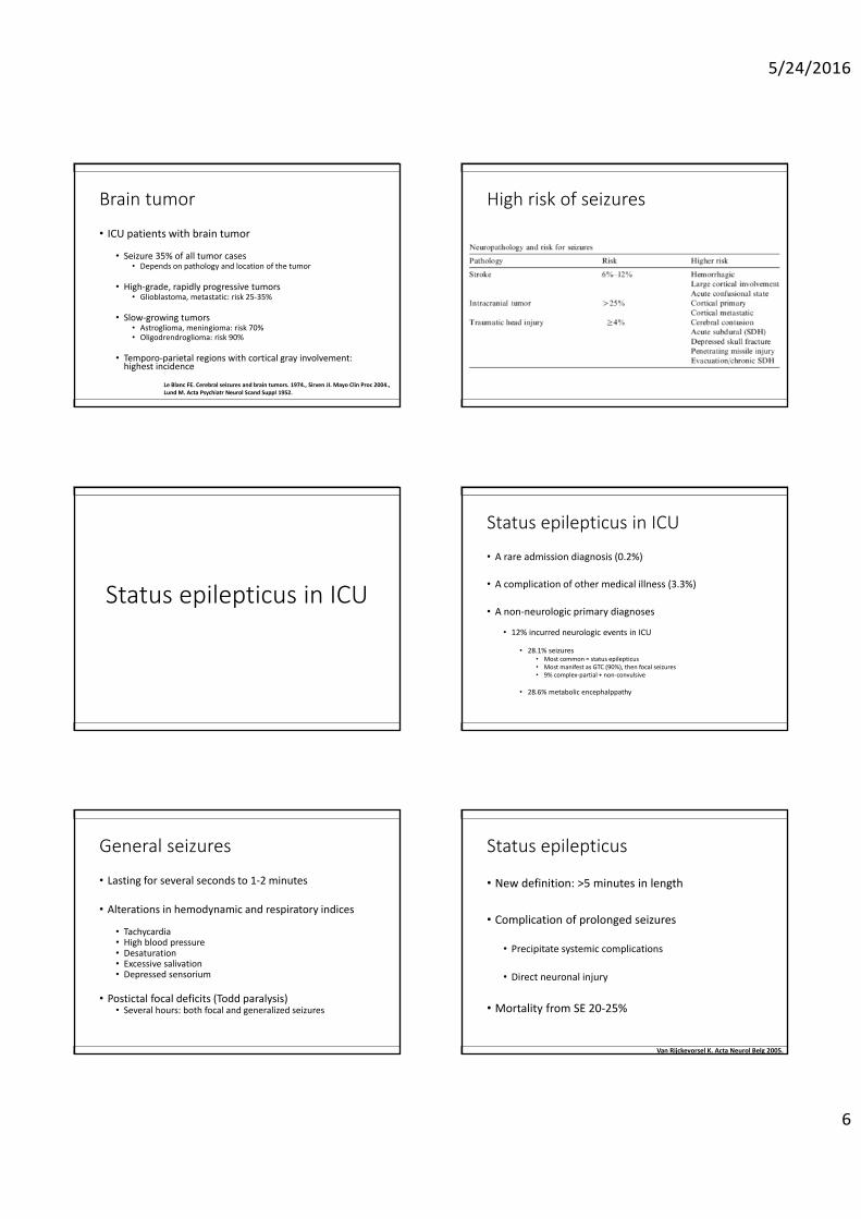
Brain tumor
• ICU patients with brain tumor
• Seizure 35% of all tumor cases• Depends on pathology and location of the tumor
• High‐grade, rapidly progressive tumors• Glioblastoma, metastatic: risk 25‐35%
• Slow‐growing tumors • Astroglioma, meningioma: risk 70%• Oligodrendroglioma: risk 90%
• Temporo‐parietal regions with cortical gray involvement: highest incidence
Le Blanc FE. Cerebral seizures and brain tumors. 1974., Sirven JI. Mayo Clin Proc 2004., Lund M. Acta Psychiatr Neurol Scand Suppl 1952.
High risk of seizures
Status epilepticus in ICU
Status epilepticus in ICU
• A rare admission diagnosis (0.2%)
• A complication of other medical illness (3.3%)
• A non‐neurologic primary diagnoses
• 12% incurred neurologic events in ICU
• 28.1% seizures• Most common = status epilepticus• Most manifest as GTC (90%), then focal seizures• 9% complex‐partial + non‐convulsive
• 28.6% metabolic encephalppathy
General seizures
• Lasting for several seconds to 1‐2 minutes
• Alterations in hemodynamic and respiratory indices
• Tachycardia• High blood pressure• Desaturation• Excessive salivation • Depressed sensorium
• Postictal focal deficits (Todd paralysis)• Several hours: both focal and generalized seizures
Status epilepticus
• New definition: >5 minutes in length
• Complication of prolonged seizures
• Precipitate systemic complications
• Direct neuronal injury
• Mortality from SE 20‐25%
Van Rijckevorsel K. Acta Neurol Belg 2005.
5/24/2016
7
Pathophysiology
• A reconfiguration of the excitatory and inhibitory network
• A breakdown of the Local inhibitory circuitry (GABA‐mediated): • inhibitory surround Æ spreading
• Recurrent or prolonged seizures induced a positive feedback loop• “Seizures beget seizures”• The longer duration of SE the more difficult it is to terminate
Subtypes of status epilepticus
• 1) Generalized convulsive SE (GCSE)• Most common subtype in ICU• Overt convulsion Æ then in prolonged SE Æ Subtle
• 2) Non‐convulsive status epilepticus• Awake, ambulatory or comatose• Complex partial status epilepticus
• 3) Focal motor SE (FMSE) or epilepsia partialis continua• Uncommon• Difficult to control• ? Unclear results in substantive injury to the cerebral cortex
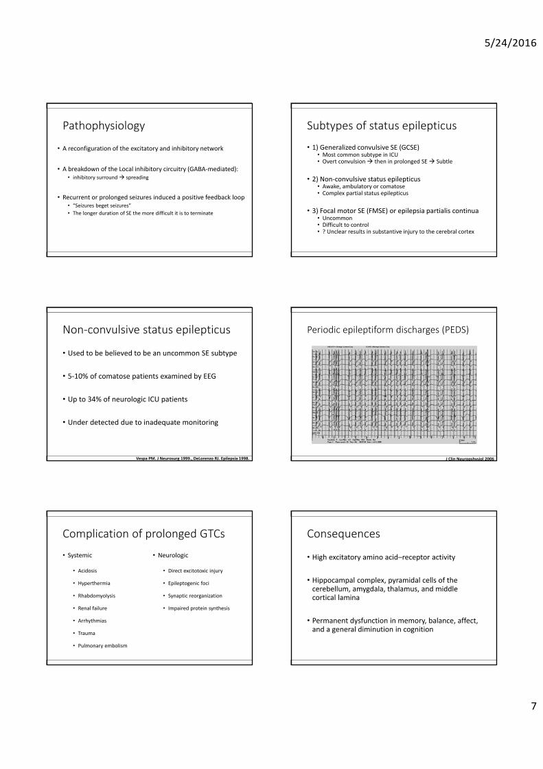
Non‐convulsive status epilepticus
• Used to be believed to be an uncommon SE subtype
• 5‐10% of comatose patients examined by EEG
• Up to 34% of neurologic ICU patients
• Under detected due to inadequate monitoring
Vespa PM. J Neurosurg 1999., DeLorenzo RJ. Epilepsia 1998.
Periodic epileptiform discharges (PEDS)
J Clin Neuropshysiol 2006
Complication of prolonged GTCs
• Systemic
• Acidosis
• Hyperthermia
• Rhabdomyolysis
• Renal failure
• Arrhythmias
• Trauma
• Pulmonary embolism
• Neurologic
• Direct excitotoxic injury
• Epileptogenic foci
• Synaptic reorganization
• Impaired protein synthesis
Consequences
• High excitatory amino acid–receptor activity
• Hippocampal complex, pyramidal cells of the cerebellum, amygdala, thalamus, and middle cortical lamina
• Permanent dysfunction in memory, balance, affect, and a general diminution in cognition

5/24/2016
8
Prognosis of status epilepticus
• Poor prognosis
• EEG: PLEDS
• Seizures lasting > 1 hour (mortality odds ratio of 10)
• Multiple seizure events
• Convulsive SE with complications• Acidosis, hyperthermia, rhabdomyolysis• Aspiration and trauma
DeLorenzo RJ. Semin Neurol 1990., Sloviter RS. Epilepsia 1999.
Investigation and Monitoring
Investigation
• Blood test & toxicology tests & septic work up (+/‐ CSF screen)
• CT, MRI brain
• Surface routine EEG recording
• Continuous EEG monitoring• ? Should perform in all ICU patients • ? impact on clinical decision‐making and outcome
• +/‐ SPECT or PET(+/‐ depth electrode)
Monitoring
• Scalp EEG (the diagnostic test of choice)
• For establishing the diagnosis of SE
• Document a possible epileptic focus
• For monitoring of the therapeutic response• Convulsive SE Æ NCSE 14%• Comatose 12 hours after initiate therapy for CSE:
• Success Rx: comatose 87%• NCSE: comatose 100%
• To prove pseudo‐seizures
Role of continuous‐EEG in ICU
• Generalized convulsive SE• To detect residual electrographic seizures (almost 50%)• 10% to 20% turning to NCSE• Unless a seizure fully resolves and the patient returns to an alert, cognitive baseline, an EEG should be obtained to exclude ongoing ictal activity
• Nonconvulsive• Esp. alteration of consciousness
• Equipment is not available in many hospitals
Nuwer M. Clin Neurophysiol 2007., Hirsch LJ. J Clin Neurophysiol 2004., Murthy JM. Neurol India 2004.
EEG in non‐convulsive status epilepticus
Brenner RP. Epilepsia 2002
• Primary
• 1) Repetitive generalized or focal spike, sharp waves, spike‐and‐wave, or sharp‐and‐slow complexes at > 3 sec
• 2) As above but <3 sec, but also meeting criteria 4 (below)• 3) Sequential rhythmic waves along with secondary criteria 1,2,3 +/‐ 4
• Secondary
• 1) Incrementing onset: increase in voltage and/or increase/decrease in frequency
• 2) Decrementing offset: decrease in voltage or frequency• 3) Postdischarge slowing or voltage attenuation• 4) Significant improvement in clinical state or EEG with anticonvulsant therapy

5/24/2016
9
Compared overnight EEG vs. first routine 30‐minutes
• Overnight EEG detected
• Overall• New or additional epileptiform abnormalities by 14%• Clinical and/or electrographic seizures 6%• Change in treatment 8%• Improvement attributed to change in treatment 4%
• In known cases with epilepsy• Treatment change with improvement 46%
• Seizures did not obviously affect outcome
Khan OI. Epileptic disorder 2014.
Controversial in EEG
• Periodic lateralizing epileptic discharges
• PLEDS if unilateral• BIPLEDS if bilateral/independent• PEDS if bilateral/uniform• Triphasic waves
• An interictal vs. ictal event
• BIPLEDS (mortality of 61%) vs. PLEDS (29%)
Status epilepticus severity scale (STESS)• Reliably identifies SE patients who will survive • Total score 0‐6, <3 – good outcome
• 1) Age• <65 years = 0 pt, ≥65 years = 2 pts
• 2) Seizure type • Simple‐partial, complex‐partial, absence, and myoclonic in the context of idiopathic/
genetic epilepsy = 0 pt• Generalized‐convulsive = 1 pt, Nonconvulsive SE in coma = 2 pts
• 3) Level of consciousness• Alert, somnolent or confused = 0 pt, Stuporous or coma‐tose = 1 pt)
• 4) History of previous seizures • Yes = 0 pt, No = 1 pt)
Rossetti AO. J Neurol 2008.
Treatment & prophylaxis
Treatment & prophylaxis
• Treatment
• Acute termination of ictal activity
• Prevention of further seizures
• Remove or correct trigger
• AEDs prophylaxis (risk vs. benefit)
Status epilepticus: initial measures• ABC & order EEG in the mean time
• CBG, thiamine, glucose
• Immediate IV BZD in 5 minutes• Lorazepam 5‐10 mg, diazepam 20‐40 mg, midazolam 5‐20 mg
• PHT: • loading 20 mg/Kg rate 50mg/min (goal level 15‐20 mg/dL)• Additional 5‐10 mg/Kg if required (goal 20‐25 mg/dL)
• VPA 20‐50 mg/Kg
• Keppra, lacozamide, topiramate, etc
5/24/2016
10
Initial drug for status epilepticus Second‐line agents
Refractory status epilepticus
• Rapid pharmacologic burst suppression/coma
• Propofol 2 mg/Kg then 150‐200 ug/Kg/min infusion
• Thiopental 4 mg/Kg then 0.3‐0.4 mg/Kg/min
• Midazolam 0.2 mg/Kg, then 0.1‐0.2 mg/hour
• Pentobarbital 5‐10mg/Kg then 1‐10 mg/Kg/h
• EEG monitoring
• Hemodynamic support
Status epilepticus: Bilateral periodic epileptiform discharges
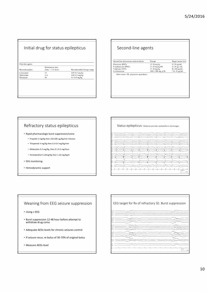
Weaning from EEG seizure suppression
• Using c‐EEG
• Burst suppression 12‐48 hour before attempt to withdraw drug coma
• Adequate AEDs levels for chronic seizures control
• If seizure recur, re‐bolus of 30‐70% of original bolus
• Measure AEDs level
EEG target for Rx of refractory SE: Burst suppression

5/24/2016
11
Treatment of seizures in ICU
• Generalized convulsive SE• A strong consensus in aggressive treatment
• Non‐convulsive SE (NCSE)• Debate
• Benign, if in patients non‐cerebral lesion, no permanent morbidity• More common following an anoxic‐ischemic event or trauma
• Most epileptologists would agree that in such scenarios the presence of continuous paroxysmal activity may accentuate injury incurred by the primary insult
Towne AR. Neurology 2000., Vespa PM. J Neurosurg 1999., DeLorenzo RJ. Epilepsia 1998.
Seizure prophylaxis in the ICU
• Medical illnesses or direct cerebral injury • Predisposed to seizures
• Medical illness• Lower risk of seizures
• CNS lesion• Higher risk of recurrence
• Prophylactic treatment• Not a guarantee the outcome• ? Risk for toxicity
Stroke and prophylaxis
• Controversial for AEDs prophylaxis
• Ischemic or hemorrhagic stroke with a large cortical involvement or causing an acute confusional state in the early aftermath of a stroke were independent predictors of future seizures
• The current ICH guidelines suggest antiepileptic treatment for 1 month in patients presenting with seizures
Arboix A, Stroke 1997., Broderick JP. Stroke 1999.
Stroke and prophylaxis
• Alcoholic patients with ICH (x3 times risk for SE)• Should be treated with GABAergic AEDs
• Late seizures occur (after 2 weeks from onset)• Long‐term anticonvulsants as greater risk for epilepsy
• SAH: controversial
Cervoni L. Neurosurg Rev 1994.
Brain tumor
• Controversial for prophylaxis
• Anticonvulsants may • Interfere with corticosteroids, chemotherapy, and radiation treatment• Induce more frequent and serious allergic reactions
• Malignant glioma• May prescribe• Even adequate anticonvulsant therapy, seizures may occur
• The AAN practice parameters “do not” support prophylactic anticonvulsants in patients who have newly diagnosed brain tumors
Vecht CJ. Lancet Neurol 2003., Aguiar D. J Neurooncol 2004.
Head injury
• Severe head injury (Cortical injury)• Strong data to support that prophylactic use of AEDs • Cerebral contusion, acute subdural hemorrhage, depressed skull fracture, penetrating missile injury
• The current practice parameters by the AAN and the Brain Trauma Foundation advocate prophylactic treatment with “phenytoin” only during “the first 7 days” from head injury
Chang BS. Neurology 2003., The Brain Trauma Foundation. J Neurotrauma 2000.

5/24/2016
12
Intracranial surgery
• The incidence of postoperative epilepsy
• 17% after supratentorial intracranial surgery• Depends on pathology, location, type of surgical approach, presence of postoperative deficit, and a previous history of epilepsy
• A meta‐analysis study: reviewing 30 publications
• AEDS prophylaxis reduce the incidence of seizures• Postoperatively, patients without seizure: “taper off” AEDs
Kuijlen JM. Seizure 199., Glantz MJ. Neurology 2000.
Summary
• Seizures in the ICU are more difficult to prevent, diagnose, and treat effectively
• Treatment of seizures and SE in an ICU is challenging
• The treatment of seizures requires a careful evaluation of toxic risk versus benefit and proper drug selection
• AEDs prophylaxis is still debated
Thank You