Embed Size (px)
Citation preview

This article appeared in a journal published by Elsevier. The attachedcopy is furnished to the author for internal non-commercial researchand education use, including for instruction at the authors institution
and sharing with colleagues.
Other uses, including reproduction and distribution, or selling orlicensing copies, or posting to personal, institutional or third party
websites are prohibited.
In most cases authors are permitted to post their version of thearticle (e.g. in Word or Tex form) to their personal website orinstitutional repository. Authors requiring further information
regarding Elsevier’s archiving and manuscript policies areencouraged to visit:
http://www.elsevier.com/copyright

Author's personal copy
Dynamic controls on accretion and lithification of modern gypsum-dominatedthrombolites, Los Roques, Venezuela
Daniel A. Petrash a,⁎, Murray K. Gingras a, Stefan V. Lalonde b, François Orange a,Ernesto Pecoits a, Kurt O. Konhauser a
a Department of Earth and Atmospheric, University of Alberta, Edmonton, Alberta, Canada T6G 2E3b Laboratoire Domaines Océaniques, Institut Universitaire Européen de la Mer, Université de Bretagne Occidentale, Place Nicholas Copernic, Technopôle Brest-Iroise, 29280 Plouzané, France
a b s t r a c ta r t i c l e i n f o
Article history:Received 29 May 2011Received in revised form 7 December 2011Accepted 7 December 2011Available online 16 December 2011
Editor: B. Jones
Keywords:Microbial matsMicrobialitesAnoxygenic photosynthesisExopolymersTrace metals
Meter-sized thrombolites coated by well developed zonally differentiated microbial mats have been foundgrowing in the shallow waters (depth b1 m) of a restricted hypersaline lagoon on the Archipelago Los Roquesin Venezuela. By contrast, within the deeper parts of the studied lagoon, sedimentation is characterized byseveral decimeters of organic-rich material containing gypsum granules lacking carbonate cementation.The lithification of the thrombolites is thought to have proceeded as follows. First, extracellular polymericsubstances (EPS) comprising the microbial mat concentrate Ca2+ and other metal cations by adsorptionfrom the hypersaline waters. Second, some of these bound metals then serve as nucleation sites for primarycalcium carbonate (CaCO3) precipitation. Third, while carbonate phases are forming in some zones of themat, in others zones they are being re-dissolved due to the acidity generated through the metabolism ofsulfide-oxidizing bacteria, Fourth, as the dissolved sulfide is oxidized into sulfate, the pore-water become sat-urated with respect to gypsum (CaSO4·2H2O). Fifth, as primary gypsum precipitates within the structures,endolithic sulfate-reducing bacteria metabolize the sulfate moiety in the mineral phase, while simultaneous-ly oxidizing the EPS trapped during accretion. Sixth, as microbial EPS degradation proceeds, the anaerobic ox-idation of specific protein fractions of the EPS matrix leads to increased alkalinity, the partial dissolution ofgypsum, supersaturation with respect to calcium carbonate, and ultimately pseudomorphic aragonite re-placement; this differs from secondary calcite cements in being enriched in 12C, and depleted in minor andtrace metals initially associated with the EPS. The biogeochemical processes occurring in this thrombolite-constructing lagoon represent a novel field site for studying the chemical and isotopic processes characteriz-ing early diagenetic gypsum and the role microbes play in its precipitation, dissolution and calcification. Inthis regard, insights gained from this modern field site will help to better understand mechanisms bywhich some Precambrian microbialites were lithified.
© 2011 Elsevier B.V. All rights reserved.
1. Introduction
Microbialites are accretionary organosedimentary structures thatform as a result of a benthic microbial community trapping and bindingdetrital sediment and/or forming the locus of mineral precipitation(Burne and Moore, 1987). Accordingly, stromatolites (Kalkowsky,1908) and thrombolites (Aitken, 1967) are distinct types of microbialitescharacterized, respectively by laminated andmesoclotted internal fabrics(Kennard and James, 1986; Riding, 2011). Both stromatolites andthrombolites typically develop in shallow subaqueous settings, such asintertidal flats and hypersaline lagoons (Jørgensen et al., 1983; DesMarais, 2003), that are characterized by higher rates of organic matterproduction relative to grazing (Stal, 2000). During diagenesis, the organic
matter serves as an electron donor to various microbial metabolisms,including sulfate reduction, that leads to (1) complex elemental cyclingwithin the structures (Des Marais, 2003), (2) the enhancement of pore-water alkalinity (Slaughter andHill, 1991), and (3) subsequent cementa-tion by calcium carbonate (Visscher et al., 1998; Riding, 2011).
Despite being less common, gypsum has also been shown to play arole in microbialite accretion (e.g., Kobluk and Crawford, 1990; Vogelet al., 2010). Morphologically, fossil gypsum microbialites may occuras domes, columns or planar structures (Rouchy and Monty, 2000).Texturally, they are more commonly distinguished by their dominantinternal thrombolitic fabric (Riding, 2000), which may locally inter-finger with crudely laminar crusts to form thrombolitic stromatolites(Riding, 2011). The sulfate mineral is a seemingly minor, yet impor-tant component of the stromatolitic record (Rouchy and Monty,2000; Pope et al., 2000; Riding, 2008), however, gypsum, as primarymineralogy, could be replaced early during diagenesis by carbonates(Vogel et al., 2010, and references therein). Hence, a more complete
Sedimentary Geology 245–246 (2012) 29–47
⁎ Corresponding author.E-mail address: [email protected] (D.A. Petrash).
0037-0738/$ – see front matter © 2011 Elsevier B.V. All rights reserved.doi:10.1016/j.sedgeo.2011.12.006
Contents lists available at SciVerse ScienceDirect
Sedimentary Geology
j ourna l homepage: www.e lsev ie r .com/ locate /sedgeo

Author's personal copy
understanding of the mechanisms by which modern subaqueousgypsum-dominated thrombolites form today would be useful to un-derstand their ancient diagenetically modified analogs; thereby theproblem of the significance of dubious pseudomorphic features inthe highly fragmented stromatolitic record could be addressed (seeRiding, 2008, p. 77–78, for details).
The bacterial role in gypsum precipitation, as well as the effects thatearly gypsum precipitation may have in microbialite fabric develop-ment has yet to be fully explored, mostly because gypsum is rarelyfound in modern microbialites (Kobluk and Crawford, 1990; Rouchyand Monty, 2000). By using transmitted and reflected light, scanningelectron microscopy (SEM), electron probe microanalysis (EPMA) andlaser ablation-inductively coupled plasma mass spectrometry (LA-ICPMS), we describe here the biogeochemical and mineralogical pro-cesses occurring in an alkaline and hypersaline lagoon in theArchipelago Los Roques, Venezuela. In the shallower areas (to a depthof b1 m) complex photosynthetic microbial mats are associated withthrombolites. The mineralogy and texture of the decimeter- to meter-scale thrombolites from Los Roques differ from most modern occur-rences because of the abundance of gypsum,which is being pseudomor-phically replaced by aragonite; while the lithification of the structures ismostly due to micritization of aragonite pseudomorphs after gypsum.
The relationships between internal microfabrics, primary andsecondary mineralogies, and the distribution of biochemical signa-tures have not been systematically studied in modern thrombolites.Specifically, we were interested in documenting: (1) the affinity ofextracellular polysaccharide (EPS)-rich organic matrices for certaindissolved metals which may serve as “biological proxies” (Fazioet al., 1982; Decho, 1990; Decho et al., 2005; Perry et al., 2005;Braissant et al., 2007; Ortega-Morales et al., 2007; Flemming andWingender, 2010); and (2) if the degradation of EPS trapped duringaccretion produces complex physicochemical conditions and influ-ences cation fluxes and internal diffusion pathways towards activesites of reactive carbonate precipitation (e.g., Kamber et al., 2004;Braissant et al., 2009; Decho, 2010). Importantly, if unique tracemetal trends are preserved in these modern thrombolites, it mightthen be possible to identify whether biological processes similarlyled to the accretion of ancient thrombolites, and for that matter, stro-matolites exhibiting heterogeneous internal fabrics.
2. Geological and environmental settings
The Archipelago comprising Los Roques National Park is located inthe southern Caribbean Sea, approximately 150 km offshore of theVenezuelan mainland. The archipelago has 42 low-laying islands anda large number of sand banks distributed in an isolated, oval-shapedrimmed shelf (Fig. 1), approximately 1250 km2 in extent. The innershelf consists of a composite lagoon which has maximum and averagewater depths of 50 m and 5 m, respectively (Amend, 1992). The com-posite lagoon is partially enclosed by coral reefs, which are better de-veloped along the eastern and southern margins of the archipelago(Fig. 1). The reef grades to sand banks that form supratidal islandswhich shelter the shelf from open ocean circulation and are frequentlycolonized by mangroves that partially confine the shallow seawatersby increasing sedimentation rates. In many of the low-laying emergedislands there are perennial hypersaline lagoons and/or ephemerallagoons, both accompanied by supratidal sabkha deposits.
Climate in Los Roques is warm and humid with average air tem-peratures and daily humidity of 28 °C and 80%, respectively. Rainfallis scarce and rarely exceeds 270 mm yr−1, the bulk of which fallsduring October through to January. Evaporation rates are up to225 mm yr−1 (as estimated by Sonnenfeld et al., 1977), thus, evapo-ration exceeds precipitation most of the year and salinities in therestricted lagoons may reach values of up to 175‰.
The studied thrombolites were observed at Laguna Pirata, which isan anchialine lagoon restricted by a permeable Quaternary beach rock
and a modern sandbank; thereby the evaporatively concentratedwater is constantly fed by seepage seawater reflux (i.e. Babel, 2007).The lagoon has maximum and average depths of 1.6 and 0.5 m,respectively. The water body is density stratified and its hypolimnionconsists of poorly oxygenated to sulfidic brine (Fig. 2). The averagetemperature of the shallow margins of the lagoon is 34 °C.
Sedimentation at the lagoon margins consists of whitish, finelycrystalline crusts formed by evaporation. These crusts, which can beup to 3 cm thick, are polymineralic and contain halite, gypsum andaragonite, thus when dry they are easily erodible by ablation orcan be partially dissolved by sporadic showers. Subaqueously, lithi-fied domal-shaped thrombolites, with sizes ranging from less than10 cm in diameter to larger coalesced structures up to 4 m large,develop along the shallow margins in a zone where water depthsrange from tens of centimeters down to about 1 m (Fig. 3A, B).The narrow actively-growing thrombolite zone is subparallel tothe shore, where most of the subaqueous sedimentary substratehas been colonized by complex microbial mats up to 2 cm thick(Figs. 3C-F, 5). The actively growing thrombolite zone laterally givesway basinwards within a few ten of meters to a thick deposit, consist-ing on an unlaminated organic-rich gelatinous mud that supportsthe growth of millimeter-scale gypsum granules (Fig. 3A), here re-ferred as gypsooids (after Vogel et al., 2009). With increasing deep,the authigenic sediment becomes less organic-rich and the gypsooidsform a coarser chicken wire-like depositional fabric (Fig. 3G).
3. Methods
3.1. Collection of samples
3.1.1. Sediment samplesTwo thrombolites (~10 cm×15 cm) were randomly sampled by
hand from the shallow lagoon (0.15 to 0.45 m). A thick microbial matcoating from one of the specimenswas collected and its different layers(Fig. 3A) were carefully separated with tweezers and sampled. In addi-tion, the organic sediments in the deepest part of the lagoon (Fig. 3G)were retrieved using a PVC dredging device and sampled. In order topreserve the microorganisms for scanning and transmission electronmicroscopy all the organic samples were placed in sterile 0.5 mlEppendorf PCR polypropylene tubes containing filtered lagoon waterand aqueous glutaraldehyde to a final concentration of 2% v/v.
3.1.2. Water samplesWater samples were collected directly from the lagoon using a 60-
ml sterile syringe. The syringe was then fitted with a 0.2 μmmicroporefilter through which water was expelled into metal-free polycarbonatescrew cap sample bottles. Filtered water samples were collected induplicate, with one being treated with analytical grade nitric acid (toa final concentration of 10% v/v) for cation analysis and the other leftunacidified for anion analysis. Overlying water temperatures andpH values were measured immediately upon collection using a Ross(Orion) combination pH electrode. Total alkalinity was measured insitu by using acid titration.
3.2. Biogeochemical profile
A rough estimation of the non-static biogeochemical zoning of themicrobial mat overlying the thrombolites (e.g. Revsbech et al., 1983)was obtained by microsensing the unlithified part of the specimenshown in Fig. 3E, which was growing with an overlying water depthof around 10 cm (Fig. 3D). O2 and H2S microsensors (UniSense A/S,Denmark, 50 μm, response time, t90, of ~0.3 s), connected to apicoammeter (UniSense), were simultaneously inserted in the matvia a micromanipulator mounted on a heavy solid stand. Measure-ments were performed at ambient temperature of ~31 °C in 100 μmincrements of vertical depth and recorded by using the computer
30 D.A. Petrash et al. / Sedimentary Geology 245–246 (2012) 29–47

Author's personal copy
software Profix (UniSense A/S, Denmark). Prior to the measurements,the O2 microsensor was linearly calibrated by using picoamperesreadings in the O2-saturated overlying water and in ascorbic acid(0.1 M, for 0% O2 saturation). The calibration of the H2S microsensorwas performed using a sulfide standard with an ionic strength of0.5 M buffered to the pH of the overlying water (~9.1).
3.3. Aqueous geochemistry
3.3.1. SO42− and Cl− ions chemistry
Quantitative analyses of SO42− and Cl− anions were performed
using a Dionex DX600 Ion Chromatograph (IC) at the University ofAlberta. Samples were diluted as required before analysis to reducesalinity to operational values adequate for the analytical machine.
3.3.2. Major and trace metal concentrationsFor determination of major and trace metal compositions of the
Los Roques lagoon water, the samples were digested with HNO3
(8 N) and analyzed using a Perkin-Elmer Elan6000 quadrupole ICP-
MS coupled to a New Wave UP-213 laser ablation system at theUniversity of Alberta. The ICP-MS instrument parameters were asfollows: RF power 1200 W, peak hopping acquisition, 50 ms dwelltime. Quantitative results were obtained via calibration of relativeelement sensitivities against a solution containing 10 ppm of Br, In,and Sc as internal standards. Relative standard deviations (2σ level)were between 3% for Na and Fe, 0.35% for Al and Zn, and between0.005 and 0.06% for most analyzed elements. Detection limits werebetween 0.006 ppb (e.g., Mn and U) and 0.75 ppb (e.g., Fe).
3.3.3. Speciation modelingThe degree of saturation of dissolved mineral species in the lagoon
water was determined using the geochemical speciation programPHREEQC (Parkhurst and Appelo, 1999). Theoretical speciation re-sults are presented in terms of the saturation index (SI) for predictedminerals, where SI is defined by SI=log (IAP/Ksp), with IAP and Ksp
being the ion activity product of the dissolved constituents and solu-bility product for the minerals considered, respectively.
Fig. 1. Location of the study area. Inset A: Oblique aerial view of Laguna Pirata. Inset B: Map of Pirata island.
31D.A. Petrash et al. / Sedimentary Geology 245–246 (2012) 29–47

Author's personal copy
3.4. Solid phase mineralogical/geochemical analyses
3.4.1. Major element geochemistryWe systematically analyzed various levels from the two thrombolite
samples for major element geochemistry by electron microprobe anal-ysis (EPMA) after the samples were mounted in epoxy. Wavelengthdispersive analyses were conducted using a JEOL 8900 Superprobe(15 kV accelerating voltage; 10 μm beam diameter, and 15 nA currentand 30 s counting time). The following mineral and synthetic crystalswere used for calibration: F-93(Mg), diopside (Ca, Si), willemite (Mn),Hem639 (Fe), magnesite (Mg), barite (SO4) and pyrope (Al2O3). Analyti-cal precisionwasmonitored through triplicate analyses at each spot area.Data are presented in a 100% carbonate basis.
3.4.2. Trace element geochemistryThe samples analyzed by EPMA were also analyzed for trace
element composition by ICP-MS. Quantitative results were obtainedvia the calibration of relative element sensitivities against the NIST612 standard with analyses being normalized to [Ca], previouslydetermined by electron microprobe analysis for the unknowns(Jarosewich, 2002). The spot size was 30 μm in diameter, with theaverages of least three laser spots reported. Concentrations belowthe detection limits are indicated where appropriate. Trace elementswere determined for the basal, middle, and uppermost crusts ofthe specimen (for a total of 94 laser spots), and in the authigenic car-bonate phases present in the microbial mat coating the same speci-men (40 laser spots).
Data reduction and concentration determinations were obtainedusing the GLITTER® (New Wave Research) laser ablation software.Repeated analysis (n=6) of the standard yielded relative standarddeviations of between 0.01 and 0.5% (2σ level) and detection limitsbetween 0.02 ppm (e.g., Zn, Cd, Cu) and 0.06 ppm (e.g., Mg, Sc, Sr)for most elements (except for elements like, B, Yb, Lu).
3.4.3. Stable C- and O-isotope analysesIn order to evaluate isotopic shifts within the thrombolites, four
calcified crusts occurring at different depths within the selected spec-imen were sub-sampled in duplicate. In addition, ~20 mg of authi-genic carbonate grains were carefully picked from freeze-driedbiofilm material. The authigenic nature of the mineral componentsof the mats was determined through the examination of the size,morphology and distinctive optical features of the grains by usingreflected light and blue light excitation microscopy (after Dravisand Yurewicz, 1985).
Prior to analysis, the subsamples were treated for 48 h with H2O2
(30%) to remove residual organic matter, rinsed three times usingultrapure water, and dried overnight in a vacuum oven at 30 °C.Stable C-isotope analyses of carbonates were performed by immers-ing whole-rock powders in 100% phosphoric acid while under vacu-um (McCrea, 1950) and analyzing the released CO2 on a FinneganMAT 252 mass spectrometer at the University of Alberta. The dataare reported in δ-notation with respect to V-PDB for carbon and oxy-gen (Craig, 1957, 1961).
3.4.4. Scanning electron microscopy (SEM)Thin sections and grain mounts were sputter coated with gold.
SEM observations were performed at the University of Alberta on aJEOL JSM-6301FXV instrument, which has an attached Norvarenergy-dispersive spectrometer system (PGT). Images were taken at5 kV, while EDX analyses were done at 20 kV; a constant working dis-tance of 15 mm was used throughout our SEM analyses. The texturalrelationships between the exopolymeric matrices and their authi-genic phases were visualized by using both low-temperature (Cryo-SEM) and the critical point drying methods.
3.5. Biological SEM and TEM
Millimeter-size samples were rapidly frozen by immersion in liq-uid nitrogen (−200 °C), then introduced in an Emitek K1250 cryo-chamber where they were fractured at −180 °C, superficially subli-mated at −90 °C, gold coated at −180 °C, and then introduced inthe refrigerated column of the JEOL 6301F at −180 °C to conducthigh-resolution examination of internal biofilm structure and mineral/microbe textural features.
In addition to the techniques describe above, the correlative TEMpreparatory scheme of Liss et al. (1996) was used to analyze individ-ual portions of the microbial mat that coated the microbialite speci-men under examination. Samples were embedded in standard low-viscosity epoxy resin, and subsequently polymerized (Spurr, 1969).The resulting blocks of embedded biofilm were sectioned with a dia-mond knife mounted in an ultramicrotome (Reichert Ultracut E).Ultrathin sections for morphological studies were cut at 50 to 70 nmand mounted on formvar-covered TEM grids (Marivac Canada)with, and without, counterstaining. Sections of biofilm were observedand images captured in transmission mode using a Philips/FEI (Mor-gani) Transmission Electron Microscope with CCD camera (TEM-CCD) at an accelerating voltage of 80 kV at the Department of Biology,University of Alberta.
Fig. 2. Schematic hydrological model of Laguna Pirata. SE: seepage seawater influx, SU sporadically, during storm events, surface seawater influx may occur.Adapted from Babel (2004), Fig. 2.
32 D.A. Petrash et al. / Sedimentary Geology 245–246 (2012) 29–47

Author's personal copy
4. Results
4.1. Lagoon water chemistry
At the time of the field work (February 2009), measured pH valuesof the surface waters of Laguna Pirata varied between 8.7 and 9.3. Thewater temperature fluctuated between 28 and 35 °C and the averagesalinity was approximately 90‰, reaching up to 140‰ in the adjacentsabkha flat (Fig. 3D). In terms of major ion chemistry the Mg/Camolarratio was found to be 6.1, showing that the lagoonal surface waterswere relatively enriched in Mg as compared with Los Roques seawa-ter (Mg/Ca=5.6). Other relevant chemical features of the lagoonwater are summarized in Table 1. Laguna Pirata waters are also slight-ly enriched in Co, Fe, Ni, Mn with regards to mean seawater; conser-vative with respect to Ti, Cu, Cr, Mo, U and V, and depleted in Zn, Zr,Ti, Nb and Sn (Fig. 4).
The hypersaline superficial waters are oversaturated with respectto dolomite (SI=3.8), calcite (SI=1.4), and aragonite (SI=1.28),
whereas gypsum, the most abundant mineral phase in Laguna Pirata,is slightly undersaturated (SI=−0.29). Undersaturation of the sur-face waters with respect to gypsum may be due to temperature, oxy-genation and mixing (e.g., Last and Schweyen, 1983) or its highnatural alkalinity (e.g., Thompson and Ferris, 1990).
Table 1Chemical features of surface Pirata's water. The mean chemical composition of LosRoques seawater (sampled in two different sites in the unrestricted lagoon) is alsoshown for comparison.
Sample Ca2+
(mmol)Mg2+
(mmol)Sr2+
(μmol)Salinity(‰)
SO42−
(mmol)Total alkalinity(mM/L)
Laguna Pirataevaporatedseawater
17.5 108.9 97.12 90 66.8 169
Seawater atLos Roques
10.1 52.2 96.1 40 31.6 134
Fig. 3. General macroscopic features of the microbially-influenced sedimentation in Laguna Pirata. (A) Microbial mat coating a thrombolite, the mat exhibit distinctive layeringrelated to vertical array of bacterial communities. Note the masses of microcrystalline gypsum. Scale bar is 10 mm. (B) Close-up of thrombolites, notice microbial mat coatingthe structures; water depth 10 cm. (C) Dense coalescent growth of thrombolites, water column may vary from 15 cm to 45 cm. (D) In situ microsensing of the bacterial mat;view from the sabkha (E) A collected thrombolites specimen. Note that the thrombolitic fabric (clotted) is dominant over the stromatolitic fabric (laminated zone); more evidentin the base of the structure. (F) Distribution of carbonate–evaporite subfacies in Laguna Pirata. (G) The organic-gypsum rich sediment in the deepest part of the lagoon.
33D.A. Petrash et al. / Sedimentary Geology 245–246 (2012) 29–47

Author's personal copy
4.2. Geobiology of the microbial mats
4.2.1. The thrombolite-constructing matMicrobial mats growing on the thrombolites reach up to 2 cm in
overall thickness (Fig. 5A). The uppermost photosynthetic layer con-sists of a 4 mm thick yellowish to green zone that is dominated byfilamentous cyanobacteria (e.g., Oscilatoria sp.) and algae (mostlyunidentified diatoms) (Fig. 6). During daylight hours, these oxygenicphotosynthesizers account for an increase in the partial pressure ofoxygen at the water/mat interface from 0.45 to 1.05 atm.
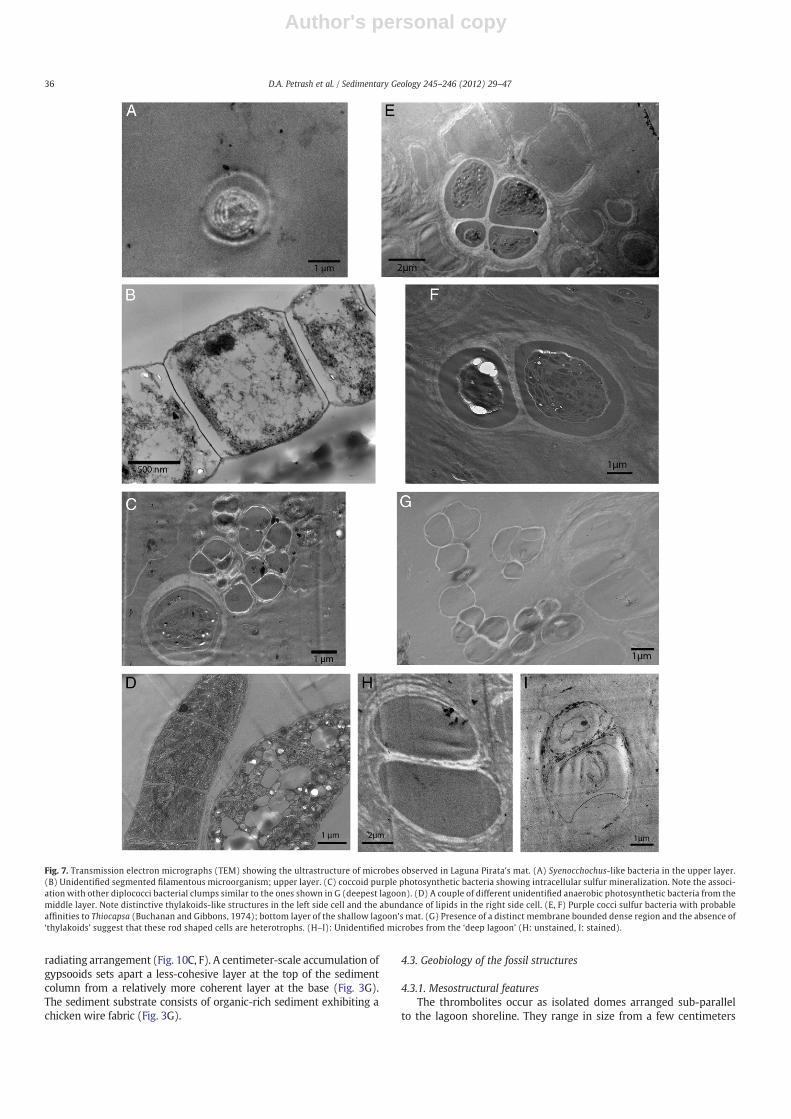
Below the uppermost layer, a green layer extends from 4 to about10 mm deep within the mat. It contains a high diversity of organisms,ranging from filamentous cyanobacteria, including a sulfide tolerantSpirulina-like cyanobacterium to clump-forming non-identified micro-colonies of rod-shaped cells (~3–5 μm in diameter) enclosed by multi-laminate sheaths (Figs. 6, 7A–C). Solitary Beggiatoa-like cells, commonlyexhibiting intracellular sulfur accumulations (Fig. 7D, left cell), werealso found. In the case of the latter, microsensors data shows that thislayer coincides with a zone exhibiting steep opposing gradients ofdecreasing O2 and increasing HS− (Fig. 5B); in this zone, white filamen-tous laminae (Fig. 5A) are suggestive of elemental sulfur accumulationdue to incomplete microbially-mediated oxidation of HS− (i.e., vanGemerden, 1993).
At depths >10–12 mm, the microbial mat becomes increasinglysulfidic as evidenced by peaks in dissolved HS− (Fig. 5B). At night,the top of the sulfidic zone shifts upwards. Fine scale microscopy ofthis layer reveals that it is made up mostly of rod-shaped bacteriaencapsulated by EPS. Morphologically the cells resemble some ofthose occurring in thick EPS in the overlying layer (cf. Fig. 7C and G).
Storm-derived sediment grains are randomly dispersed within thelagoon's microbial mat. The grains are mostly subangular in shape andconsist of millimeter-scale fragments of the calcareous algae Halimedasp.; other storm-transported bioclasts, such as ooids and shell frag-ments, are also present. The allochems are frequently affected bymicro-boring (Fig. 8), which could represent biogeochemical dissolutionproduced by endolithic microbial communities (i.e., those betweenand upon mineral grains) (e.g., Kobluk and Risk, 1977; Reid andMacintyre, 2000). Microprobe analysis indicates that Mg-calcite maybe formed as an internalmicrocrystalline precipitateswithin themicro-bore cavities; it also occurs as part of a thin microcrystalline cementoften encrusting primary grains coated by EPS microfilms (Fig. 8).
As for the inorganic constituents of the thrombolite-constructingmat, calcium carbonate accumulation consists dominantly of crystalclusters (15–25 nm in diameter), which forms small peloids up toa few hundred micrometers in diameter (Fig. 9). Mineralogically,the authigenic precipitates consist of: (1) micrometer-scale aragoniteneedles usually forming peloidal aggregates of up to 400 μm in diam-eter (Fig. 9A, B); (2) ellipsoidal aggregates formed by blocky calcitecrystals (Fig. 9C); (3) spheroidal forms of Mg-calcite, with grainsizes ranging from 2 μm up to 10 μm and which may, or may not, becalcified bacterial cells and their EPS; and (4) spherical Ca-phosphateparticles up to 20 μm in diameter which are encrusted by calcifiedMg-rich precipitates (Fig. 9E, F).
4.2.2. The gelatinous gypsooid-supporting matrixIn contrast to its shallower counterpart, the gelatinous organic
matrix forming in the deepest lagoon (water depth >1.0 m) lacks a
Fig. 4. Logarithmic plot showing the enrichment/depletion factors of selected trace elements in the surface waters Laguna Pirata, normalized respect Los Roques seawater (LRsw).
Fig. 5.Macroscopic and microsensing data from the bacterial mat associated with the accreting thrombolitic structures. (A) This bacterial mat was coating the thrombolite specimenshown in Fig. 3E. The upper layer is dominated by cyanobacteria, with algae and diatoms also present; underneath the upper layer, between 4 and 10 mm depth, a purple to greenlayer can be distinguished, the layer has a white fibrous lamina of elemental sulfur. (B) Daylight microelectrode profile of the bacterial mat shown in (A). The partial pressure ofoxygen in the mat/water interface rises dramatically near the surface, from 0.25 to 0.95 atm. From 4 to 10 mm there is a zone of steep opposing gradients of oxygen (fromabove) and sulfide produced by sulfate reducing bacteria.
34 D.A. Petrash et al. / Sedimentary Geology 245–246 (2012) 29–47

Author's personal copy
multicolored mat and consists of a non-lithifying, purplish slime ofabout 30–40 cm thick. The organic matrix material at the hypolimnionis dominated by coccoid bacteria (~2–4 μm in diameter) that resemblethose of the Chromatiaceae family (e.g., Thiocapsa sp.) (Figs. 6F, 7H)and generally exhibit intracellular S0 mineralization (Fig. 6H, I). Thecells occur singly or in hemispheroidal cell pairs, forming irregularlyto ordered EPS-enclosed colonies (2–8, or more cells). Bacterial cellsexhibiting similar morphologies were also observed in the suboxiclayer of the thrombolite-constructing mat (Fig. 7D), but there theyoccur in lower population densities (Fig. 6F).
Authigenic gypsum is present at the bottom of the deepest lagoonwhere it forms centimeter-scale aggregates or gypsooids. The gypsooids
are comprised of individual crystals with sizes ranging from 50 to400 μm (Fig. 10A, B). Petrographical analyses of the gypsooids showthat individual crystallites develop mostly as equant-shaped and dis-torted forms (Fig. 10). In addition, delicate gypsum rosettes wereobserved in the intracrystalline spaces of the distorted gypsoids. Thesepolycrystalline aggregates were commonly observed coated by EPS,suggesting that their formation is associated with the presence ofpolysaccharide-rich organic matrix as has been previously demonstrat-ed elsewhere (e.g., Cody and Cody, 1988).
Aragonite is subordinate in abundance to gypsum; when observed,aragonite grows rarely as delicate needles embedded in cracks parallelto the {010} cleavage of the host Ca-sulfate or as peloids exhibiting a
Fig. 6. Delicate microbial features from the biofilm associated with the microbialites of the Laguna Pirata. (A) Autofluorescence in the upper layer of the microbial mat shown inFig. 4A. Photosynthetic bacteria (red autofluorescence excitation) within an EPS matrix (blue autofluorescence). (B) and (C) Filamentous bacteria (in their amorphous exopolymericsheath). (D) Middle layer of the thrombolite-constructing mat showing high microbial diversity, spirochetes and other unidentified filamentous bacteria, presumably sulfatetolerant, are evident. (E) Shows abundant coccoid bacteria associated to a filamentous community. The arrow shows a micrometer-scale mineralized spheroid shown in detailin Fig. 9F; bottom layer at the shallow lagoon's mat. (F) Bacterial community from the deepest thick-mat, these rod-shaped bacteria usually form aggregates within a continuoussheath (arrow).
35D.A. Petrash et al. / Sedimentary Geology 245–246 (2012) 29–47
Author's personal copy
radiating arrangement (Fig. 10C, F). A centimeter-scale accumulation ofgypsooids sets apart a less-cohesive layer at the top of the sedimentcolumn from a relatively more coherent layer at the base (Fig. 3G).The sediment substrate consists of organic-rich sediment exhibiting achicken wire fabric (Fig. 3G).
4.3. Geobiology of the fossil structures
4.3.1. Mesostructural featuresThe thrombolites occur as isolated domes arranged sub-parallel
to the lagoon shoreline. They range in size from a few centimeters
Fig. 7. Transmission electron micrographs (TEM) showing the ultrastructure of microbes observed in Laguna Pirata's mat. (A) Syenocchochus-like bacteria in the upper layer.(B) Unidentified segmented filamentous microorganism; upper layer. (C) coccoid purple photosynthetic bacteria showing intracellular sulfur mineralization. Note the associ-ation with other diplococci bacterial clumps similar to the ones shown in G (deepest lagoon). (D) A couple of different unidentified anaerobic photosynthetic bacteria from themiddle layer. Note distinctive thylakoids-like structures in the left side cell and the abundance of lipids in the right side cell. (E, F) Purple cocci sulfur bacteria with probableaffinities to Thiocapsa (Buchanan and Gibbons, 1974); bottom layer of the shallow lagoon's mat. (G) Presence of a distinct membrane bounded dense region and the absence of‘thylakoids’ suggest that these rod shaped cells are heterotrophs. (H–I): Unidentified microbes from the ‘deep lagoon’ (H: unstained, I: stained).
36 D.A. Petrash et al. / Sedimentary Geology 245–246 (2012) 29–47

Author's personal copy
to >1 m in diameter, often forming the biggest coalesced structuresbasinwards. In general, the thrombolites exhibit an internal meso-clotted to crudely laminated fabric similar to what is typically definedas thrombolitic stromatolites (Kennard and James, 1986; Shapiro,2000; Riding, 2011), but much more porous (Fig. 3E). It is plausiblethat the cavities associated with the gypsum-dominated frameworkwere formed by entrapment of biomass material followed by its deg-radation and mineralization.
The local laminar framework consists of isolated, discontinuous,millimeter-thick convex upward zones formed mostly by Ca-carbonatesreplacing gypsum (Fig. 11). The crude laminar zones intergrade withthe dominant grain-supported framework consisting of nodular gyp-sum cemented by clotted microcrystalline carbonates. Together, theseminerals sustain the structural integrity of the thrombolitic fabric.Numerous cavities, up to a few centimeters wide, break up the continu-ity of the grain-supported framework to yield an overall porosity of 35–40% (Fig. 3E). The larger vugs lack internal sediment; their boundariesare pervasively replaced and cemented by carbonate (Fig. 9C).
4.3.2. Microstructural featuresGypsum accounts for more than 40–55% of the bulk mineralogy.
Individual gypsum crystals (c-axis ranging from 200 to 450 μm) arepresent as mosaics of: (1) equant forms, with well developed l{111}prisms; (2) lenticular forms; and (3) distorted tabular habits (Fig. 11).Individual crystals within the gypsummosaics exhibit randomly orient-ed optical axes (Figs. 10, 11). Bulk X-ray diffraction analyses (not shown)indicate that the mineral assemblage accompanying gypsum consistsof 20–35% aragonite, 15–20% calcite, and 5% halite.
In thin section, 5 to 20% of the calcium carbonate is present as afinely crystalline cement that accumulates in the porous spaces (i.e.Reid et al., 1990), and about 15 to 25% occurs as pseudomorphic ara-gonite after gypsum. As observed by composite electron S and Camaps (Fig. 12A, B), the pseudomorphic replacement of gypsum (seebelow) is typically accompanied by elemental sulfur accumulations,and in areas having a relatively higher pseudomorphic aragonite thecontent of S0 increase from less than 1% to up to 5% of the local bulkmineralogy (Fig. 13). As determined by XRD, halite can also be pre-sent within these thrombolites. Rare clays were also found present
in trace amounts by EPMA. Fabrics formed by microcrystalline car-bonates are laminar and clotted/peloidal, the latter forming patchescomprised by clusters of finely crystalline cements infilling the inter-stices between partially replaced gypsum grains (see below) and theframework voids (Fig. 11A, B). Discrete calcified filamentous bacteriaoccur as tubules that are preserved by occlusion of the mineralizedtubules by internal micrite precipitates (e.g., Turner et al., 2000).They appear suspended in the partially replaced gypsum grains, andare more commonly observed at peripheries of gypsum dominatedzones and the clotted/peloidal microfabrics (Fig. 11C, F).
Gypsum pseudomorphs. Aragonite, as verified by X-ray diffraction,occurs as a pervasive replacement phase that often mimics the crys-talline pattern of gypsum, i.e., it is a pseudomorph (Fig. 9D). Gypsumreplacement occurs along crystal boundaries and cleavage/fractureplanes. The interface between gypsum and aragonite commonly oc-curs nearly orthogonal to the dissolution fronts along gypsum crystals(Fig. 9F). Elemental maps show that pseudomorphic crystals are fre-quently associated with traces of elemental sulfur (Fig. 13).
As determined by laser ablation, strontium in gypsum crystals(n=23) approaches concentrations of 995±153 ppm. No inclusiontreatment or corrections for celestite were conducted, but only crys-tals previously identified as gypsum in the microprobe were analyzedby LA-ICP-MS. The Sr content of aragonite replacing gypsum (n=11)averaged 4,763±1,460 ppm.
Microcrystalline cements. Backscattered electron images and elemen-tal maps indicate that the cements consist dominantly of Mg-richmicrite with scattered microsparite crystals (approximately 5 μm insize). Microspar crystals form an irregular interlocked mosaic withmicritic cements (i.e., Reid et al., 1990) and they often exhibit fuzzygrain boundaries with partially replaced gypsum. The microcrystal-line cements form patches in which isolated filamentous and coccoidshapes, of approximately 2 μm in diameter, probably representingmineralized bacteria-like remains, can be distinguished (Fig. 11B).
Although the porosity is typically high, within the crudely lami-nated zones porosity may decrease to nearly 3% as the fenestral fea-tures are progressively infilled by microcrystalline cements. In such
Fig. 8.Microtextural features in sediments from the thrombolite-constructing mat. X-ray maps (CaO (A) and MgO (B)) are shown above; BSE images are shown below. (A) Unfilledmicrobores and obliteration of authigenic/trapped sediments within the microbial mat. Note the thin EPS coat that serve as the locus of microcrystalline carbonate precipitation(arrow). (B): the X-ray maps (above) show rims enriched in Mg2+ that suggest in situ recrystallization. The enrichment of Mg is locally associated to the presence of organicbiofilms (arrow). The presence of Mg-zoning in primary carbonate grains suggest that these overgrowths may lead to grain fusion.
37D.A. Petrash et al. / Sedimentary Geology 245–246 (2012) 29–47

Author's personal copy
zones a fine regular micrometer-scale lamination, largely defined byits relative abundance of magnesium, develops to form stromatoliticfabrics that are locally distributed (Figs. 9A, F; 12). By contrast, ingypsum-dominated zones the microcrystalline peloidal cements aremore abundant, thus these zone show a clotted thrombolitic fabric(Fig. 9B).
4.4. Carbon and oxygen stable isotopes composition
The oxygen- and carbon-isotopic data for the thrombolites areshown in Table 2. The δ18O ratios fall between −1.1 and −0.1. Theδ13C ratios range from −8.3 to −2.7. In specimens with two typesof carbonates, there exists (1) an isotopically lighter carbonate(δ13C=−8.3 to −5.3‰; δ18O=−0.1 to+1.1‰) replacing gypsum,
and (2) a Mg-enriched and isotopically heavier dissolution-enhancedcement (δ13C=−2.7 to −3.2‰; δ18O=−0.2 to −0.1‰). The carbonisotope ratios also partially correlate (RS=0.525, p=0.01) with theMgCO3 content (weight %). Furthermore, the Mg- and trace metal-contents of these phases seem to correlate with the degradation statesof microbial biomass trapped during accretion and areas exhibitingMg enrichments typically show a significant decrease in the relativeabundance of S0.
4.4.1. Carbon isotopes
Inorganic carbon. Fig. 14 shows the carbon isotopic composition fromthe lithified and unlithified parts of a thrombolite specimen. A com-parison between the average δ13Cinorg values from the unlithified
Fig. 9. Cryo-SEM showing authigenic carbonate held by the mat coating the thrombolites. (A) calcite spheroidal aggregates. (B) Aragonite aggregates (peloids) are enclosed by theEPS matrix. The star shows an area amplified in C. (C) The tightly packed peloids show EPS strands crossing the porous spaces of the aragonite aggregates. (D) Detail of a micro-crystalline spheroid. Observe the blocky calcite crystallites that compose these spheroids. (E) A solid Ca-phosphate spheroid surrounded by a calcite sheath; EDX analysis revealedthat these sheaths are composed of Mg-enriched carbonate. (F) Detailed view of “E”, note the presence of nan(n)obacteria-like textures (Folk, 1993) within the sheath surface(arrow).
38 D.A. Petrash et al. / Sedimentary Geology 245–246 (2012) 29–47

Author's personal copy
mat (δ13C=−6.1±1.0‰) and the cements stabilizing the structure(−5.4±2.5‰, Table 2), shows that the heavier values occur on theinitial accretionary surfaces at the base of the lithified thrombolite.On the other hand, δ13C values of subsamples taken at the center ofthe specimen (−7.8±0.2) are about −2.8‰ lighter than those atthe thrombolite's interface, the last in turn are slightly heavier thanthat of the authigenic carbonates precipitated in the mat (Fig. 14).These values suggest that towards the center there is an additionalinput of isotopically light carbon to the local pore-water reservoir,probably associated with heterotrophic decomposition of organic ma-terial trapped during accretion.
For comparison, Table 2 shows the isotopic ratios obtained fordispersed Halimeda sp. flakes fragments trapped by the microbial
mat in the shallow lagoon. The δ13C of the Halimeda sp. bioclastsaveraged +2.1±0.25‰. The 13C content of calcareous algae usuallyreflects significant metabolic effects and thus may exhibit a wide rangeof δ13C values (Lee and Carpenter, 2001); this variability, however,was not observed in the carbonates cements stabilizing the thrombolite.
Organic carbon. The organic carbon isotopic composition (δ13Corg) ofthe purple layer (Fig. 5) averaged a value of −15.6±0.9‰ (V-PDB).Accordingly, the magnitude of the isotopic shift during organic toinorganic carbon fixation within the specimen under examination,Δδ13COrg-Inorg, is estimated in about 10‰. Such fractionation factor isconsistent with the preferential removal of 12C by the microbialbiomass.
Fig. 10. Biological SEM of theorganic sediment supporting gypsum crystals. Cryo-SEM (A, B, D) and critical point dried (C, E, F) samples showing morphologies of gypsum crystalsembedded in the organic slime from the deepest part of Pirata Lagoon. (A) General view of the EPS network. Note subhedral gypsum crystals held in the mucous matrix. (B) Looselypacked subhedral gypsum within the EPS network. Note EPS strands crossing the porous space. (C) Gypsum crystal surface exhibiting a replacement feature composed of randomlyoriented bladed aragonite (lower part of the photomicrography). (D) A subhedral lenticular gypsum, the prisms faces {110}, {010}, and {111} are present. (E) Gypsum rosettes crys-tallized within the fibrous spaces of the crystal aggregates shown in “D”. The individual “petals” of these rosettes are lenticular crystals with minor twinning. (F) A plate-likeaggregate with minor twin formation. These crystal aggregates are arranged sub-parallel to {103}, the face {111} is also present. The arrow shows fibrous interstitial aragonite.
39D.A. Petrash et al. / Sedimentary Geology 245–246 (2012) 29–47

Author's personal copy
4.4.2. Oxygen isotopesThe mean δ18OPDB (0.4±0.14‰) of carbonates from the thrombo-
lite are comparable with the values observed in the authigeniccarbonates from the living mat, −0.9±0.25‰ (Table 2, Fig. 14).Such values contrast with the mean δ18OPDB of the trapped Halimedasp. flakes, +2.2±0.13‰. The narrow range of δ18O values in Hali-meda sp., which is known to be relatively stable (Lee and Carpenter,2001), reflects values close to the predicted for equilibrium with LosRoques's seawater and is considered to represent the baseline forthe δ18O value of the bioclast material precipitated in equilibrium
with seawater from the unrestricted setting, as such trapping seemsto contribute marginally to microbialite accretion.
From the temperature rangemeasured in situ (28 to 35 °C), and byusing the equation of Friedman and O'Neil (1977) with a fractionationfactor calculated as described in Faure (1991, p. 351), the lagoonwater from which carbonates precipitated yields δ18Ow values be-tween +1.8‰ and +3.8 (SMOW). The oxygen isotopic values(V-PDB) of the samples are consistent with carbonate phases precip-itated in isotopic equilibrium with the lagoon's partially evaporatedseawater in a relatively humid setting.
Fig. 11. Replacement of gypsum by carbonate. Microcrytalline cements can be found infilling inter- and intragranular spaces. Standard (A–C), backscattered electron (D) and SEM(E, F) petrography. (A) Base of the thrombolite, the image shows a subhedral, corroded, gypsum crystal (Gy) draped over by concentric micrite/microspar laminae. (B) Micrite/microsparite aggregates around corroded and partially dissolved gypsum crystals. Dark areas dominantly represent accumulation of organic material. (C) Irregular fenestral voidenclosed by clotted-laminated micrite/microspar cements. (D) Calcification of gypsum leads to the formation of pseudomorphs. In the pseudomorphs, calcite replacement mimicsthe {001} crystallographic planes of gypsum although calcification appears to preferentially occur perpendicular to {010} cleavage of gypsum, preserving the (010) cleavage surface.(E) Poikilotopic gypsum enclosed by Mg-enriched cement. F: Solution fronts on gypsum crystals often show aragonite crystals sub-perpendicularly orientated to the gypsumsurfaces.
40 D.A. Petrash et al. / Sedimentary Geology 245–246 (2012) 29–47

Author's personal copy
Fig. 12. BSE and composite X-ray maps (S, Mg and Ca) of different zones of the thrombolite specimen examined. (A–B) approximately 2 cm from the top of the structure; (C–D)approximately 7 cm from the top of the structure. (E–F) at the laminar basal part (~12 cm from the top) of the structure. Note S0 and Mg2+ accumulations in B and D and therelative abundance of Mg-rich calcites towards the base (F).
41D.A. Petrash et al. / Sedimentary Geology 245–246 (2012) 29–47

Author's personal copy
4.5. Trace elements
Table 3 shows the enrichment factors of some minor and tracemetals incorporated into carbonates from one lithified thrombolitespecimen as compared with the authigenic carbonates from the asso-ciated microbial mat. There are striking similarities between the tracemetal patterns of the carbonates providing structural integrity to thethrombolites and the trends exhibited by the authigenic fraction pre-cipitated at the mat (Fig. 15), suggesting a common mechanism oftrace element incorporation operating in both lithified and non-lithified zones of these microbialites.
Carbonates within the lithified structures are slightly enriched (upto 2×) in Li, Na, V, Cr, Fe, and Pb. Enrichment factors are higher for V,Co, Sb, U (2× to 4×), while Cu and Mo are concentrated between 7×and 12×. Ni exhibits a conservative behavior and its concentrationin the authigenic carbonates within the mat match those valuesobserved in the thrombolites. A high-resolution minor and trace
element analysis of the thrombolite specimen is shown in Fig. 16. Anincrease in redox-sensitive elements approximately 6–8 cm below themat/thrombolite interface is indicative of the local occurrence of morestrongly reducing redox microenvironments as reported elsewhere(e.g., Tribovillard et al., 2006), and accounts for the maximum oxygendiffusion zone within the porous fabric.
5. Discussion
As discussed below, the dynamics of phototrophic and heterotro-phic bacteria, together with local environmental factors such asthe availability of accommodation space and hydrodynamics of thedepositional settings, exert a strong control over the growth, mineral-ogy and fabric of the thrombolites in Laguna Pirata (i.e., Des Marais,2003). The carbon isotopic signatures, as well as the minor andtrace element distribution, within these structures may have rele-vance for better interpreting the mechanisms by which ancientthrombolites may have formed.
5.1. Accretion model and mineral paragenesis
The accretion of microbialites, in general, is a process sensitive toexternal or internal influences (e.g., Feldmann and McKenzie, 1998;Andres and Reid, 2006; Planavsky and Ginsburg, 2009), and evensmall changes in the local environment can yield notable ecological ef-fects, including the reorganization of the entire microbial mat system(i.e., Gerdes et al., 2000; Des Marais, 2003). In this regard, the throm-bolites in Laguna Pirata likely reflects a period of gradual lagoon waterlevel decline which results in slow accretion, mostly linked to authi-genic carbonate accumulation in the mats, that forms stromatoliticfabrics. As the structures reach the maximum water level, accommo-dation decreases and vertical accretion is substituted by lateral accre-tion forming the coalescedmeter-scale thrombolites described herein.
Concomitant with mineral authigenesis, the motile photosyntheticmicroorganisms comprising the mat migrate vertically in order to
Fig. 13. BSE and X-ray map of the basal part of the thrombolite specimen.(A) fine scale lamination, largely defined by the relative abundance of magnesium (darker areas in thebottom image). (B) BSE and X-ray may show Mg-enriched zones and its textural relationship with aragonite (darker areas in the bottom image). The arrow shows infilledmicroborings-like as observed in the lower lithified carbonate crusts. (C) Quantitative compositional data (mol%) for calcite cements and mat authigenics. The cements are Mg-enriched relative to the mat's authigenics.
Table 2Stable C and O isotopes values from Los Roques microbialites (V-PDB), together with itsMg oxide content (wt. %). Halimeda sp. values are shown for comparison. See Fig. 14Afor approximate location of the subsamples within the thrombolite.
Subsample Location MgO δ13C δ18O
1a Base 21.51 −2.70 −0.101b Base 20.40 −1.93 −0.552 Base 7.82 −3.20 −0.203a Middle 4.35 −7.40 −0.103b Middle 6.00 −8.36 −0.103c Middle 5.36 −7.70 −0.554 Interface 1.94 −5.30 −1.15a Mat auth 4.81 −5.05 −0.605b Mat auth 6.55 −7.17 −1.146a Halimeda sp. Open lagoon N/D 1.75 −3.057a Allochem. (Halimeda sp.) N/D 2.44 −1.517b Allochem. (Halimeda sp.) N/D 2.42 −1.547c Allochem. (Halimeda sp.) N/D 1.70 −1.92
42 D.A. Petrash et al. / Sedimentary Geology 245–246 (2012) 29–47

Author's personal copy
access zones of optimal light conditions (Buczynski and Chafetz, 1993;Grotzinger and Knoll, 1999; Des Marais, 2003). As primary producersmigrate upwards and away from the lithified substrate, the pore-
water composition in the space formed between the detached photo-synthetic mat and the thrombolite surface then becomes temporarilycontrolled by the activity of anoxygenic sulfide oxidizers. Their meta-bolic activity induces physicochemical conditions that lead to fastgypsum growth and further cementation that favor the developmentof the mososcale thrombolitic fabric. Because the gypsum exhibits dis-torted and equant forms, it is thought that the precipitation and growthof the primary sulfate crystals occurred under the influence of organicEPS matrices (e.g., Cody and Cody, 1988; Cody and Cody, 1991; Vogelet al., 2010).
As the EPS trapped within the gypsum crystals are partially de-graded by endolithic sulfate reducers, which facilitates an increasein pore-water pH (Slaughter and Hill, 1991), the precipitation of0gypsum is progressively inhibited because the alkalinisation reac-tions decrease the saturation state of gypsum (Sonnenfeld, 1984;Cody and Cody, 1988; Thompson and Ferris, 1990; Cody and Cody,1991; Doğanp et al., 2004; Vogel et al., 2009, 2010). Concomitantly,these conditions lead to the early replacement of gypsum by arago-nite (Sonnenfeld, 1984; Thompson and Ferris, 1990; Cody and Cody,1991; Amjad, 1996; Vogel et al., 2010) as the high magnesiumconcentrations in the pore-water exerts an inhibitory effect over3precipitation of calcite (Peckman et al., 1999):
CaSO4·H2O þ 2ðCH2OÞ þ 2OH−→HS
−
þ CaCO3ðaragoniteÞþHCO−3 þ 3H2O ð1Þ
Local extracellular accumulations of S0 seems to be associated topseudomorphic gypsum replacement, suggesting that the replace-ment process involves the coupled activity of sulfate reducing bacteria(SRB) and sulfide oxidizing bacteria (SOB) (Kah et al., 2001; Visscher
Fig. 14. Carbon and oxygen stable isotopes andmajor element distribution in one of the specimens collected. (A) Sub-sampled zones for isotopic analysiswithin the specimen. (B) Down-depth molar variation of MgCO3 and SrCO3 as obtained by LA-ICP-MS. (C) The δ13C versus δ18O plot differentiates microbial carbonate from aeolian transported Halimeda sp. fragments.(D) Carbon isotopic data with depth in the specimen; a strong positive correlation was found to exist between the average δ13C values and the MgCO3 content of both microbial matcarbonates and the thrombolite cements.
Table 3Comparison of trace elements concentrations of the authigenic carbonates in themicrobialites and in the mat.
Element Averageconcentration (ppm)in thrombolitecements
Average concentration(ppm) in matauthigenic samples
Degree ofenrichment
Degree ofdepletion
Li 4.1 2.4 1.7Na 6,603.4 5,584.2 1.2Ti 171.9 177.2 – –
V 3.0 1.6 1.9Cr 12.8 10.7 1.2Mn 7.7 9.0 0.9Fe 124.4 79.1 1.6Co 0.9 0.2 3.6Ni 1.7 1.7 1.0Cu 0.008 0.001 7.6Zn 10.9 4.9 2.2Sr 5,389.3 4,585.3 1.2Y 0.3 0.4 0.6Zr 0.4 0.4 – –
Nb 0.1 0.1 0.6Mo 3.6 0.3 12.2Cd 0.3 0.4 0.9Sn 0.8 1.1 0.7Sb 0.4 0.1 2.6Pb 1.0 0.6 1.6U 5.2 2.1 2.5
43D.A. Petrash et al. / Sedimentary Geology 245–246 (2012) 29–47

Author's personal copy
and Stolz, 2005; Fernández-Díaz et al., 2009; Vogel et al., 2009). Dis-solved sulfide from reaction 1 may partially diffuse upwards and be-come chemically oxidized by O2, which was formed within thecyanobacterial layers of the mat:
2HS− þ 0:5O2→2S
0 þ H2O ð2Þ
Reaction 2 leads to the characteristic elemental sulfur lamina thatare often observed underlying the cyanobacteria and overlying thepurple anoxygenic photosynthesizers (see van den Ende and vanGemerden, 1993; van Gemerden, 1993; Stefess et al., 1996 for details)(Fig. 17).
In addition, by forming dissolved sulfide and contributing to theformation of ammonia, the activity of SRB drives important changesin pore-water alkalinity, pH and metal activities within the thrombo-lite itself (Slaughter and Hill, 1991; Dupraz and Visscher, 2005;Wright and Wacey, 2005; Decho, 2010). Increased alkalinity leads toan increase in the activity of the Mg and carbonate ions (Berner,1975) which co-precipitate with dissolved Ca to form Mg-enrichedcalcite phases that form mostly as microcrystalline cements, butmay locally replaces the metastable aragonite precursors (Fig. 17).Importantly, the early diagenetic microcrystalline cements providestructural coherence to the porous structures. A potential outcomefrom the ongoing diagenetic process is the total occlusion of theporous spaces by secondary carbonate cements and the completecalcification of primary sulfates. This may result in mesoclots separat-ed by patches of mudstone and sparry cements, which, as discussedin the next section, have bioactive metal enrichment trends that
correlate well with the degradation states of the organic mattertrapped during accretion.
Interestingly, the thrombolites do not occur in the deep portion ofthe lagoon (water column>~1 m) where sedimentation consists ofchicken-wire gypsum nodules growing in a organic matrix thatlacks carbonate cementation. In this zone, tidally-controlled seepageinflux and scarce light penetration lead to stratification of the watercolumn, which in turn prevents the development of photosyntheticmicrobial mats in the deepest part of the basin. As a consequence,the cascade of catalytic reactions controlling the accretion and lithifi-cation of sediments in the shallow lagoon realm do not take place,and the H2S produced in the bottom sediment can be oxidized toelemental sulfur and sulfate (Canfield et al., 1993; van Gemerden,1993; Kaufman et al., 1996; Visscher and Stolz, 2005) that rapidlyreact with excess Ca2+ ions in solution leading to bottom water gyp-sum supersaturation.
5.2. Chemical signatures
The degradation of the EPS releases to the pore-waters a numberof redox sensitive bioactive elements that were previously sorbed tothe functional groups of the trapped microbial biomass (e.g., V, Cr,Mo, Co, etc.; Hunter et al., 1998). Upon release, some of these metalsform hydrated complexes with the reactive calcite surfaces andbecome incorporated into growing carbonate cements throughout avariety of mechanism enhanced by increased alkalinities (see Brand,1994 for details). This process can lead to localized enrichment trends
Fig. 15. Logarithmic plot showing relevant metal-enrichment/depletion trends between carbonates phases. We calculated the sediment enrichment/depletion factors by comparingthe ratio of the concentration of elements of interest (Tr) and Ca relative to their concentration ratios as observed in the overlying partially evaporated seawater.
Fig. 16. Summary of relevant vertical enrichment trends within the specimen under examination. The covariance of certain metals within the vertical profile can be interpreted interms of the microbial process involved in local metal enrichments. To the right of each elemental contour there is an accumulative % vertical profile showing a striking enrichmenttowards the center of the specimen. See text for details.
44 D.A. Petrash et al. / Sedimentary Geology 245–246 (2012) 29–47

Author's personal copy
that are consistent with the availability and degradation states of theorganic matter within the structures (Fig. 16).
Similarly, the carbon and oxygen isotopic composition of the carbon-ate phases within the thrombolites reflect the timing of precipitationand the interplay of organic and inorganic carbon reservoirs; early-diagenetic carbonate cements (average δ13CPDB≈−2‰) are close toequilibrium with respect to inorganic carbon, whereas deeper inthe thrombolites, gypsum-replacing carbonates are relatively enrichedin 12C (average δ13CPDB≈−5‰), likely incorporating carbon releasedby SRB heterotrophy of organic matter (Fig. 14). The uniformity ofthe δ18O values, as compared with allochemical grains in the basin(aeolian-transported Halimeda sp. flakes, δ18OSMOW=−3.0‰, Fig. 14),suggests that authigenicmicrobially-induced precipitation exceeds allo-chemical trapping and neomorphism.
The lack of correlation between carbon and oxygen isotopes(Fig. 14) suggests that the dissolution/lithification cycles leadingto stabilization of these structures occurs entirely under continuoushypersaline conditions. Accordingly, the influence of major stormevents, which would result in correlative C and O isotopic signatures,indicative of kinetic fractionation (Craig et al., 1963), seems unlikelyto contribute to the lithification and stabilization of these structures.By contrast, the correlation between some trace metal concentrationand δ13C in the thrombolites reflects the interplay between bioticand abiotic processes. The heavier carbon isotopic composition is in-dicative of a progressive depletion of available organic matter asit is used as an energy source by chemoheterotrophic microorganismswithin the structure. Depletion of labile organic matter locally ledto higher pore water alkalinities and CO3
2− activities which promotedthe incorporation of Mg2+ and other cations having similar enthalpiesof hydration into secondary isotopically-heavier carbonate phases(e.g., Nissenbaum et al., 1972; Wright, 1999).
6. A modern analog to ancient gypsum-dominated microbialites
The microbialites from Laguna Pirata are comparable with mas-sive, gypsified stromatolitic structures from the mid-Miocene (Bade-nian) of the eastern Ukraine and Poland (Peryt, 1996; Peryt et al.,2004; Babel, 2007), and also with less spectacular centimeter-scaleexamples of stromatolitic build-ups associated with gypsum that areknown in the Messinian of western Cyprus, Sicily and Crete; Paleo-gene of Bresse, France, and Neogene of Egypt (Pierre and Rouchy,1988; Rouchy and Monty, 2000, and references therein). Becausegypsum is rarely found forming domal thrombolitic structures today(Rouchy and Monty, 2000), the thrombolites from Laguna Pirataoffer a rare opportunity to examine the microbial role in the forma-tion and preservation of gypsum-dominated microbialites.
These microbialites also allow us to extrapolate the biogeochemi-cal processes that may have led to the development of microbialitesin Late Archean to Proterozoic evaporitic environments, where com-parable pseudomorphic fabrics occurred (El Tabakh et al., 1999; Popeand Grotzinger, 2003; Hardie, 2004; Sumner, 2004; Schröder et al.,2008), but for which the primary mineralogy remains largelyunknown. For instance, the primary mineralogy of some meter-scale domes exhibiting pseudomorphic fabrics could have been eithergypsum or aragonite, the latter being proposed based on petrographicand trace element data (Sumner and Grotzinger, 2000; Sumner, 2004;but see Hardie, 2003, 2004 for a discussion). In this regard, the relativelyhigh concentrations of Sr in pseudomorphic aragonite after gypsumin the thrombolites from Laguna Pirata (Table 3) suggests that usingstrontium concentrations to delineate primary mineralogy may onlyapply if the source of Sr in the pseudomorphic calcite was seawater. Ifmetastable aragonite forms after gypsum then the Sr of calcite replacingthe aragonite may completely mask that of the older sulfate precursor.
Fig. 17. Paragenetic diagram and relevant metabolic pathways determining mineral associations within Pirata's thrombolites. SO: sulfide oxidation; SR: sulfate reduction; Arag: Aragonite;HMC: High-Mg Calcite.
45D.A. Petrash et al. / Sedimentary Geology 245–246 (2012) 29–47

Author's personal copy
Thus, the use of strontiummay not be clear-cut in resolving the issue ofprimary mineralogy of pseudomorphic fabrics in ancient microbialites.
7. Conclusions
1. In Laguna Pirata the production of EPS by photoautotrophic micro-bial mats and its subsequent degradation by an associated hetero-trophic community promotes the accretion of gypsum-dominatedthrombolites that grow along the shallow rim of the lagoon.
2. Within the thrombolites, spatially and temporally complexdissolution/precipitation reactions, mediated by the metabolicactivity of anoxygenic bacterial communities, lead to pseudomorphicreplacement of gypsum by aragonite, accompanied by the occur-rence of extracellular elemental sulfur accumulations and intragra-nular Mg-enriched microcrystalline cementation.
3. As a result of the ongoingmicrobially catalyzed early diagenetic reac-tions, metastable aragonite is prone to become successively replacedby more stable carbonate phases. These also lead to most extracellu-lar S0 deposits within the fossil porous thrombolitic fabric to becycled by sulfide dependent communities interacting with BSR.
4. Among the most influential parameters potentially affecting theprogressive calcification of gypsum microbialites is the amount oforganic compounds trapped during accretion because its degrada-tion resulted in localized increased zones of alkalinity in whichMg-enriched calcites may subsequently precipitate.
5. The trace metal signatures in the secondary cements are correla-tive with degradation states of organic matter as inferred bymineralogical assemblages: aragonite+elemental sulfur vs. arago-nite+Mg-calcite, and their elemental distribution.
6. By contrast, the abundance of gypsum and the deficiency of car-bonate in the unlithified organic sediment formed in the deepestpart of the lagoon suggest that conditions in this zone preventedthe formation of complex photosynthetic microbial mats andthus, thrombolite development. As a consequence, the dissolvedsulfide produced in the bottom sediment is likely to be oxidizedto form sulfate, which in turn, reacts with dissolved Ca2+ to formgypsum granules (or gypsooids).
7. The timing and processes of microbially-induced replacement andlithification of modern gypsum-rich microbialites, and their result-ing biogeochemical signatures, are crucial in trying to ascertainbiogeochemical signatures in ancient microbialites, particularlyconsidering that the ancient counterparts have been subjected tobillions of years of post-depositional modifications.
Acknowledgments
We thank Dr Karlis Muehlenbachs who provided us with valuablesupport for stable isotope data acquisition and Dee-Ann Rollings forher help with the SEM. The careful and insightful reviews of Dr R.Pamela Reid and Dr Brian Jones improved significantly the quality ofthis manuscript, in this regard their editorial effort, and that of anon-ymous reviewers are greatly acknowledged Dr Robert Riding and DrAubrey Zerkle provided helpful suggestions and corrections. Field-work was undertaken with the permission of the Instituto Nacionalde Parques,Venezuela (INPARQUES) that granted access to the studysite and allowed the collection of samples. Funding for this workwas provided by the Natural Sciences and Engineering ResearchCouncil to Canada (NSERC) to K.O.K and M.K.G.
References
Aitken, J.D., 1967. Classification and environmental significance of crypt- algal lime-stones and dolomites, with illustrations from the Cambrian and Ordovician ofsouthwestern Alberta. Journal of Sedimentary Petrology 37, 1163–1178.
Amend, T., 1992. Los habitantes del archipiélago en el pasado y el presente. In: Amend,T. (Ed.), Parque Nacional Archipiélago Los Roques: Parques Nacionales y Conserva-ción Ambiental No. 3 (Caracas, Editorial Torino) 1–42 pp.
Amjad, Z., 1996. Scale inhibition in desalination applications: an overview. Corrosion,NACE 96–230.
Andres, M., Reid, R.P., 2006. Growth morphologies of modern marine stromatolites: acase study from Highborne Cay, Bahamas. Sedimentary Geology 185, 319–328.
Babel, M., 2004. Models for evaporite, selenite and gypsum microbialite deposition inancient saline basins. Acta Geologica Polonica 54, 219–249.
Babel, M., 2007. Depositional environments of a salina-type evaporite basin recorded inthe Badenian gypsum facies in the northern Carpathian Foredeep. Geological Society,London, Special Publication 285, 107–142.
Berner, R.A., 1975. The role of magnesium in the crystal growth of calcite and aragonitefrom seawater. Geochimica et Cosmochimica Acta 39, 489–604.
Braissant, O., Decho, A.W., Dupraz, C., Glunk, C., Przekop, K.M., Visscher, P.T., 2007.Exopolymeric substances of sulfate-reducing bacteria, interactions with calciumat alkaline pH and implication for formation of carbonate minerals. Geobiology 5,401–411.
Braissant, O., Decho, A.W., Przekop, K.M., Gallagher, K.L., Glunk, C., Dupraz, C., Visscher,P.T., 2009. Characteristics and turnover of exopolymeric substances in a hypersa-line microbial mat. FEMS Microbiology Ecology 67, 293–307.
Brand, U., 1994. Morphochemical and replacement diagenesis of carbonates. In: Wolf,K.H., Chilingarian, G.V. (Eds.), Diagenesis IV Developments Sedimentology 51.Elsevier, Amsterdam, pp. 217–282.
Buczynski, C., Chafetz, H.S., 1993. Habit of bacterially induced precipitates of calciumcarbonate: examples from laboratory experiments and recent sediments. In:Rezak, R., Lavoie, D.L. (Eds.), Carbonate Microfacies. Springer-Verlag, New York,pp. 105–116.
Burne, R.V., Moore, L.S., 1987. Microbialites: organosedimentary deposits of benthicmicrobial communities. PALAIOS 2, 241–254.
Canfield, D.E., Thamdrup, B., Hansen, J.W., 1993. The anaerobic degradation of organicmatter in Danish coastal sediments: iron reduction, manganese reduction, andsulfate reduction. Geochimica et Cosmochimica Acta 57, 3867–3883.
Cody, R.D., Cody, A.M., 1988. Gypsum nucleation and crystal morphology in analogsaline terrestrial environments. Journal of Sedimentary Research 58, 247–255.
Cody, R.D., Cody, A.M., 1991. Crystal habit modifications of gypsum from epitaxial-likeadsorption of stereospecific growth inhibitors. Journal of Crystal Growth 113,508–519.
Craig, H., 1957. Isotopic standards for carbon and oxygen and correction factors formass-spectro-metric analysis of carbon dioxide. Geochimica et CosmochimicaActa 12, 133–149.
Craig, H., 1961. Standard for reporting concentrations of deuterium and oxygen-18 innatural waters. Science 133, 1833–1834.
Craig, H., Gordon, L.I., Horibe, Y., 1963. Isotopic exchange effects in the evaporation ofsea water. Journal of Geophysical Research 68, 5079–5087.
Decho, A.W., 1990. Microbial exopolymer secretions in ocean environments: theirrole(s) in food webs and marine processes. Oceanography and Marine Biology:An Annual Review 28, 73–153.
Decho, A.W., 2010. Overview of biopolymer-induced mineralization: what goes on inbiofilms? Ecological Engineering 36, 137–144.
Decho, A.W., Visscher, P.T., Reid, R.P., 2005. Production and cycling of natural microbialexopolymers (EPS) within a marine stromatolite. Palaeogeography, Palaeoclima-tology, Palaeoecology 219, 71–86.
Des Marais, D.J., 2003. Biogeochemistry of hypersaline microbial mats illustrates thedynamics of modern microbial ecosystems and the early evolution of the bio-sphere. The Biological Bulletin 204, 160–167.
Doğanp, Ö., Akyolp, E., Önerp, M., 2004. Polyelectrolytes inhibition effect on crystalliza-tion of gypsum. Crystal Research and Technology 39, 1108–1114.
Dravis, J., Yurewicz, D., 1985. Enhanced carbonate petrography using fluorescencemicroscopy. Journal of Sedimentary Petrology 55, 795–804.
Dupraz, C., Visscher, P.T., 2005. Microbial lithification in marine stromatolites and hyper-saline mats. Trends in Microbiology 13, 429–438. Available at: http://linkinghub.elsevier.com/retrieve/pii/S0966842X05001976 [Accessed March 25, 2011].
El Tabakh, M., Grey, K., Pirajno, F., Schreiber, B.C., 1999. Pseudomorphs after evaporiticminerals interbedded with 2.2 Ga stromatolites of the Yerrida basin, WesternAustralia: origin and significance. Geology 27, 871–874.
Fazio, S.A., Uhlinger, D.J., Parker, J.H., White, D.C., 1982. Estimations of uronic acids asquantitative measures of extracellular and cell wall polysaccharide polymersfrom environmental samples. Applied and Environmental Microbiology 43,1151–1159.
Feldmann, M., McKenzie, J.A., 1998. Stromatolite–thrombolite associations in a modernenvironment, Lee Stocking Island, Bahamas. PALAIOS 13, 201–212.
Fernández-Díaz, L., Pina, C.M., Astilleros, J.M., Sánchez-Pastor, N., 2009. The carbonation ofgypsum: pathways and pseudomorph formation. American Mineralogist 94,1223–1234.
Flemming, H.C., Wingender, J., 2010. The biofilm matrix. Nature Reviews Microbiology8, 623–633.
Folk, R.L., 1993. S.E.M. imaging of bacteria and nannobacteria in carbonate sedimentsand rocks. Journal of Sedimentary Petrology 63, 990–999.
Friedman, I., O'Neil, J.R., 1977. Compilation of stable isotope fractionation factors ofgeochemical interest, In: Fleisher, M. (Ed.), Data of Geochemistry, 6th ed. : USGSProf. Paper 440.
Gerdes, G., Krumbein, W.E., Noffke, N., 2000. Evaporite microbial sediments. In: Riding,R.E., Awramik, S.M. (Eds.), Microbial sediments. Springer-Verlag, Berlin, pp. 196–208.
Grotzinger, J.P., Knoll, A.H., 1999. Stromatolites in Precambrian carbonates, evolutionarymileposts or environmental dipsticks? Annual Reviewof Earth and Planetary Sciences27, 313–358.
Hardie, L.A., 2003. Secular variations in Precambrian seawater chemistry and the timing ofPrecambrian aragonite seas and calcite seas. Geology 31, 785–788.
46 D.A. Petrash et al. / Sedimentary Geology 245–246 (2012) 29–47

Author's personal copy
Hardie, L.A., 2004. Secular variations in Precambrian seawater chemistry and the timing ofPrecambrian aragonite seas and calcite seas. Reply to Dr. Dawn Sumner Comment.Geology 32, e1–e2.
Hunter, K.S., Wang, Y.F., Van Cappellen, P., 1998. Kinetic modeling of microbially drivenredox chemistry of subsurface environments: coupling transport, microbial metab-olism and geochemistry. Journal of Hydrology 209, 53–80.
Jarosewich, E., 2002. Smithsonian microbeam standards. Journal of Research of theNational Institute of Standards and Technology 107, 681–685.
Jørgensen, B.B., Revsbech, N.P., Cohen, Y., 1983. Photosynthesis and structure of benthicmicrobial mats: microelectrode and SEM studies of four cyanobacterial communi-ties. Limnology and Oceanography 28, 1075–1093.
Kah, L.C., Lyons, T.W., Chesley, J.T., 2001. Geochemistry of a 1.2 Ga carbonate–evaporitesuccession, northern Baffin and Bylot Islands: implications for Mesoproterozoicmarine evolution. Precambrian Research 111, 203–234.
Kalkowsky, E., 1908. Oolith und Stromatolith im norddeutschen Buntsandstein.Deutschen Geologische Gesellschaft 60, 68–125.
Kamber, B.S., Bolhar, R., Webb, G.E., 2004. Geochemistry of late Archaean stromatolitesfrom Zimbabwe: evidence for microbial life in restricted epicontinental seas. Pre-cambrian Research 132, 379–399. Available at: http://linkinghub.elsevier.com/retrieve/pii/S0301926804000816 [Accessed February 9, 2011].
Kaufman, E.N., Little, M.H., Selvaraj, P.T., 1996. Recycling of FGD gypsum to calcium car-bonate and elemental sulfur using; mixed sulfate-reducing bacteria with sewagedigest as a carbon source. Journal of Chemical Technology and Biotechnology 66,365–374.
Kennard, J.M., James, N.P., 1986. Thrombolites and stromatolites: two distinct types ofmicrobial structures. PALAIOS 1, 492–503.
Kobluk, D.R., Crawford, D.R., 1990. A modern hypersaline organic mud- and gypsum-dominated basin and associated microbialites. PALAIOS 5, 134–148.
Kobluk, D.R., Risk, M.J., 1977. Calcification of exposed filaments of endolithic algae,micrite envelope formation and sediment production. Journal of SedimentaryPetrology 47, 517–528.
Last, W.M., Schweyen, T.H., 1983. Sedimentology and geochemistry of saline lakes ofthe Great Plains. Hydrobiologia 105, 245–263.
Lee, D., Carpenter, S.J., 2001. Isotopic disequilibrium in marine calcareous algae. ChemicalGeology 172, 307–329.
Liss, S.N., Droppo, I.G., Flannigan, D.T., Leppard, G.G., 1996. Floc architecture inwastewaterand natural riverine systems. Environmental Science and Technology 30, 680–686.
McCrea, J.M., 1950. On the isotopic chemistry of carbonates and a paleotemperaturescale. Journal of Chemistry and Physics 18, 849–857.
Nissenbaum, A., Baedecker, M., Kaplan, I., 1972. Organic geochemistry of Dead Seasediments. Geochimica et Cosmochimica Acta 36, 709–727.
Ortega-Morales, B.O., Santiago-García, J.L., Chan-Bacab, M.J., Moppert, X., Miranda-Tello, E., Fardeau, M.L., Carrero, J.C., Bartole-Perez, P., Valandez-Gonzalez, A.,Guezennec, J., 2007. Characterization of extracellular polymers synthesized bytropical intertidal biofilm bacteria. Journal of Applied Microbiology 102, 254–264.
Parkhurst, D.L., Appelo, C.A.J., 1999. User's guide to PHREEQC (version 2)-A computerprogram for speciation, batch-reaction, one-dimensional transport, and inversegeochemical calculations. U.S. Geological Survey Water-Resources InvestigationsReport 99-4259, 312 p.
Peckman, J., Paul, J., Thiel, V., 1999. Bacterially mediated formation of diagenetic aragoniteand native sulphur in Zechstein carbonates (Upper Permian, Central Germany).Sedimentary Geology 126, 205–222.
Perry, T., Klepac-Ceraj, V., Zhang, X.V., McNamara, C.J., Polz, M.F., Martin, S.T., Berke, N.,Mitchell, R., 2005. Binding of harvested bacterial exopolymers to the surface ofcalcite. Environmental Science and Technology 39, 8770–8775.
Peryt, T.M., 1996. Sedimentology of Badenian (Middle Miocene) gypsum in eastern Ga-licia, Podolia and Bukovina (West Ukraine). Sedimentology 43, 571–588.
Peryt, T.M., Peryt, D., Jasionowski, M., Poberezhskyy, A.V., Durakiewicz, T., 2004. Post-evaporitic restricted deposition in the Middle Miocene Chokrakian-Karaganian ofeast Crimea (Ukraine). Sedimentary Geology 170, 21–36.
Pierre, C., Rouchy, J.M., 1988. Carbonate replacements after sulphate evaporates in theMiddle Miocene of Egypt. Journal of Sedimentary Petrology 58, 446–456.
Planavsky, N., Ginsburg, R.N., 2009. Taphonomy of modern marine Bahamian microbia-lites. PALAIOS 24, 5–17.
Pope, M.C., Grotzinger, J.P., 2003. Paleoproterozoic Stark Formation, Athapuscow Basin,Northwest Canada: record of cratonic-scale salinity crisis. Journal of SedimentaryResearch 73, 280–295.
Pope, M.C., Grotzinger, J.P., Schreiber, B.C., 2000. Evaporitic Subtidal Stromatolites Pro-duced by in situ Precipitation: Textures, Facies Associations, and Temporal Signif-icance. Journal of Sedimentary Research 70, 1139–1151.
Reid, R.P., Macintyre, I.G., 2000. Microboring versus recrystallization: further insightinto the micritization process. Journal of Sedimentary Research 70, 24–28.
Reid, R.P., Macintyre, I., James, N., 1990. Internal precipitation of microcrystalline car-bonate: a fundamental problem for sedimentologists. Sedimentary Geology 68,163–170.
Revsbech, N.P., Jørgensen, B.B., Blackhurn, T.H., Cohen, Y., 1983. Microelectrode studiesof photosynthesis and O2, H2S and pH profiles of microbial mat. Limnology andOceanography 28, 1062–1074.
Riding, R., 2000. Microbial carbonates: the geological record of calcified bacterial-algalmats and biofilms. Sedimentology 47, 179–214.
Riding, R., 2008. Abiogenic, microbial and hybrid authigenic carbonate crusts: compo-nents of Precambrian stromatolites. Geologia Croatica 61, 73–103.
Riding, R., 2011. Microbialites, stromatolites , and thrombolites. In: Reitner, V., Thiel, J.(Eds.), Encyclopaedia of Geobiology. : (Encyclopaedia of Earth Sciences Series).Springer, Heidelberg, pp. 635–654.
Rouchy, J.M., Monty, C., 2000. Gypsum microbial stromatolites: neogene and modernexamples. In: Riding, R.E., Awramik, S.M. (Eds.), Microbial sediments. Springer,Berlin, pp. 209–216.
Schröder, S., Bekker, A., Beukes, N.J., Strauss, H., van Niekerk, H.S., 2008. Rise in seawa-ter sulphate concentration associated with the Paleoproterozoic positive carbonisotope excursion: evidence from sulphate evaporites in the 2.2–2.1 Gyr shallow-marine Lucknow Formation, South Africa. Terra Nova 20, 108–117.
Shapiro, R.S., 2000. A comment on the systematic confusion of thrombolites. PALAIOS15, 166–169.
Slaughter, M., Hill, R.J., 1991. The influence of organic matter in organogenic dolomiti-zation. Journal of Sedimentary Research 61, 296–303.
Sonnenfeld, P., 1984. Brines and Evaporite. Academic Press Inc.. 613 pp.Sonnenfeld, P., Hudec, P.P., Turek, A., Boon, J.A., 1977. Base-metal concentration in a
density stratified evaporite pan. In: Fischer, J.H. (Ed.), Reefs and evaporites—concepts and depositional models: AAPG Studies in Geology, pp. 181–187.
Spurr, A.R., 1969. A low-viscosity epoxy resin embedding medium for electron micros-copy. Journal of Ultrastructure Research 26, 31–43.
Stal, L.J., 2000. Cyanobacterial mats and stromatolites. In: Whitton, B.A., Potts, M.(Eds.), The Ecology of Cyanobacteria: Their Diversity in Time and Space. KluwerAcademic Publishers, Dordrecht, pp. 61–120.
Stefess, G.C., Torremans, R.A.M., De Schrijver, R., Robertson, L.A., Kuenen, J.G., 1996.Quantitative measurement of sulphur formation by steady-state and transient-state continuous cultures of autotrophic Thiobacillus species. Applied Microbiologyand Biotechnology 45, 169–175.
Sumner, D.Y., 2004. Secular variations in Precambrian seawater chemistry and the tim-ing of Precambrian aragonite seas and calcite seas-Comment. Geology 32, e1–e2.
Sumner, D.Y., Grotzinger, J.P., 2000. Late Archean aragonite precipitation: petrography,facies associations, and environmental significance. In: Grotzinger, J.P., James, N.P.(Eds.), Carbonate Sedimentation and Diagenesis in the Evolving PrecambrianWorld. SEPM Special Publication 67, pp. 123–1443. Available at: http://sp.sepmonline.org/content/sepspecpub/sepspcab/1/SEC8.body.pdf [Accessed March25, 2011].
Thompson, J.B., Ferris, F.G., 1990. Cyanobacterial precipitation of gypsum, calcite, andmagnesite from natural alkaline lake water. Geology 18, 995–998.
Tribovillard, N., Algeo, T., Lyons, T., Riboulleau, A., 2006. Trace metals as paleoredox andpaleoproductivity proxies: an update. Chemical Geology 232, 12–32.
Turner, E.C., James, N.P., Narbonne, G.M., 2000. Taphonomic control on microstructurein early neoproterozoic reefal stromatolites and thrombolites. PALAIOS 15, 87–111.
van den Ende, F.P., van Gemerden,H., 1993. Sulfide oxidationunder oxygen limitation by aThiobacillus thioparus isolated from a marine microbial mat. FEMS MicrobiologyEcology 13, 69–77.
van Gemerden, H., 1993. Microbial mats: a joint venture. Marine Geology 113, 3–25.Visscher, P.T., Stolz, J.F., 2005. Microbial mats as bioreactors, populations, processes,
and products. Palaeogeography, Palaeoclimatology, Palaeoecology 219, 87–100.Visscher, P.T., Reid, R.P., Bebout, B.M., Hoeft, S.E., Macintyre, I.G., Thompson, J.A., 1998.
Formation of lithified micritic laminae in modern marine stromatolites (Bahamas):the role of sulfur cycling. American Mineralogist 83, 1482–1493.
Vogel, M.B., Des Marais, D.J., Turk, K.A., Parentau, M.N., Jahnke, L.L., Kubo, M.D.Y., 2009.The role of biofilms in the sedimentology of actively forming gypsum deposits atGuerrero Negro, Mexico. Astrobiology 9, 875–893.
Vogel, M., Des Marais, D., Parenteau, M.N., et al., 2010. Biological influences on modernsulfates: textures and composition of gypsum deposits from Guerrero Negro, BajaCalifornia Sur, Mexico. Sedimentary Geology 223, 265–280.
Wright, D.T., 1999. The role of sulphate-reducing bacteria and cyanobacteria in dolomiteformation in distal ephemeral lakes of the Coorong region, South Australia. Sedimen-tary Geology 126, 147–157.
Wright, D.T., Wacey, D., 2005. Precipitation of Dolomite Using Sulphate-ReducingBacteria from the Coorong Region, South Australia: Significance and Implications.
47D.A. Petrash et al. / Sedimentary Geology 245–246 (2012) 29–47