Embed Size (px)
Citation preview
Copyright © 2016 International Anesthesia Research Society. Unauthorized reproduction of this article is prohibited.588 www.anesthesia-analgesia.org February 2017 • Volume 124 • Number 2
Copyright © 2016 International Anesthesia Research SocietyDOI: 10.1213/ANE.0000000000001721
Survivors of severe brain injury may pass into a coma: a state of absent brain arousal (level of alertness) and awareness (content of awareness).1 Coma usually lasts
no longer than 3 weeks, after which a patient may recover some brain arousal.1,2 Because still no awareness can be detected, the name unresponsive wakefulness syndrome (previously known as vegetative state; VS/UWS) is used.3 Brain awareness may return partially (minimally conscious
state; MCS) or completely.4 Coma, VS/UWS, and MCS are collectively known as disorders of consciousness (DOC).
Neuroimaging in DOC is applied to improve accuracy of the differential diagnosis by helping to detect signs of awareness in patients with limited or absent body control. With active task paradigms, some patients, whose aware-ness is spared to some extent, can modulate brain activity in a way that is similar to that observed in healthy control
BACKGROUND: To reduce head movement during resting state functional magnetic resonance imaging, post-coma patients with disorders of consciousness (DOC) are frequently sedated with propofol. However, little is known about the effects of this sedation on the brain connectivity patterns in the damaged brain essential for differential diagnosis. In this study, we aimed to assess these effects.METHODS: Using resting state functional magnetic resonance imaging 3T data obtained over several years of scanning patients for diagnostic and research purposes, we employed a seed-based approach to examine resting state connectivity in higher-order (default mode, bilateral external control, and salience) and lower-order (auditory, sensorimotor, and visual) resting state networks and connectivity with the thalamus, in 20 healthy unsedated controls, 8 unsedated patients with DOC, and 8 patients with DOC sedated with propofol. The DOC groups were matched for age at onset, etiology, time spent in DOC, diagnosis, standardized behavioral assessment scores, movement intensities, and pattern of structural brain injury (as assessed with T1-based voxel-based morphometry).RESULTS: DOC were associated with severely impaired resting state network connectivity in all but the visual network. Thalamic connectivity to higher-order network regions was also reduced. Propofol administration to patients was associated with minor further decreases in thalamic and insular connectivity.CONCLUSIONS: Our findings indicate that connectivity decreases associated with propofol sedation, involving the thalamus and insula, are relatively small compared with those already caused by DOC-associated structural brain injury. Nonetheless, given the known importance of the thalamus in brain arousal, its disruption could well reflect the diminished movement obtained in these patients. However, more research is needed on this topic to fully address the research question. (Anesth Analg 2017;124:588–98)
Sedation of Patients With Disorders of Consciousness During Neuroimaging: Effects on Resting State Functional Brain ConnectivityMuriëlle Kirsch, MD,*† Pieter Guldenmund, PhD,* Mohamed Ali Bahri, PhD,* Athena Demertzi, PhD,* Katherine Baquero, MSc,‡§ Lizette Heine, PhD,*‖ Vanessa Charland-Verville, PhD,*‖ Audrey Vanhaudenhuyse, PhD,*¶ Marie-Aurélie Bruno, PhD,*‖ Olivia Gosseries, PhD,*‖# Carol Di Perri, PhD,* Erik Ziegler, PhD,* Jean-François Brichant, MD, PhD,† Andrea Soddu, PhD,** Vincent Bonhomme, MD, PhD,*†† and Steven Laureys, MD, PhD,*‖
From the *Coma Science Group and §MoVeRe Group, Cyclotron Research Center, University of Liège, Liège, Belgium; †Department of Anesthesia and Intensive Care Medicine, CHU Sart Tilman Hospital, University of Liège, Liège, Belgium; ‡Computer Imaging and Medical Applications Laboratory, National University of Colombia, Bogotá, Colombia; ‖Department of Neurology, CHU Sart Tilman Hospital University of Liège, Liège, Belgium; ¶Department of Algology and Palliative Care, University Hospital of Liège, University of Liège, Liège, Belgium; #Center for Sleep and Consciousness and Postle Laboratory, Department of Psychiatry, University of Wisconsin, Madison, Wisconsin; **Department of Physics and Astronomy, Brain & Mind Institute, University of Western Ontario, London, Ontario, Canada; and ††Department of Anesthesia and Intensive Care Medicine, CHR Citadelle and CHU Liège, University of Liège, Liège, Belgium.
Accepted for publication October 5, 2016.
Funding: This research was funded by the Belgian National Funds for Scientific Research (Brussels, Belgium), the European Commission (Brussels, Belgium), the James McDonnell Foundation (Saint Louis,
ReseaRch RepoRt
Missouri), the Mind Science Foundation (San Antonio, Texas), the French Speaking Community Concerted Research Action (ARC, 06/11–340, Brussels, Belgium), the Fondation Médicale Reine Elisabeth (Brussels, Belgium), the University of Liège (Liège, Belgium), the University Hospital of Liège (Liège, Belgium), the Belgian American Educational Foundation and Wallonie-Bruxelles International, and a discovery grant (A.S.) from the Natural Sciences and Engineering Research Council (NSERC).
The authors declare no conflicts of interest.
Muriëlle Kirsch and Pieter Guldenmund contributed equally to this work (shared first authors); Vincent Bonhomme and Steven Laureys contributed equally to this work (shared last authors).
Reprints will not be available from the authors.
Address correspondence to Pieter Guldenmund, Coma Science Group, Cyclotron Research Center, B30, Allée du 6 août, Sart Tilman, 4000 Liège, Belgium. Address e-mail to [email protected]; Steven Laureys, Coma Science Group, Cyclotron Research Center, B30, Allée du 6 août, Sart Tilman, 4000 Liège, Belgium. Address e-mail to [email protected].

Copyright © 2016 International Anesthesia Research Society. Unauthorized reproduction of this article is prohibited.
February 2017 • Volume 124 • Number 2 www.anesthesia-analgesia.org 589
subjects performing the tasks. This brain activity can be picked up with functional magnetic resonance imaging (fMRI) or electroencephalography and can even be used to establish a form of limited communication.5,6 However, such active brain modulation relies on high degrees of cognitive control, which is rare in patients in MCS. Passive neuroim-aging paradigms, for instance, the application of a salient sound such as the subject’s own name, test the brain’s response to the stimulus and do not need great patient cooperation. Unfortunately, common sensory problems like aphasia could render these paradigms uninformative. Task-free neuroimaging of the brain during the resting state (a nonsleep, mind-wandering state7,8) does not have these shortcomings. It is used to compare brain metabolism (with positron emission tomography) or functional brain con-nectivity (with resting state fMRI) in patients with healthy controls, inferring consciousness when high similarities are found.9
Resting state fMRI looks at spontaneous, oscillatory neuronal activity at the low-frequency range (<0.1 Hz). Brain regions that show a synchronized activity oscillation are thought to be functionally connected in a resting state network (RSN). A set of robustly detected RSNs includes the default mode network (DMN, consisting of the poste-rior cingulate cortex/precuneus, inferior parietal lobules, medial prefrontal cortex, medial temporal lobes, dorso-lateral frontal cortex, pontine tegmental area, and thala-mus), external control networks (ECNs, consisting of the inferior parietal, dorsolateral and medial prefrontal cor-tices, and thalamus), and the salience network (consisting of the anterior cingulate cortex, bilateral anterior insulae, and thalamus).10–14 These higher-order RSNs are associated with internal awareness, external awareness, and saliency detection, respectively.11 Lower-order RSNs include the auditory RSN (consisting of the bilateral insulae/superior temporal cortices and thalamus), sensorimotor RSN (con-sisting of bilateral sensorimotor cortices and thalamus), and visual RSN (consisting of bilateral visual cortices and thala-mus).10,11 The integrity of higher-order RSNs, and especially the DMN, has been shown to be indicative of the level of consciousness during sleep15 and anesthesia13,14,16–18 and in DOC.7,19–24 Current diagnostic examinations of DOC also include the other RSNs to gain a more complete picture of disturbed brain function.11,20 Additionally, thalamocortical connectivity changes have also been shown to play a role in DOC.19,21,25
Acquisition of resting state fMRI in patients with DOC is a challenging operation. One of the main problems is restraining patient head motion, because (resting state) fMRI is exceptionally sensitive to movement. This could induce false-positive “activations” when motion artifacts are correlated with neuronal activation. False-negative “activations” can result from reduced detection sensitiv-ity due to motion-induced noise.26 Furthermore, head dis-placement leads to an altered head position in the scanner, changing slice orientation. Post-scan preprocessing steps cannot fully repair such severely damaged data, although this has become an active field of research.27–30 The prob-lem of movement is especially present in patients with DOC.31,32 Traditional physical head restraint techniques include the placement of foam cushions around the head.
Unfortunately, this precaution is often unable to cope with strong motion impulses in patients with DOC. Therefore, an often applied method for head motion reduction is seda-tion.33 However, little is known about the possible effects of sedation on resting state connectivity, which is important for differential diagnosis.
In the present study, we examined the effect of sedation on long-range resting state connectivity in patients with DOC, using resting state fMRI. We chose propofol, because this is one of the most well-studied, most applied, and saf-est anesthetic agents available.34 It is the drug of choice for immobilization purposes in MRI research, because it has short induction and recovery times, and does not usually require additional sedatives.35 We compared RSN integrity in 8 unsedated patients with DOC that had limited move-ment in the scanner with 8 patients with DOC who had been sedated to reduce their high-intensity movement. These groups were matched for age at onset, etiology, time spent in DOC, diagnosis, and the Coma Recovery Scale Revised (CRS-R) total score.36 Equality of structural brain injury was also assessed (using voxel-based morphometry (VBM8 [http://dbm.neuro.uni-jena.de/vbm/] for SPM8 [www.fil.ion.ucl.ac.uk/spm]), as was movement intensity.23 RSN integrity was furthermore compared with 20 age-matched healthy controls. Although a comparison of brain connectiv-ity in unsedated patients with brain connectivity in the same patients during sedation would have allowed for a more powerful analysis, it would have relied on sedating patients who would not need the sedation. As the patients them-selves were not in a condition to make an informed decision, in contrast to previous propofol studies with healthy sub-jects,13,14,17,18 we deemed such an approach unethical.
We expected patients with DOC to have severely dis-rupted higher-order RSNs.11,20 Given previous examinations of the effect of mild propofol sedation on RSN connectivity in healthy controls,13,14,17 and the fact that patients with DOC might already have severe disruption of RSNs,7,19–24 we pre-dicted that propofol sedation would not greatly affect RSN integrity in patients with DOC.
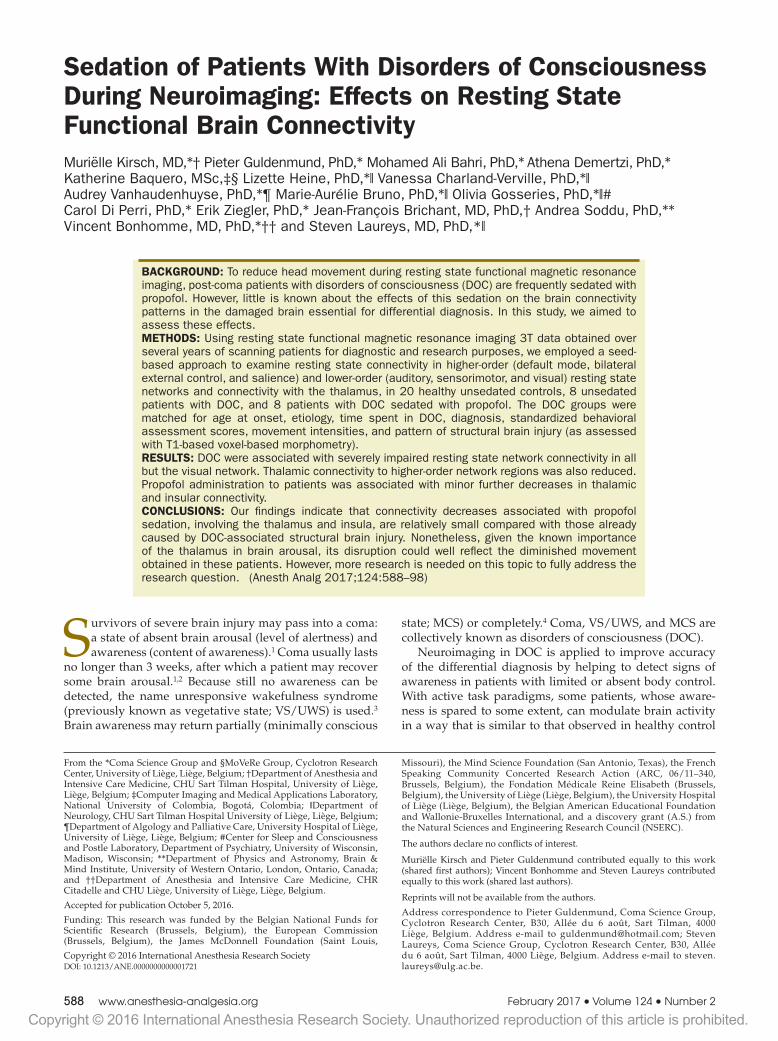
METHODSSubjectsOf an initial resting state fMRI database of 180 patients with DOC (coma, VS/UWS, and MCS) and patients with the locked-in syndrome and those that had recently recovered from the MCS (exit MCS), constructed over several years of scanning patients for diagnostic and research purposes, 126 patients were diagnosed as being in the VS/UWS or MCS. Of these, 36 patients had been scanned while being sedated solely with propofol, and 49 patients had been scanned with-out any sedative. Eventually, resting state fMRI data from only 8 unsedated patients with DOC (mean age = 44 ± 16 years; time in DOC = 695 ± 1169 days, median = 91 days, interquartile range = 23–693 days; 2 traumatic MCS and 6 nontraumatic VS/UWS; mean CRS-R total score = 7 ± 3), and 8 patients with DOC sedated with propofol (mean age = 43 ± 21 years; time in DOC = 867 ± 1426 days, median = 50 days, interquartile range = 25–1086 days; 2 traumatic MCS and 6 nontraumatic VS/UWS; mean CRS-R total score = 7 ± 4) were selected (Table 1). This was a direct result of our
Copyright © 2016 International Anesthesia Research Society. Unauthorized reproduction of this article is prohibited.
Sedation in Disorders of Consciousness
590 www.anesthesia-analgesia.org aNesthesia & aNalgesia
strict patient inclusion criteria for this study. These were: diagnosed as being in the VS/UWS or MCS, as assessed with the CRS-R; scanning occurred more than 2 weeks after initial brain injury in stabilized patients; propofol was used as the sole sedative agent (for the sedated group); an absence of large hemorrhage effects, movement artifacts, foreign body artifacts, midline shifts, acquisition artifacts, low gray–white matter contrast, or exceptionally severe structural brain injury, as assessed by careful visual inspection of the T1 images by an expert. Both DOC groups were matched for age at onset, etiology, time spent in DOC, diagnosis, CRS-R total score, and movement intensities, as assessed both parametrically and nonparametrically. The diagno-sis of MCS or VS/UWS was based on behavioral analysis with the CRS-R, which was repeated several times during a week and performed by trained professionals.37 The CRS-R is a standardized scale that is currently considered to be the most trustworthy behavioral diagnosis tool for patients with DOC available.9,38 The control group consisted of 20 healthy, unsedated control subjects (mean age = 46 ± 18 years). The study was approved by the Ethics Committee of the Medical School of the University of Liège and the IRB. Written informed consent to participate in the study was obtained from the subjects themselves in the case of healthy controls, and from the legal surrogates of the patients.
Sedation ProtocolThe decision to sedate the patient or not was taken by an MRI scanning expert and was based on the severity of patient movement when placed in the MRI scanner. The propofol concentration was kept to a minimum (Table 1). Foam cushion head restraints were placed. Before fMRI data acquisition, all subjects fasted for at least 6 hours for solids, and 2 hours for liquids. During scanning, they wore headphones and earplugs. Propofol sedation was administered through intravenous infusion, using a target-controlled infusion system (Diprifusor, pharmacokinetic model of Marsh et al39, AlarisTM, Alaris Medical Belgium B.V., Strombeek-Bever, Belgium), to obtain constant plasma concentrations. To ensure adequate ventilation, some patients received assisted mechanical ventilation through a tracheostomy or through an endotracheal tube when already in place. Additional oxygen was delivered, either through a facemask or through the airway instrumentation device. Parameters of all patients were closely and continu-ously monitored during the procedure, including arterial blood pressure, electrocardiogram, breathing frequency, and pulse oxymetry (Spo2). All parameters remained sta-ble during data acquisition. When administered, sedation was titrated to achieve immobility in the scanner. Once obtained, the necessary plasma concentration of propofol was kept constant throughout the procedure. Scanning started 5 minutes after having reached the desired clinical state (immobility), hence allowing time for the equilibra-tion of propofol concentrations between pharmacokinetic compartments. For post-hoc confirmation of propofol plasma concentrations, a blood sample was drawn in some patients during the steady-state period of sedation (before and after the sequence). Sedation characteristics are sum-marized in Table 1. Throughout the procedure, a certified Ta
ble
1.
Pat
ient
Cha
ract
eris
tics
Pat
ient
Dia
gnos
is
Acc
ordi
ng t
o C
RS-R
Hig
hest
CR
S-R
To
tal S
core
Etio
logy
Age
at
Ons
et
(Yea
rs)
Tim
e in
DO
C
(Day
s)Ta
rget
Pro
pofo
l C
once
ntra
tion
(μg
/m
L)A
irw
ay D
evic
eVe
ntila
tion
Hea
rt R
ate,
Med
ian
(Ran
ge)
Bre
athi
ng
Freq
uenc
y, m
edia
n (R
ange
)U
1VS
/UW
S3
nT (an
oxia
)44
23
0En
dotr
ache
al t
ube
Con
trol
led
103 (102–1
07)
20
U2
VS/U
WS
5nT
(m
enin
gitis
)54
52
0Tr
ache
osto
my
Spo
ntan
eous
74 (60–1
00)
25 (14–3
3)
U3
VS/U
WS
4nT
(an
oxia
)44
20
0Tr
ache
osto
my
Con
trol
led
144 (140–1
46)
12
U4
VS/U
WS
7nT
(C
VA)
74
22
0Tr
ache
osto
my
Spo
ntan
eous
94 (93–9
5)
12 (10–1
4)
U5
VS/U
WS
4nT
(an
oxia
)48
129
0Tr
ache
osto
my
Spo
ntan
eous
113 (112–1
13)
20 (17–2
1)
U6
VS/U
WS
6nT
(an
oxia
)31
2031
0N
oS
pont
aneo
us70 (67–7
4)
15 (13–1
8)
U7
MC
S12
T26
3034
0N
oS
pont
aneo
us64 (60–7
0)
13 (10–1
4)
U8
MC
S11
T30
247
0N
oS
pont
aneo
us67 (57–7
2)
15 (11–2
0)
Mea
n (S
D)
–7 (3)
–44 (16)
695 (1169)
––
–91 (28)
16 (5)
S1
VS/U
WS
3nT
(an
oxia
)73
47
1.8
Trac
heos
tom
yS
pont
aneo
us91 (89–9
2)
21 (19–2
1)
S2
VS/U
WS
5nT
(hy
pogl
ycem
ia)
53
20
1.0
Endo
trac
heal
tub
eC
ontr
olle
d94 (93–1
00)
16
S3
VS/U
WS
6nT
(an
oxia
)48
52
2.0
Trac
heos
tom
yS
pont
aneo
us92 (91–9
3)
28 (25–2
8)
S4
VS/U
WS
6nT
(C
VA)
69
17
1.5
Endo
trac
heal
tub
eC
ontr
olle
d75 (74–7
5)
14
S5
VS/U
WS
5nT
(an
oxia
)16
27
2.5
Trac
heos
tom
yS
pont
aneo
usM
DM
DS
6VS
/UW
S6
nT (an
oxia
)33
456
1.5
No
Spo
ntan
eous
69 (65–7
2)
16 (11–1
7)
S7
MC
S13
T22
2977
1.8
No
Spo
ntan
eous
59 (56–6
1)
16 (16–1
7)
S8
MC
S13
T30
3342
1.5
No
Spo
ntan
eous
66 (64–6
7)
9 (8–1
0)
Mea
n (S
D)
–7 (4)
–43 (21)
867 (1426)
1.7
(0.4
)–
–78 (14)
17 (6)
Twel
ve p
atie
nts
in V
S/U
WS
(al
l non
trau
mat
ic)
and
4 p
atie
nts
in M
CS
(al
l tra
umat
ic).
Abbr
evia
tions
: C
RS
-R, C
oma
Rec
over
y S
cale
Rev
ised
(ba
sed
on a
uditi
ve, vi
sual
, m
otor
, ve
rbal
, co
mm
unic
atio
n, a
nd a
rous
al t
estin
g35);
CVA
, ce
rebr
ovas
cula
r ac
cide
nt;
DO
C, di
sord
ers
of c
onsc
ious
ness
; M
CS
, m
inim
ally
co
nsci
ous
stat
e; V
S/U
WS
, veg
etat
ive
stat
e/un
resp
onsi
ve w
akef
ulne
ss s
yndr
ome;
T, t
raum
atic
; nT
, non
trau
mat
ic;
MD
, mis
sing
dat
a; U
, uns
edat
ed s
ubje
ct; S
, sed
ated
sub
ject
.
Copyright © 2016 International Anesthesia Research Society. Unauthorized reproduction of this article is prohibited.
February 2017 • Volume 124 • Number 2 www.anesthesia-analgesia.org 591
anesthesiologist and complete resuscitation equipment were present.
Data AcquisitionStructural MRI T1 data (T1-weighted 3-dimensional gradient echo images using 120 slices, repetition time = 2300 milliseconds, echo time = 2.47 milliseconds, voxel size = 1.0 × 1.0 × 1.2 mm3, flip angle = 9°, field of view = 256 × 256 mm2) and resting state fMRI data (Echo planar imaging sequence using 32 slices, repetition time = 2000 milliseconds, echo time = 30 milliseconds, voxel size = 3.0 × 3.0 × 3.0 mm3, flip angle = 78°, field of view = 192 × 192 mm2, 300 volumes) were acquired on a 3T scanner (Siemens, Munich, Germany).
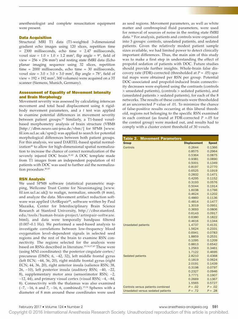
Assessment of Equality of Movement Intensity and Brain MorphologyMovement severity was assessed by calculating interscan movement and total head displacement using 6 rigid-body movement parameters, and a t test was applied to examine potential differences in movement severity between patient groups.23 Similarly, a T1-based voxel-based morphometry analysis of brain structure (VBM8 [http://dbm.neuro.uni-jena.de/vbm/] for SPM8 [www.fil.ion.ucl.ac.uk/spm]) was applied to search for potential morphological differences between both patient groups. For this analysis, we used DARTEL-based spatial normal-ization40 to allow for high-dimensional spatial normaliza-tion to increase the chance of correct normalization of the severely injured DOC brain.41,42 A DOC template made from T1 images from an independent population of 61 patients with DOC was used to further aid the normaliza-tion procedure.40,43
RSN AnalysisWe used SPM8 software (statistical parametric map-ping, Wellcome Trust Centre for Neuroimaging [www.fil.ion.ucl.ac.uk]) to realign, normalize, smooth (8 mm), and analyze the data. Movement artifact reduction soft-ware was applied (ArtRepair44, software written by Paul Mazaika, Center for Interdisciplinary Brain Science Research at Stanford University, http://cibsr.stanford.edu/tools/human-brain-project/artrepair-software.html), and data were temporally bandpass filtered (0.007–0.1 Hz). We performed a seed-based analysis to investigate correlations between low-frequency blood oxygenation level–dependent signals in selected seed regions and the rest of the brain to examine RSN con-nectivity. The regions selected for the analysis were based on RSNs described in literature.10,14,17,45 These were (using MNI coordinates): the posterior cingulate cortex/precuneus (DMN; 6, −42, 32), left middle frontal gyrus (left ECN; −44, 36, 20), right middle frontal gyrus (right ECN; 44, 36, 20), right anterior insula (salience RSN; 38, 26, −10), left posterior insula (auditory RSN; −40, −22, 8), supplementary motor area (sensorimotor RSN; −2, −12, 44), and primary visual cortex (visual RSN; −4, −84, 8). Connectivity with the thalamus was also examined (−7, −16, 6 and 7, −16, 6, combined).13,14 Spheres with a diameter of 8 mm around these coordinates were used
as seed regions. Movement parameters, as well as white matter and cerebrospinal fluid parameters, were used for removal of sources of noise in the resting state fMRI data.14 For analysis, patients and controls were organized into 3 groups: controls, unsedated patients, and sedated patients. Given the relatively modest patient sample sizes available, we had limited power to detect clinically important differences. Thus, the main aim of this study was to make a first step in understanding the effect of propofol sedation of patients with DOC. Future studies should provide further insights. Whole-brain false dis-covery rate (FDR)-corrected (thresholded at P = .05) spa-tial maps were obtained per RSN per group. Potential DOC-associated and propofol-induced brain connectiv-ity decreases were explored using the contrasts (controls > unsedated patients), (controls > sedated patients), and (unsedated patients > sedated patients), for each of the 8 networks. The results of these contrasts were thresholded at an uncorrected P value of .01. To minimize the chance of false-positive results occurring at this liberal thresh-old, regions not belonging to the specific RSN examined in each contrast (as found at FDR-corrected P =.05 for the control group) were masked out, and results had to comply with a cluster extent threshold of 30 voxels.
Table 2. Movement ParametersGroup Displacement SpeedControls 0.2644 0.1366
0.8575 0.29640.5364 0.08750.9381 0.08900.5001 0.13490.8197 0.27700.6525 0.19190.2832 0.14710.4295 0.11520.3193 0.05760.5044 0.19141.6038 0.17660.4624 0.12020.4860 0.11360.4814 0.14771.3019 0.09510.3693 0.08060.6143 0.09170.6380 0.18220.4616 0.1344
Unsedated patients 1.4737 0.41311.5624 0.23310.6941 0.07831.8859 0.25310.1095 0.12090.8813 0.65421.2563 0.34611.7122 0.4695
Sedated patients 2.8210 0.43680.1819 0.06242.0191 0.14390.3198 0.07970.2327 0.09463.7771 0.19070.2669 0.13671.5565 0.5727
Controls versus patients combined P = .02 P = .02Unsedated versus sedated patients P = .72 P = .28
Copyright © 2016 International Anesthesia Research Society. Unauthorized reproduction of this article is prohibited.
Sedation in Disorders of Consciousness
592 www.anesthesia-analgesia.org aNesthesia & aNalgesia
RESULTSAssessment of Equality of Movement Intensity and Brain MorphologyA t test assessed potential differences in movement severity between the groups of unsedated and sedated patients, as well as between controls and the combined group of patients, using speed and displacement parameters.23 No significant difference in movement severity between the patient groups was found, although a potential difference was found between controls and the combined group of patients (displacement: P = .02, speed: P = .02; Table 2).
Differences in gray matter volume (using a threshold of FDR-corrected P < .05) between healthy controls and the combined group of patients with DOC (contrast: controls > patients combined), and between unsedated and sedated patients with DOC (contrasts: unsedated DOC > sedated DOC; sedated DOC > unsedated DOC) were examined. Differences between controls and patients were found to be widespread (P < .001; Table 3).42 No structural differences could be observed between unsedated and sedated patients with DOC.
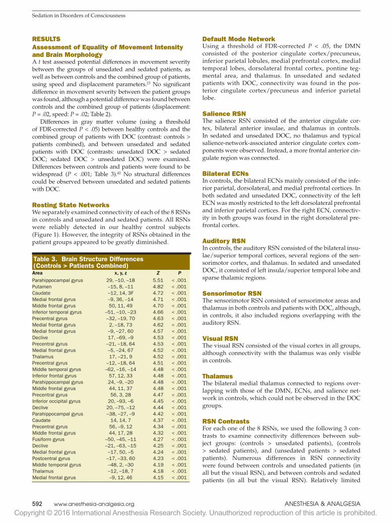
Resting State NetworksWe separately examined connectivity of each of the 8 RSNs in controls and unsedated and sedated patients. All RSNs were reliably detected in our healthy control subjects (Figure 1). However, the integrity of RSNs obtained in the patient groups appeared to be greatly diminished.
Default Mode NetworkUsing a threshold of FDR-corrected P < .05, the DMN consisted of the posterior cingulate cortex/precuneus, inferior parietal lobules, medial prefrontal cortex, medial temporal lobes, dorsolateral frontal cortex, pontine teg-mental area, and thalamus. In unsedated and sedated patients with DOC, connectivity was found in the pos-terior cingulate cortex/precuneus and inferior parietal lobe.
Salience RSNThe salience RSN consisted of the anterior cingulate cor-tex, bilateral anterior insulae, and thalamus in controls. In sedated and unsedated DOC, no thalamus and typical salience-network-associated anterior cingulate cortex com-ponents were observed. Instead, a more frontal anterior cin-gulate region was connected.
Bilateral ECNsIn controls, the bilateral ECNs mainly consisted of the infe-rior parietal, dorsolateral, and medial prefrontal cortices. In both sedated and unsedated DOC, connectivity of the left ECN was mostly restricted to the left dorsolateral prefrontal and inferior parietal cortices. For the right ECN, connectiv-ity in both groups was found in the right dorsolateral pre-frontal cortex.
Auditory RSNIn controls, the auditory RSN consisted of the bilateral insu-lae/superior temporal cortices, several regions of the sen-sorimotor cortex, and thalamus. In sedated and unsedated DOC, it consisted of left insula/superior temporal lobe and sparse thalamic regions.
Sensorimotor RSNThe sensorimotor RSN consisted of sensorimotor areas and thalamus in both controls and patients with DOC, although, in controls, it also included regions overlapping with the auditory RSN.
Visual RSNThe visual RSN consisted of the visual cortex in all groups, although connectivity with the thalamus was only visible in controls.
ThalamusThe bilateral medial thalamus connected to regions over-lapping with those of the DMN, ECNs, and salience net-work in controls, which could not be observed in the DOC groups.
RSN ContrastsFor each one of the 8 RSNs, we used the following 3 con-trasts to examine connectivity differences between sub-ject groups: (controls > unsedated patients), (controls > sedated patients), and (unsedated patients > sedated patients). Numerous differences in RSN connectivity were found between controls and unsedated patients (in all but the visual RSN), and between controls and sedated patients (in all but the visual RSN). Relatively limited
Table 3. Brain Structure Differences (Controls > Patients Combined)Area x, y, z Z P
Parahippocampal gyrus 29, –10, –18 5.51 < .001Putamen –15, 8, –11 4.82 < .001Caudate –12, 14, 3F 4.72 < .001Medial frontal gyrus –9, 36, –14 4.71 < .001Middle frontal gyrus 50, 11, 49 4.70 < .001Inferior temporal gyrus –51, –10, –23 4.66 < .001Precentral gyrus –32, –19, 70 4.63 < .001Medial frontal gyrus 2, –18, 73 4.62 < .001Medial frontal gyrus –9, –27, 60 4.57 < .001Declive 17, –69, –9 4.53 < .001Precentral gyrus –21, –18, 64 4.53 < .001Medial frontal gyrus –5, –24, 67 4.52 < .001Thalamus 17, –21, 9 4.52 < .001Precentral gyrus –12, –18, 64 4.51 < .001Middle temporal gyrus –62, –16, –14 4.48 < .001Inferior frontal gyrus 57, 12, 33 4.48 < .001Parahippocampal gyrus 24, –9, –20 4.48 < .001Middle frontal gyrus 44, 11, 37 4.48 < .001Precentral gyrus 56, 3, 28 4.47 < .001Inferior occipital gyrus 20, –93, –6 4.45 < .001Declive 20, –75, –12 4.44 < .001Parahippocampal gyrus –38, –27, –9 4.42 < .001Caudate 14, 14, 7 4.37 < .001Precentral gyrus 56, –9, 12 4.34 < .001Middle frontal gyrus 44, 17, 28 4.32 < .001Fusiform gyrus –50, –45, –11 4.27 < .001Declive –21, –63, –15 4.25 < .001Medial frontal gyrus –17, 50, –5 4.24 < .001Postcentral gyrus –17, –33, 60 4.23 < .001Middle temporal gyrus –48, 2, –30 4.19 < .001Thalamus –12, –18, 7 4.18 < .001Medial frontal gyrus –9, 12, 46 4.15 < .001
Copyright © 2016 International Anesthesia Research Society. Unauthorized reproduction of this article is prohibited.
February 2017 • Volume 124 • Number 2 www.anesthesia-analgesia.org 593
further connectivity decreases, concerning the thalamus and salience RSN, were found in sedated as compared with unsedated patients (Figure 2).
Contrasts “Controls > Unsedated Patients” and “Controls > Sedated Patients”Unsedated patients with DOC, as compared with con-trols, had less connectivity in the DMN (posterior cingu-late cortex/precuneus, medial prefrontal cortex, bilateral medial temporal lobes, left inferior parietal lobe, and dorsolateral prefrontal cortex), salience RSN (left insula and anterior cingulate cortex), left ECN (bilateral dor-solateral prefrontal cortices and medial frontal cortex), right ECN (left inferior parietal lobe and left dorsolateral prefrontal cortex), auditory RSN (right insula/superior
temporal lobe and sensorimotor cortex), and sensorimo-tor RSN (right sensorimotor cortex and left medial tem-poral lobe). Less connectivity with the thalamus was also found (regions overlapping with the DMN, ECNs, and salience RSN). No differences were found for the visual RSN. Highly comparable results were found for the con-trast (controls > sedated patients).
Contrast “Unsedated Patients > Sedated Patients”Sedated patients with DOC, as compared with unsedated patients with DOC, had less long-range connectivity (con-nectivity with regions distant from the seed region) in the salience network (left anterior insula: x = −24, y = 23, z = −5), and with the thalamus (posterior cingulate cortex: x = −4,
Figure 1. RSN connectivity in healthy con-trols and unsedated and sedated patients with DOC. Results were thresholded at FDR-corrected P < .05. DMN indicates default mode network; DOC, disorders of consciousness; ECN, external control network; FDR, false discovery rate; RSN, resting state network.
Copyright © 2016 International Anesthesia Research Society. Unauthorized reproduction of this article is prohibited.
Sedation in Disorders of Consciousness
594 www.anesthesia-analgesia.org aNesthesia & aNalgesia
y = −31, z = 34; medial prefrontal cortex: x = −8, y = 38, z = 36; left caudate: x = −12, y = 14, z = 13; right caudate: x = 15, y = 11, z = 16; medial prefrontal cortex: x = −8, y = 41, z = 32; and left dorsolateral prefrontal cortex: x = −54, y = 20, z = 13). No differences were found in the other 6 RSNs.
DISCUSSIONIn this study, we aimed to examine the effect of propo-fol on brain RSNs in the damaged brains of patients with DOC. Because these RSNs are used to examine the level of remaining consciousness, any disruption induced by pro-pofol could interfere with this assessment. As expected, we observed decreases in RSN connectivity in patients com-pared with controls. Importantly, minor further thalamic and insular disconnections were found to be associated with propofol sedation.
RSNs in Controls and Unsedated Patients With DOCIn our healthy controls, connectivity in the 7 RSNs we exam-ined was comparable to that found in literature.10,11 We also added a seed analysis using the thalamus as seed region, because previous studies on the effect of propofol in healthy subjects have shown thalamic involvement.13,14 The thala-mus was found to connect most to regions of the higher-order RSNs. This thalamocortical connectivity was lost in DOC. We, furthermore, found strong DOC-associated con-nectivity decreases in all 4 higher-order networks (DMN, bilateral ECNs, salience RSN). Similar disruptions have been reported in previous resting state fMRI studies and link the functioning of cortical higher-order RSNs and their connectivity with the thalamus with the generation of con-sciousness.7,9,19–24 Structural brain injury in patients with
Figure 2. RSN connectivity decreases in patients with and without sedation as com-pared with healthy controls. Unsedated and sedated DOC were also contrasted against each other. Results were masked with RSN regions obtained in the healthy controls using an FDR-corrected threshold of P < .05 (Figure 1), and thresholded at P < .01 (uncor-rected; cluster extent threshold = 30 voxels). DMN indicates default mode network; DOC, disorders of consciousness; ECN, external control network; FDR, false discovery rate; RSN, resting state network.
Copyright © 2016 International Anesthesia Research Society. Unauthorized reproduction of this article is prohibited.
February 2017 • Volume 124 • Number 2 www.anesthesia-analgesia.org 595
DOC was found to be widespread, which is in line with findings from previous post-mortem46–51 and MRI stud-ies.42,52–56 Higher-order RSNs depend heavily on long-range connectivity, and these connections might thus be especially vulnerable to structural brain injury.54,57 Similarly, long-range connectivity decreases were observed in auditory and sensorimotor RSNs and appear to further reflect the associa-tion between reduced brain integration of information and loss of consciousness.1,58
Possible Effects of Propofol in DOCAlthough propofol-induced loss of consciousness in healthy controls has been found to lead to decreases in connectiv-ity in higher-order RSNs, mild propofol sedation is associ-ated with relatively minor connectivity decreases.13,14,16,17 In healthy subjects, the quantity of propofol administered to the patients with DOC in this study would have induced a state of mild sedation.13 However, because little is known about the effect of propofol on RSN connectivity in the dam-aged brain, a direct comparison between the mildly sedated state in healthy controls13,14 and the state resulting from pro-pofol administration to patients with DOC cannot be read-ily made.
Comparing sedated with unsedated patients in DOC, we found reduced long-range connectivity in the salience RSN and with the thalamus to be associated with propofol administration. As such, induction of these disconnections appears to be sufficient to reduce movement in excessively moving patients with DOC. One of the observed thalamic connectivity decreases involved the striatum, a finding previously reported for healthy subjects sedated with pro-pofol.59,60 This underlines the close association between the thalamus and these nuclei61 and could partially explain dif-ferences in thalamocortical connectivity. Striatal modula-tion of thalamocortical connectivity has been shown to be strongly implicated in the regulation of motor control,62 depending on the pivotal role of the thalamus in brain arousal.63 Although it is unclear which are the exact causes of the uncontrolled high-intensity movement observed in a large portion of patients with DOC, some parallels might be drawn between DOC and movement disorders, such as Parkinson's disease, where known disruption of communi-cation between thalamus, striatum, and cortex is also likely to underlie movement abnormalities.64,65
Thalamostriatal and thalamocortical connectivity decreases thus appear to at least partly underlie propofol-induced reduction of patient movement. However, our other findings also hint at a possibly broader affection of brain function and awareness. The thalamus and striatum have been shown to be implicated in the regulation of alert-ness66–68 and switching behaviors.69 We also found propofol-induced decreased connectivity between the thalamus and (dorsal) posterior cingulate cortex, similar to findings from previous propofol studies with healthy controls.13,14,70 This dorsal part of the posterior cingulate cortex, a key hub in the DMN, has been suggested to play a role in orchestrating the switch between internal and external awareness.71,72 In this context, the reduced connectivity of the thalamus with regions of the salience RSN and ECNs, again found in pro-pofol-induced loss of consciousness in healthy subjects,13
is also interesting.7 All these connectivity changes might represent minor alterations in the functioning of higher-order RSNs. Although the disruption of these higher-order networks, and their connectivity with the thalamus, could play a role in loss of patient movement control, it has also been associated with disrupted awareness in general.7,9,13,18 Therefore, propofol application might affect remaining awareness in patients with DOC.1
Although the detected propofol-induced reductions include connectivity in the salience network and with the thalamus, which might be used for neuroimaging-based diagnosis,9,13 the decreases were relatively minor, especially compared with those decreases caused by DOC-associated structural brain injury. Furthermore, considering the fact that we here present group-level results, intersubject vari-ability might very well overshadow the connectivity reduc-tions we here associated with propofol administration. It is, however, interesting to find that long-range thalamo-cortical connectivity might still be affected by propofol in patients with DOC, in which only low-level or absence of consciousness is assumed. Theoretically, such a response to propofol might in the future be used as a biomarker in itself, although there are several ethical problems with this idea. Most importantly, patients will only be sedated when abso-lutely necessary, mostly when the patients move too much to produce analyzable data, given the fact that no unnec-essary potential health risks should be taken. Therefore, no analyzable data set will be available for the unsedated state to compare with. In addition, the found reductions in connectivity appear to be too small to produce a reli-able biomarker at the single-subject level. However, the great reduction of connectivity found between thalamus and regions of higher-order RSNs observed with our con-trast between healthy controls and unsedated patients with DOC, as previously found between thalamus and DMN,19 as well as during propofol-induced anesthesia,13,14 warrants a further examination of this connectivity pattern as a bio-marker of consciousness.
Methodological ConsiderationsA clear limitation in our study is the fact that brain injury differs from one patient to the other, and consequently, to some extent, so will the effect of propofol on brain resting state connectivity. This limitation was a direct consequence of our ethics-based policy of sedating patients only when absolutely necessary. However, great care was taken to try to match groups of sedated and unsedated patients for age, time spent in DOC, etiology, diagnosis, CRS-R total score, head movement severity, and structural brain injury. As such, even with our relatively modest patient group sizes, resulting from our great care taken in matching groups, we feel confident that our analysis gives insight into the effects of propofol on RSN connectivity in patients with DOC. It should be noted that, before sedation, there was a difference in the severity of movement between the patient groups. Sedation of the intensively moving group removed this movement difference. However, the initial movement dif-ference, with its underlying mechanisms, might be a poten-tial factor influencing our results for which we could not adjust. For ethical reasons, no Pco2 values were obtained
Copyright © 2016 International Anesthesia Research Society. Unauthorized reproduction of this article is prohibited.
Sedation in Disorders of Consciousness
596 www.anesthesia-analgesia.org aNesthesia & aNalgesia
during scanning. However, it has been shown that Pco2 lev-els do not appear to change the BOLD response to neuronal activity.73,74 Furthermore, for our analysis, we are inter-ested in correlations rather than specific regional effects and are therefore confident that Pco2 levels do not signif-icantly influence our results.75 Our choice of a seed-voxel approach instead of an independent component analysis was based on its proven robustness, as reflected in our find-ing of RSNs greatly resembling those mentioned in litera-ture.10,11 Although independent component analysis holds promise in the field of resting state fMRI, issues involv-ing the component creation and selection process need to be addressed.20,76,77 Finally, the use of tracheostomy or an endotracheal tube in a number of our patients may possi-bly have had a limited effect on brain state, because these airway devices could induce a certain level of discomfort. To our knowledge, this effect has not been studied in detail, and our modest sample sizes do not allow for an exami-nation. However, although it could potentially increase brain arousal, we expect any influence on our results to be minor, especially knowing that these airway devices were already in place well before our neuroimaging procedure, and knowing that the distribution of the use of these air-way devices was relatively even among our sedated and unsedated patient groups.
CONCLUSIONSIn this study, we examined how propofol sedation might affect RSN connectivity in patients with DOC. We found minor propofol-associated decreases in connectivity, involv-ing thalamostriatal, thalamocortical, and salience network connectivity. This indicates that these patients still have a form of brain connectivity that can be modified by propo-fol, and gives insight into the brain mechanisms underlying uncontrolled patient movement. However, the major differ-ences were found between controls and (un)sedated patients with DOC, which is related to the great extent of structural brain injury in DOC. Given the known negative effects of high-intensity movement during resting state fMRI, which decreases detectability of RSNs, propofol sedation might presently be considered to be a good method to ensure ana-lyzable data in patients with DOC with strong, uncontrolled head and body movement. Future studies should further examine safety aspects associated with this procedure. E
DISCLOSURESName: Muriëlle Kirsch, MD.Contribution: This author helped design the study, conduct the study, collect the data, analyze the data, and prepare the manu-script; this author was responsible for patient care and the propofol administration and monitoring.Name: Pieter Guldenmund, PhD.Contribution: This author helped design the study, collect and ana-lyze the data, and prepare the manuscript.Name: Mohamed Ali Bahri, PhD.Contribution: This author helped analyze the data and prepare the manuscript.Name: Athena Demertzi, PhD.Contribution: This author helped collect the data and prepare the manuscript; this author was responsible for patient care.Name: Katherine Baquero, MSc.Contribution: This author helped analyze the data and prepare the manuscript.
Name: Lizette Heine, PhD.Contribution: This author helped collect the data and prepare the manuscript; this author was responsible for patient care.Name: Vanessa Charland-Verville, PhD.Contribution: This author helped collect the data and prepare the manuscript; this author was responsible for patient care.Name: Audrey Vanhaudenhuyse, PhD.Contribution: This author helped collect the data, prepare the man-uscript, and was responsible for patient care.Name: Marie-Aurélie Bruno, PhD.Contribution: This author helped collect the data and prepare the manuscript; this author was responsible for patient care.Name: Olivia Gosseries, PhD.Contribution: This author helped collect the data and prepare the manuscript; this author was responsible for patient care.Name: Carol Di Perri, PhD.Contribution: This author helped collect the data and prepare the manuscript; this author was responsible for patient care.Name: Erik Ziegler, PhD.Contribution: This author helped analyze the data and prepare the manuscript.Name: Jean-François Brichant, MD, PhD.Contribution: This author helped prepare the manuscript; this author was responsible for the propofol administration and monitoring.Name: Andrea Soddu, PhD.Contribution: This author helped design the study, conduct the study, and prepare the manuscript.Name: Vincent Bonhomme, MD, PhD.Contribution: This author helped design the study, conduct the study, prepare the manuscript, and was responsible for patient care and the propofol administration and monitoring.Name: Steven Laureys, MD, PhD.Contribution: This author helped design the study, conduct the study, and prepare the manuscript.This manuscript was handled by: Gregory J. Crosby, MD.
REFERENCES 1. Laureys S, Owen AM, Schiff ND. Brain function in coma, vege-
tative state, and related disorders. Lancet Neurol. 2004;3:537–546. 2. Bruno MA, Vanhaudenhuyse A, Thibaut A, Moonen G, Laureys
S. From unresponsive wakefulness to minimally conscious PLUS and functional locked-in syndromes: recent advances in our under-standing of disorders of consciousness. J Neurol. 2011;258:1373–1384.
3. Laureys S, Celesia GG, Cohadon F, et al; European Task Force on Disorders of Consciousness. Unresponsive wakefulness syndrome: a new name for the vegetative state or apallic syn-drome. BMC Med. 2010;8:68.
4. Giacino JT, Ashwal S, Childs N, et al. The minimally con-scious state: definition and diagnostic criteria. Neurology. 2002;58:349–353.
5. Monti MM, Vanhaudenhuyse A, Coleman MR, et al. Willful modulation of brain activity in disorders of consciousness. N Engl J Med. 2010;362:579–589.
6. Schnakers C, Perrin F, Schabus M, et al. Detecting conscious-ness in a total locked-in syndrome: an active event-related par-adigm. Neurocase. 2009;15:271–277.
7. Guldenmund P, Vanhaudenhuyse A, Boly M, Laureys S, Soddu A. A default mode of brain function in altered states of con-sciousness. Arch Ital Biol. 2012;150:107–121.
8. Raichle ME, Snyder AZ. A default mode of brain function: a brief history of an evolving idea. Neuroimage. 2007;37:1083–1090.
9. Guldenmund P, Stender J, Heine L, Laureys S. Mindsight: diagnostics in disorders of consciousness. Crit Care Res Pract. 2012;2012:624724.
10. Damoiseaux JS, Rombouts SA, Barkhof F, et al. Consistent rest-ing-state networks across healthy subjects. Proc Natl Acad Sci U S A. 2006;103:13848–13853.
11. Heine L, Soddu A, Gómez F, et al. Resting state networks and consciousness: alterations of multiple resting state network connectivity in physiological, pharmacological, and pathologi-cal consciousness States. Front Psychol. 2012;3:295.
Copyright © 2016 International Anesthesia Research Society. Unauthorized reproduction of this article is prohibited.
February 2017 • Volume 124 • Number 2 www.anesthesia-analgesia.org 597
12. Meindl T, Teipel S, Elmouden R, et al. Test-retest reproducibility of the default-mode network in healthy individuals. Hum Brain Mapp. 2010;31:237–246.
13. Guldenmund P, Demertzi A, Boveroux P, et al. Thalamus, brainstem and salience network connectivity changes during propofol-induced sedation and unconsciousness. Brain Connect. 2013;3:273–285.
14. Boveroux P, Vanhaudenhuyse A, Bruno MA, et al. Breakdown of within- and between-network resting state functional mag-netic resonance imaging connectivity during propofol-induced loss of consciousness. Anesthesiology. 2010;113:1038–1053.
15. Horovitz SG, Braun AR, Carr WS, et al. Decoupling of the brain’s default mode network during deep sleep. Proc Natl Acad Sci U S A. 2009;106:11376–11381.
16. Schrouff J, Perlbarg V, Boly M, et al. Brain functional integra-tion decreases during propofol-induced loss of consciousness. Neuroimage. 2011;57:198–205.
17. Greicius MD, Kiviniemi V, Tervonen O, et al. Persistent default-mode network connectivity during light sedation. Hum Brain Mapp. 2008;29:839–847.
18. Guldenmund P, Gantner IS, Baquero K, et al. Propofol-induced frontal cortex disconnection: a study of resting-state networks, total brain connectivity, and mean BOLD signal oscillation fre-quencies. Brain Connect. 2016;6:225–237.
19. Vanhaudenhuyse A, Noirhomme Q, Tshibanda LJ, et al. Default network connectivity reflects the level of conscious-ness in non-communicative brain-damaged patients. Brain. 2010;133:161–171.
20. Demertzi A, Gómez F, Crone JS, et al. Multiple fMRI system-level baseline connectivity is disrupted in patients with con-sciousness alterations. Cortex. 2014;52:35–46.
21. Crone JS, Soddu A, Höller Y, et al. Altered network properties of the fronto-parietal network and the thalamus in impaired consciousness. Neuroimage Clin. 2014;4:240–248.
22. Huang Z, Dai R, Wu X, et al. The self and its resting state in con-sciousness: an investigation of the vegetative state. Hum Brain Mapp. 2014;35:1997–2008.
23. Soddu A, Vanhaudenhuyse A, Bahri MA, et al. Identifying the default-mode component in spatial IC analyses of patients with disorders of consciousness. Hum Brain Mapp. 2012;33:778–796.
24. Soddu A, Vanhaudenhuyse A, Demertzi A, et al. Resting state activity in patients with disorders of consciousness. Funct Neurol. 2011;26:37–43.
25. Gili T, Saxena N, Diukova A, Murphy K, Hall JE, Wise RG. The thalamus and brainstem act as key hubs in alterations of human brain network connectivity induced by mild propofol sedation. J Neurosci. 2013;33:4024–4031.
26. Power JD, Barnes KA, Snyder AZ, Schlaggar BL, Petersen SE. Spurious but systematic correlations in functional connec-tivity MRI networks arise from subject motion. Neuroimage. 2012;59:2142–2154.
27. Baquero K, Gómez F, Cifuentes C, Guldenmund P, Demertzi A. A multiscale method for a robust detection of the default mode network. Proc SPIE. 2013;8922.
28. Patel AX, Kundu P, Rubinov M, et al. A wavelet method for modeling and despiking motion artifacts from resting-state fMRI time series. Neuroimage. 2014;95:287–304.
29. Spisák T, Jakab A, Kis SA, et al. Voxel-wise motion artifacts in population-level whole-brain connectivity analysis of resting-state FMRI. PLoS One. 2014;9:e104947.
30. Murphy K, Birn RM, Bandettini PA. Resting-state fMRI con-founds and cleanup. Neuroimage. 2013;80:349–359.
31. Stender J, Gosseries O, Bruno MA, et al. Diagnostic precision of PET imaging and functional MRI in disorders of consciousness: a clinical validation study. Lancet. 2014;384:514–522.
32. Cruse D, Thibaut A, Demertzi A, et al. Actigraphy assessments of circadian sleep-wake cycles in the Vegetative and Minimally Conscious States. BMC Med. 2013;11:18.
33. Galanaud D, Naccache L, Puybasset L. Exploring impaired consciousness: the MRI approach. Curr Opin Neurol. 2007;20:627–631.
34. Machata AM, Willschke H, Kabon B, Kettner SC, Marhofer P. Propofol-based sedation regimen for infants and children
undergoing ambulatory magnetic resonance imaging. Br J Anaesth. 2008;101:239–243.
35. Young C, Knudsen N, Hilton A, Reves JG. Sedation in the inten-sive care unit. Crit Care Med. 2000;28:854–866.
36. Giacino J KK, Whyte J. The JFK Coma Recovery Scale-Revised: measurement characteristics and diagnostic utility. Arch Phys Med Rehabil. 2004;85:2020–2029.
37. Giacino JT, Kalmar K, Whyte J. The JFK Coma Recovery Scale-Revised: measurement characteristics and diagnostic utility. Arch Phys Med Rehabil. 2004;85:2020–2029.
38. Seel RT, Sherer M, Whyte J, et al. Assessment scales for dis-orders of consciousness: evidence-based recommendations for clinical practice and research. Arch Phys Med Rehabil. 2010;91:1795–813.
39. Marsh B, White M, Morton N, Kenny GN. Pharmacokinetic model driven infusion of propofol in children. Br J Anaesth. 1991;67:41–48.
40. Ashburner J. A fast diffeomorphic image registration algo-rithm. Neuroimage. 2007;38:95–113.
41. Takahashi R, Ishii K, Miyamoto N, et al. Measurement of gray and white matter atrophy in dementia with Lewy bodies using diffeomorphic anatomic registration through exponentiated lie algebra: a comparison with conven-tional voxel-based morphometry. AJNR Am J Neuroradiol. 2010;31:1873–1878.
42. Guldenmund P, Soddu A, Baquero K, et al. Structural brain injury in patients with disorders of consciousness: A voxel-based morphometry study. Brain Inj. 2016;30:343–352.
43. Peelle JE, Cusack R, Henson RN. Adjusting for global effects in voxel-based morphometry: gray matter decline in normal aging. Neuroimage. 2012;60:1503–1516.
44. Mazaika P, Hoeft F, Glover GH, Reiss AL. Paper presented at the 15th Annual Meeting of the Organization for Human Brain Mapping (June, San Francisco, CA). Methods and software for fMRI analysis for clinical subjects. 2009.
45. Seeley WW, Menon V, Schatzberg AF, et al. Dissociable intrin-sic connectivity networks for salience processing and executive control. J Neurosci. 2007;27:2349–2356.
46. Jellinger KA. Neuropathology of prolonged unresponsive wakefulness syndrome after blunt head injury: review of 100 post-mortem cases. Brain Inj. 2013;27:917–923.
47. Kinney HC, Korein J, Panigrahy A, Dikkes P, Goode R. Neuropathological findings in the brain of Karen Ann Quinlan. The role of the thalamus in the persistent vegetative state. N Engl J Med. 1994;330:1469–1475.
48. Adams JH, Graham DI, Jennett B. The neuropathology of the vegetative state after an acute brain insult. Brain. 2000;123(pt 7):1327–1338.
49. Adams JH, Jennett B, McLellan DR, Murray LS, Graham DI. The neuropathology of the vegetative state after head injury. J Clin Pathol. 1999;52:804–806.
50. Maxwell WL, Pennington K, MacKinnon MA, et al. Differential responses in three thalamic nuclei in moderately disabled, severely disabled and vegetative patients after blunt head injury. Brain. 2004;127:2470–2478.
51. Maxwell WL, MacKinnon MA, Smith DH, McIntosh TK, Graham DI. Thalamic nuclei after human blunt head injury. J Neuropathol Exp Neurol. 2006;65:478–488.
52. Fernández-Espejo D, Junque C, Bernabeu M, Roig-Rovira T, Vendrell P, Mercader JM. Reductions of thalamic volume and regional shape changes in the vegetative and the minimally conscious states. J Neurotrauma. 2010;27:1187–1193.
53. Fernández-Espejo D, Bekinschtein T, Monti MM, et al. Diffusion weighted imaging distinguishes the vegetative state from the minimally conscious state. Neuroimage. 2011;54:103–112.
54. Fernández-Espejo D, Soddu A, Cruse D, et al. A role for the default mode network in the bases of disorders of conscious-ness. Ann Neurol. 2012;72:335–343.
55. Newcombe VF, Williams GB, Scoffings D, et al. Aetiological dif-ferences in neuroanatomy of the vegetative state: insights from diffusion tensor imaging and functional implications. J Neurol Neurosurg Psychiatry. 2010;81:552–561.
56. Juengling FD, Kassubek J, Huppertz HJ, Krause T, Els T. Separating functional and structural damage in persistent
Copyright © 2016 International Anesthesia Research Society. Unauthorized reproduction of this article is prohibited.
Sedation in Disorders of Consciousness
598 www.anesthesia-analgesia.org aNesthesia & aNalgesia
vegetative state using combined voxel-based analysis of 3-D MRI and FDG-PET. J Neurol Sci. 2005;228:179–184.
57. Khalsa S, Mayhew SD, Chechlacz M, Bagary M, Bagshaw AP. The structural and functional connectivity of the posterior cin-gulate cortex: comparison between deterministic and probabi-listic tractography for the investigation of structure-function relationships. Neuroimage. 2014;102(pt 1):118–127.
58. Tagliazucchi E, Chialvo DR, Siniatchkin M, et al. Large-scale signatures of unconsciousness are consistent with a departure from critical dynamics. J R Soc Interface. 2016;13:20151027.
59. Mhuircheartaigh RN, Rosenorn-Lanng D, Wise R, Jbabdi S, Rogers R, Tracey I. Cortical and subcortical connectivity changes during decreasing levels of consciousness in humans: a functional magnetic resonance imaging study using propofol. J Neurosci. 2010;30:9095–9102.
60. Hudetz AG, Liu X, Pillay S. Dynamic repertoire of intrinsic brain states is reduced in propofol-induced unconsciousness. Brain Connect. 2015;5:10–22.
61. Haber SN, Calzavara R. The cortico-basal ganglia inte-grative network: the role of the thalamus. Brain Res Bull. 2009;78:69–74.
62. Jankowski J, Scheef L, Hüppe C, Boecker H. Distinct stria-tal regions for planning and executing novel and automated movement sequences. Neuroimage. 2009;44:1369–1379.
63. Saper CB, Scammell TE, Lu J. Hypothalamic regulation of sleep and circadian rhythms. Nature. 2005;437:1257–1263.
64. Bu LL, Yang K, Xiong WX, et al. Toward precision medicine in Parkinson’s disease. Ann Transl Med. 2016;4:26.
65. Revell MA. Deep brain stimulation for movement disorders. Nurs Clin North Am. 2015;50:691–701.
66. Yeo SS, Chang PH, Jang SH. The ascending reticular activating system from pontine reticular formation to the thalamus in the human brain. Front Hum Neurosci. 2013;7:416.
67. Kundishora AJ, Gummadavelli A, Ma C, et al. Restoring con-scious arousal during focal limbic seizures with deep brain stimulation [published online ahead of print March 3, 2016). Cereb Cortex. doi: 10.1093/cercor/bhw035.
68. Paus T. Functional anatomy of arousal and attention systems in the human brain. Prog Brain Res. 2000;126:65–77.
69. Smith Y, Galvan A, Ellender TJ, et al. The thalamostriatal system in normal and diseased states. Front Syst Neurosci. 2014;8:5.
70. Hudetz AG. General anesthesia and human brain connectivity. Brain Connect. 2012;2:291–302.
71. Leech R, Kamourieh S, Beckmann CF, Sharp DJ. Fractionating the default mode network: distinct contributions of the ventral and dorsal posterior cingulate cortex to cognitive control. J Neurosci. 2011;31:3217–3224.
72. Leech R, Braga R, Sharp DJ. Echoes of the brain within the pos-terior cingulate cortex. J Neurosci. 2012;32:215–222.
73. Birn RM, Diamond JB, Smith MA, Bandettini PA. Separating respiratory-variation-related fluctuations from neuro-nal-activity-related fluctuations in fMRI. Neuroimage. 2006;31:1536–1548.
74. Wise RG, Ide K, Poulin MJ, Tracey I. Resting fluctuations in arterial carbon dioxide induce significant low frequency varia-tions in BOLD signal. Neuroimage. 2004;21:1652–1664.
75. Corfield DR, Murphy K, Josephs O, Adams L, Turner R. Does hypercapnia-induced cerebral vasodilation modulate the hemodynamic response to neural activation? Neuroimage. 2001;13:1207–1211.
76. De Martino F, Gentile F, Esposito F, et al. Classification of fMRI independent components using IC-fingerprints and support vector machine classifiers. Neuroimage. 2007;34:177–194.
77. Margulies DS, Böttger J, Long X, et al. Resting developments: a review of fMRI post-processing methodologies for spontane-ous brain activity. MAGMA. 2010;23:289–307.