Embed Size (px)
Citation preview

Chapter – IV
SECTION – C
STUDIES ON
HEPATOPROTECTIVE
ACTIVITY

Introduction
Carbon tetrachloride as a hepatotoxin
Plant materials in hepatoprotective activity
Pharmacological screening :
ethanolic extract of seeds of Ricinus communis
Pharmacological screening :
chloroform extract of leaves of Ricinus communis
Results and discussions
Pharmacology
91
HEPATOPROTECTIVE ACTIVITY
Introduction
Uncontrolled environmental pollution, poor sanitary conditions,
xenobiotics, alcohol intoxications and the indiscriminate use of potent drugs
predispose the liver to a vast array of disorders. However, infection by virus still
remains as the major cause of liver disease. Global estimates1
indicate that there
are about 18,000 deaths every year due to liver cirrhosis caused by hepatitis.
Hepatocellular carcinoma ranks among the top ten common tumors of the world,
with an average of over 2, 50,000 new cases reported every year.
The liver, weighing between 1200-1500 g is the largest solid organ in the
body. Essentially the liver has four quite distinct functions.
1. It supplies bile salts and bicarbonate to assist in digestion.
2. It acts as a buffer between the gut and the systemic circulation maintaining
stable levels of amino acids and glucose.
3. It synthesizes a large number of specialized proteins, carbohydrates and
lipids.
4. It is a major excretory pathway for the larger and more hydrophobic
metabolites, foreign substances and drugs.
Liver disorders may be classified as hepatitis (Inflammation of the liver),
hepatotosis (non-inflammatory disorders or degeneration of the liver
parenchyma), chronic hepatitis and liver cirrhosis2. However, there is no strict
hepatological delineation of these disorders, making a similar classification of
hepatoprotective agents virtually impossible.

Pharmacology
92
Table – 4
Types of Hepatotoxic Agents
Inorganic agents
Metals and
metalloids:
Antimony, arsenic, beryllium, bismuth, boron, cadmium,
chromium, cobalt, copper, iron, lead, manganese, mercury,
gold, phosphorous, selenium, tellurium, thallium, zinc.
Organic agents
Natural plant
toxins:
Albotocin, cycasin, icterogenin, indospicins, lantana, agaione,
pyrrolizidines, safrole, tannic acid.
Mycotoxins:
Aflatoxin, cyclochlorotine, ethanol, luteoskyrin, chratoxins,
rubratoxins, sterignatocystins, griseofulvin, sporidesmin,
tetracycline and other antibiotics.
Bacterial
toxins:
Exotoxins (C.diphtheriae, Cl.botulinum, Str.hemolyticus),
endotoxins, ethionine.
Synthetic Non-
Medicinal:
Haloalkanes and haloolefins, nitroalkanes chloroaromatic
compounds, nitroaromatic compounds, organic amines, azo
compounds, phenol and derivatives, various other organic
compounds.
Medicinal
agents: Over 100 drugs used for treatment and diagnosis.
The agents listed in this table vary considerably in their potential for
causing hepatic injury.

Pharmacology
93
Table –5
Classification of hepatotoxic agents and major characteristics of each group.
Category of
agent Mechanism
Histological
lesion Example
Intrinsic toxicity
Direct
Direct physico-
chemical distortion
and destruction of
structural basic cell
metabolism.
Necrosis (zonal)
and/or steatosis.
CCl4, CHCl3,
Phosphorous
Indirect
cytotoxic
Interference with
specific metabolic
pathways leading
to structural injury.
Steatosis
or
necrosis
Ethionine, Mycotoxins
Cholestatic
Interference with
hepatic excretory
pathways leading
to cholestasis
Bile casts
Ictirogenin C-17
alklylated anabolic and
contraceptive steroids.
Host idiosyncrasy
Hyper
sensitivity Drug allergy
Necrosis or
cholestasis
Sulphonamides, PAS,
Halothane
Metabolic
abnormality
Production of
hepatic metabolites
Necrosis or
cholestasis
Iproniazid,
Isoniazid,Halothane

Pharmacology
94
Carbon tetrachloride as a hepatotoxin
During the early part of this century, carbon tetrachloride was first found
to produce hepatic injury in man and experimental animals3. The intervening
years have seen thousands of reports regarding adverse effects to human health
due to this particular compound carbon tetrachloride 3, 4, 5
. Poisoning with carbon
tetrachloride has been a well-accepted and widely used model to study the
pathophysiology of inflammation, liver injury or hepatic inflammation4, 5
. In the
course of unraveling the mechanism by which it produces fatty liver, carbon
tetrachloride has served to elucidate the pathogenesis of fatty metamorphosis
induced by other etiological factors4, 5
. While it can lead to a damage of number
of tissues it is particularly responsible for damaging to the liver and kidneys
of animals / human beings 6, 7, 8, 9, 10
. The agent is a potent hepatotoxin. Single
doses lead promptly to centrizonal necrosis and steatosis6, 4, 8, 9
. Within a few
minutes, there will be an injury to the endoplasmic reticulum, which leads to
functional defects of the hepatocyte and multiple biochemical manifestations of
hepatic injury6, 4, 10, 11
.
Carbon tetrachloride12
because of its high lipid solubility is well
distributed in the body, but produces toxic effects that are largely confined to the
liver and kidneys. The toxicity is increased by agents (e.g. phenobarbitone),
which induce microsomal drug metabolizing enzymes and reduced by the
inhibitors of microsomal enzymes. The microsomal mixed function oxidase
system withdraws an electron from CCl4 leaving the reactive trichloromethyl
radical CCl•3. This free radical has life time of only about 100 microseconds and

Pharmacology
95
so has time to diffuse for only a short distance within the liver cell before
undergoing secondary reactions. The secondary reactions, which are responsible
for the biochemical damage may be of various kinds:
a) Oxidation of thiols to disulphide bonds.
b) Saturation of double bonds in lipids, proteins or nucleotides, resulting
covalent attachment of free radical group of those sites.
c) Lipid peroxidation reaction in which polyunsaturated membrane lipids are
converted to peroxide derivative and eventually to aldehydes and other
products leading to a further cascade of reaction, which results in irreversible
membrane damage.
Prolonged administration of carbon tetrachloride can lead to cirrhosis13
and hepatic carcinoma14
. Most of the acute and chronic hepatic injuries appear to
result from the action of metabolite of the toxin4.Chemically carbon tetrachloride
is a simple, strongly non-polar molecule6, which undergoes metabolism in the
smooth endoplasmic reticulum. A chemical characteristic that is the relative low
energy for C-Cl bond which may be highly responsible to brand carbon
tetrachloride as a potential hepatotoxin has been emphasized by Recknagel and
Glende4 and Slater
15. The bond association energy is progressively higher in less
toxic haloalkanes and tends to be lower in more toxic haloalkanes4, 11, 15
.
Plant materials in hepatoprotective activity
Despite tremendous advances in modern medicine, there is hardly any
drug that stimulate liver function, offer protection to the liver from damage or
help the regeneration of the parenchyma cells16
. While corticosteroids are
Pharmacology
96
immunosuppressive agents, the side effects of which are alarming, are the only
drugs of choice in modern medicine for the management of liver ailments. Plants
and natural products are proving to be good hepatoprotectants. This is evident
from the voluminous work published on such materials and their hepatoprotective
activity17
. The importance of plant products in modern medicine even in a highly
advanced society as that of USA can be seen from the data of natural surveys18
,
where in it was found that 25% of all the prescriptions dispensed contained crude
plant materials or crude plant extracts. About 170 phytoconstituents isolated from
around 100 plants belonging to 55 families have been reported to possess liver
protective activity and about 600 commercial herbal formulations with claimed
hepatoprotective activity are being sold world wide. Of these about forty patent
poly herbal formulations, representing various Indian herbs are available in the
Indian market. For centuries, indigenous drugs, either alone or in combination
were have advocated in the traditional systems of medicine especially Ayurveda
for the treatment of liver disorders.
It was the isolation of silymarin19-23
, a flavanolignan from Silybum
marianum an widespread research on hepatoprotective agents all over the world.
Other important antihepatotoxic drug discoveries from plant sources include
cynarin from Cynara scolymus24
. The discovery of diverse chemical compounds
from the natural products and synthetic compounds used in protective liver
therapy such as phospholipids, sugar alcohols, pyrimidine, purine derivatives,
vitamins, cysteine, glutathione, corticoids, androgens, penicillamine, ricinin etc.,
does not confine the activity to any particular class of compounds25
, but

Pharmacology
97
emphasizes once again the complexity of liver disorders in addition to the
different action, mechanisms of different pharmaceutical preparations. However,
it may be noted that many of the anti hepatotoxic compounds mentioned in
literature are phenolic and phenol propane derivatives. Systematic
pharmacological studies are therefore well conceived and justified especially in
the lignans, neolignans, higher condensed flavonoids and cinnamic acid
derivatives.
It may be noted that though hepatoprotective activity was reported for
various classes of phyotoconstituents such as flavonoids, triterpenes, steroids,
lignans, polyphenols, glycosides, saponins, volatile oils, coumarins etc., the
reports on hepatoprotective activity of triterpenes, flavonoids, steroids and
lignans were comparatively more26.
Extensive work was carried out on the antihepatotoxic activity of
flavonoids as a class of compounds. The hepatoprotective activity of
flavonoids like quercetin, luteolin, apigenin, quercitroside etc is well
documented in literature27, 28
. Though their mechanism of action has not
been clearly understood, certain flavonoids like silybin, cyanidanol-3,
quercetin and taxifolin are believed to act as antioxidants and therefore
may be useful in the treatment of liver disorders, where lipid peroxidation
is an important process29, 30
.

Pharmacology
98
PHARMACOLOGICAL SCREENING-
HEPATOPROTECTIVE ACTIVITY
Effect of the ethanolic extract of the seeds of Ricinus communis
Preparation of extract
Fresh seeds of the plant (2 kg) were collected, washed and Sun dried.
They were then crushed to a fine powder. The dried powder (600 g) was
exhaustively extracted with ethanol in batches of 200 g each in the soxhlet
extractor (40 cycles for each batch). The ethanolic extract was collected. The
ethanol was distilled off and the concentrate was evaporated on a water bath to
get a sticky mass (10 g). This residue was used for screening various
pharmacological activities.
Selection of animals
Male Wistar albino rats weighing between 200-250 g were obtained from
M/s Maharvir Enterprises, Hyderabad, Andhra Pradesh, India. The animals were
housed in polypropylene cages in an adequately ventilated room. (At a
temperature of 25± 02 with an alternating 12 hr light – dark cycle and relative
humidity of 50 ±15%), one week before the start and also during the experiment
as per the rules and regulations of the Institutional Animal Ethics committee and
by the regulatory body of the government (Regd.No.1048/a/07/CPCSEA). The
rats were allowed to have standard rat feed pellets supplied by Hindustan Lever
Co., Bombay and water ad libitum throughout the course of the study.

Pharmacology
99
Preparation of Tween 80 (1%)
Stock suspension of Tween 80 was prepared by triturating 1gm of Tween 80
in 100ml of distilled water, was used for suspending the test compound and
standard drug.
Acute Toxicity Study
Animals weighing between 200-250 g were used for the study. These
were divided into 6 groups of 6 animals each. The test extract was administered
orally as a suspension in Tween 80 (3 ml of 1% solution) to the different groups
in increasing dose levels of 10, 40, 100, 400, 1000 and 4000mg/kg body weight.
The animals were then observed continuously for 3 hours for general
behavior, neurological and autonomic profiles for every 30 minutes for next 3
hours and finally till death after 24 hours 31
.
Hepatotoxin
A dose of CCl4 (0.4 ml /kg body weight, i.p.) was used as hepatotoxin to
induce liver damage.
Standard drug
A dose of silymarin (1.25 mg per/kg body weight, p.o) was used as a
standard drug as hepatoprotective agent. It was obtained as a gift sample from
M/S Himalaya Drug Company, Bangalore.
Selection of Dose
From preliminary toxicity studies it was observed that the animals were
safe for maximum dose of 4000 mg/kg body weight. But there were a few
changes in the behavioral responses like alertness, touch response and

Pharmacology
100
restlessness. Therefore 1/10th
of maximum tolerated dose i.e. 400 mg /kg body
weight was chosen for the study.
Hepatoprotective activity
Modified method of Chandra et al32
, was adopted in this study. 24 albino
rats weighing 200–250 g were used for the evaluation of the hepatoprotective
activity of the ethanolic extract of the seeds of Ricinus Communis. These were
divided into 4 groups of 6 animals each.
Group I
The animals served as normal control. These were orally dosed with
Tween 80 (3 ml of 1% solution) daily for seven days. Blood samples were
collected by direct heart puncture on 8th
day to estimate serum enzyme levels of
SGOT, SGPT and alkaline phosphatase. In the process animals will die as a
consequence due to over dose of pentobarbitone. Liver was excised off and kept
in formalin for histopathological studies.
Group II
The animals served as CCl4 control. They were injected intraperitoneally
with CCl4 (0.4ml/kg body weight) followed after 10 minutes by the oral
administration of Tween 80 (3 ml of 1% solution) daily for seven days. Blood
samples were collected on 8th
day to estimate serum enzyme levels of SGOT,
SGPT and alkaline phosphatase. In the process animals will die as a consequence
due to over dose of pentobarbitone. Liver was excised off and kept in formalin
for histopathological studies.
Pharmacology
101
Group III
The animals were treated with CCl4 (0.4 ml/kg body weight, i.p.) for
seven days. Then from the 8th
day to 14th
day silymarin (1.25 mg/kg body weight,
p.o) was administered as a suspension in Tween 80 (3 ml of 1% solution). Blood
samples were withdrawn on the 15th
day by direct heart puncture and their
enzyme levels of SGOT, SGPT and alkaline phosphatase were estimated. In the
process animals will die as a consequence due to over dose of pentobarbitone.
Liver was excised off and kept in formalin for histopathological studies.
Group IV
The animals were treated with CCl4 (0.4 ml/kg body weight i.p.) for seven
days and ethanolic extract of the seeds of Ricinus communis (0.4 g/kg body
weight, orally) as a suspension in Tween 80 (3ml of 1% solution) was
administered from 8th
day to 14th
day. Blood samples were withdrawn on the 15th
day by direct heart puncture. The enzyme levels of SGOT, SGPT and alkaline
phosphatase were estimated. The animals were sacrificed by pentobarbitone
overdose. Liver was excised off and kept in formalin for histopathological
studies.
Biochemical studies
The blood samples were collected from the animals of all 4 groups by
direct cardiac puncture of the anaesthetized animals. The serum was separated by
centrifugation and kept at-15°C; using an autoanalyzer, the following
biochemical parameters were studied.

Pharmacology
102
a) Serum Glutamic Oxaloacetic Transaminase (SGOT)
b) Serum Glutamic Pyruvic Transaminase (SGPT)
c) Alkaline Phosphatase (ALP)
Histopathological studies
The liver of animals of all the four groups were taken and liver sections
were performed by using microtome. The sections were then stained with
haematoxylin and eosin and were observed for degeneration and necrotic changes
which were graded as follows:
a) Degeneration :
O : No degeneration
+ : Few vacuolated cells per lesion
++ : More than 10 cells per lesion
+++ : More than 2 rows of vacuolated cells around necrotic zone per lesion.
b) Necrosis:
O : No necrosis
+ : Focal necrosis of one or two cells per lesion.
++ : Focal necrosis of more than two cells per lesion.
+++ : Massive centrilobular necrosis.
Statistical analysis
Unpaired student„t‟ test was used to analyse the difference in biochemical
parameters between control groups and treated groups.

Pharmacology
103
RESULTS AND DISCUSSION
Hepatoprotective activity
Group I
Biochemical studies
The serum glutamic oxaloacetic transaminase (SGOT) and serum
glutamic pyruvic transaminase (SGPT) levels stood at 44.43±1.02 U/L, and
45.02±1.1 U/L respectively. While the alkaline phosphatase level was 412.1±1.09
U/L (Table-6).
Histopathological studies
The section of liver which was exposed to Tween 80 showed normal
structure of the hepatic cells (Fig-2a).
Group II
Biochemical studies
The animals of this group displayed a significant increase in SGOT (157.5
± 1.43 U/L), SGPT (145 ± 2.1 U/L) and alkaline phosphatase (472.66
±2.63 U/L) respectively, in comparison to those of the normal animals of group I
(Table-6).
Histopathological studies
The section of liver which was exposed to CCl4 showed centrilobular fatty
degeneration, cloudy swelling and necrosis of hepatic cells (Fig-2b).

Fig 2a: Section of the rat liver (normal control).
Fig 2b: Section of the rat liver treated with CCl4 .
Pharmacology
104
Group III
Biochemical studies
When compared to group II, this group exhibited a significant fall in
SGOT (64.3±0.932 U/L), SGPT (62.0±0.57 U/L) and alkaline phosphatase
(423.5±1.04 U/L) (Table-6).
Histopathological studies
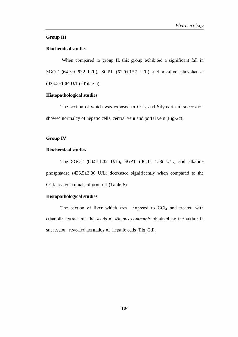
The section of which was exposed to CCl4 and Silymarin in succession
showed normalcy of hepatic cells, central vein and portal vein (Fig-2c).
Group IV
Biochemical studies
The SGOT (83.5±1.32 U/L), SGPT (86.3± 1.06 U/L) and alkaline
phosphatase (426.5±2.30 U/L) decreased significantly when compared to the
CCl4 treated animals of group II (Table-6).
Histopathological studies
The section of liver which was exposed to CCl4 and treated with
ethanolic extract of the seeds of Ricinus communis obtained by the author in
succession revealed normalcy of hepatic cells (Fig -2d).
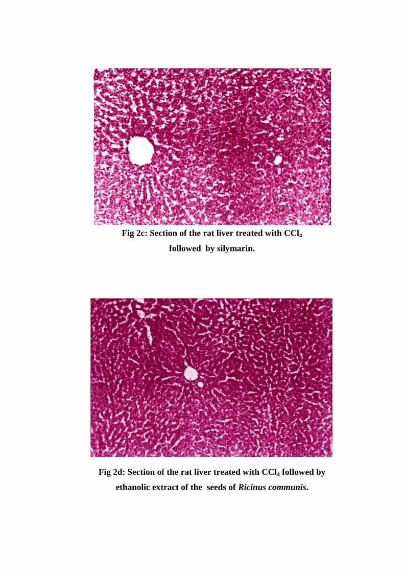
Fig 2c: Section of the rat liver treated with CCl4
followed by silymarin.
Fig 2d: Section of the rat liver treated with CCl4 followed by
ethanolic extract of the seeds of Ricinus communis.

Pharmacology
105
Table - 6
Effect of ethanolic extract of the seeds of Ricinus communis
on CCl4 induced biochemical changes in male albino rats
Groups SGOT
(U/L)
SGPT
(U/L)
ALP
(U/L)
Group I
(Normal) 44.43±1.02 45.02±1.1 412.1±1.09
Group II
(CCl4) 157.5±1.43* 145±2.1* 472.66±2.63*
Group III
(CCl4 +Silymarin) 64.3±0.932* 62.0±0.57* 423.5±1.04*
Group IV
(CCl4 +Ethanolic
extract)
83.5±1.32* 83.6±1.06* 426.5±2.30*
*P < 0.001 compared to control
Values are mean ±SEM, of six animals in each group.
Data was analysed by unpaired„t‟ test.

Pharmacology
106
Fig 3 : Variation in enzyme upper levels for all the treated groups
Fig 4 : Variation in enzyme lower levels for all the treated groups
0
50
100
150
200
250
300
350
400
450
500
Group I (Normal) Group II (CCl4) Group III (CCl4 +Silymarin)
Group IV (CCl4 +Ethanolic extract)
SGOT (U/L) SGPT (U/L) ALP (U/L)
0
0.5
1
1.5
2
2.5
3
Group I (Normal) Group II (CCl4) Group III (CCl4 +Silymarin)
Group IV (CCl4 +Ethanolic extract)
SGOT (U/L) SGPT (U/L) ALP (U/L)
Group I (Normal) Group II (CCl4) Group III (CCl4 + Silyamarin)
Group IV (CCl4 + Ethanolic extract)
Group I (Normal) Group II (CCl4) Group III (CCl4 + Silyamarin)
Group IV (CCl4 + Ethanolic extract)

Pharmacology
107
Histopathological studies
Group Degeneration Necrosis
Normal 0 0
CCl4 +++ +++
CCl4 + Silymarin + +
CCl4 + Ethanolic extract 0 0
No degeneration as well as no indication of necrosis was observed for the
section of the liver treated with Tween 80. The section of liver treated with
carbon tetrachloride showed more than two rows of vacuolated cells around
necrotic zone per lesion and massive centrilobular necrosis. In case of the section
of liver treated with carbon tetrachloride and sylimarin in succession exhibited a
few vacuolated cells per lesion and focal necrosis of one or two cells per lesion.
The section of liver treated with carbon tetrachloride and ethanolic extract of the
seeds of Ricinus communis obtained by the author in succession does not
revealed any degeneration and as well as necrotic changes, which indicates that
the ethanolic extract of the seeds of Ricinus communis has shown fruitful results
in the hepatotoxic protective activities.
Pharmacology
108
Effect of the chloroform extract of the leaves of Ricinus communis
Modified Chandra et al25
method was adopted for this study. The
chloroform extract obtained as per the procedure described in chemical
investigation was taken up for this study. The processes of evaluation of
hepatoprotective activities of seeds of Ricinus communis was carried out with
details of the activities like - acute toxicity study, hepatotoxin, selection of dose,
hepatoprotective activity, biochemical and histopathological studies. Based on
the above studies results and discussions pertaining to the observations were
narrated here under.

Pharmacology
109
RESULTS AND DISCUSSION
Hepatoprotective activity
Group I
Biochemical studies
The Serum Glutamate Oxaloacetate Transaminase (SGOT) and Serum
Glutamate Pyruvate Transaminase (SGPT) values were found to be 89.88±
1.25 U/L and 35.16 ± 0.92 U/L respectively whilst the alkaline phosphatase
value was found to be 100.05±1.125 U/L (Table-7).
Histopathological studies
The liver exposed to Tween 80 revealed the following facts: The black
spots appearing at the central portion represent glycogen where as white spots
indicate the presence of vacuoles. A prominent blood vessel with normal
appearance having fewer vacuoles can also be witnessed from (Fig-5a).
Group II
Biochemical studies
The animals of this group displace significant increase in values of SGOT
(141.4 ± 17.286 U/L), SGPT (77.5±1.79 U/L) and alkaline phosphatase (379.98 ±
48.786 U/L) respectively in comparison to those of the normal animals of Group
I (Table-7).
Histopathological studies
The liver exposed to CCl4 revealed the following facts: The cells were
ruptured, vacuoles are clearly visible covering large area showing necrosis of
liver cells. Number and size of vacuoles are much more than normal. The walls
of blood vein, bile ductioles are in the form of waves and broken at several
places.The epidermis is also swollen and wavy which shows the swelling and
oedema in the liver tissues. Several cells are not nucleated and cytoplasm of a
few cells is dissolved (Fig-5b).
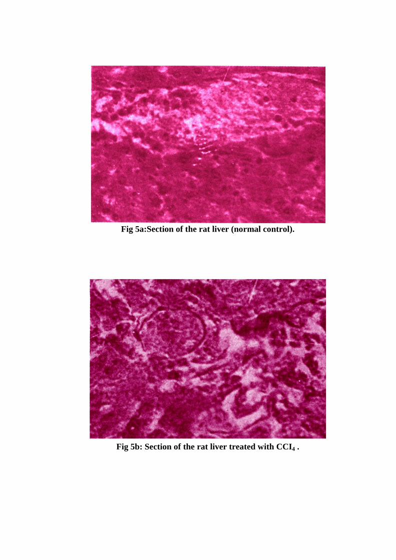
Fig 5a:Section of the rat liver (normal control).
Fig 5b: Section of the rat liver treated with CCI4 .

Pharmacology
110
Group III
Biochemical studies
When compared to group II, this group exhibited a significant fall in the
values of SGOT (119.58±4.86 U/L), SGPT ((53.0±1.450 U/L) and alkaline
phosphatase (290.98±1.26 U/L) (Table-7).
Histopathological studies
Liver exposed to CCl4 and treated with silymarin in succession revealed
normal hepatocytes and mild inflammation (Fig-5c).
Group IV
Biochemical studies
The values of SGOT (126.4±1.34 U/L), SGPT (60.83±0.976 U/L) and
alkaline phosphatase (304.88 ±2.708 U/L) decreased significantly when
compared to the CCl4 treated animals of group II (Table-7).
Histpathological studies
In the case of liver exposed to CCl4 and treated with chloroform extract of
the leaves of Ricinus communis obtained by the author in succession led to the
following observations: vacuoles or necrosis visible but the size and number of
vacuoles are less than that of controls. The walls of blood vessel are slightly
damaged (Fig-5d).
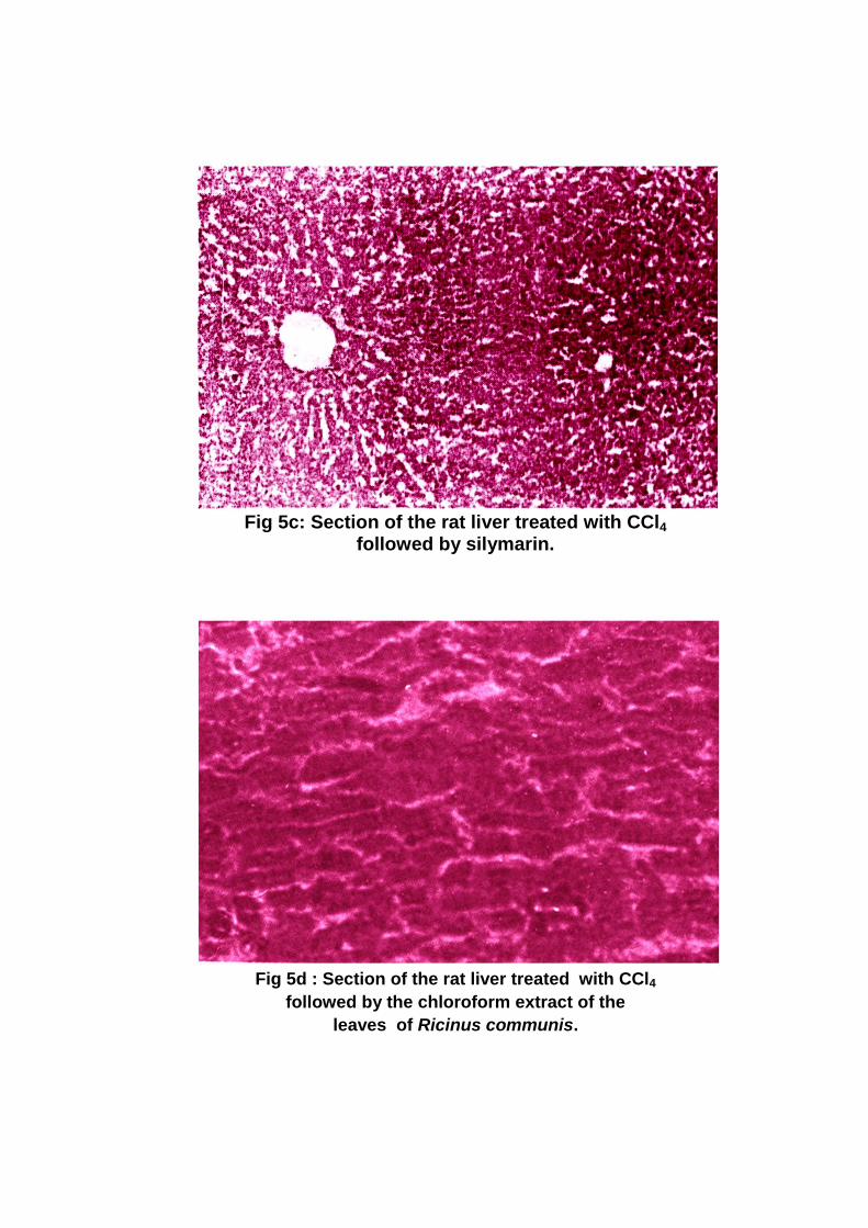
Fig 5c: Section of the rat liver treated with CCl4
followed by silymarin.
Fig 5d : Section of the rat liver treated with CCl4
followed by the chloroform extract of the
leaves of Ricinus communis.

Pharmacology
111
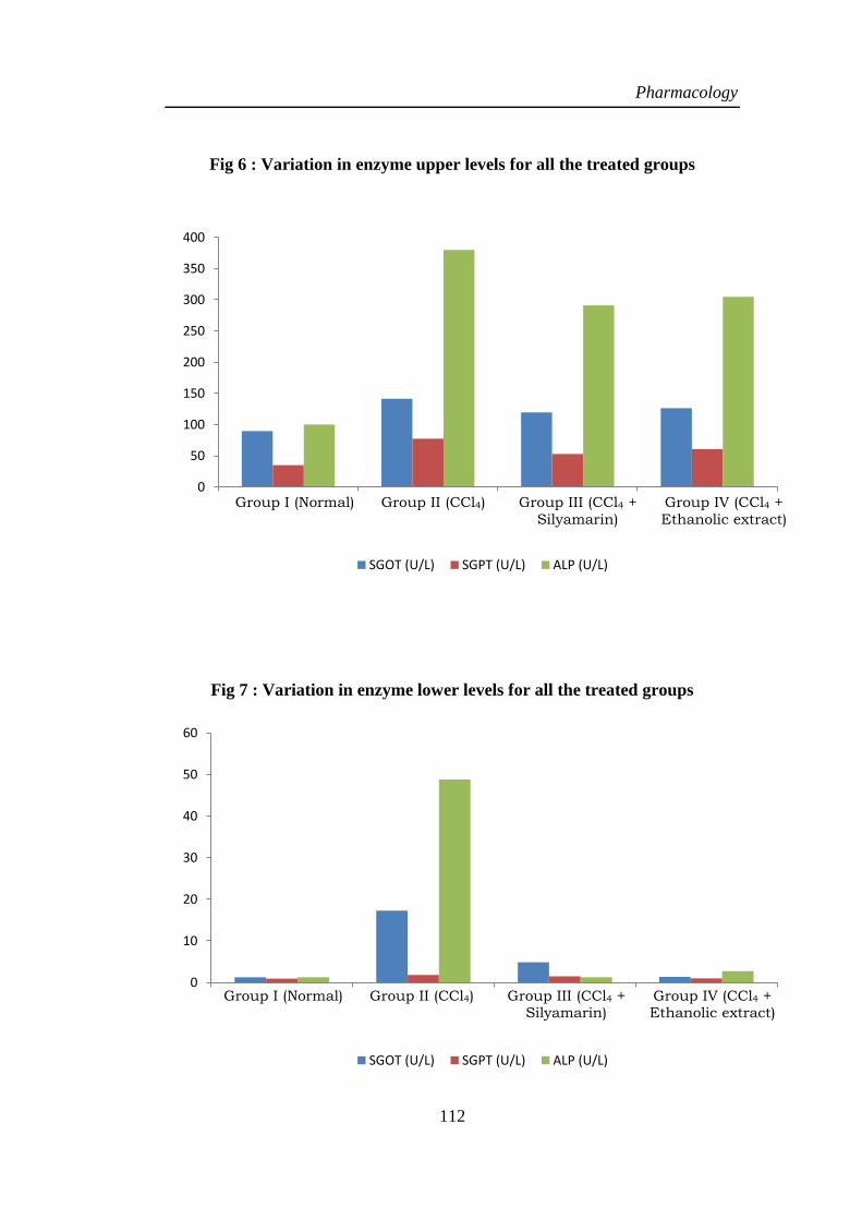
Table –7
Effect of chloroform extract of the leaves of Ricinus communis
on CCl4 induced biochemical changes in male albino rats.
Groups SGOT
(U/L)
SGPT
(U/L)
ALP
(U/L)
Group I (Normal) 89.88±1.256 35.16±0.92 100.05±1.25
Group II (CCl4) 141.4±17.286* 77.5±1.79* 379.98±48.786*
Group III
(CCl4 +Silymarin) 119.58±4.86* 53.01±1.45* 290.98±1.26*
Group IV
(CCl4 +Chloroform
extract)
126.4±1.34* 60.83±0.976* 304.88±2.708*
*P < 0.001 compared to control
Values are mean ±SEM, of six animals in each group.
Data was analysed by unpaired„t‟ test.
Pharmacology
112
Fig 6 : Variation in enzyme upper levels for all the treated groups
Fig 7 : Variation in enzyme lower levels for all the treated groups
0
50
100
150
200
250
300
350
400
Group I (Normal) Group II (CCl4) Group III (CCl4 +Silymarin)
Group IV (CCl4 +Ethanolic extract)
SGOT (U/L) SGPT (U/L) ALP (U/L)
0
10
20
30
40
50
60
Group I (Normal) Group II (CCl4) Group III (CCl4 +Silymarin)
Group IV (CCl4 +Ethanolic extract)
SGOT (U/L) SGPT (U/L) ALP (U/L)
Group I (Normal) Group II (CCl4) Group III (CCl4 +
Silyamarin)
Group IV (CCl4 +
Ethanolic extract)
Group I (Normal) Group II (CCl4) Group III (CCl4 + Silyamarin)
Group IV (CCl4 + Ethanolic extract)

Pharmacology
113
Histopathological studies
Group Degeneration Necrosis
Normal 0 0
CCl4 +++ +++
CCl4 + Silymarin + +
CCl4 + Chloroform extract + +
No degeneration as well as no indication of necrosis was observed for the
section of liver treated with Tween 80. The section of liver treated with carbon
tetrachloride showed more than two rows of vacuolated cells around necrotic
zone per lesion and massive centrilobular necrosis. In case of the section of liver
treated with carbon tetrachloride and sylimarin in succession exhibited a few
vacuolated cells per lesion and focal necrosis of one or two cells per lesion.
However the section of liver treated with carbon tetrachloride and chloroform
extract of the leaves of Ricinus communis obtained by the author in succession
revealed a few vacuolated cells per lesion and focal necrosis of one or two cells
per lesion. The above findings indicate that the ethanolic extract of the seeds of
Ricinus communis is effective in the hepatoprotective activity.