Embed Size (px)
Citation preview
SECTION – I(Course Content)
GROSS ANATOMY
UPPER LIMB
Schedule IPECTORAL REGION AND AXILLA
Lecture: 03 hrsDissection/ Prosection: 10 hrs
Tutorials: 01 hr
LECTURE TOPICS: Mammary gland. Axillary vessels; axillary lymph nodes; lymphatic drainage of the breast. Brachial plexus.
DISSECTION/ PROSECTION:
Identification of relevant skeletal features: thoracic cage sternum; costal cartilages; ribs and thoracic vertebrae. sternum manubrium; body; xiphoid process; jugular (suprasternal
notch; sternal angle (angle of Louis). first rib surfaces; borders; ends. clavicle medial end; shaft; lateral end. scapula surfaces; borders; processes spine, acromion, coracoid. humerus head; greater and lessor tubercles; intertubercular groove;
surgical neck. Subcutaneous structures:
Mammary gland (should include skin also); supraclavicular nerves; anterior andlateral branches of intercostal nerves and accompanying arteries; veins.Deep fascia: pectoral; clavipectoral and axillary.Muscles: pectoralis major; obliqus externus abdominis; serratus anterior;pectoralis minor; subclavius; subscapularis; teres major; latissimus dorsi;coracobrachialis; short head of biceps; long head of triceps; deltoid. Boundaries of axilla:Contents of axilla: Nerves roots; trunks; divisions; cords and branches of brachial plexus. Arteries: axillary artery and its branches. Veins: axillary veins and its tributaries Lymph nodes: axillary lymph nodes. Surface anatomy: axillary artery. Applied anatomy: injuries to the brachial plexus; lymphatic drainage of
the breast.
TUTORIAL TOPICS FOR THE WEEK Relevant osteology. Relevant radiological anatomy. Relevant living anatomy. Relevant crosssectional anatomy.
Schedule 2FRONT OF ARM AND CUBITAL FOSSA.
Lecture: 01 hrsDissection/ Prosection: 10 hrs
Tutorials: 01 hr LECTURE TOPICS: 1. Brachial artery. DISSECTION/ PROSECTION: Identification of relevant skeletal features:humerus: deltoid tuberosity; supracondylar ridge; epicondyles.radius: head; radial tuberosity.ulna: coronoid process. Subcutaneous structures:medial cutaneous nerve of arm and forearm; upper and lower cutaneous nerves of the arm; lateral and posterior cutaneous nerves of the forearm;cephalic, basilic and median cubital veins; epitrochlear lymph nodes. Deep fascia: medial and lateral intermuscular septa; (flexor and extensor compartments)
Muscles: biceps brachii; brachialis; coracobrachialis; pronator teres;brachioradialis. Nerves:axillary, musculocutaneous; ulnar; median; radial. Veins: venae commitantes of brachial artery. Surface anatomy: brachial artery. Applied anatomy: suitability of antecubital veins for intravenoius injections and taking blood for analysis and for transfusion; supracondylarfracture and complications.
TUTORIAL TOPICS FOR THE WEEK Relevant osteology. Relevant radiological anatomy. Relevant living anatomy. Relevant crosssectional anatomy.
Schedule 3.
SUPERFICIAL DISSSECTION OF THE BACK OF THE TRUNK, SCAPULAR REGION AND BACK OF THE ARM.Lecture: 01 hrs
Dissection/ Prosection: 10 hrsTutorials: 01 hr
LECTURE TOPICS: 1. Radial nerve.
DISSECTION/ PROSECTION:Identification of relevant skeletal features:skull mastoid process; superior nuchal line; external occipital
protruberance and crest. vertebral column spines of the vertebrae; vertebra prominence C7 (or T1); sacrum; coccyx. hip bone iliac crest; supracristal plane at the level of L4 spine;
posterior iliac spine at the level of S2 spine. scapula medial, superior and lateral (axillary) borders; scapular notch; spine of
scapula; supra and infraspinous fossae; spinoglenoidnotch; glenoidcavity; infraglenoid tubercle; superior angle at the level of T2 spine;spine of the scapula at the level of T3 spine; inferior angle at thelevel of T7 spine;
humerus greater and lesser tubercles; deltoid tuberosity; radial groove. ulna olecranon process.
.Subcutaneous structures:
Cutaneous branches of the dorsal rami;posterior cutaneous nerve of the arm.Deep fascia: thoracolumbar fascia.Ligaments:ligamentum nuchae; supraspinous ligaments; coracoacromial lgament; superior transverse acromial ligament.Muscles:trapezius; lattisimus dorsi; levator scapulae; rhomidoideus major and minor; deltoid; supraspinatus; infraspinatus; teres major and minor;inferior belly of omohyoid; subscapularis; serratus anterior; triceps Boundaries of quadrangular and triangular spaces; triangle of auscultation and lumbar triangles.Nerves:accessory; suprascapular; axillaruy; other nerves supplying muscles.Surface anatomy: axillary nerve; radial nerve.Applied anatomy: fracture of the neck of the humerus; fracture of themiddle of the shaft of the humerus.
TUTORIAL TOPICS FOR THE WEEK Relevant osteology. Relevant radiological anatomy. Relevant living anatomy. Relevant crosssectional anatomy.
Schedule4.JOINTS OF THE SHOULDER GIRDLE; BACK OF FOREARM AND HAND.
Lecture: 02 hrs
Dissection/ Prosection: 10 hrsTutorials: 01 hr
LECTURE TOPICS: Shoulder girdle
Elbow jointDISSECTION/ PROSECTION:Sternoclavicular jointIdentification of relevant skeletal features:manubrium; medial end of the clavicle;first costal cartilage.Muscles in relation to the joint: pectoralis major; sternomastoid; subclavius.Capsule: attachments.Ligaments: anterior and posterior sternoclavicular; interclavicular; costoclavicular.Synovial membrane: reflection.Intraarticular structures: articular disc.Articular surface: size of sternal and clavicular articular surfaces.Movements: gliding; rotation.Nerve supply: medial supraclavicular; nerve to subclavius.
Acromioclavicular jointIdentification of relevant skeletal features: lateral end of clavicle; acromion processs of scapula.Muscles in relation to the capsule of joint: trapezius; deltoid.Capsule: attachments.Ligaments: coracoclavicular.Synovial membrane: reflection.Intraarticular structures: articular disc sometime present.Articular surfaces: shape.Movements: gliding; rotation.Nerve supply: suprascapular; lateral pectoral.Applied anatomy: dislocation.Shoulder jointIdentification of relevant skeletal features: glenoid cavity; head of humerus.Muscles in relation to the joint: deltoid; rotator cuff muscles; long head of biceps; long head of triceps.Capsule: attachments.Ligaments: coracoacromial; coracohumeral; glenohumeralIntracapsular structures: tendon of long head of biceps.Synovial membrane: reflection.Intraarticular structures: articular disc.Articular surface: humeral and glenoidal articular surfaces; labrum glenoidale.Movements: flexion; extension; abduction; adduction; medial and lateral rotation; circumduction.Nerve supply: suprascapular; axillary and lateral pectoral.Applied anatomy: dislocation.Back of forearm and handIdentification of relevant Skeletal features: radius posterior surface; dorsal tubercle; styloid process; ulna supinator crest; posterior surface;head; styloid process; metacarpals; phalanges.Subcutaneous structures: posterior cutaneous nerve of forearm; superficial branch of radial nerve; dorsal branch of ulnar nerve; dorsal venousarch; basilic and cephalic veins.Deep Fascia: extensor retinaculum; (osteofascial compartments).Muscles: brachioradialis; extensor carpi radialis longus and brevis; extensor digitorum; extensor digiti minimi; extensor carpi ulnaris; supinator;abductor pollicis longus; extensor pollicis longus; extensor indicis.Anatomical snuff boxNerves: deep branch of radial posterior interossoeus.Arteries: posterior interosseous; dorsal carpal arch and branches.Applied anatomy: radial nerve palsy; fracture of lower end of radius (Colle's fracture).
TUTORIAL TOPICS FOR THE WEEK Relevant osteology. Relevant radiological anatomy. Relevant living anatomy. Relevant crosssectional anatomy.
Schedule5.FRONT OF FOREARM AND HAND
Lecture: 03 hrs
Dissection/ Prosection: 10 hrsTutorials: 01 hr
LECTURES:
Ulnar nerve, median nerve, radial nerve. Wrist joint. Palmar spaces
DISSECTION/ PROSECTION:Identification of relevant skeletal features:humerus medial epicondyle; medial supracondylar ridge.radius surfaces; borders; styloid process.ulna surfaces; borders; styloid process.carpus hook of the hamate; tubercle of the scaphoid; pisiform;
tubercle and groove of trapezium; metacarpus; phalanges.
Subcutaneous structures: medial cutaneous nerve of the forearm; lateral cutaneous nerve of forearm; palmar cutaneous branch of the ulnar nerve;palmar cutaneous branch of the median nerve; digital nerves and vessels; cephalic, basilic and median cubital veins.Deep fascia: flexor retinaculum; palmar aponeurosis; fascial septa of the hand.Ligaments: superficial ands deep transverse metacarpal ligaments.Muscles: flexor carpi ulnaris; palmaris longus; flexor carpi radialis; pronator teres; flexor digitorum superficialis; flexor digitorum profundus; flexorpollicis longus; pronator quadratus; thenar and hypothenar muscles; lumbricals; adductor pollicis; interossei.Synovial sheaths of long flexor tendons.Nerves: median, ulnar, superficial radial.Arteries: radial and ulnar arteries and their branches; superficial and deep palmar arches.Surface anatomy: radial and ulnar arteries; median nerve; superficial and deep palmar arches.Applied anatomy: Volkman's ischaemic contracture; Duptyren's contracture; claw hand; fascial spaces of hand.
TUTORIAL TOPICS FOR THE WEEK Relevant osteology. Relevant radiological anatomy. Relevant living anatomy. Relevant crosssectional anatomy.
Schedule6.ELBOW, RADIOULNAR, WRIST AND JOINTS OF THE HAND.
Lecture: 04 hrs
Dissection/ Prosection: 10 hrsTutorials: 01 hr
LECTURES: Radioulnar joints (supination and pronation); articulated hand. Venous drainage and lymphatic drainage of the upper limb. Movements of the thumb. Sectional anatomy of arm, forearm and hand.
PRACTICALS AND TUTORIALS:
Elbow joint
Identification of relevant skeletal features: humerus trochlea;capitulum; radial, coronoid and olecranon fossae; ulna trochlear notch; coronoidand olecranon processes; radius head; neck; tuberosity.Muscles in relation to the capsule of the joint: brachialis; biceps; triceps; anconeus.Capsule: attachment.Ligaments: ulnar collateral; radial collateral; quadrate.Synovial membrane: reflection.Articular surfaces: shape; carrying angle.Movements: flexion; extension.Nerve supply: musculocutaneous; radial.Blood supply: anastamosis around elbow joint.Applied anatomy: dislocations; fractures. Proximal and distal radioulnar joints Identification of relevant skeletal features: radius head; ulnar notch; ulnaradial notch; head.Capsule: attachments.Ligaments: annular (proximal joint).Intraarticular structures: articular disc (distal joint).Synovial membrane: reflection.Movements: pronation; supination. Middle radioulnar joint Ligaments: oblique cord; interosseous membrane. Wrist joint Identification of relevant skeletal features: distal end of radius; articular disc; scaphoid; lunate; triquetrum.Capsule: attachments.Ligaments:palmar radiocarpal; palmar ulnar carpal; dorsal radiocarpal; radial and ulnar collateral.Synovial membrane: reflection.Articular surfaces: shapeMovements: flexion, extension; adduction; abduction; circumduction.
Intercarpal, Midcarpal, Carpometacarpal, Metacarpophalangeal and Interphalangeal joints.
Identification of relevant skeletal features: carpus; metacarpus; phalanges.Capsule: attachmentsLigaments: dorsal and palmar; collateral; interosseous.Synovial membrane: reflection.
Movements: flexion, extension (all joints); adduction, abduction (midcarpal, metacarpophalangealjoints and carpometacarpal joint of the thumb);rotation and circumduction (carpometacarpal joint of the thumb).
TUTORIAL TOPICS FOR THE WEEK Relevant osteology. Relevant radiological anatomy. Relevant living anatomy. Relevant crosssectional anatomy.
SECTION – II(Course Content under Level – I, II, III)
OUTLINE OF LECTURES
S.No TOPIC MUST KNOW SHOULD KNOW COULD KNOW
1. MAMMARY GLAND 1. Architecture of gland 3. Blood supply4. Nerve supply5. Lymphatics & Ca. Breast
2. Relations 6. Metastasis of Ca
Peau De Orange Krukenberg's
Tumour Prognosis
7. Milk line & anomalies
2. AXILLA 1. Boundaries 3. Contents with special emphasis toaxillary lymph nodes
2. Clavipectoral fascia
4. Palpation of axillarylymph nodes
3. AXILLARY ARTERY 1. Course3. Relations4. Branches
2. Axillary sheath5. Anastamosis around scapula
4. BRACHIAL PLEXUS
1. Formation, components & extent 2. Branches3. Relations of cords & branches 4. Applied: Erb's paralysis Klumpke's paralysis
5. Prefixation & Brachial plexus6. Cervical rib syndrome7. Level of injury from trunks tobranches and associated clinicalpicture
5. SHOULDER JOINT
1. Classification2. Interior of the joint3. Capsules & Ligaments4. Synovial membrane5. Movements & group of muscles6. Nerve supply7. Overhead abduction8. Rotator cuff9. Secondary socket10. a. Relations b. Bursae c. Surgical approaches anatomical basis
11.Dislocations
12. Painful arc syndrome13. Frozen shoulder
6. RADIAL NERVE 1. Root value2. Course3. Relations4. Motor distribution 6. Applied :
Crutch paralysis Saturday night palsy Wrist drop
5. Articular branches & dermatomaldistribution
7. # shaft of Radius /Humerus8. Compression of radial

nerve in plaster cast.
7. ARM & BRACHIALARTERY
1. Course2. Major branches 5. Palpation of brachial artery
3. Compartments & relationship
of vessels & nerve with r/o T.S4. Anastamosis around elbow joint
6. Brachiofemoral delay7. Supracondylar spur8. Application of tourniquetin stopping bleeding.
8.
FRONT OF FOREARM &RADIAL ARTERY
CUBITAL FOSSA
1. Group of muscles in forearm 2. Boundaries & contents of cubital
fossa3. Radial artery Course in forearm snuff box palm4. Branches of Radial artery 5. Applied anatomy
IV injections B.P measurement Palpation of brachial & radial
artery
6. Volkmann's Ischaemiccontracture
7. Use of radial artery incoronary bypass surgery8. Use of radial artery in skinflaps
9.
HAND
1. Cutaneous innervation2. Intrinsic muscles & palmar spaces3. Palmar arterial arches4. Flexor retinaculum
5. Dorsal digital expansion6. Movements of thumb joints 7. Evolution of thumb / functions of
hand and Grip 9. Dupuytren's contracture
8. Clinical considerations of
palmar spaces
10.
ULNAR & MEDIAN NERVES
1. Course & relations2. Motor distribution in forearm & palm3. Palpation of ulnar nerve4. Flexor retinaculum
5. Carpal tunnel syndrome6. Ape thumb deformity7. Ulnar claw hand8. True claw hand
11.
ELBOW JOINT
1. Classification2. Capsules & ligaments3. Synovial membrane 5. Movements & group of muscles
4. Relations 5. Applied:
Tennis elbow Students elbow
Subluxation of head of radiusTennis elbowCubitus valgusPulled elbowCarrying angle
12.
RADIO ULNAR JOINTS
1. Classification2. Capsules & ligaments3. Synovial membrane4. Movements & group of muscles5. Interosseous membrane
6. Weight transmission7. Colle's fracture
8. Axis of supnation andpronation
9. Changing axes duringsupination and pronation
SECTION – II(Course Content under Level – I, II, III)
DISSECTION INCISIONS
DISSECTION
Learning Objectives of Dissection
S.No
TOPIC
DISSECTION
STEPS
WHAT IS EXPECTED FROM THE STUDENTS
SUMMARY
LEVEL 1 LEVEL 2 LEVEL 3 IDENTIFY UNDERSTAND 1. PECTORAL
REGION &MAMMARYGLAND
Incision nos. 14 Reflect theskin flaps laterally leavingthe
nipple & the surroundingskin in position Divide the deepfascia in theDeltopectoral
groove Remove thefascia from theant.part of the
Pect .Major &Deltoid &definetheir attachments Detach theclavicular &sternal heads of P.Major & reflect
Mammarygland
Pectoralismajor
Cephalic
vein PectoralisMinor
Subclavius
Anteriorcutaneous .branches. ofIntercostalnerves.
SupraclavicularNerve.
Latl. Cutaneous. Branches ofIntercostalNerve
Muscles of thepectoralregion
Mammarygland
Cephalic vein
Actions ofpectoral ms.
Blood supply &lymphaticdrainage ofmammary gland& its appliedanatomy
APPLIED ASPECT
Developmental anamolies Gynaecomastia Cancer breast
it towards itsinsertion &
identify: While reflectingthe PectoralisMajor, identify:
Medial pectoralN. (piercing theP. minor&supplying theP. major)
S.No
TOPIC
DISSECTIONSTEPS
WHAT IS EXPECTED FROM THE STUDENTS
SUMMARY
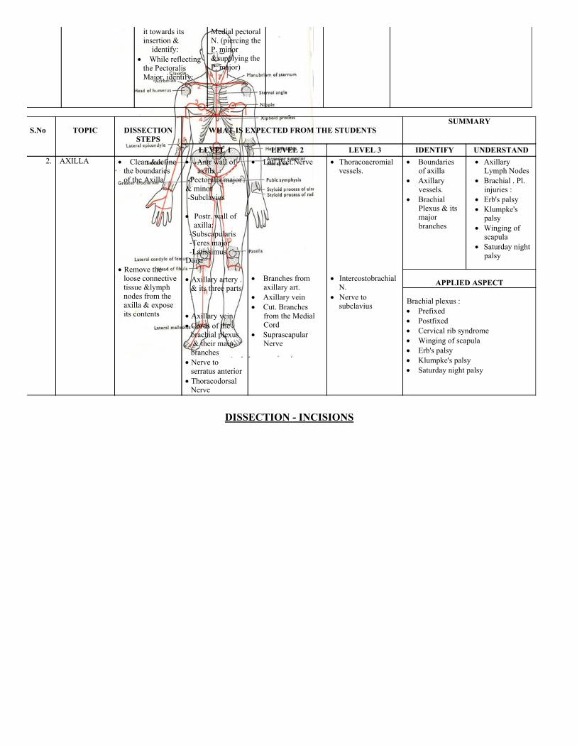
LEVEL 1 LEVEL 2 LEVEL 3 IDENTIFY UNDERSTAND 2. AXILLA Clean &define
the boundariesof the Axilla
Remove theloose connectivetissue &lymphnodes from theaxilla & exposeits contents
Antr wall ofaxilla
Pectoralis major& minor Subclavius Postr. wall ofaxilla:
Subscapularis Teres major LatissimusDorsi Axillary artery .& its three parts:
Axillary vein Cords of thebrachial plexus.& their mainbranches
Nerve toserratus anterior
ThoracodorsalNerve
Latl.Pect.Nerve Branches from
axillary art. Axillary vein Cut. Branches
from the MedialCord
SuprascapularNerve
Thoracoacromialvessels.
IntercostobrachialN.
Nerve tosubclavius
Boundariesof axilla
Axillaryvessels.
BrachialPlexus & itsmajorbranches
AxillaryLymph Nodes
Brachial . Pl.injuries :
Erb's palsy Klumpke'spalsy
Winging ofscapula
Saturday nightpalsy
APPLIED ASPECT Brachial plexus : Prefixed Postfixed Cervical rib syndrome Winging of scapula Erb's palsy Klumpke's palsy Saturday night palsy
DISSECTION INCISIONS
DISSECTION
S.No
TOPIC
DISSECTION
STEPS
WHAT IS EXPECTED FROM THE STUDENTS
SUMMARY
LEVEL 1 LEVEL 2 LEVEL 3 IDENTIFY UNDERSTAND 3. DISSECTION OF
BACK Incision 1,3,4& 5
Reflect skinflaps laterally
Strip sup.Fascia from thedeep fascia
Remove thefascia from thesurface of theTrapezius &
Trapezius Deltoid
Cutaneous
Nerves ofthe back(dorsal ramiof spinalnerves)
Muscles of theback
Triangle ofauscultation
Arrangement ofthese muscles.
Action of thesemuscles.
Movements ofscapula & ms.causing them.
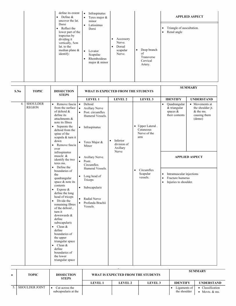
define its extent Define &uncover the lat.Dorsi
Reflect thelower part of thetrapezius bydividing itvertically, 5cmlat. to themedian plane &identify:
Infraspinatus Teres major &
minor Latissimus
Dorsi Levator
Scapulae Rhomboideus
major & minor
Accessory
Nerve. Dorsal
scapularNerve.
Deep branch
ofTransverse CervicalArtery.
APPLIED ASPECT
Triangle of auscultation. Renal angle
S.No
TOPIC
DISSECTIONSTEPS
WHAT IS EXPECTED FROM THE STUDENTS
SUMMARY
LEVEL 1 LEVEL 2 LEVEL 3 IDENTIFY UNDERSTAND 4. SHOULDER
REGION Remove fasciafrom the surfaceof deltoid &define itsattachments ¬e its fibres
Separate thedeltoid from thespine of thescapula & turn itdown
Remove fasciaoverinfraspinatusmuscle &identify the twoteres ms.
Define theboundaries ofthequadrangularspace & note itscontents
Expose &define the longhead of triceps
Divide theremaining fibresof the deltoid ,turn itdownwards &definesubscapularis
Clean &defineboundaries ofthe uppertriangular space
Clean &defineboundaries ofthe lowertriangular space
Deltoid Axillary Nerve Post. circumflex
Humeral Vessels. Infraspinatus Teres Major &
Minor Axillary Nerve. Postr.
CircumflexHumeral Vessels.
Long head of
Triceps Subscapularis Radial Nerve Profunda Brachii
Vessels.
Inferior
division ofAxillaryNerve
Upper Lateral .CutaneousNerve of thearm
Circumflex
Scapularvessels.
Quadrangular& triangularspaces &their contents
Movements atthe shoulder jt.& the ms.causing them(demo)
APPLIED ASPECT
Intramuscular injections Fracture humerus Injuries to shoulder.
S.No
TOPIC
DISSECTIONSTEPS
WHAT IS EXPECTED FROM THE STUDENTS
SUMMARY
LEVEL 1 LEVEL 2 LEVEL 3 IDENTIFY UNDERSTAND 5. SHOULDER JOINT Cut across the
subcapsularis at the
Ligaments ofthe shoulder
Classification Movts. & ms.
neck of the scapula& reflect it down
Expose & cleanthecoracoclavicularlig.
Medial tocoracoclavicularlig. identify:
Give a vertical
incision throughthe capsule of thejoint
Rotate the armmedially.Disarticulate thehead of thehumerus throughthe cut in thecapsule & identify
Coracoclavicularlig.
Articular
capsule ofshoulder joint
Intracapsular
tendon of longhead of biceps
Glenoid labrum
Suprascapularvessels &Nerve
Two parts of thecoracoclavicularlig.
Conoid part trapezoid Glenohumeral
lig. Trans humeral
ligament Coracohumeral
ligament
joint
causing them Overheadabduction
Frozen shoulder
APPLIED ASPECT
Injuries shoulder joint Weight /force transmission
S.No
TOPIC
DISSECTIONSTEPS
WHAT IS EXPECTED FROM THE STUDENTS
SUMMARY
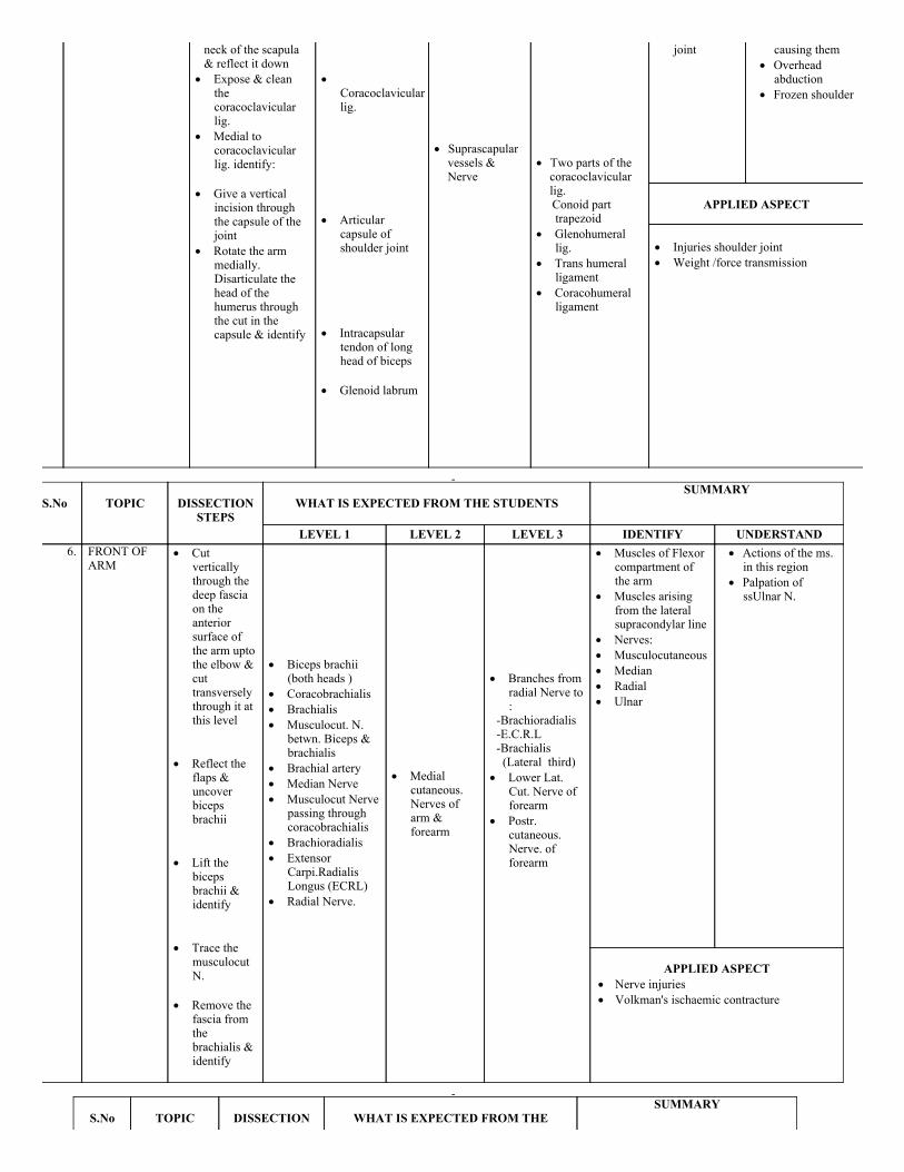
LEVEL 1 LEVEL 2 LEVEL 3 IDENTIFY UNDERSTAND 6. FRONT OF
ARM Cut
verticallythrough thedeep fasciaon theanteriorsurface ofthe arm uptothe elbow &cuttransverselythrough it atthis level
Reflect the
flaps &uncoverbicepsbrachii
Lift the
bicepsbrachii &identify
Trace the
musculocutN.
Remove the
fascia fromthebrachialis &identify
Biceps brachii
(both heads ) Coracobrachialis Brachialis Musculocut. N.
betwn. Biceps &brachialis
Brachial artery Median Nerve Musculocut Nerve
passing throughcoracobrachialis
Brachioradialis Extensor
Carpi.RadialisLongus (ECRL)
Radial Nerve.
Medial
cutaneous.Nerves ofarm &forearm
Branches from
radial Nerve to:
Brachioradialis E.C.R.L Brachialis (Lateral third) Lower Lat.
Cut. Nerve offorearm
Postr.cutaneous.Nerve. offorearm
Muscles of Flexorcompartment ofthe arm
Muscles arisingfrom the lateralsupracondylar line
Nerves: Musculocutaneous Median Radial Ulnar
Actions of the ms.in this region
Palpation ofssUlnar N.
APPLIED ASPECT
Nerve injuries Volkman's ischaemic contracture
S.No
TOPIC
DISSECTION
WHAT IS EXPECTED FROM THESUMMARY

STEPS STUDENTS
LEVEL 1 LEVEL 2 LEVEL 3 IDENTIFY UNDERSTAND 7. BACK OF
ARM Remove the
deep fasciafrom the backof the arm &expose &define thethree heads ofthe triceps
Find the
Radial N. inthe axilla post.to axillary art.
Trace theRadial N. inthe triceps &separate thetriceps alongthe line of thenerve in themuscle
Divide &reflect parts ofthe lateralhead toexpose:
Follow the
ulnar N. post.compt. & traceit to the backof the medialepicondyle
Triceps Radial N Ulnar N
Profunda
BrachiVessels.
Branches of
the radialNerve inthe radialgroove:
Postr. cut.Nerve offorearmNerve toanconeus
Boundaries &contents ofthe lowertraingularspace
Muscles of theposteriorcompartment
Actions of themuscles of thepostr. compt.
Injury to theradial N.
Saturday nightpalsy
Crutch paralysis Wrist drop
APPLIED ASPECT
Nerve injuries Intramuscular injections
S.No
TOPIC
DISSECTIONSTEPS
WHAT IS EXPECTED FROM THE
STUDENTS
SUMMARY
LEVEL 1 LEVEL 2 LEVEL 3 IDENTIFY UNDERSTAND 8. CUBITAL
FOSSA Clean &define theboundaries ofthe cubitalfossa
Clean &
define thestructures inthe roof
Clean &
define thecontents ofthe fossa
Clean the
muscularfloor of thefossa
Boundaries : BaseImaginary line Joining the Epicondylesof the Humerus Medial PronatorteresLatlBrachioradialisApex meetingof the abovetwo.From Medl. ToLatl. Median nerve, Passingbetween 2 Heads ofP.Teres Brachial art &its 2 Terminal branches:1. Radial
artery2. Ulnar
Mediancubital vein
Lat. Cut.
N. offorearm
Med. Cut.N. offorearm
Boundariesincludingfloor andcontents ofthe cubitalfossa
Applied imp.Of:Brachial art.Median cubitalvein
APPLIED ASPECT
Brachial artery Median cubital vein
arteryTendon ofbicepsRadial N.(passingbetween thetwo heads ofsupinator)
Brachialis(medl.)Supinator (latl)
S.No
TOPIC
DISSECTIONSTEPS
WHAT IS EXPECTED FROM THE STUDENTS
SUMMARY
LEVEL 1 LEVEL 2 LEVEL 3 IDENTIFY UNDERSTAND 9. FRONT OF
FOREARM(Flexorcompartment)
Give a verticalmidline incisionat the elbowextending up tothe wrist &extend ittransverselyacross the wrist (continuation ofincision 5)
Divide the deepfascia of theforearm &expose the sup.Ms
Clean & definethe Ulnar Vessels & N.between theF.C.U & F.D.S
Pull thebrachioradialislaterally toexpose E.C.R.L
Between theB.R. & E.C.R.L.identify:
Cut tendon of F.C.B. & P.L,about 5 cmabove the wrist.Expose & define
Push aside theF.D.S & identifythe deep flexors& median N.
Clean & definethe flexorretinaculum
Supfl. Group (medl.To latl):
F.C.U;P.L;F.C; P.T Middle group: F.D.S. Ulnar vs. Ulnar N. Radial art flexor
digitorum superficialis
Deep group: F.P.L;F.D.P;P.Q. Median N. Flexor
Retinaculum Structures passing
deep to it
Sup. Br. of
radial N Anterior.
InterosseousNerve.
Lat. Cut. N.
of forearm Med. Cut. N.
of forearm Anterior
Interosseousartery.
Muscles of thefl.compartmentof the forearm& theirgrouping
Median N. Ulnar Vessels. Actions of
these muscles. Flexor
retinaculum
Attachment Function Structures
passing deepto it
APPLIED ASPECTS
Carpal tunnel syndrome Effect of nerve injuries
S.No
TOPIC
DISSECTIONSTEPS
WHAT IS EXPECTED FROM THE
STUDENTS
SUMMARY
LEVEL 1 LEVEL 2 LEVEL 3 IDENTIFY UNDERSTAND 10. FRONT OF
FOREARM(Continued)& PALM
Proximal to theFI. Retinac tracethe radial art.Lat. To thetendon of F.C.R.
Identify theUlnar Vs. & N.betwn the FCU& FDS & tracethem sup. ToFlex. Retinac.
Clean & trace
Radial artery. Ulnar Vessels
and Nerve. From latl. to
med: EPL;MedianNerve;FDS;FD
Palmaraponeurosis
Thenar &
Palmar
cut. Br.of Ulnar
Thenar &Hypothenar ms.
Palmararches:
Sup Deep Median N. Ulnar N.
Actions ofthenar &Hypothenar ms.
Actions of thelong flexor ms.
Actions oflumbricals
Flexor synovialsheath
Ulnar bursa Radial bursa
the structuresdeep to the FI.Retinac.
Continueincision 5 overthe palm
Reflect the skinflaps
Separate thepalmar apo.From the thenar& hypothenarms.
Cut the apo.Proximally turnit distally &identify:
Clean & definethe thenar &hypothenar ms.
Cut the palmarisbrevis
Trace thetendons of theFDS & FDP uptotheir insertions &identify
Hypothenarmuscles.
Supfl. Palmararch
Thenarmuscles.(4):
AbPBHypothenarms.(4): PB,ABDMB,FDMB &OppDMB
Deep br. ofUlnar Nerve
Lumbricals inthe tendsonsof FDP
Synovialsheaths
Deep palmararch
Sup. Br.of ulnar N
Flexor
synovialsheath oflongflexors
N Dorsal
br. ofUlnar N
Distrib. Of: Median N Ulnar N
APPLIED ASPECTS
Claw hand Ape thumb deformity Mid palmar space
S.No
TOPIC
DISSECTIONSTEPS
WHAT IS EXPECTED FROM THE STUDENTS
SUMMARY
LEVEL 1 LEVEL 2 LEVEL 3 IDENTIFY UNDERSTAND 11. BACK OF FOREARM
(EXTENSORCOMPARTMENT)
Remove theskin & fasciafrom theforearm ,leaving theextensorretinaculumintact & defineits attachments
Separateextensor ms.from each otherat the wrist
Separate BR,ECRL & ECRBfrom extensordigitorum
Expose &clean supinator(lying deep tothe above ms.)
Expose thePostr.Interosseous N.emerging fromsupinator
Brachioradialis ECRL & ECRB ED Postr.
InterosseousNerve.
Superficial
Branch ofradial Nerve.
Posterior
interosseousart.
Branches. ofposteriorinterosseousNerve to thevariousmuscles.
Extensorretinaculum itsvariouscompartments &their contents
Actions of thems. ofextensorcompartment
Dorsal digitalexpansion
Cutaneousinnervation ofdorsum ofhand
APPLIED ASPECTS
Wrist drop
S.No
TOPIC
DISSECTION
STEPS
WHAT IS EXPECTED FROM THE STUDENTS
SUMMARY
LEVEL 1 LEVEL 2 LEVEL 3 IDENTIFY UNDERSTAND12. 12.ELBOW
JOINT Separate allthe muscles fromthe epicondyles & reflect themdistally
Divide thebiceps,brachialis &triceps about 34cm proximal to
Fibrouscapsule
Radialcollateral lig.
Ulnarcollateral lig.
Classification ofthe joint.
MovementsPermitted &muscles causingthem
Carrying angle Relations & Nerve
supply of ElbowJoint.
the elbow & turnthem distally
Separate allsurroundingmuscles. Fromthe fibrouscapsule ofElbow Joint,retaining thebrachial vessels.& nerves
Median Radial Ulnar Make atransverseincision throughthe anterior &posterior part ofthe fibrouscapsule examinethe synovialmembrane
Fibrous
capsule
Radial
collateralligament.
Ulnarcollaterallig.
Anterior lig. Posterior lig.
APPLIED ASPECTS
Dislocation Tennis elbow Students elbow Golfer's elbow
S.No
TOPIC
DISSECTIONSTEPS
WHAT IS EXPECTED FROM THE
STUDENTS
SUMMARY
LEVEL 1 LEVEL 2 LEVEL 3 IDENTIFY UNDERSTAND 13. WRIST JOINT Remove the
remain of thethenar & thehypothenar ms.from the bones
Reflect allflexor &extensor tendonsdistally
Clean & definethe fibrouscapsule
Fibrous
capsule
Radial
coll. Lig. Ulnar
coll. Lig
Anterior
lig. Posterior
lig.
Fibrouscapsule
Radial &ulnarcollateralligs.
Classificationof wrist joint
Movts.Permitted & thems. causingthem
Relations &nerve supply
APPLIED ASPECTS Wrist drop Colle's Fracture Smith's Fracture
SECTION – II(Course Content under Level – I, II, III)
TUTORIALS
OUTLINE OF TUTORIALS
S.No TOPIC MUST KNOW SHOULD KNOW COULDKNOW
1. SCAPULA 1. Type of bone2. Parts of bone felt
subcutaneously3. Side determination4. Anatomical position5. Vertebral levels6. General attachment of muscles 8. Capsular attachment 10. Clinical: Winging of scapula
7. Ligament
attachment 9. Ossification &Ossification centres
10. Clinical:
Fracture of Scapula.
11. Pulsatingscapula
2. CLAVICLE 1. Type of bone2. Parts of bone3. Side determination4. Features5. General attachment of muscles6. a. Ligament attachment
coracoclavicular 9. Functions10. Ossification11. Clinical: Fracture of shaft Weight transmission
7. Peculiarities of
clavicle8. Sex differences
6b. Capsules &Ligamentsexceptcoracoclavicular
3. HUMERUS 1. Type of bone2. Parts of bone3. Side determination4. Features5. Attachment of muscles6. Capsular attachment
shoulderjoint elbow joint
7. Glenohumeral lig,coracohumeral & transversehumeral lig.
10. Clinical: Ulnar nerve palpation Dislocation of shoulder Fracture of surgical neck
8. Ligaments 9. Ossification 1. Clinical:
Supracondylar #Volkman'sischaemiccontractureSaturday nightparalysisTennis elbow
2. Struther's
ligament
4. RADIUS &ULNA
1. Type of bone2. Parts of bone3. Side determination4. Features5. Muscles acting on elbow & RU
joint6. Capsule & Ligament of elbow
joint 7. Radial & Ulnar collateral
ligament of wrist joint 9. Clinical: # staff of radius &
ulna Colle's # Pulled elbow
8. Articular disc of
Inf. RU joints.11. Clinical:Student's elbow
8. Smith
Peterson's #9. # Midshaft
ulna
5. ARTICULATEDHAND
1. Names of carpal bones : Proximal row Distal row 3. Muscle attachment4. Flexor retinaculum5. Relation of FCR; FCU
2. Individual boneIdentification ofcarpal bones in anarticulated skeletonor in a XRay film 6. Relation of Ulnar
nerve to hook ofhamate
7. ClinicalAnatomy: Avascular necrosisof scaphoiddislocation oflunate.
6. LIVINGANATOMY
1. Movements of joints2. Anatomical snuff box4. Palpate ulnar nerve, radial
artery, bony prominences,brachial artery
5. Demonstration of actions ofmuscles
3. Relative positionof styloid process ofradius & ulna
7. RADIOLOGY 1. Bones & Joints identification 2. Ossification
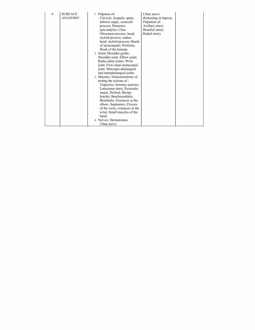
8 SURFACEANATOMY
1. Palpaton of:Clavicle, Scapula: spine,inferior angle, coracoidprocess; Humerus:epicondyles; UlnaOlecranon process, head,styloid process; radiushead, styloid process; Haedsof metacarpals; Pisiform;Hook of the hamate.
2. Joints Shoulder girdle;Shoulder joint, Elbow joint;Radioulnar joints; Wristjoint; First carpometacarpaljoint; Metcarpophalangealand interphalangeal joints
3. Muscles: Demonstrations oftesting the actions of :Trapezius; Serratus anterior,Latissimus dorsi, Pectoralismajor, Deltiod, Bicepsbrachii, Brachioradialis,Brachialis, Extensors at theelbow, Supinators, Flexorsof the wrist, extensors at thewrist, Small muscles of thehand.
4. Nerves: Dermatomes Ulnar nerve
Ulnar nervethickening in leprosyPalpation of:Axillary arteryBrachial arteryRadial artery


![Vasa aberrantia connecting the brachial and radial arteries aberrantia connecting the brachial and radial arteries 77 References [1] Hollinshead WH. Anatomy for surgeons: The back](https://img.pdfslide.us/doc/110x75/5a9f52337f8b9a62178c8d5c/pdfvasa-aberrantia-connecting-the-brachial-and-radial-arteries-aberrantia-connecting.jpg)


![1. brachial plexus & its applied anatomy[1]](https://img.pdfslide.us/doc/110x75/554b284fb4c905da088b492a/1-brachial-plexus-its-applied-anatomy1.jpg)