Embed Size (px)
Citation preview
Objectives• Explain how the two parts of the
nervous system work together.• Describe the structure and function
of the central nervous system.• Describe the structure and function
of the peripheral nervous system.
Section ResourcesUnit Resource Book
Study Guide pp. 35–36Power Notes p. 37Reinforcement p. 38
Interactive Reader Chapter 29Spanish Study Guide pp. 295–296
Biology Toolkit pp. C11, C20, C26, C39, D8
TechnologyPower Presentation 29.4Media Gallery DVDOnline Quiz 29.4
Activate Prior Knowledge Remind students that the body has many functions that occur simultaneously. Ask, Can you walk and chew gum at the same time? yes Explain that coopera-tion between the central and peripheral nervous systems enables processing of many forms of sensory input.
VocabularyAcademic Vocabulary The prefix peri- in peripheral comes from a root meaning “to carry around.” The related words periphery and perimeter refer to an outer boundary. Discuss how this description matches with the peripheral nervous system shown in FIGURE 29.9.
AnswersA Summarize The PNS sensory
neurons pick up signals from all parts of the body and transmit them to the CNS, which interprets those signals. The CNS relays a response to motor neurons of the PNS, which stimulate a response.
Plan and PreparePlan and Prepare
TeachTeach
8/18/09 12:03:338/18/09 12:03:338/18/09 12:03:330003:33 P3:33 P3:33 PMMM
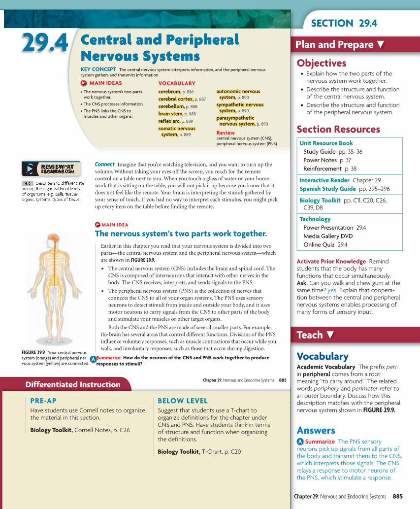
FIGURE 29.9 Your central nervous system (orange) and peripheral ner-vous system (yellow) are connected.
Connect Imagine that you’re watching television, and you want to turn up the volume. Without taking your eyes off the screen, you reach for the remote control on a table next to you. When you touch a glass of water or your home-work that is sitting on the table, you will not pick it up because you know that it does not feel like the remote. Your brain is interpreting the stimuli gathered by your sense of touch. If you had no way to interpret each stimulus, you might pick up every item on the table before finding the remote.
MAIN IDEA
The nervous system’s two parts work together.Earlier in this chapter you read that your nervous system is divided into two parts—the central nervous system and the peripheral nervous system—which are shown in FIGURE 29.9.
• The central nervous system (CNS) includes the brain and spinal cord. The CNS is composed of interneurons that interact with other nerves in the body. The CNS receives, interprets, and sends signals to the PNS.
• The peripheral nervous system (PNS) is the collection of nerves that connects the CNS to all of your organ systems. The PNS uses sensory neurons to detect stimuli from inside and outside your body, and it uses motor neurons to carry signals from the CNS to other parts of the body and stimulate your muscles or other target organs.
Both the CNS and the PNS are made of several smaller parts. For example, the brain has several areas that control different functions. Divisions of the PNS influence voluntary responses, such as muscle contractions that occur while you walk, and involuntary responses, such as those that occur during digestion.
Summarize How do the neurons of the CNS and PNS work together to produce responses to stimuli?
29.4 Central and Peripheral Nervous SystemsKEY CONCEPT The central nervous system interprets information, and the peripheral nervous system gathers and transmits information.
MAIN IDEAS• The nervous system’s two parts
work together.
• The CNS processes information.
• The PNS links the CNS to muscles and other organs.
VOCABULARYcerebrum,cerebrum, p. 886
cerebral cortex,cerebral cortex, p. 887
cerebellum,cerebellum, p. 888
brain stem,brain stem, p. 888
reflex arc,reflex arc, p. 889
somatic nervous somatic nervous system,system, p. 889
autonomic nervous autonomic nervous system,system, p. 890
sympathetic nervous sympathetic nervous system,system, p. 890
parasympathetic parasympathetic nervous system,nervous system, p. 890
Reviewcentral nervous system (CNS), peripheral nervous system (PNS)
Chapter 29: Nervous and Endocrine Systems 885
4.c Describe and differentiate among the organizational levels of organisms (e.g., cells, tissues, organs, systems, types of tissue).
b10hspe-092904.indd 885b10hspe-092904.indd 885b10hspe-092904.indd 885 9/9/09 7:51:52 9/9/09 7:51:52 9/9/09 7:51:52 PPP
A
Differentiated Instruction
SECTION 29.4
PRE-APHave students use Cornell notes to organize the material in this section.
Biology Toolkit, Cornell Notes, p. C26
BELOW LEVELSuggest that students use a T-chart to organize definitions for the chapter under CNS and PNS. Have students think in terms of structure and function when organizing the definitions.
Biology Toolkit, T-Chart, p. C20
Chapter 29: Nervous and Endocrine Systems 885
bbb111000hhhsssttteee---000999222999...iiinnndddddd 888888555b10hste-0929.indd 885b10hste-0929.indd 885b10hste-0929.indd 885 999///111000///000999 111:::555666:::444111 PPPMMM9/10/09 1:56:41 PM9/10/09 1:56:41 PM9/10/09 1:56:41 PM
Time 5 minutes
Lab Binder Human Bio, p. 22
Purpose Compare the relative amounts of space given to touch reception in a fingertip and a forearm.Teacher Note “It’s a fun, interactive, and relevant activity for the classroom.”
LAB MANAGEMENT• Use toothpicks with pointed ends.• Straightened paper clips can be used
instead of toothpicks.
AnswersSample Data
Area Tested Number of Toothpicks
Used Reported
fingertip 1 1
fingertip 2 2
fingertip 1 1
forearm 2 1
forearm 2 1
forearm 1 1
Teacher Note “A lot of students believe there is less “meat” in the finger than the forearm and therefore this affects results.”
Analyze and Conclude 1. Students should observe that the
fingertip received more sensory information. The number of tooth-picks reported and the number of toothpicks used were the same in more instances when the fingertip was tested than when the forearm was tested.
2. The fingertip has more space devoted to it in the primary sensory cortex.
Teach continuedTeach continued
QUICK LAB QUICK LAB
D E S I G N YO U R O W N
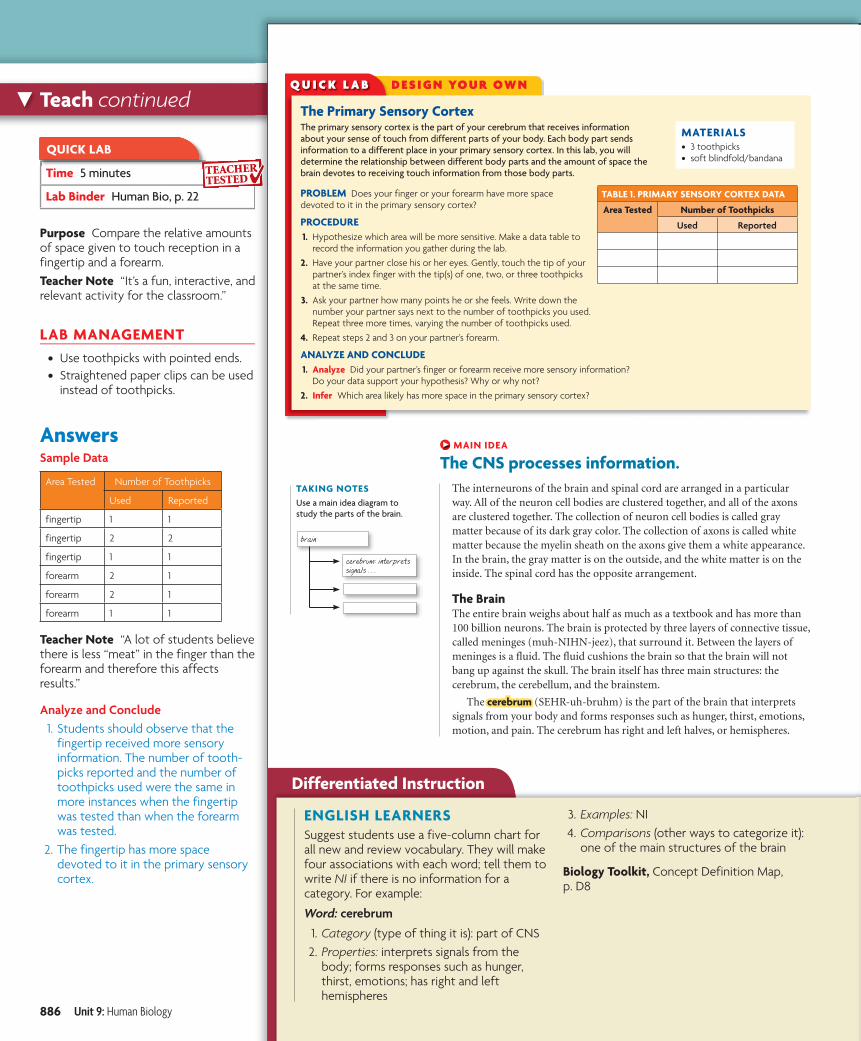
The Primary Sensory CortexThe primary sensory cortex is the part of your cerebrum that receives information about your sense of touch from different parts of your body. Each body part sends information to a different place in your primary sensory cortex. In this lab, you will determine the relationship between different body parts and the amount of space the brain devotes to receiving touch information from those body parts.
PROBLEM Does your finger or your forearm have more space devoted to it in the primary sensory cortex?
PROCEDURE 1. Hypothesize which area will be more sensitive. Make a data table to
record the information you gather during the lab.
2. Have your partner close his or her eyes. Gently, touch the tip of your partner’s index finger with the tip(s) of one, two, or three toothpicks at the same time.
3. Ask your partner how many points he or she feels. Write down the number your partner says next to the number of toothpicks you used. Repeat three more times, varying the number of toothpicks used.
4. Repeat steps 2 and 3 on your partner’s forearm.
ANALYZE AND CONCLUDE 1. Analyze Did your partner’s finger or forearm receive more sensory information?
Do your data support your hypothesis? Why or why not?
2. Infer Which area likely has more space in the primary sensory cortex?
MATERIALS• 3 toothpicks• soft blindfold/bandana
Q U I C K L A B
TABLE 1. PRIMARY SENSORY CORTEX DATA
Area Tested Number of Toothpicks
Used Reported
TAKING NOTESUse a main idea diagram to study the parts of the brain.
cerebrum: interprets signals . . .
brain
MAIN IDEA
The CNS processes information.The interneurons of the brain and spinal cord are arranged in a particular way. All of the neuron cell bodies are clustered together, and all of the axons are clustered together. The collection of neuron cell bodies is called gray matter because of its dark gray color. The collection of axons is called white matter because the myelin sheath on the axons give them a white appearance. In the brain, the gray matter is on the outside, and the white matter is on the inside. The spinal cord has the opposite arrangement.
The BrainThe entire brain weighs about half as much as a textbook and has more than 100 billion neurons. The brain is protected by three layers of connective tissue, called meninges (muh-NIHN-jeez), that surround it. Between the layers of meninges is a fluid. The fluid cushions the brain so that the brain will not bang up against the skull. The brain itself has three main structures: the cerebrum, the cerebellum, and the brainstem.
The cerebrumcerebrum (SEHR-uh-bruhm) is the part of the brain that interprets signals from your body and forms responses such as hunger, thirst, emotions, motion, and pain. The cerebrum has right and left halves, or hemispheres.
886 Unit 9: Human Biology
b10hspe-092904.indd 886 9/2/08 1:50:42 PM
Differentiated Instruction
ENGLISH LEARNERSSuggest students use a five-column chart for all new and review vocabulary. They will make four associations with each word; tell them to write NI if there is no information for a category. For example:
Word: cerebrum
1. Category (type of thing it is): part of CNS 2. Properties: interprets signals from the
body; forms responses such as hunger, thirst, emotions; has right and left hemispheres
3. Examples: NI 4. Comparisons (other ways to categorize it):
one of the main structures of the brain
Biology Toolkit, Concept Definition Map, p. D8
886 Unit 9: Human Biology
b10hste-0929.indd 886b10hste-0929.indd 886 9/10/08 1:56:46 PM9/10/08 1:56:46 PM
The Inside StoryIn 1848, Phineas Gage was a young foreman of a railroad crew whose job was to use explosives to clear ground for laying tracks. On September 13, an accidental explosion blew an iron rod, more than 1 meter (1 yd) long, com-pletely through his head. The rod entered Gage’s face below the cheek-bone, traveled behind his eye, and exited the top of his head. He recov-ered, but the once capable foreman became obstinate, fitful, and vulgar. He lost his job.
The extensive injury to the frontal lobe of his brain and his subsequent change in behavior enabled the medical world to make a connection between the frontal lobe and psychological processes involved in emotion, personal-ity, and problem-solving. Gage lived his life generally out of the public eye, yet stories abounded of him exhibiting himself in fairgrounds and circuses around the country. All sorts of charac-teristics were attributed to him, such as drunkenness, slovenliness, and being sexually uninhibited, though there is little evidence to support any of it. He died in 1860 after suffering a seizure. The iron rod went to the grave with him.
VocabularyWord Origins The roots of the words lobe and cortex both come from the language of botany. Lobe comes from a root meaning “hull” or “pod,” and describes a structure that projects outward, as does an ear lobe. Cortex comes from a root meaning “bark” and refers to an outer layer.
AnswersA Apply (1) vision; (2) hearing, speech;
(3) reasoning, speech, movement
cerebrum
M8/06 2:37:07 PM
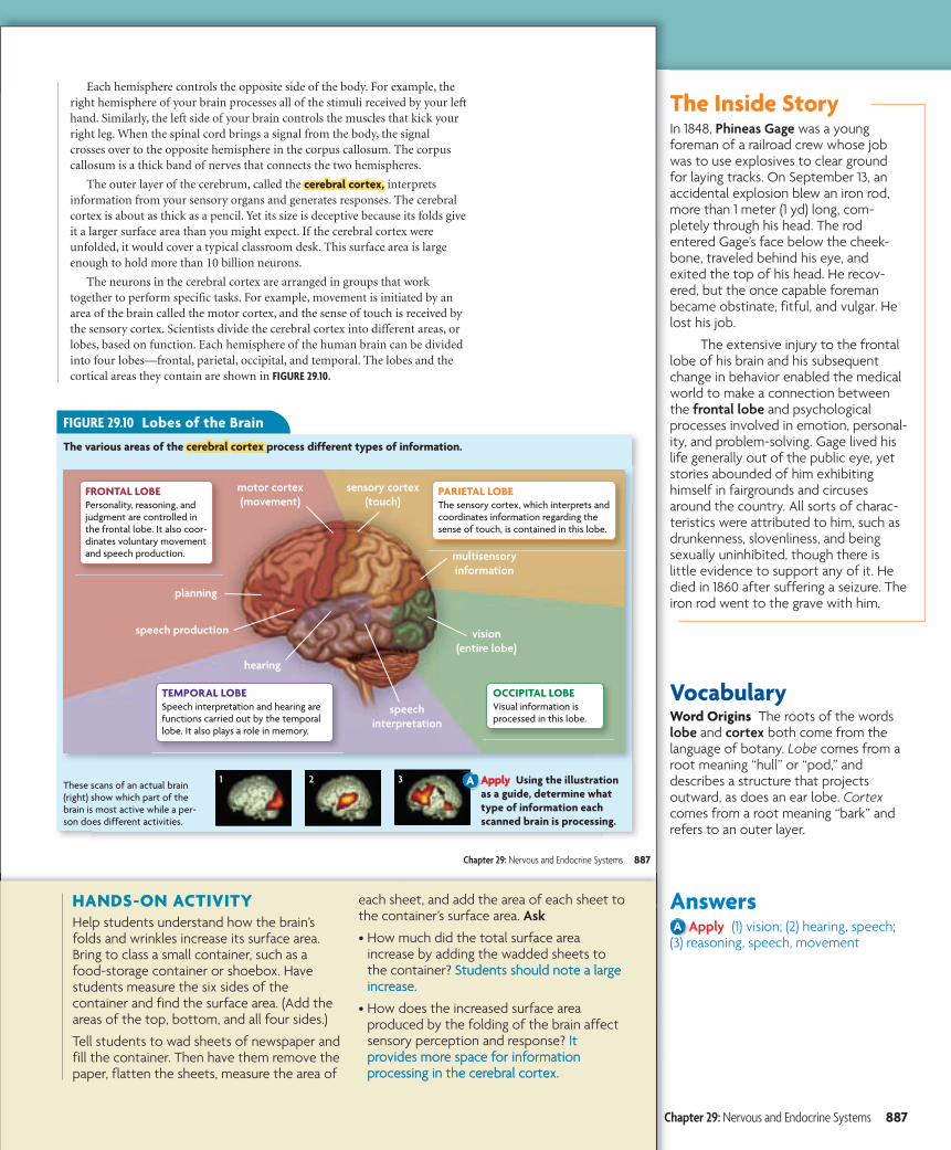
FIGURE 29.10 Lobes of the Brain
The various areas of the cerebral cortexcerebral cortex process different types of information.
Apply Using the illustration as a guide, determine what type of information each scanned brain is processing.
These scans of an actual brain (right) show which part of the brain is most active while a per-son does different activities.
OCCIPITAL LOBEVisual information is processed in this lobe.
PARIETAL LOBEThe sensory cortex, which interprets and coordinates information regarding the sense of touch, is contained in this lobe.
FRONTAL LOBEPersonality, reasoning, and judgment are controlled in the frontal lobe. It also coor-dinates voluntary movement and speech production.
TEMPORAL LOBESpeech interpretation and hearing are functions carried out by the temporal lobe. It also plays a role in memory.
vision(entire lobe)
multisensory information
sensory cortex (touch)
speech production
planning
hearing
motor cortex (movement)
speech interpretation
1 2 3
Each hemisphere controls the opposite side of the body. For example, theright hemisphere of your brain processes all of the stimuli received by your lefthand. Similarly, the left side of your brain controls the muscles that kick yourright leg. When the spinal cord brings a signal from the body, the signalcrosses over to the opposite hemisphere in the corpus callosum. The corpuscallosum is a thick band of nerves that connects the two hemispheres.
The outer layer of the cerebrum, called the cerebral cortex,cerebral cortex, interpretsinformation from your sensory organs and generates responses. The cerebralcortex is about as thick as a pencil. Yet its size is deceptive because its folds giveit a larger surface area than you might expect. If the cerebral cortex wereunfolded, it would cover a typical classroom desk. This surface area is largeenough to hold more than 10 billion neurons.
The neurons in the cerebral cortex are arranged in groups that worktogether to perform specific tasks. For example, movement is initiated by anarea of the brain called the motor cortex, and the sense of touch is received bythe sensory cortex. Scientists divide the cerebral cortex into different areas, orlobes, based on function. Each hemisphere of the human brain can be dividedinto four lobes—frontal, parietal, occipital, and temporal. The lobes and thecortical areas they contain are shown in FIGURE 29.10.
Chapter 29: Nervous and Endocrine Systems 887
bhspe-092904.indd Sec4:887 6/28/06 2:37:12
A
HANDS-ON ACTIVITYHelp students understand how the brain’s folds and wrinkles increase its surface area. Bring to class a small container, such as a food-storage container or shoebox. Have students measure the six sides of the container and find the surface area. (Add the areas of the top, bottom, and all four sides.)
Tell students to wad sheets of newspaper and fill the container. Then have them remove the paper, flatten the sheets, measure the area of
each sheet, and add the area of each sheet to the container’s surface area. Ask
• How much did the total surface area increase by adding the wadded sheets to the container? Students should note a large increase.
• How does the increased surface area produced by the folding of the brain affect sensory perception and response? It provides more space for information processing in the cerebral cortex.
Chapter 29: Nervous and Endocrine Systems 887
ONLINE BIOLOGY Go to the chapter Resource Center at ClassZone.com for additional resources and information on the nervous system.
Science Trivia• The brain of an adult human weighs
between 1300 and 1400 grams.• The brain of a bottle-nosed dolphin
weighs about 1600 grams, an elephant’s about 6000 grams.
• The brain of a cat weighs about 30 grams, a dog’s about 70 grams.
• The brain of a hamster weighs about 1.4 grams, a goldfish’s 0.097 gram.
Vocabularyhypothalamus The prefix hypo- comes from the Greek word hypo, meaning “under.” The hypothalamus is the part of the brain under the thalamus.
Take It FurtherResearchers have tried to define the biological basis for intelligence for decades. Many attempts have been made to correlate IQ, a measure of intelligence known as the intelligence quotient, to brain size. A recent study by Richard Haier at the University of California in Irvine suggests that there may be some connection.
A study of MRI brain scans showed that people with high test scores had significantly more gray matter in 24 regions of the brain than did people with lower scores. Haier thinks that different types of intelligence may correlate to the amount of gray matter in a particular part of the brain. Still the study showed that only about 6 percent of total gray matter in the brain is related to IQ.
Teach continuedTeach continued
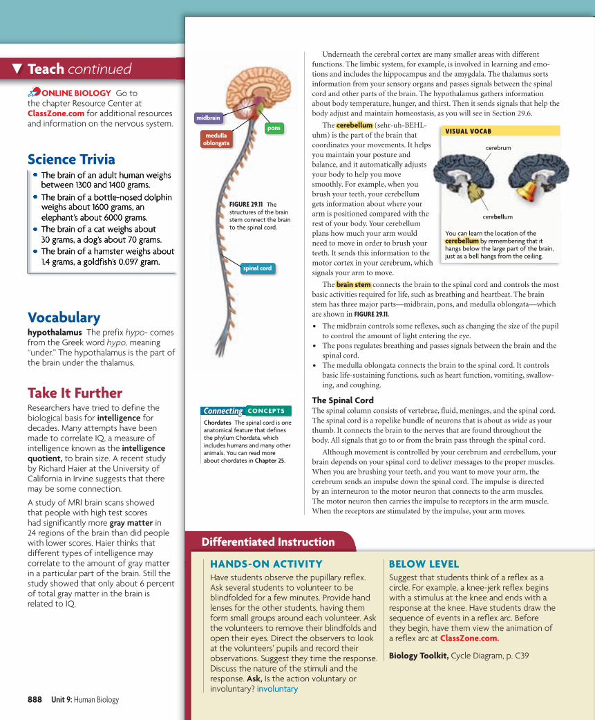
You can learn the location of the cerebellumcerebellum by remembering that it hangs below the large part of the brain, just as a bell hangs from the ceiling.
VISUAL VOCAB
cerebellum
cerebrum
FIGURE 29.11 The structures of the brain stem connect the brain to the spinal cord.
pons
midbrain
medulla oblongata
spinal cord
ConnectingChordates The spinal cord is one anatomical feature that defines the phylum Chordata, which includes humans and many other animals. You can read more about chordates in Chapter 25.
CONCEPTS
Underneath the cerebral cortex are many smaller areas with differentfunctions. The limbic system, for example, is involved in learning and emo-tions and includes the hippocampus and the amygdala. The thalamus sortsinformation from your sensory organs and passes signals between the spinalcord and other parts of the brain. The hypothalamus gathers informationabout body temperature, hunger, and thirst. Then it sends signals that help thebody adjust and maintain homeostasis, as you will see in Section 29.6.
The cerebellumcerebellum (sehr-uh-BEHL-uhm) is the part of the brain thatcoordinates your movements. It helpsyou maintain your posture andbalance, and it automatically adjustsyour body to help you movesmoothly. For example, when youbrush your teeth, your cerebellumgets information about where yourarm is positioned compared with therest of your body. Your cerebellumplans how much your arm wouldneed to move in order to brush yourteeth. It sends this information to themotor cortex in your cerebrum, whichsignals your arm to move.
The brain stembrain stem connects the brain to the spinal cord and controls the mostbasic activities required for life, such as breathing and heartbeat. The brainstem has three major parts—midbrain, pons, and medulla oblongata—whichare shown in FIGURE 29.11.
• The midbrain controls some reflexes, such as changing the size of the pupilto control the amount of light entering the eye.
• The pons regulates breathing and passes signals between the brain and thespinal cord.
• The medulla oblongata connects the brain to the spinal cord. It controlsbasic life-sustaining functions, such as heart function, vomiting, swallow-ing, and coughing.
The Spinal CordThe spinal column consists of vertebrae, fluid, meninges, and the spinal cord.The spinal cord is a ropelike bundle of neurons that is about as wide as yourthumb. It connects the brain to the nerves that are found throughout thebody. All signals that go to or from the brain pass through the spinal cord.
Although movement is controlled by your cerebrum and cerebellum, yourbrain depends on your spinal cord to deliver messages to the proper muscles.When you are brushing your teeth, and you want to move your arm, thecerebrum sends an impulse down the spinal cord. The impulse is directedby an interneuron to the motor neuron that connects to the arm muscles.The motor neuron then carries the impulse to receptors in the arm muscle.When the receptors are stimulated by the impulse, your arm moves.
888 Unit 9: Human Biology
l C d 2 03240 Fil N bh 092904 i dd U l L M difi d 6/26/06 7 37 PMhspe-092904.indd Sec4:888 6/28/06 2:37:19 PM
Reflex arcs,
somatic nervous system
bhspe-092904.i
A
Differentiated Instruction
HANDS-ON ACTIVITYHave students observe the pupillary reflex. Ask several students to volunteer to be blindfolded for a few minutes. Provide hand lenses for the other students, having them form small groups around each volunteer. Ask the volunteers to remove their blindfolds and open their eyes. Direct the observers to look at the volunteers’ pupils and record their observations. Suggest they time the response. Discuss the nature of the stimuli and the response. Ask, Is the action voluntary or involuntary? involuntary
BELOW LEVELSuggest that students think of a reflex as a circle. For example, a knee-jerk reflex begins with a stimulus at the knee and ends with a response at the knee. Have students draw the sequence of events in a reflex arc. Before they begin, have them view the animation of a reflex arc at ClassZone.com.
Biology Toolkit, Cycle Diagram, p. C39
888 Unit 9: Human Biology
Integrating GeneticsCIPA, Congenital Insensitivity to Pain with Anhydrosis, is a rare genetic disorder that affects the nervous system. It is caused by mutations on the neurotrophic tyrosine kinase receptor gene (NTRK) and prevents the formation of nerve cells that transmit signals to the brain registering pain and temperature. People who suffer from CIPA are unable to feel pain, detect temperature, or even sweat. Their other senses are intact, however.
If a person suffering from CIPA were to inadvertently place their hand on a hot stove, they would not be able to detect the heat or feel any pain. No impulse would cause them to remove their hand. Instead they would first need to recog-nize the danger, then respond. Individuals with CIPA commonly suffer from injuries to the arms and legs, mouth, lips, tongue, and gums. Because of their inability to sweat, they commonly suffer fevers, and even hyperthermia. There is no treatment for this disorder.
AnswersA Summarize Unlike a voluntary
muscle movement, a muscle movement caused by a reflex arc does not involve the brain.
cerebellum
cerebellum
brain stem
M8/06 2:37:19 PM
Watch a refl ex arc in action at ClassZone.com.
BIOLOGY
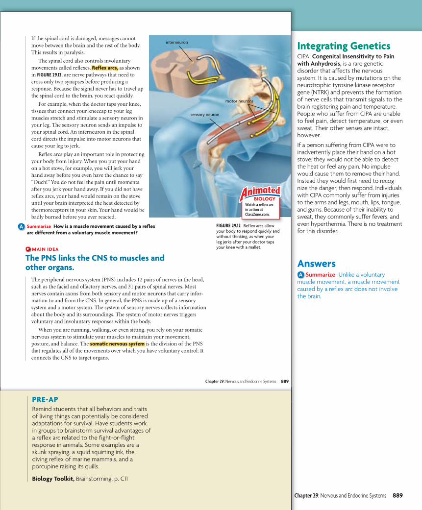
FIGURE 29.12 Reflex arcs allow your body to respond quickly and without thinking, as when your leg jerks after your doctor taps your knee with a mallet.
interneuron
sensory neuron
motor neurons
If the spinal cord is damaged, messages cannotmove between the brain and the rest of the body.This results in paralysis.
The spinal cord also controls involuntarymovements called reflexes. Reflex arcs,Reflex arcs, as shownin FIGURE 29.12, are nerve pathways that need tocross only two synapses before producing aresponse. Because the signal never has to travel upthe spinal cord to the brain, you react quickly.
For example, when the doctor taps your knee,tissues that connect your kneecap to your legmuscles stretch and stimulate a sensory neuron inyour leg. The sensory neuron sends an impulse toyour spinal cord. An interneuron in the spinalcord directs the impulse into motor neurons thatcause your leg to jerk.
Reflex arcs play an important role in protectingyour body from injury. When you put your handon a hot stove, for example, you will jerk yourhand away before you even have the chance to say“Ouch!” You do not feel the pain until momentsafter you jerk your hand away. If you did not havereflex arcs, your hand would remain on the stoveuntil your brain interpreted the heat detected bythermoreceptors in your skin. Your hand would bebadly burned before you ever reacted.
Summarize How is a muscle movement caused by a reflex arc different from a voluntary muscle movement?
MAIN IDEA
The PNS links the CNS to muscles and other organs.
The peripheral nervous system (PNS) includes 12 pairs of nerves in the head,such as the facial and olfactory nerves, and 31 pairs of spinal nerves. Mostnerves contain axons from both sensory and motor neurons that carry infor-mation to and from the CNS. In general, the PNS is made up of a sensorysystem and a motor system. The system of sensory nerves collects informationabout the body and its surroundings. The system of motor nerves triggersvoluntary and involuntary responses within the body.
When you are running, walking, or even sitting, you rely on your somaticnervous system to stimulate your muscles to maintain your movement,posture, and balance. The somatic nervous systemsomatic nervous system is the division of the PNSthat regulates all of the movements over which you have voluntary control. Itconnects the CNS to target organs.
Chapter 29: Nervous and Endocrine Systems 889
bhspe-092904.indd Sec4:889 6/28/06 2:37:26
A
PRE-APRemind students that all behaviors and traits of living things can potentially be considered adaptations for survival. Have students work in groups to brainstorm survival advantages of a reflex arc related to the fight-or-flight response in animals. Some examples are a skunk spraying, a squid squirting ink, the diving reflex of marine mammals, and a porcupine raising its quills.
Biology Toolkit, Brainstorming, p. C11
Chapter 29: Nervous and Endocrine Systems 889
Take It FurtherToday, our fight-or-flight mechanism is oftentimes activated not because there is a threat to our lives but rather because we are experiencing a stressful situation. Our inability to fight or flee from stresses can lead to stress-related illnesses, such as heart disease, high blood pressure, migraines, and insomnia.
AnswersA Analyze Reflex arcs are part of the
autonomic nervous system because they produce involuntary muscle movement.
Assess Use the Online Quiz or Section Quiz (Assessment Book, p. 574).Reteach Have students work in groups to construct crossword puzzles using the vocabulary words and other important words in this section. Have groups exchange puzzles and fill them out.
Teach continuedTeach continued
Assess and ReteachAssess and Reteach
U
29.4 ASSESSMENT
Connecting CONCEPTS
ONLINE QUIZClassZone.com
Central nervous system (CNS)
Peripheral nervous system (PNS)
Autonomic nervous system
(involuntary)
Somatic nervous system
(voluntary)
Parasympathetic nervous system
(calm and relaxation)
Sympathetic nervous system
(action and stress)
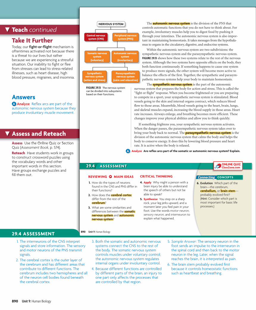
FIGURE 29.13 The nervous system can be divided into subsystems based on their functions.
NERVOUS SYSTEM The autonomic nervous systemautonomic nervous system is the division of the PNS that controls automatic functions that you do not have to think about. For example, involuntary muscles help you to digest food by pushing it through your intestines. The autonomic nervous system is also impor-tant in maintaining homeostasis. It takes messages from the hypothala-mus to organs in the circulatory, digestive, and endocrine systems.
Within the autonomic nervous system are two subdivisions: the sympathetic nervous system and the parasympathetic nervous system. FIGURE 29.13 shows how these two systems relate to the rest of the nervous system. Although the two systems have opposite effects on the body, they both function continuously. If something happens to cause one system to produce more signals, the other system will become more active to balance the effects of the first. Together, the sympathetic and parasym-pathetic nervous systems help your body to maintain homeostasis.
The sympathetic nervous systemsympathetic nervous system is the part of the autonomic nervous system that prepares the body for action and stress. This is called the “fight or flight” response. When you become frightened or you are preparing to compete in a sport, your sympathetic nervous system is stimulated. Blood vessels going to the skin and internal organs contract, which reduces blood flow to those areas. Meanwhile, blood vessels going to the heart, brain, lungs, and skeletal muscles expand, increasing the blood supply in those areas. Heart rate increases. Airways enlarge, and breathing becomes more efficient. These changes improve your physical abilities and allow you to think quickly.
If something frightens you, your sympathetic nervous system activates. When the danger passes, the parasympathetic nervous system takes over to bring your body back to normal. The parasympathetic nervous systemparasympathetic nervous system is the division of the autonomic nervous system that calms the body and helps the body to conserve energy. It does this by lowering blood pressure and heart rate. It is active when the body is relaxed.
Analyze Are reflex arcs part of the somatic or autonomic nervous system? Explain.
REVIEWING MAIN IDEAS
1. How do the types of neurons found in the CNS and PNS differ in their functions?
2. How does the cerebral cortexcerebral cortex differ from the rest of thecerebrumcerebrum?
3. What are some similarities and differences between the somatic somatic nervous systemnervous system and autonomic autonomic nervous systemnervous system?
CRITICAL THINKING
4. Apply Why might a person with a brain injury be able to understand the speech of others but not be able to speak?
5. Synthesize You step on a sharp rock, your leg jerks upward, and a moment later you feel pain in your foot. Use the words motor neuron, sensory neuron, and interneuron to explain what happened.
6. Evolution Which part of the brain—the cerebrum, cerebellum,cerebellum, or brain stembrain stem—probably evolved first? (Hint: Consider which part is most important for basic life processes.)
890 Unit 9: Human Biology
b10hspe-092904.indd 890 9/2/08 1:51:02 PM
A
addiction,desensitization,tolerance,sensitization,
stimulant,depressant,
b10h
29.4 ASSESSMENT 1. The interneurons of the CNS interpret
signals and store information. The sensory and motor neurons of the PNS transmit signals.
2. The cerebral cortex is the outer layer of the cerebrum and has different areas that contribute to different functions. The cerebrum includes two hemispheres and all of the neuron cell bodies found beneath the cerebral cortex.
3. Both the somatic and autonomic nervous systems connect the CNS to the rest of the body. The somatic nervous system controls muscles under voluntary control; the autonomic nervous system regulates internal organs under involuntary control.
4. Because different functions are controlled by different parts of the brain, an injury to one part only affects the processes that are controlled by that region.
5. Sample Answer: The sensory neuron in the foot sends an impulse to the interneuron in the spinal cord and then back to the motor neuron in the leg. Later, when the signal reaches the brain, it is interpreted as pain.
6. The brain stem probably evolved first because it controls homeostatic functions such as heartbeat and breathing.
890 Unit 9: Human Biology
b10hste-0929.indd 890b10hste-0929.indd 890 9/10/08 1:56:28 PM9/10/08 1:56:28 PM