Embed Size (px)
Citation preview

57
SECTION 2 CHAPTER 5 IMAGING AND NEW TECHNOLOGY
IMA
GIN
G
AN
D N
EW
T
EC
HN
OLO
GY
59
5.0 IMAGING AND NEW TECHNOLOGY
5.1 RELATIONSHIP TO CERTIFICATE OF NEED
Connecticut General Statutes Section 19a-638(a)(9) specifies a Certificate of Need is required for the acquisition of computed tomography scanners, magnetic resonance imaging scanners, positron emission tomography scanners or positron emission tomography-computed tomography scanners, by any person, physician, provider, short-term acute care general hospital, or children’s hospital. Entities seeking authorization to acquire such imaging equipment are required to demonstrate that they meet clear public need as well as other criteria set forth in Connecticut General Statutes Section 19a-639.
5.2 GENERAL BACKGROUND
In 2009, OHCA formed an industry workgroup to review provider experience with the operation of the 2005 law governing CT scanners, with the goal of providing recommendations for possible use in the development of criteria and guidelines with which to evaluate CT scanners. Building upon those recommendations, OHCA formed a second workgroup in 2010 comprised of representatives from various segments of the Connecticut health care industry. It included independent and hospital radiologists, members of the Radiological Society of Connecticut, Connecticut Hospital Association, Connecticut Dental Association, Connecticut State Medical Society, Connecticut health care industry attorneys and OHCA staff. This workgroup helped OHCA develop proposed imaging standards and guidelines, which OHCA intends to promulgate as regulations. The imaging equipment definitions, standards and guidelines in this chapter have been developed through collaboration with the imaging workgroup.
This chapter focuses on magnetic resonance imaging (MRI), computed tomography (CT), and positron emission tomography-computed tomography (PET-CT). The standards and guidelines for PET scanners have been combined with PET-CT, the two have become imaging technologies intertwined to the point that providers are replacing PET scanners with PET-CT scanners, and vendors no longer support, sell, or lease PET scanners.
5.3 CURRENT IMAGING LANDSCAPE
Pursuant to CGS 19a-634(c), in January 2012, OHCA surveyed all imaging providers in Connecticut to establish an inventory of all MRI, CT, PET and PET-CT scanners statewide. Based on survey responses, Connecticut currently has 114 MRI scanners, 123 CT scanners, 6 PET scanners and 21 PET-CT scanners.As stipulated in CGS 19a-690, all imaging equipment operating in Connecticut must be accredited through the American College of Radiology. The locations of these MRI, CT and PET and PET/CT scanners are shown in Appendices M-Q.
The majority of imaging scanners were acquired through the Certificate of Need application process; however, some scanners were acquired without CON approval, as providers could acquire imaging equipment costing less than $400K without CON approval, Public Act 05-93 removed this threshold, in an effort to deter the acquisition of substandard equipment. Providers still were allowed to acquire the equipment without CON approval if they could demonstrate that it was acquired prior to July 1, 2005 and was in operation before July 1, 2006, pursuant to Public Acts 05-93 and 06-28. Many of these providers sought a CON determination stating that they were in compliance with the law, without having to demonstrate the need for the equipment.Under current law, any previously authorized imaging scanner (acquired through a CON Determination, a CON Decision or an Agreed Settlement with OHCA) can be replaced with a similar scanner (e.g., a 1.5T MRI scanner with a 3.0 MRI scanner, or a 4-slice CT scanner with a 16-slice CT scanner) and the owner or the authorized holder of the CON can relocate the imaging equipment anywhere statewide. Other than a notification to OHCA of the relocation and disposition of the replaced imaging scanner, no additional action is required.
Market trends over the past several years have affected the environment in which hospital and free-standing imaging centers operate. In the past, there was a steady and ongoing migration of imaging services out of the hospital setting, mostly to physician-owned free-standing imaging centers. Today however, reimbursement issues, access to capital,
60
vendor relationships and physician employment are initiating a wave of acquisitions of imaging equipment at free-standing imaging centers by hospitals.79 CON approval is required for these acquisitions and purchasers must demonstrate clear public need for the equipment.
5.4 STANDARDS/GUIDELINES
DEFINITIONS
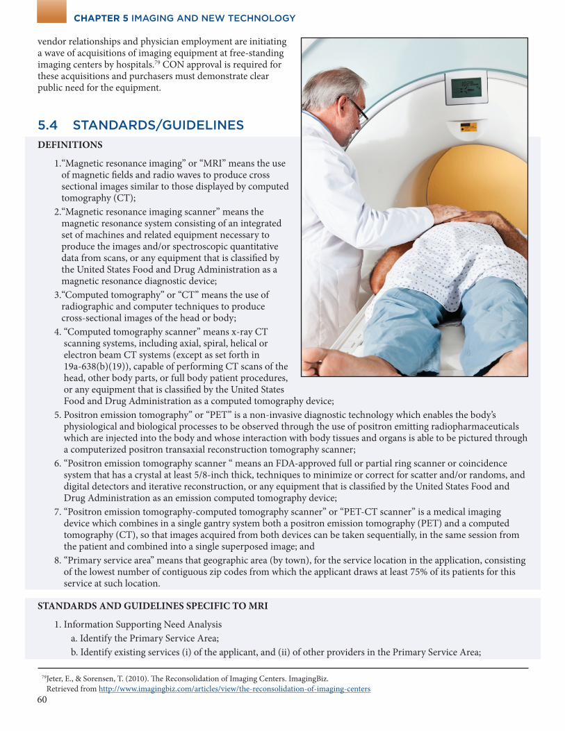
1. “Magnetic resonance imaging” or “MRI” means the use of magnetic fields and radio waves to produce cross sectional images similar to those displayed by computed tomography (CT);
2. “Magnetic resonance imaging scanner” means the magnetic resonance system consisting of an integrated set of machines and related equipment necessary to produce the images and/or spectroscopic quantitative data from scans, or any equipment that is classified by the United States Food and Drug Administration as a magnetic resonance diagnostic device;
3. “Computed tomography” or “CT” means the use of radiographic and computer techniques to produce cross-sectional images of the head or body;
4. “Computed tomography scanner” means x-ray CT scanning systems, including axial, spiral, helical or electron beam CT systems (except as set forth in 19a-638(b)(19)), capable of performing CT scans of the head, other body parts, or full body patient procedures, or any equipment that is classified by the United States Food and Drug Administration as a computed tomography device;
5. Positron emission tomography” or “PET” is a non-invasive diagnostic technology which enables the body’s physiological and biological processes to be observed through the use of positron emitting radiopharmaceuticals which are injected into the body and whose interaction with body tissues and organs is able to be pictured through a computerized positron transaxial reconstruction tomography scanner;
6. “Positron emission tomography scanner “ means an FDA-approved full or partial ring scanner or coincidence system that has a crystal at least 5/8-inch thick, techniques to minimize or correct for scatter and/or randoms, and digital detectors and iterative reconstruction, or any equipment that is classified by the United States Food and Drug Administration as an emission computed tomography device;
7. “Positron emission tomography-computed tomography scanner” or “PET-CT scanner” is a medical imaging device which combines in a single gantry system both a positron emission tomography (PET) and a computed tomography (CT), so that images acquired from both devices can be taken sequentially, in the same session from the patient and combined into a single superposed image; and
8. “Primary service area” means that geographic area (by town), for the service location in the application, consisting of the lowest number of contiguous zip codes from which the applicant draws at least 75% of its patients for this service at such location.
STANDARDS AND GUIDELINES SPECIFIC TO MRI
1. Information Supporting Need Analysis a. Identify the Primary Service Area; b. Identify existing services (i) of the applicant, and (ii) of other providers in the Primary Service Area;
79 Jeter, E., & Sorensen, T. (2010). The Reconsolidation of Imaging Centers. ImagingBiz. Retrieved from http://www.imagingbiz.com/articles/view/the-reconsolidation-of-imaging-centers
CHAPTER 5 IMAGING AND NEW TECHNOLOGY
61
CHAPTER 5 IMAGING AND NEW TECHNOLOGY
c. Provide capacity of existing services identified in subsection (1)(b), if available; d. Explain the likely impact on existing services identified in subsection (1)(b); e. Provide actual and proposed hours of operation for services; f. Provide 3 year projection of utilization, with reasonable assumptions on MRI scan volume and capacity; and g. Demonstrate need as described in 2 and 3 below.
2. Need Analysis – Statewide Benchmark
Assumptions
a. “Utilization Rate per Capita” means the number of scans/1,000 population as determined by data collected and published by the Office of Health Care Access division of the Department of Public Health through its data collection and survey processes. If such data is not available from the Office, the applicant is responsible for including reliable statistics, with citations, to establish the utilization rate;
b. “Utilization Rate” means procedure per year for the PSA calculated by multiplying the Utilization Rate per Capita by the population in the PSA using the most recently available census data;
c. “Current Estimated Capacity” means 4,000 scans/year multiplied by the number of scanners in the PSA at the time of the application; and
d. “Percent Utilization of Current Capacity” means the “Utilization Rate/Current Estimated Capacity.”
For current estimated capacity to remain in effect, it must be updated and such update published by the Office of Health Care Access not less than every two years based on the Statewide Facilities and Services Plan. If the Office does not publish an update, the applicant may present reliable capacity estimates for consideration by OHCA to establish the capacity.
3. Need Methodology
The Applicant shall demonstrate that the proposed scanner meets either of the following criteria:
a. The applicant is expected to demonstrate that the Percent Utilization of Current Capacity in the Primary Service Area exceeds 85%.
b. If the applicant has an MRI scanner in the Primary Service Area, the applicant is expected to demonstrate that its Percent Utilization of Current Capacity exceeds 85%.
If the applicant is unable to demonstrate a clear public need for the proposed scanner based upon the assumptions and need methodology in subsection (3)(a) and the requirements of subsection (3)(b) have been met, the Applicant may rely upon any other relevant factors, including those described in subsection 7, to demonstrate need among the population it intends to serve.
4. Quality and Accessibility
The Applicant shall demonstrate that the proposal meets the following criteria:
a. Hospital applicants shall be accredited by the Joint Commission on Accreditation of Healthcare Organizations or certified by Medicare directly or through a deeming agency;
b. Non-hospital facilities shall obtain accreditation from the American College of Radiology within eighteen months of the date on which imaging activities are first conducted;
c. A full-time board certified radiologist, who is a member in good standing with the American College of Radiology, shall be responsible for managing the operation of the MRI scanner and for the written interpretation of the MRI scan;
d. Personnel shall be trained, consistent with guidance of the American College of Radiology, in the use of the MRI scanner and the safety procedures to follow in the event of an emergency;
e. When imaging is performed a physician must be available either on-site or with immediate access to remote viewing of images as they are acquired. The physician in this case must be qualified to interpret images, make adjustments to imaging parameters or protocols, make decisions regarding magnetic field strength risks, and consult with the technologists on technical factors related to the study acquisition. This physician must be board certified to perform and interpret the examinations so produced;
62
f. When contrast is administered, a physician capable of addressing any contrast reactions or adverse events must be on site and immediately physically available to assist in the imaging suite. This physician must be in proximity such that he/she can respond immediately if called. This is not intended to require the physical presence of a physician in the room or suite at all times;
g. The facility or provider must have a policy that explains what steps will be taken to respond in the event of a medical emergency for patients undergoing MRI scans, including the plan for responding to allergic reactions related to contrast media or other drugs or biologicals used in connection with the scan; and
h. The facility or provider shall not deny MRI scanner services to any individual based upon the ability to pay or source of payment, including uninsured, underinsured and Medicaid patients.
5. Financial criteria
The Applicant shall demonstrate that it has sufficient capital to finance the project and provide projections concerning the revenue and expenses for the first three years of the proposal.
6. Other Factors for Consideration
The office may also take the following criteria into consideration during its review of an application:
a. The capabilities of the proposed CT scanner as compared to existing scanners; b. The ability of the applicant to serve an underserved population and not jeopardize the financial viability of the project; c. The impact on existing services, including avoiding delays in timely diagnosis or treatment; d. The use of the scanner for clinical research; e. The history of the applicant in running accredited, financially successful facilities; f. The applicant’s ability to make radiation dose exposure decisions; and g. For hospital applicants only, unique patient populations or specific clinical needs for specialty scanners or
specific clinical applications, including scanners with multiple use applications; complexity of scanning procedures, including the impact on available scanner access due to lengthy procedures; necessity for back-up and redundant equipment to meet the needs of emergency departments.
STANDARDS AND GUIDELINES SPECIFIC TO CT SCANS
1. Information Supporting Need Analysis
a. Identify the Primary Service Area; b. Identify existing services (i) of the applicant, and (ii) of other providers in the Primary Service Area; c. Provide capacity of existing services identified in subsection (1)(a), if available; d. Explain the likely impact on existing services identified in subsection (1)(b); e. Provide actual and proposed hours of operation for services; f. Provide 3 year projection of utilization, with reasonable assumptions on CT scan volume and capacity; and g. Demonstrate need as described in 2 and 3 below.
2. Need Analysis – Statewide Benchmark
Assumptions:
a. “Utilization Rate per Capita” means the number of scans/1,000 population as determined by data collected and published by the Office of Health Care Access, a division of the Department of Public Health through its data collection and survey processes. If such data is not available from the Office, the applicant is responsible for including reliable statistics, with citations, to establish the utilization rate;
b. “Utilization Rate” means the procedure per year for the PSA calculated by multiplying the “Utilization Rate per Capita” by the population in the PSA using the most recently available census data.
c. “Current Estimated Capacity” is 12,000 scans per year multiplied by the number of hospital based scanners in
CHAPTER 5 IMAGING AND NEW TECHNOLOGY
63
CHAPTER 5 IMAGING AND NEW TECHNOLOGY
the PSA at the time of the application for the acquisition of a hospital based scanner and 3,700 scans per year multiplied by the number of outpatient scanners in the PSA at the time of the application for the acquisition of an outpatient scanner; and
d. “Percent Utilization of Current Capacity” means “Utilization Rate/Current Estimated Capacity”.
For current estimated capacity to remain in effect, it must be updated and such update published by the Office of Health Care Access not less than every two years based on the Statewide Facilities and Services Plan. If the Office does not publish an update, the applicant may present reliable capacity estimates for consideration by OHCA to establish the capacity.
3. Need Methodology
The Applicant shall demonstrate that the proposed scanner meets either of the following criteria:
a. The applicant is generally expected to demonstrate that the Percent Utilization of Current Capacity in the Primary Service Area exceeds 85%.
b. If the applicant has a CT scanner in the Primary Service Area, the applicant is expected demonstrate that its Percent Utilization of Current Capacity exceeds 85%.
If the applicant is unable to demonstrate a clear public need for the proposed scanner based upon the assumptions and need methodology in subsection 3(a) and subsection 3(b) have been met, the Applicant may rely upon any other relevant factors, including those described in subsection (7), to demonstrate need among the population it intends to serve.
4. Quality and Accessibility
The Applicant shall demonstrate that the proposal meets the following criteria:
a. Hospital applicants shall be accredited by The Joint Commission or certified by Medicare directly or through a deeming agency;
b. Non-hospital facilities shall obtain accreditation from either the American College of Radiology or the Intersocietal Commission on the Accreditation of Computed Tomography Laboratories within eighteen months of that date on which the imaging activities are first conducted;
c. The CT unit shall be operated safely by trained physicians and/or radiologic technologists who are licensed in Connecticut and who meet the minimum criteria set forth by the appropriate accrediting organization including but not limited to the American College of Radiology, the American Registry of Radiologic Technologists, and the American Registry of Clinical Radiography;
d. All applicants must employ or contract with a radiation physicist to review the quality and safety of the operation of the CT scanner;
e. When imaging is performed a physician must be available either on-site or with immediate access to remote viewing of images as they are acquired. The physician must be qualified to interpret images, make adjustments to imaging parameters or protocols, make decisions regarding radiation dose, and consult with the technologists on technical factors related to the study acquisition. This physician must be board certified to perform and interpret the examinations so produced;
f. When contrast is administered, a physician capable of addressing any contrast reactions or adverse events must be on site and immediately physically available to assist in the imaging suite. This physician must be in proximity such that he/she can respond immediately if called. This is not intended to require the physical presence of a physician in the room or suite at all times;
g. The facility or provider must have a policy that explains what steps will be taken to respond in the event of a medical emergency for patients undergoing CT scans, including the plan for responding to allergic reactions related to contrast media or other drugs or biologicals used in connection with the scan; and
h. The facility or provider shall not deny CT scanner services to any individual based upon the ability to pay or source of payment, including uninsured, underinsured and Medicaid patients.
64
5. Financial criteria
The Applicant shall demonstrate that it has sufficient capital to finance the project and provide projections concerning the revenue and expenses for the first three years of the proposal.
6. Other Factors for Consideration
The office may also take the following criteria into consideration during its review of an application:
a. The capabilities of the proposed CT scanner as compared to existing scanners; b. The ability of the applicant to serve an underserved population and not jeopardize the financial viability of the
project; c. The impact on existing services, including avoiding delays in timely diagnosis or treatment; d. The use of the scanner for clinical research; e. The history of the applicant in running accredited, financially successful facilities; f. The applicant’s ability to make radiation dose exposure decisions; and g. For hospital applicants only, unique patient populations or specific clinical needs for specialty scanners or
specific clinical applications, including scanners with multiple use applications; complexity of scanning procedures, including the impact on available scanner access due to lengthy procedures; necessity for back-up and redundant equipment to meet the needs of emergency departments.
STANDARDS AND GUIDELINES SPECIFIC TO PET and PET/CT SCANS
1. Information Supporting Need Analysis
a. Identify the Primary Service Area; b. Identify existing services (i) of the applicant, and (ii) of other providers in the Primary Service Area; c. Provide capacity of existing services identified in subsection 1(b), if available; d. Explain the likely impact on existing services identified in subsection 1(b); e. Provide actual and proposed hours of operation for services; f. Provide 3 year projection of utilization, with reasonable assumptions on PET or PET-CT scan volume and
capacity; and g. Demonstrate need as described in 2 and 3 below.
2. Need Analysis – Statewide Benchmark Assumptions:
a. Utilization Rate per Capita” means the number of scans/1000 population as determined by data collected and published by the Office of Healthcare Access Division of the Department of Public Health through its data collection and survey processes. If such data are not available from the Office, the applicant is responsible for including reliable statistics, with citations, to establish the utilization rate;
b. “Utilization Rate” means procedure per year for the PSA calculated by multiplying the Utilization Rate Per Capita by the population in the PSA using the most recently available census data;
c. “Current estimated capacity” means 700 scans per year multiplied by the number of scanners in the service area; and
d. “Percent Utilization of Current Capacity” means “Utilization rate/Current Estimated Capacity.”
For current estimated capacity to remain in effect, it must be updated and such update published by the Office of Health Care Access not less than every two years based on the Statewide Facilities and Services Plan. If the Office does not publish an update, the applicant may present reliable capacity estimates for consideration by OHCA to establish the capacity.
3. Need Methodology The Applicant shall demonstrate that the proposed scanner meets either of the following criteria:
CHAPTER 5 IMAGING AND NEW TECHNOLOGY
65
CHAPTER 5 IMAGING AND NEW TECHNOLOGY
a. The applicant is expected to demonstrate that the Percent Utilization of Current Capacity in the Primary Service Area exceeds 85%.
b. If the applicant has a PET or PET/CT scanner in the Primary Service Area, the applicant is expected to demonstrate that its Percent Utilization of Current Capacity exceeds 85%.
If the applicant is unable to demonstrate a clear public need for the proposed scanner based upon the assumptions and need methodology in subsection 3(a) and subsection 3(b) have been met, the Applicant may rely upon any other relevant factors, including those described in subsection 7, to demonstrate need among the population it intends to serve.
4. Quality and Accessibility The Applicant shall demonstrate that the proposal meets the following criteria:
a. Hospital applicants shall be accredited by the Joint Commission or certified by Medicare directly or through a deeming agency;
b. Non-hospital facilities shall obtain accreditation from either the American College of Radiology or the Intersocietal Commission on the Accreditation of Nuclear Laboratories within eighteen months of the date on which imaging activities are first conducted;
c. A physician who is board-certified, shall be available during service hours; d. Qualified engineering and physics personnel with training in the operation and maintenance of PET
equipment shall be available to the facility during service hours; e. Qualified radiation safety personnel with training and experience in the handling of short-lived position
emitting nuclides shall be available during services hours; f. The facility must have a policy that explains what steps will be taken to respond in the event of a medical
emergency for patients undergoing PET or PET-CT scans, including the plan for responding to allergic reactions related to contrast media or other drugs or biologicals used in connection with the scan; and
g. The facility or provider shall not deny PET or PET-CT scanner services to any individual based upon the ability to pay or source of payment, including uninsured, underinsured and Medicaid patients.
5. Financial Criteria The Applicant shall demonstrate that it has sufficient capital to finance the project and provide projections
concerning the revenue and expenses for the first three years of the proposal.
6. Other Factors for Consideration
The office may also take the following criteria into consideration during its review of an application:
a. The capabilities of the proposed PET or PET-CT scanner as compared to existing PET or PET-CT scanners; b. The ability of the applicant to serve an underserved population and not jeopardize the financial viability of the
project; c. The impact on existing services, including avoiding delays in timely diagnosis or treatment; d. The use of the PET or PET-CT scanner for clinical research; e. The history of the applicant in running accredited, financially successful facilities; f. The applicant’s ability to make radiation dose exposure decisions; and
For hospital applicants only, unique patient populations or specific clinical needs for specialty scanners or specific clinical applications, including scanners with multiple use applications; complexity of scanning procedures, including the impact on available scanner access due to lengthy procedures; necessity for back-up and redundant equipment to meet the needs of emergency departments.
7. Replacement of PET scanners
a. A facility or provider may replace a PET scanner with a PET-CT scanner, without obtaining a CON, provided that the CT scanner will not be used independently of the PET component of the PET-CT scanner.
b. A facility or provider may replace a mobile PET scanner or PET/CT scanner, without obtaining a CON, with a fixed PET or PET/CT scanner.
66
CHAPTER 5 IMAGING AND NEW TECHNOLOGY
5.5 NEW TECHNOLOGY
5.5.1 RELATIONSHIP TO CERTIFICATE OF NEED
Connecticut General Statutes Section 19a-638(a)(12) specifies a Certificate of Need is required for the acquisition of equipment utilizing new technology. The acquisition of new technology requires clear public need as well as other criteria set forth in Connecticut General Statutes Section 19a-639 are met.
5.5.2 DETERMINATION OF NEW TECHNOLOGY
Any person, provider or vendor looking to introduce or acquire technology or service in Connecticut should first file a CON Determination Request Form with OHCA for an official determination if the proposed technology is considered to be new technology.
67
CHAPTER 5 IMAGING AND NEW TECHNOLOGY
5.5.3 STANDARDS/GUIDELINES
DEFINITIONS
“New Technology” means equipment or services not previously provided in the state of Connecticut for the treatment of patients.80
REVIEW CRITERIA
A CON application (application) for new technology shall be consistent with the Plan if the following criteria are met:
1. The applicant shall document that the proposed new technology is efficacious; 2. The applicant shall document that the equipment is certified for its proposed use by the United States Food and
Drug Administration (FDA); 3. If applicable, preference shall be given to proposals that involve multi-institutional arrangements by contract,
agreement, ownership, or other means between two (2) or more agencies to coordinate services, share support services, or provide services on a geographically integrated basis. A party to a multi-institutional arrangement shall not establish its own service or participate in another arrangement for the service until the original service is operating at sufficient capacity for adequate efficiency and quality of care. If the projected use of the new service includes expected referrals from others, the referring parties should be included in the multi-institutional arrangement, if possible;
4. If applicable, preference shall be given to proposals that place the new technology in a medical school or other teaching or research facility. New technology designed for pediatric use or proposed for use by pediatric patients shall be approved only in pediatric teaching facilities which have the availability of physician specialty support and specialized ancillary support services;
5. Before acquiring new technological equipment, applicants shall have complementary diagnostic and treatment services available to support the new program;
6. In cases where specific professional standards have not yet been formulated, applicants shall demonstrate that personnel who will staff the new technology are qualified and adequately trained. The applicant shall specify how personnel will be trained in the use of the specific equipment and safety procedures to follow in the event of an emergency. The institution providing the new services shall document its plan for providing continuing education for referring physicians and institutions in the use of the new technology; and
7. Applicants acquiring new technological equipment shall report utilization and demographic data necessary to evaluate the technology and to facilitate State planning.
80 The definition does not specify types of technology, but historically, OHCA reviewed new and developing technologies such as MRI and lithotripsy in the 1980s, extracorporeal shock wave therapy in the 1990s, and Robotic Assisted Surgeries, such as the Da Vinci Robotic Surgery system, in the early 2000s. As these new technologies were introduced in Connecticut, they were first acquired by research and teaching institutions, then by other institutions and private practitioners.