Embed Size (px)
Citation preview

CHAPTER 1
12/4/2017 PATHOLOGY(reform)-dh 1
SECTION 2 CELL INJURY

CHAPTER 1
12/4/2017 PATHOLOGY(reform)-dh 2
Reversible
Irreversible
Cellular Swelling
Fatty Change
Hyaline Change
Amyloid Change
Mucoid Change
Pathologic Pigmentation
Pathologic Calcification
Cell Death
Degeneration

CHAPTER 1
12/4/2017 PATHOLOGY(reform)-dh 3
Reversible Cell Injury
Intracellular &/or extracellular abnormal
accumulation:
Excess amounts of various normal
substances (water,lipids,proteins,pigments)
Abnormal substances (exogenous,
endogenous)
Degeneration

CHAPTER 1
12/4/2017 PATHOLOGY(reform)-dh 4
Cellular Swelling
Intracellular accumulation
Sodium
Water
(hydropic degeneration)
(1)

CHAPTER 1
12/4/2017 PATHOLOGY(reform)-dh 5
Cellular Swelling (2)
Morphology
NE:
LM:
EM:
Cloudy swelling
Increase in the weight
the organs

CHAPTER 1
12/4/2017 PATHOLOGY(reform)-dh 6
Cellular Swelling (3)
Morphology
NE:
LM:
EM:
Large
Small & fine granules in the cytoplasm
the cells
Ballooning change

CHAPTER 1
12/4/2017 PATHOLOGY(reform)-dh 7
Cellular Swelling (4)
Morphology
NE:
LM:
EM:
Swelling
Endoplasmic reticulum
Mitochondria

CHAPTER 1
12/4/2017 PATHOLOGY(reform)-dh 8
Injurious agents
Mitochondria damage
Mechanisms
Water & sodium
within the cells
Cellular swelling
ATP

CHAPTER 1
12/4/2017 PATHOLOGY(reform)-dh 9
Fatty Change
Intracellular abnormal accumulation:
Triglycerides
(1)
Steatosis
Often occurred in the liver and the heart

CHAPTER 1
12/4/2017 PATHOLOGY(reform)-dh 10
Morphology
Fatty Change (2)
NE:
LM:
EM:
Large
Yellow
Soft
Greasy

CHAPTER 1
12/4/2017 PATHOLOGY(reform)-dh 11
Morphology
Fatty Change (3)
NE:
LM:
EM:
Round, clear vacuoles
Orange-red color by staining with
Sudan Ⅲ or Oil Red O (Frozen tissue
sections!)
Fat vacuoles
CHAPTER 1
12/4/2017 PATHOLOGY(reform)-dh 12
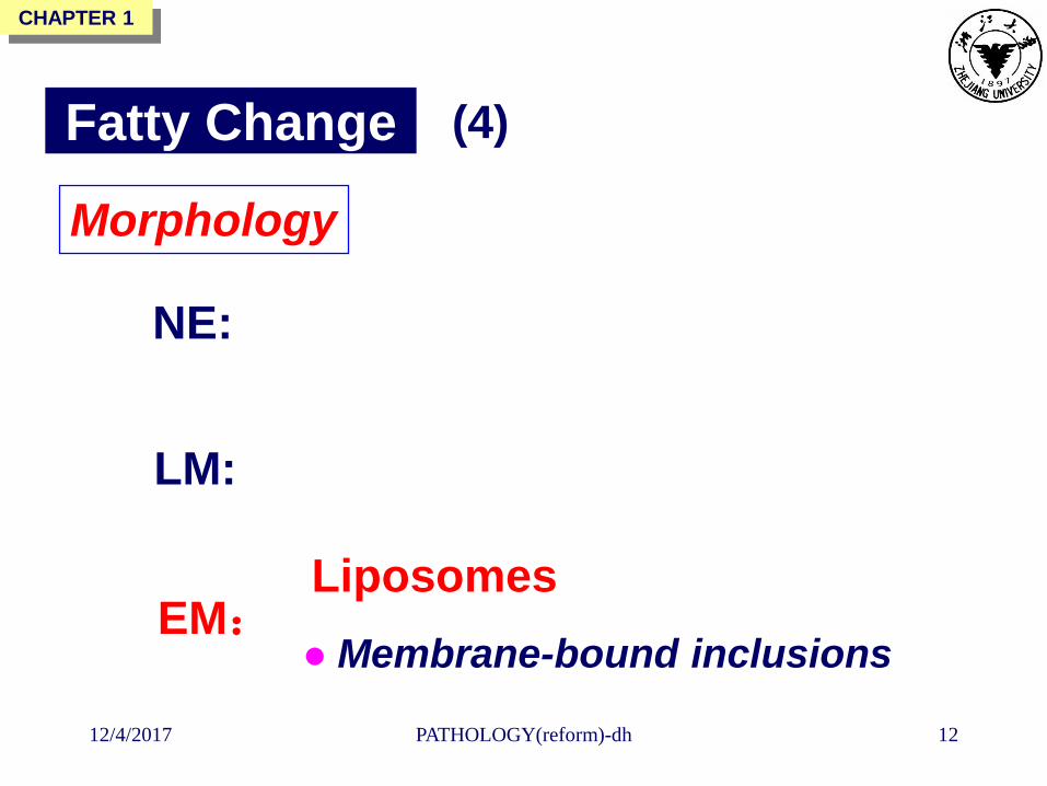
Morphology
Fatty Change (4)
NE:
LM:
EM: Membrane-bound inclusions
Liposomes

CHAPTER 1
12/4/2017 PATHOLOGY(reform)-dh 13
Fatty Change of the Liver
Mild fatty change:
Not affect the gross appearance
With progressive accumulation:
NE
Large
Yellow
Soft
Greasy
Fatty Liver: Severe & diffuse fatty change
(1)

CHAPTER 1
12/4/2017 PATHOLOGY(reform)-dh 14
Fatty liver

CHAPTER 1
12/4/2017 PATHOLOGY(reform)-dh 15
LM
Fatty Change of the Liver (2)
Fat vacuoles
Small , in the cytoplasm
around the nucleus
Displacing the nucleus
to the cell periphery
Fatty cysts

CHAPTER 1
12/4/2017 PATHOLOGY(reform)-dh 16Fatty change of the liver

CHAPTER 1
12/4/2017 PATHOLOGY(reform)-dh 17
Fatty change of the liver(Sudan Ⅲ)

CHAPTER 1
12/4/2017 PATHOLOGY(reform)-dh 18
Mechnisms
Fatty Change of the Liver (3)
Overproduction of triglycerides
Excessive entry of free fatty acids into liver
Enhanced fatty acid synthesis from acetate
Decreased fatty acid oxidation
Decreased apoprotein synthesis

CHAPTER 1
12/4/2017 PATHOLOGY(reform)-dh 19
Fatty Change of the Myocardium
Mild fatty change:
Not affect the gross appearance
With progressive accumulation:
NE
Tigered effect
Apparent bands of yellowed myocardium
alternating with bands of
dark,red-brown,uninvolved myocardium

CHAPTER 1
12/4/2017 PATHOLOGY(reform)-dh 20
Fatty change
of heart
muscle
(tigered effect)

CHAPTER 1
12/4/2017 PATHOLOGY(reform)-dh 21
Fatty change of
heart muscle
(tigered effect)
Sudan-
hematoxylin

CHAPTER 1
12/4/2017 PATHOLOGY(reform)-dh 22

CHAPTER 1
12/4/2017 PATHOLOGY(reform)-dh 23
A homogeneous, translucent, pink
appearance in HE staining
Hyaline Change
Intracellular or extracellular abnormal
accumulation:
Proteins
A descriptive morphologic term
(1)

CHAPTER 1
12/4/2017 PATHOLOGY(reform)-dh 24
Hyaline change in arteriolosclerosis
e.g. Hypertension, Diabetes
Hyaline change in connective tissues
e.g. Old scars
Hyaline change within the cytoplasm
e.g. Nephrotic syndrome, Russell
bodies, Mallory body
Hyaline Change (2)

CHAPTER 1
12/4/2017 PATHOLOGY(reform)-dh 25
Hyaline change in arteriolosclerosis

CHAPTER 1
12/4/2017 PATHOLOGY(reform)-dh 26
Vascular pathology in hypertension. A, Hyaline arteriolosclerosis. The
arteriolar wall is hyalinized, and the lumen is markedly narrowed.

CHAPTER 1
12/4/2017 PATHOLOGY(reform)-dh 27
Chronic glomerulonephritis

CHAPTER 1
12/4/2017 PATHOLOGY(reform)-dh 28
Protein reabsorption droplets in the renal tubular epithelium

CHAPTER 1
12/4/2017 PATHOLOGY(reform)-dh 29
Mallory bodies in liver cells

CHAPTER 1
12/4/2017 PATHOLOGY(reform)-dh 30
Amyloidosis
Extracellular abnormal accumulation:
Amyloid

CHAPTER 1
12/4/2017 PATHOLOGY(reform)-dh 31
Physicochemical characteristics of amyloid
+Iodine--- a brown color--- +H2SO4 --- blue
Staining: Congo red--- red,
HE--- homogeneous pink
EM: nonbranching fibrils 7.5-10 nm wide
X-ray: a pleated –sheet structure
(rendering protein very resistant to
enzymatic degradation, contributing
to its accumulation in tissues)

CHAPTER 1
12/4/2017 PATHOLOGY(reform)-dh 32
Mucoid Change
Extracellular abnormal accumulation:
Mucopolysaccharide
(Glycosaminoglycans, Hyaluronic Acid)

CHAPTER 1
12/4/2017 PATHOLOGY(reform)-dh 33
Pathologic Pigmentation
Intracellular & extracellular abnormal
accumulation:
Exogenous
Endogenous
Colored substances

CHAPTER 1
12/4/2017 PATHOLOGY(reform)-dh 34
Pathologic Pigmentation
Exogenous
Endogenous
✓ Hemosiderin
✓ Lipofuscin
✓ Melanin
✓ Carbon

CHAPTER 1
12/4/2017 PATHOLOGY(reform)-dh 35
Carbon

CHAPTER 1
12/4/2017 PATHOLOGY(reform)-dh 36
Hemosiderin granules in macrophages in the alveolus
Hemosiderin

CHAPTER 1
12/4/2017 PATHOLOGY(reform)-dh 37
Hemosiderin granules in liver cells. A, H&E B, Prussian blue reaction

CHAPTER 1
12/4/2017 PATHOLOGY(reform)-dh 38
Lipofuscin

CHAPTER 1
12/4/2017 PATHOLOGY(reform)-dh 39
Melanin

CHAPTER 1
12/4/2017 PATHOLOGY(reform)-dh 40
Pathologic Calcification
Intracellular & extracellular abnormal
accumulation:
Calcium salts
1. Except for the bones and teeth
2. Pathologic conditions
(1)

CHAPTER 1
12/4/2017 PATHOLOGY(reform)-dh 41
Metastatic calcification in the wall of the stomach

CHAPTER 1
12/4/2017 PATHOLOGY(reform)-dh 42
Dystrophic Calcification
In areas of necrosis
No calcium metabolic derangements
Metastatic calcification
In normal tissues
Some calcium metabolic derangements
Pathologic Calcification (2)

CHAPTER 1
12/4/2017 PATHOLOGY(reform)-dh 43
2. Apoptosis
1. Necrosis
Cell Death
Irreversible Cell Injury
A sequence of morphologic changes
that follow cell death in living tissue
A distinctive and important mode
of cell death regulated by genes

CHAPTER 1
12/4/2017 PATHOLOGY(reform)-dh 44
Necrosis
Two essentially concurrent processes to
produce the morphologic changes :
1. Enzymatic digestion of the cell
2. Denaturation of proteins
(1)
Autolysis
Heterolysis

CHAPTER 1
12/4/2017 PATHOLOGY(reform)-dh 45
Necrosis
Basic pathologic changes
Types of necrosis
Sequences of necrosis
(2)

CHAPTER 1
12/4/2017 PATHOLOGY(reform)-dh 46
Necrosis
Basic Pathologic Changes
(3)
Nuclear changes
Karyolysis
Pyknosis
Karyorrhexis
Cytoplasm Increased eosinophilia

CHAPTER 1
12/4/2017 PATHOLOGY(reform)-dh 47
Necrosis
Types of Necrosis
(4)
Liquefactive necrosis
Fat necrosis
Coagulative necrosis
Caseous necrosis
Gangrene
Fibrinoid necrosis


![Cell Injury[1]](https://img.pdfslide.us/doc/110x75/563dba79550346aa9aa5f218/cell-injury1-56a51a5ef0c98.jpg)