Embed Size (px)
Citation preview

Second Practical Session
CNS Block
Pathology Dept, KSU
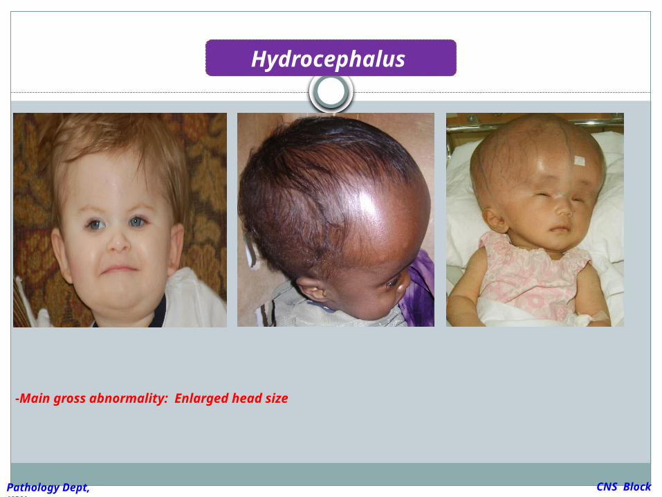
Hydrocephalus
CNS Block
Pathology Dept, KSU
CASE 1:
A 9 months infant was suffering from
enlarged head size and admitted to
hospital with convulsions, went into coma
and died. Autopsy was done and the brain
was large with dilated ventricles .
What is your provisional diagnosis?
CNS Block
Pathology Dept, KSU
Hydrocephalus
CNS Block
Pathology Dept, KSU
-Main gross abnormality: Enlarged head size
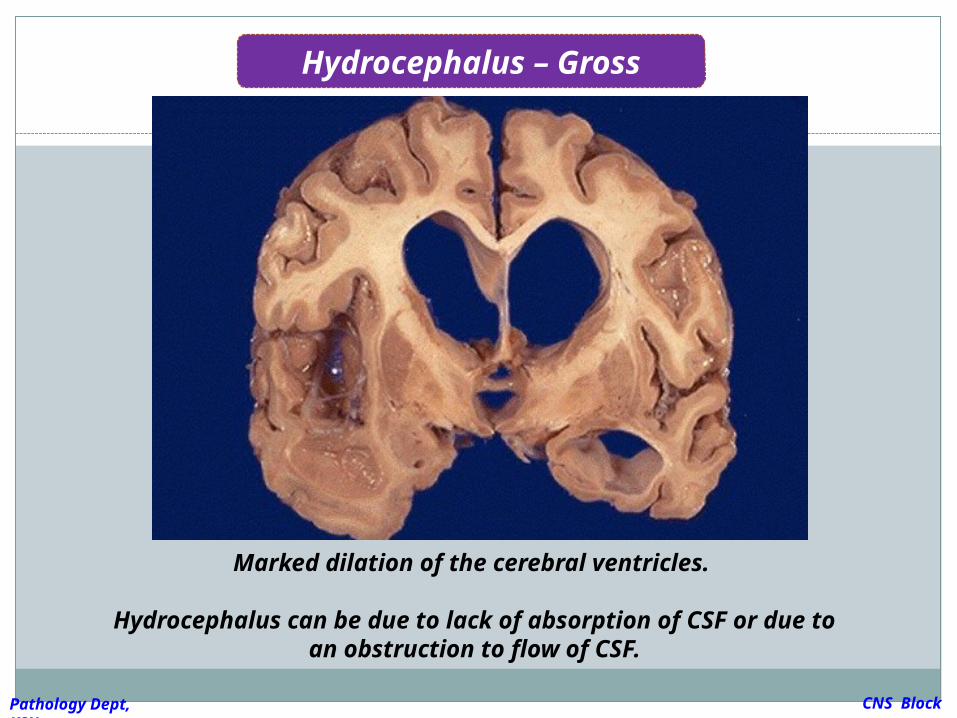
Hydrocephalus – Gross
Marked dilation of the cerebral ventricles.
Hydrocephalus can be due to lack of absorption of CSF or due to an obstruction to flow of CSF.
CNS Block
Pathology Dept, KSU
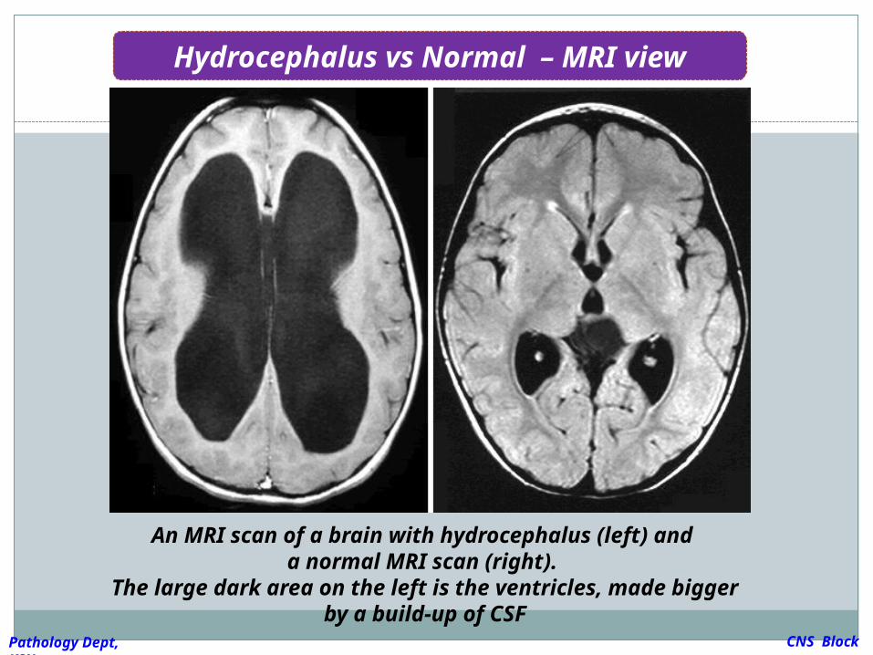
An MRI scan of a brain with hydrocephalus (left) and
a normal MRI scan (right). The large dark area on the left is the ventricles,
made bigger by a build-up of CSF
Hydrocephalus vs Normal – MRI view
CNS Block
Pathology Dept, KSU
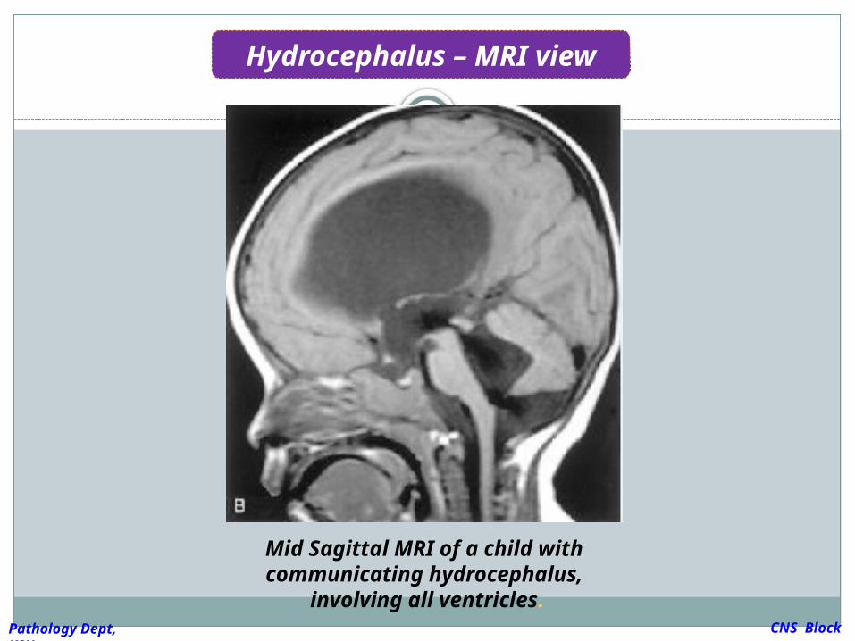
Mid Sagittal MRI of a child with communicating
hydrocephalus, involving all ventricles.
Hydrocephalus – MRI view
CNS Block
Pathology Dept, KSU
Pyogenic (Bacterial ) Meningitis
CNS Block
Pathology Dept, KSU
CASE 2 :
4 years old child who was treated from otitis
media and suddenly complained from
headache, vomiting, fever and stiff neck. CSF
was found to be clouded with abnormal
increase of neutrophils, increased protein
and absence of sugar. Gram stain of the CSF
fluid showed meningococci .
What is your
diagnosis ? CNS Block
Pathology Dept, KSU
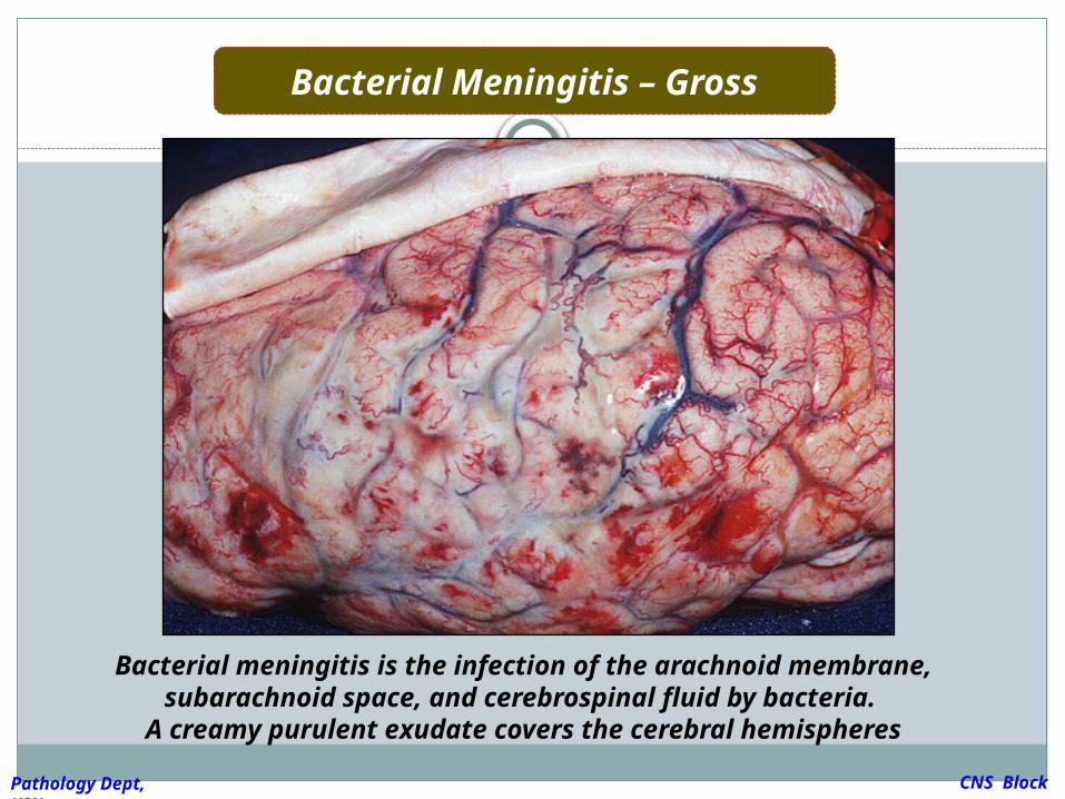
Bacterial Meningitis – Gross
Bacterial meningitis is the infection of the arachnoid membrane, subarachnoid space, and cerebrospinal fluid by
bacteria. A creamy purulent exudate covers the cerebral
hemispheres CNS Block
Pathology Dept, KSU
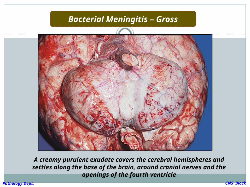
Bacterial Meningitis – Gross
A creamy purulent exudate covers the cerebral hemispheres and settles along the base of the brain, around cranial nerves and the openings of the fourth
ventricle CNS Block
Pathology Dept, KSU
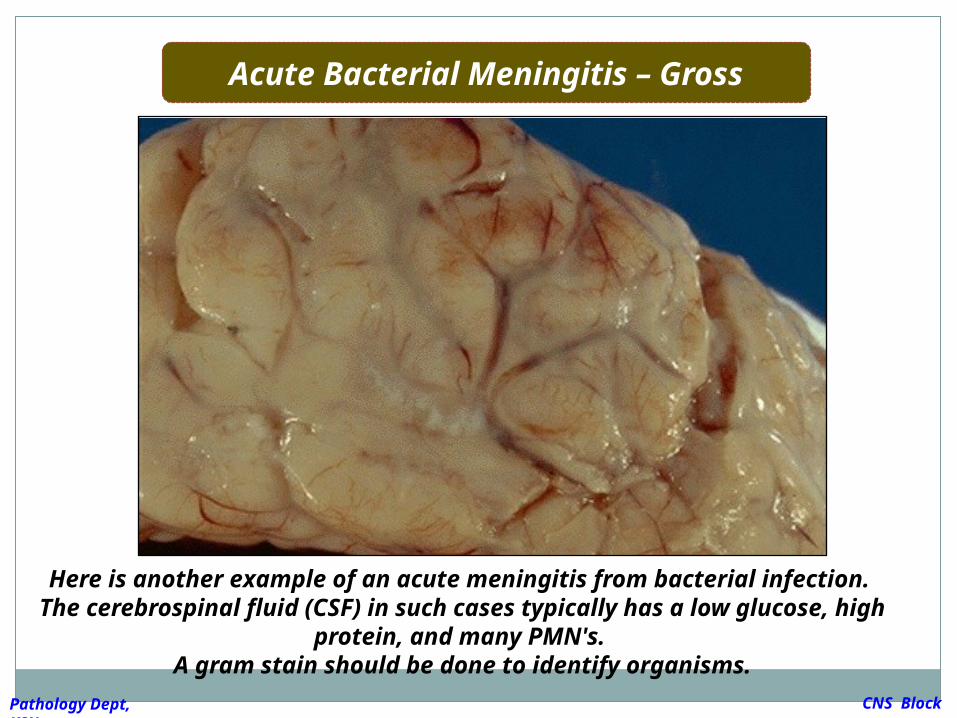
Acute Bacterial Meningitis – Gross
Here is another example of an acute meningitis from bacterial infection.
The cerebrospinal fluid (CSF) in such cases typically has a low glucose, high protein, and many PMN's.
A gram stain should be done to identify organisms. CNS Block
Pathology Dept, KSU
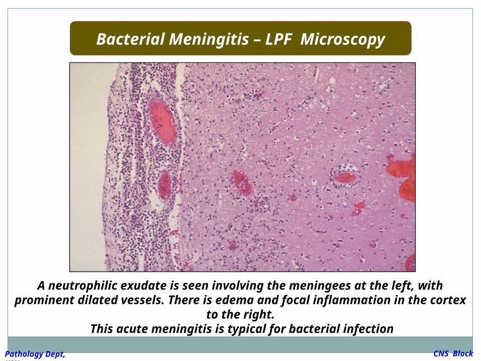
Bacterial Meningitis – LPF Microscopy
A neutrophilic exudate is seen involving the meningees at the left, with prominent dilated vessels. There is edema and focal
inflammation in the cortex to the right. This acute meningitis is typical for bacterial infection
CNS Block
Pathology Dept, KSU
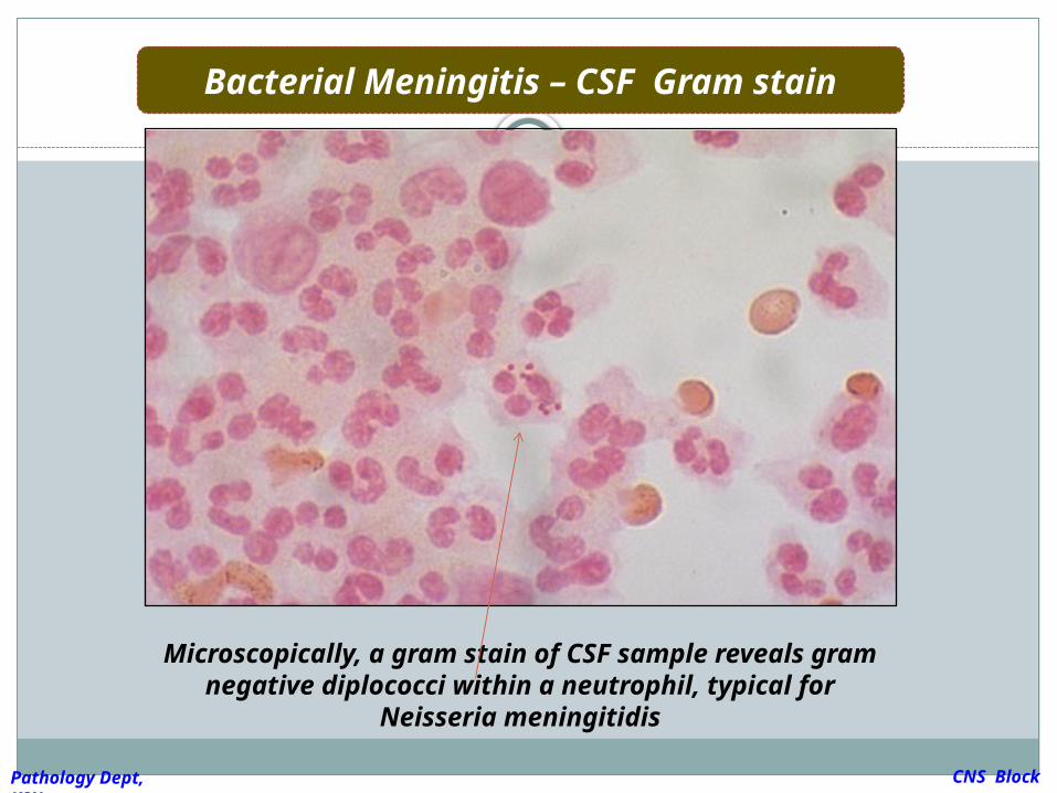
Bacterial Meningitis – CSF Gram stain
Microscopically, a gram stain of CSF sample reveals gram negative diplococci within a
neutrophil, typical for Neisseria meningitidis
CNS Block
Pathology Dept, KSU
Cerebral Abscess
CNS Block
Pathology Dept, KSU
CASE 3:
A 35 years old lady complains from otitis
media . Suddenly she suffers from headache
and convulsions. Brain MRI reveals 5 cm.
fluid filled cavity in the temporal lobe.
Examination of the CSF shows increased
pressure with lymphocytes and increased
protein but there is no change of sugar
content.
What is your diagnosis ?
CNS Block
Pathology Dept, KSU
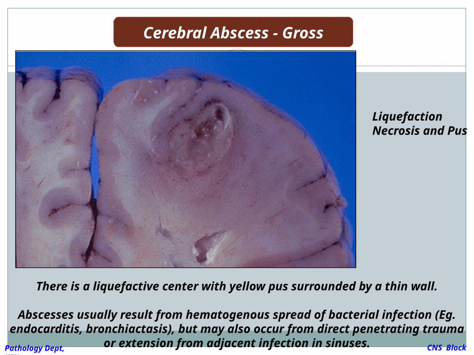
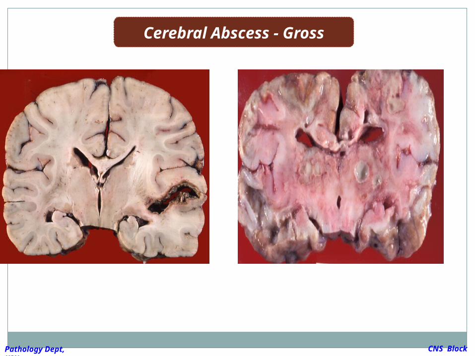
Cerebral Abscess - Gross
There is a liquefactive center with yellow pus surrounded by a thin wall.
Abscesses usually result from hematogenous spread of bacterial
infection (Eg. endocarditis, bronchiactasis), but may also occur from direct penetrating trauma or extension from adjacent infection in
sinuses.
CNS Block
Pathology Dept, KSU
Liquefaction Necrosis and Pus
Cerebral Abscess - Gross
CNS Block
Pathology Dept, KSU
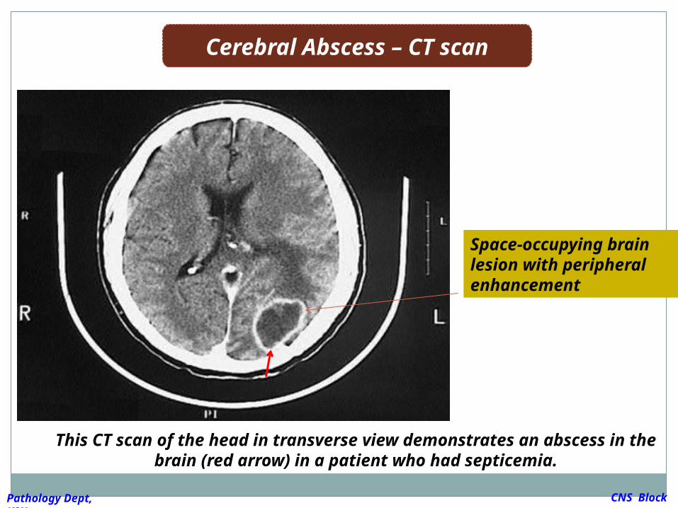
Cerebral Abscess – CT scan
This CT scan of the head in transverse view demonstrates an abscess in the brain (red arrow) in a patient who had septicemia.
CNS Block
Pathology Dept, KSU
Space-occupying brain lesion with peripheral enhancement
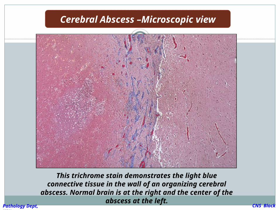
Cerebral Abscess –Microscopic view
This trichrome stain demonstrates the light blue connective tissue in the wall of an organizing cerebral abscess. Normal brain is at the right
and the center of the abscess at the left.CNS Block
Pathology Dept, KSU
Ruptured Berry Aneurysm causing subarachnoid
hemorrhage
CNS Block
Pathology Dept, KSU
A previously healthy 31-year-old woman
experiences a severe headache and loses
consciousness within an hour. An emergent head
CT scan reveals extensive subarachnoid
hemorrhage at the base of the brain. She is a
febrile. A lumbar puncture yields cerebrospinal
fluid with many red blood cells, but no white blood
cells. The CSF protein is slightly increased, but the
glucose is normal.
What is your provisional diagnosis ?
CASE 4:
CNS Block
Pathology Dept, KSU
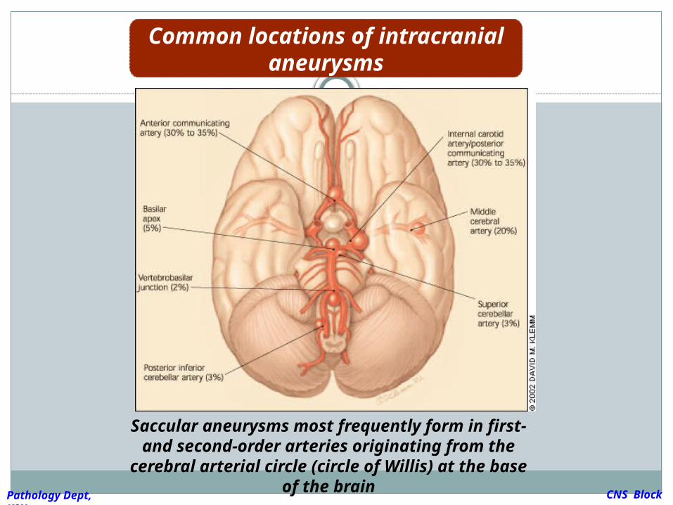
Common locations of intracranial aneurysms
Saccular aneurysms most frequently form in first- and second-order arteries
originating from the cerebral arterial circle (circle of Willis) at the base of the brain CNS
BlockPathology Dept, KSU
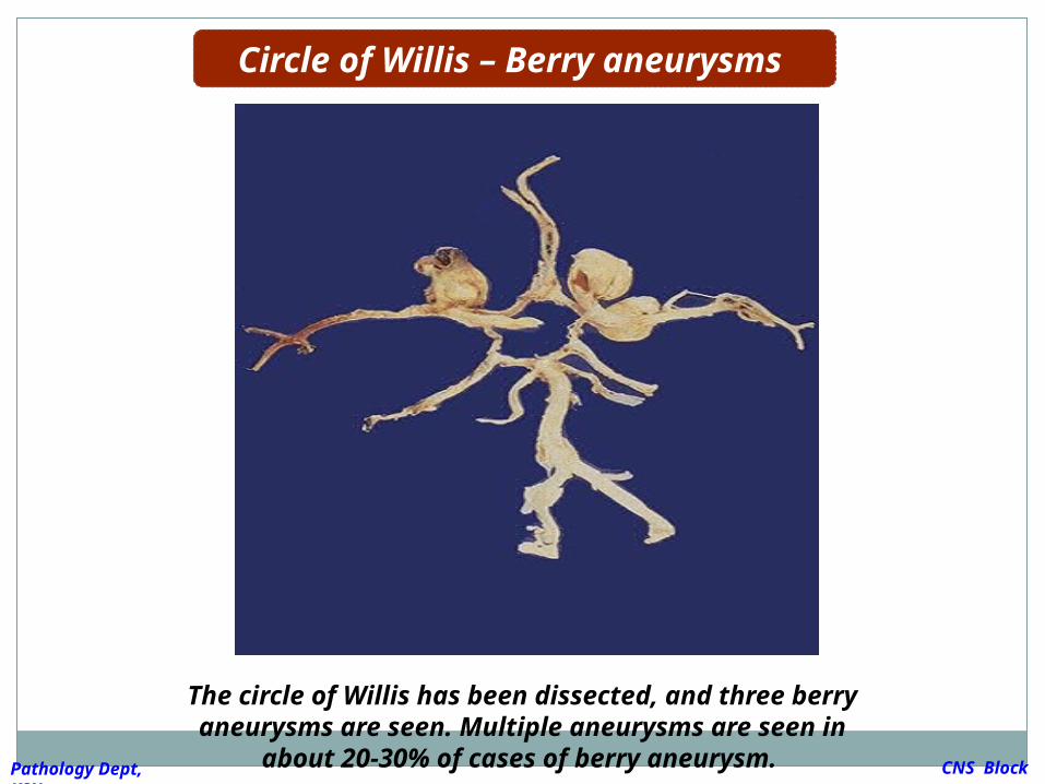
The circle of Willis has been dissected, and three berry aneurysms are seen. Multiple
aneurysms are seen in about 20-30% of cases of berry aneurysm.
Circle of Willis – Berry aneurysms
CNS Block
Pathology Dept, KSU
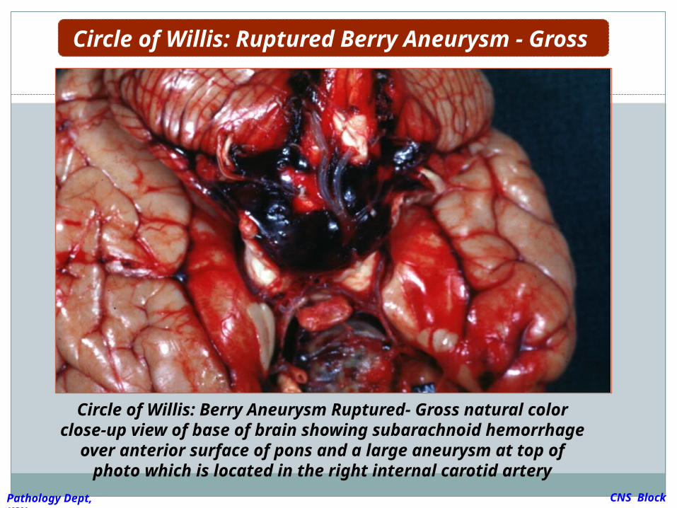
Circle of Willis: Berry Aneurysm Ruptured- Gross natural color close-up view of base of brain showing subarachnoid hemorrhage over anterior surface of pons and a large aneurysm at top of photo which is
located in the right internal carotid artery
Circle of Willis: Ruptured Berry Aneurysm - Gross
CNS Block
Pathology Dept, KSU
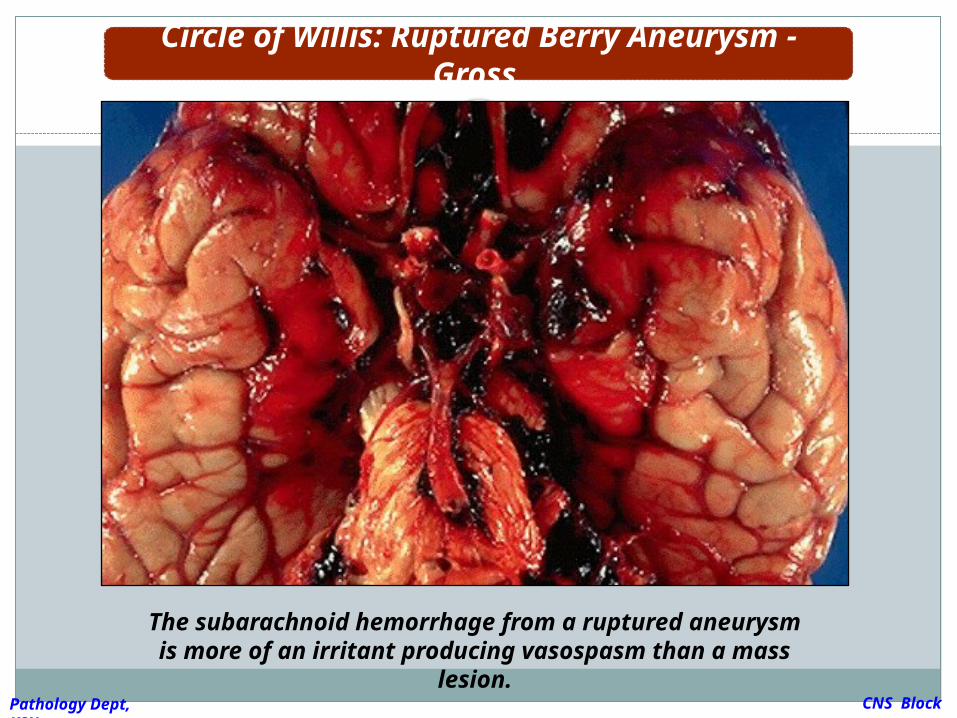
The subarachnoid hemorrhage from a ruptured aneurysm is more of an irritant producing
vasospasm than a mass lesion.
Circle of Willis: Ruptured Berry Aneurysm - Gross
CNS Block
Pathology Dept, KSU
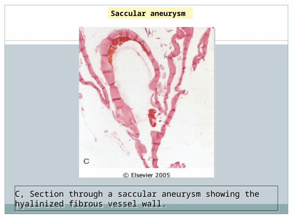
C, Section through a saccular aneurysm showing the hyalinized fibrous vessel wall.C, Section through a saccular aneurysm showing the hyalinized fibrous vessel wall.
Saccular aneurysm
Alzheimer disease
CNS Block
Pathology Dept, KSU
A 85 years old man complains of
progressive loss of memory, disorientation
and alterations in mood and behavior since
20 years. He was admitted to hospital
because he was disabled and immobile and
he died in hospital after one week of
admission. Autopsy was done and the brain
cortex was found to be atrophied.
What is your diagnosis ?
CASE 5 :
CNS Block
Pathology Dept, KSU
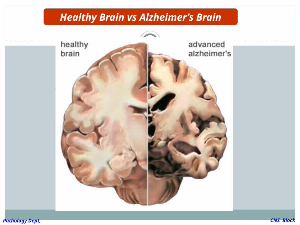
Healthy Brain vs Alzheimer’s Brain
CNS Block
Pathology Dept, KSU
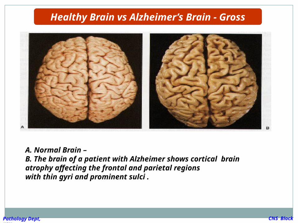
A. Normal Brain – B. The brain of a patient with Alzheimer shows cortical brain atrophy affecting the frontal and parietal regionswith thin gyri and prominent sulci .
Healthy Brain vs Alzheimer’s Brain - Gross
CNS Block
Pathology Dept, KSU
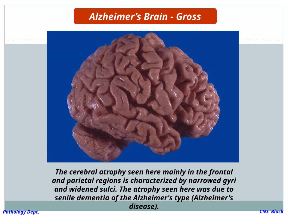
Alzheimer’s Brain - Gross
The cerebral atrophy seen here mainly in the frontal and parietal regions is characterized
by narrowed gyri and widened sulci. The atrophy seen here was due to senile dementia of the Alzheimer's type (Alzheimer's disease).
CNS Block
Pathology Dept, KSU
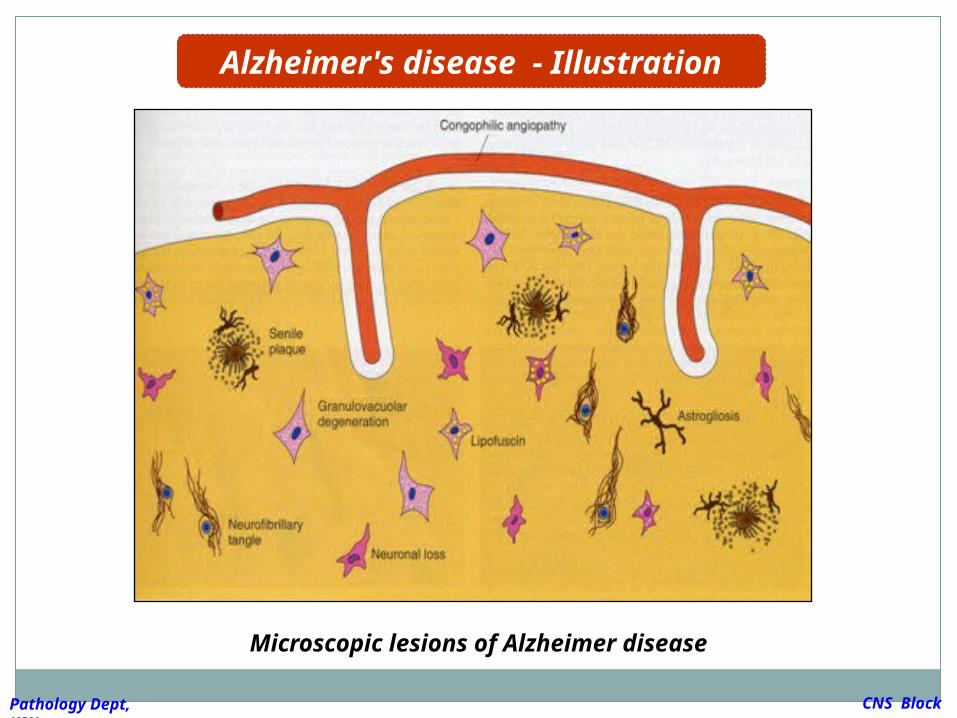
Microscopic lesions of Alzheimer disease
Alzheimer's disease - Illustration
CNS Block
Pathology Dept, KSU
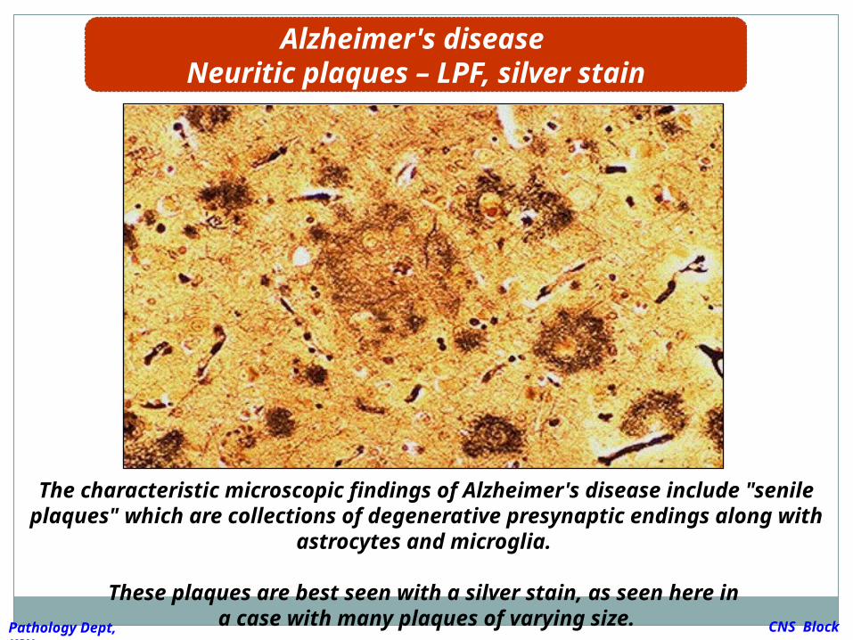
The characteristic microscopic findings of Alzheimer's disease include "senile plaques" which are collections of degenerative
presynaptic endings along with astrocytes and microglia.
These plaques are best seen with a silver stain, as seen here in a case with many plaques of varying size.
Alzheimer's disease Neuritic plaques – LPF, silver stain
CNS Block
Pathology Dept, KSU
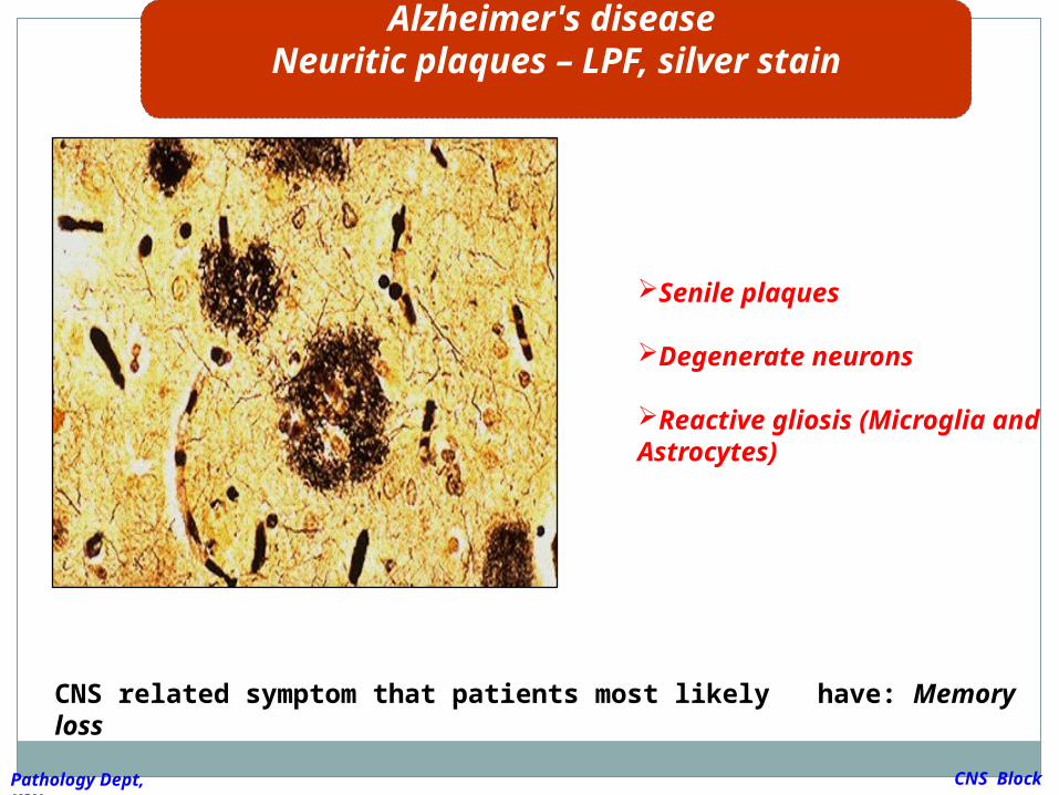
CNS related symptom that patients most likely have: Memory loss
Alzheimer's disease Neuritic plaques – LPF, silver stain
CNS Block
Pathology Dept, KSU
Senile plaques
Degenerate neurons
Reactive gliosis (Microglia and Astrocytes)
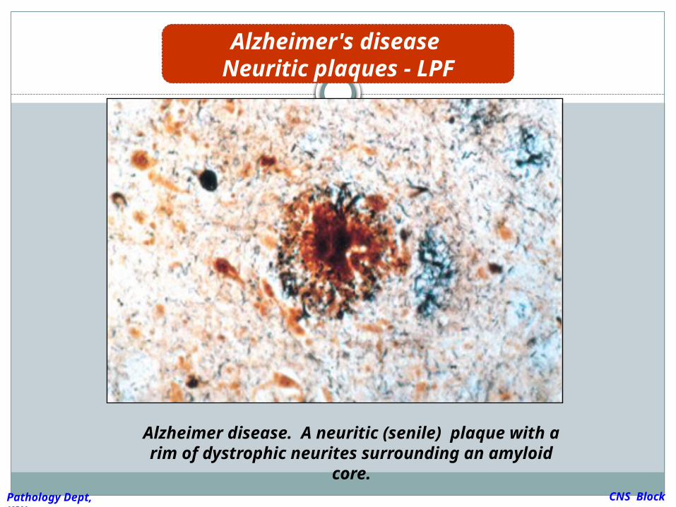
Alzheimer disease. A neuritic (senile) plaque with a rim of dystrophic neurites surrounding
an amyloid core.
Alzheimer's disease Neuritic plaques - LPF
CNS Block
Pathology Dept, KSU
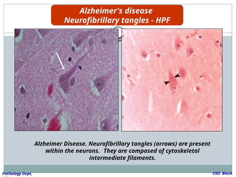
Alzheimer Disease. Neurofibrillary tangles (arrows) are present within the neurons. They are composed of
cytoskeletal intermediate filaments.
Alzheimer's disease Neurofibrillary tangles -
HPF
CNS Block
Pathology Dept, KSU

THE END