Embed Size (px)
Citation preview
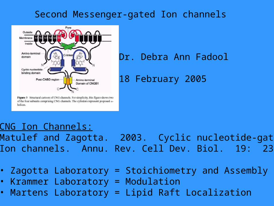
Second Messenger-gated Ion channels
CNG Ion Channels:Matulef and Zagotta. 2003. Cyclic nucleotide-gatedIon channels. Annu. Rev. Cell Dev. Biol. 19: 23-44
• Zagotta Laboratory = Stoichiometry and Assembly• Krammer Laboratory = Modulation• Martens Laboratory = Lipid Raft Localization
Dr. Debra Ann Fadool
18 February 2005
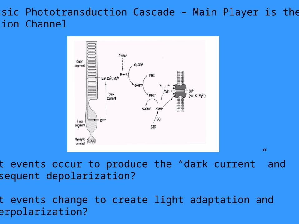
Classic Phototransduction Cascade – Main Player is theCNG ion Channel
What events occur to produce the “dark current” and subsequent depolarization?
What events change to create light adaptation and hyperpolarization?
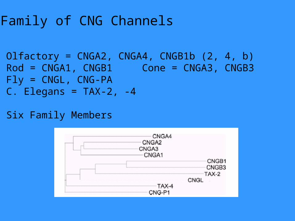
Family of CNG Channels
Olfactory = CNGA2, CNGA4, CNGB1b (2, 4, b)Rod = CNGA1, CNGB1 Cone = CNGA3, CNGB3Fly = CNGL, CNG-PAC. Elegans = TAX-2, -4
Six Family Members
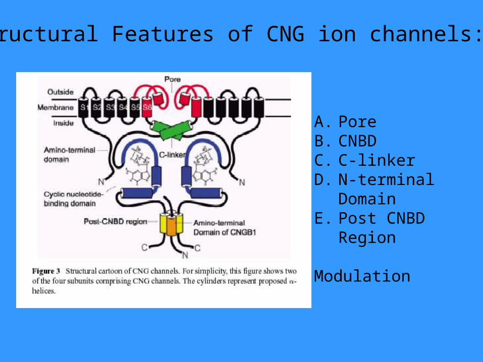
Structural Features of CNG ion channels:
A. PoreB. CNBDC. C-linkerD. N-terminal
DomainE. Post CNBD
Region
Modulation
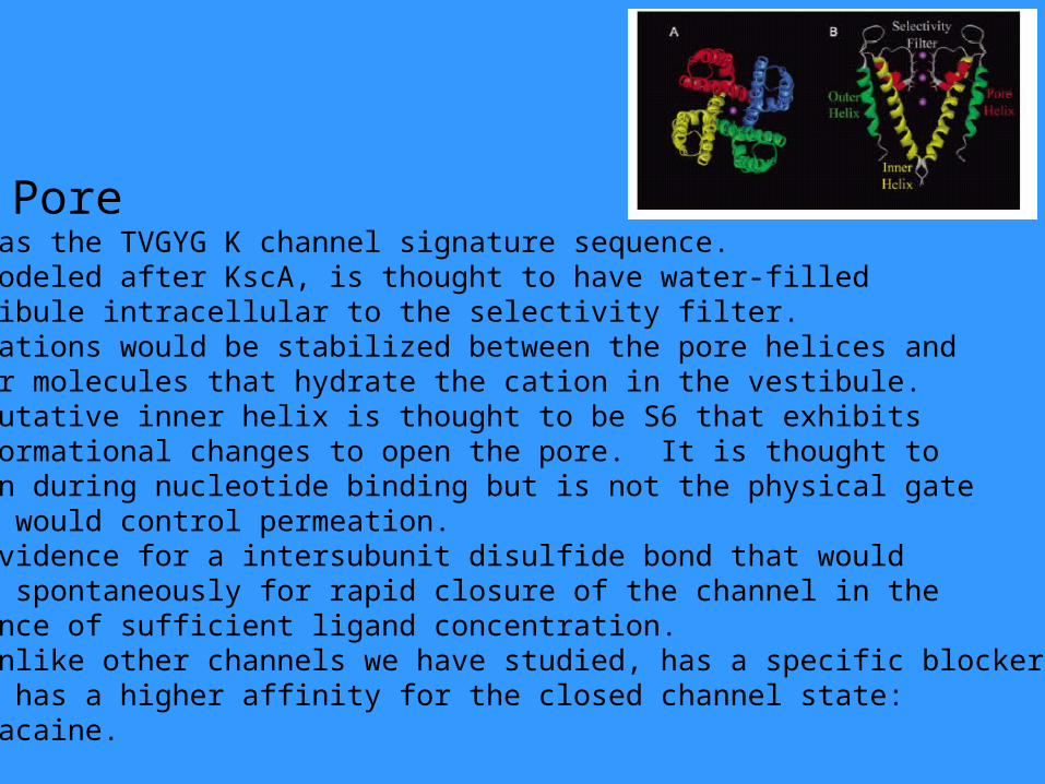
A. Pore1. Has the TVGYG K channel signature sequence.2. Modeled after KscA, is thought to have water-filledvestibule intracellular to the selectivity filter.3. Cations would be stabilized between the pore helices and water molecules that hydrate the cation in the vestibule.4. Putative inner helix is thought to be S6 that exhibitsconformational changes to open the pore. It is thought to widen during nucleotide binding but is not the physical gatethat would control permeation.5. Evidence for a intersubunit disulfide bond that would form spontaneously for rapid closure of the channel in the absence of sufficient ligand concentration.6. Unlike other channels we have studied, has a specific blockerthat has a higher affinity for the closed channel state: Tetracaine.
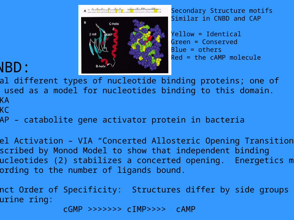
B. CNBD:1. Several different types of nucleotide binding proteins; one ofwhich is used as a model for nucleotides binding to this domain.
a. PKAb. PKCc. CAP – catabolite gene activator protein in bacteria
2. Channel Activation – VIA “Concerted Allosteric Opening Transition”First described by Monod Model to show that independent bindingof the nucleotides (2) stabilizes a concerted opening. Energetics mayvary according to the number of ligands bound.
3. Distinct Order of Specificity: Structures differ by side groups on the purine ring:
cGMP >>>>>>> cIMP>>>> cAMP
Secondary Structure motifs Similar in CNBD and CAP
Yellow = IdenticalGreen = ConservedBlue = othersRed = the cAMP molecule
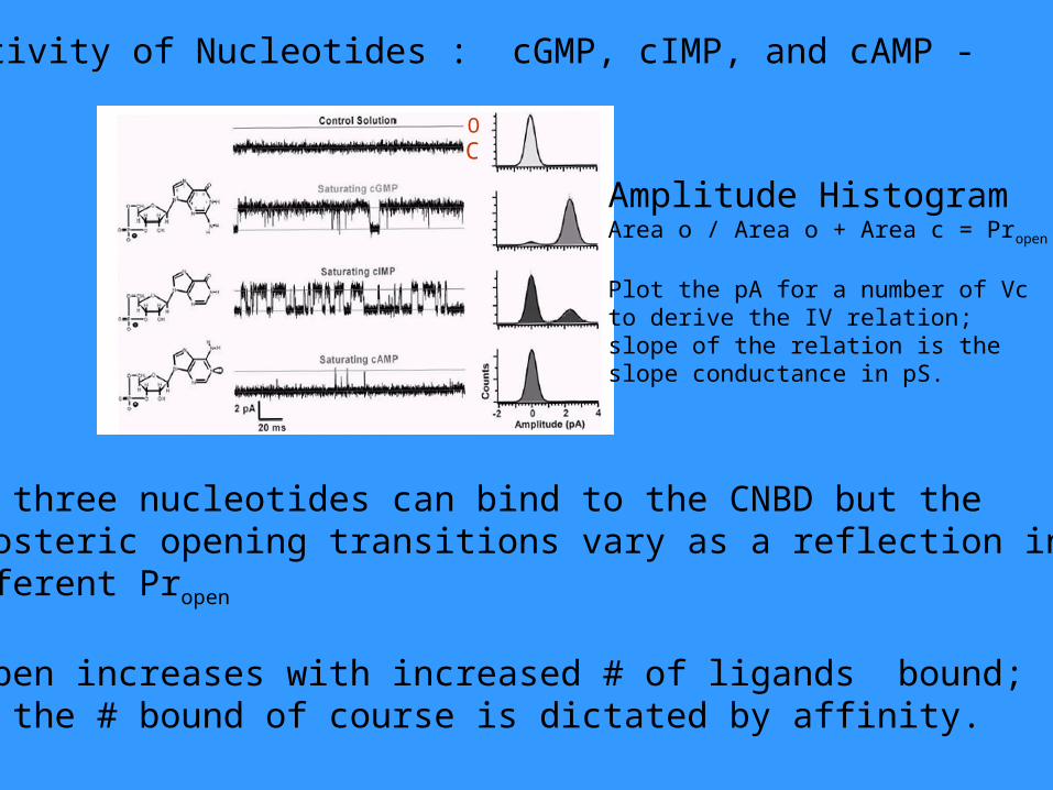
CO
Amplitude HistogramArea o / Area o + Area c = Propen
Plot the pA for a number of Vcto derive the IV relation; slope of the relation is the slope conductance in pS.
All three nucleotides can bind to the CNBD but theallosteric opening transitions vary as a reflection in different Propen
Propen increases with increased # of ligands bound; but the # bound of course is dictated by affinity.
Activity of Nucleotides : cGMP, cIMP, and cAMP -
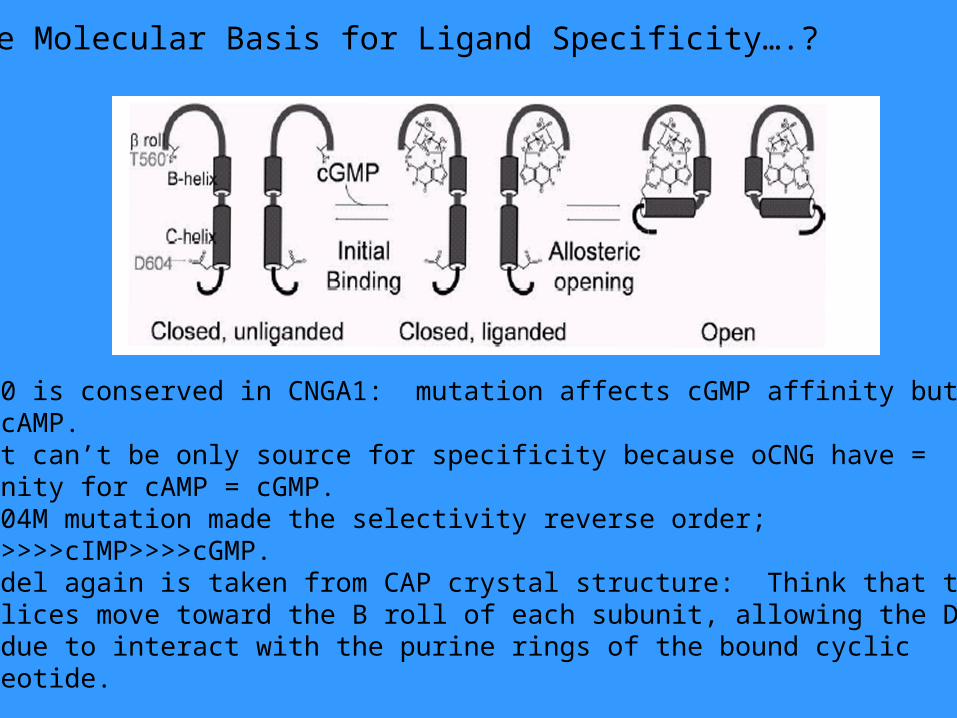
The Molecular Basis for Ligand Specificity….?
•T560 is conserved in CNGA1: mutation affects cGMP affinity butnot cAMP.• But can’t be only source for specificity because oCNG have =affinity for cAMP = cGMP.• D604M mutation made the selectivity reverse order; cAMP>>>>cIMP>>>>cGMP.• Model again is taken from CAP crystal structure: Think that the C-helices move toward the B roll of each subunit, allowing the D604 residue to interact with the purine rings of the bound cyclicnucleotide.
C.The C Linker:1. The residues of the linker are modulated by metals.2. Three residues of the linker can affect gating –
R460, I465, and N466.
D. N-Terminal Domain:1. Stabilizes the open state by decreasing the free energy (delta G)of gating: Called the “autoexcitatory effect on gating”.2. Ca/Cam binding to the N-terminal of CNGA2 causes a decrease
in Propen.3. Olfactory adaptation: negative feedback of Ca (permeant ion)
for Ca/Cam that inhibits the N-terminal domain of the channel.4. N and C terminus interact directly: Ca/Cam prevents this
interaction required for gating, and causes decreasecAMP/cGMP from binding.
E. Post-CNBD Region:1. Also mediates the Ca/Cam modulation/inhibition.2. Important for trafficking and heteromeric assembly3. RP = truncated mutation in this region.
F. Types of Modulation:1. Ca/Cam2. Metals3. PKC4. DAG5. Na/Ca K exchangers in protein-protein interatactions6. Role of circadian rhythms
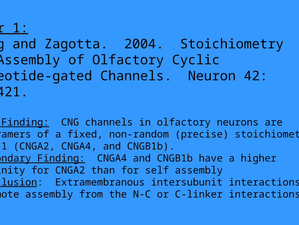
Paper 1:Zheng and Zagotta. 2004. Stoichiometry and Assembly of Olfactory Cyclic Nucleotide-gated Channels. Neuron 42: 411-421.
Key Finding: CNG channels in olfactory neurons are tetramers of a fixed, non-random (precise) stoichiometry:2:1:1 (CNGA2, CNGA4, and CNGB1b).Secondary Finding: CNGA4 and CNGB1b have a higheraffinity for CNGA2 than for self assemblyConclusion: Extramembranous intersubunit interactionspromote assembly from the N-C or C-linker interactions.
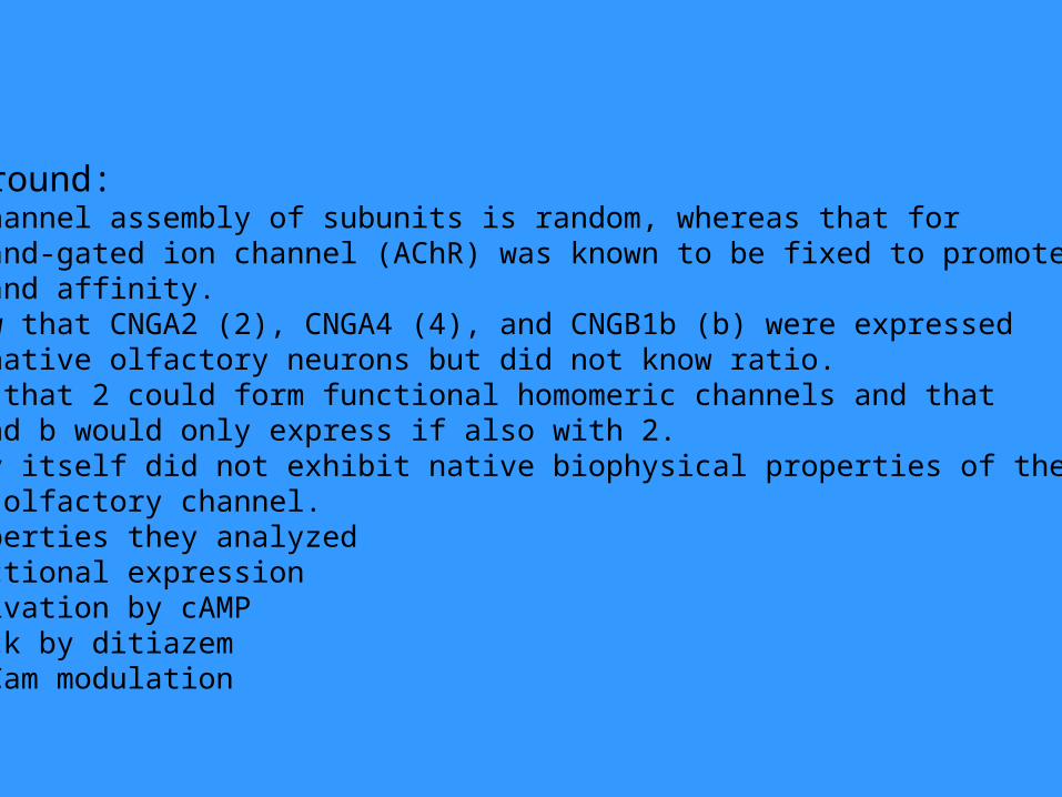
Background:1. K channel assembly of subunits is random, whereas that for
ligand-gated ion channel (AChR) was known to be fixed to promoteligand affinity.
2. Knew that CNGA2 (2), CNGA4 (4), and CNGB1b (b) were expressedin native olfactory neurons but did not know ratio.
3. New that 2 could form functional homomeric channels and that4 and b would only express if also with 2.
4. 2 by itself did not exhibit native biophysical properties of the CNG olfactory channel.
5. Properties they analyzeda. Functional expressionb. Activation by cAMPc. Block by ditiazemd. Ca/Cam modulation
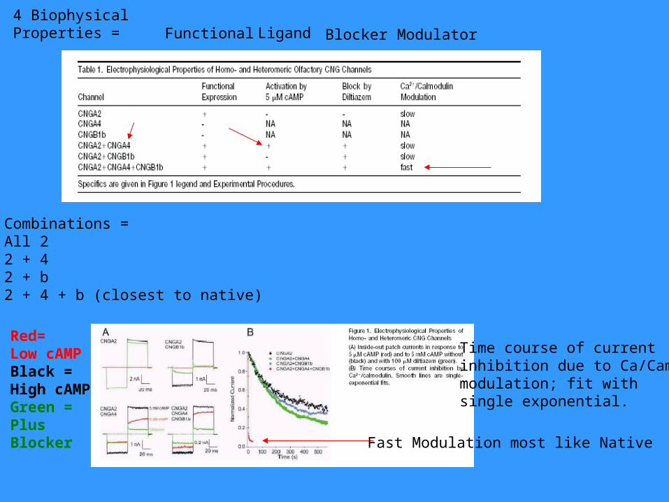
Functional Ligand Blocker Modulator4 BiophysicalProperties =
Combinations = All 22 + 42 + b2 + 4 + b (closest to native)
Time course of currentinhibition due to Ca/Cammodulation; fit withsingle exponential.
Red=Low cAMPBlack = High cAMPGreen = Plus Blocker Fast Modulation most like Native
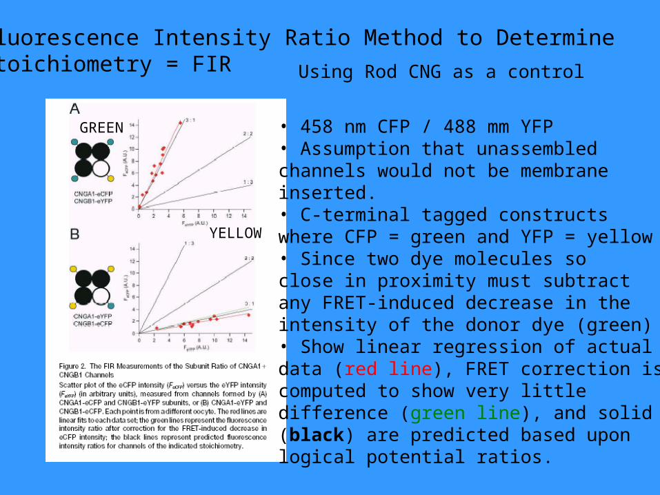
Fluorescence Intensity Ratio Method to Determine Stoichiometry = FIR
• 458 nm CFP / 488 mm YFP• Assumption that unassembledchannels would not be membraneinserted.• C-terminal tagged constructswhere CFP = green and YFP = yellow• Since two dye molecules so close in proximity must subtractany FRET-induced decrease in theintensity of the donor dye (green)• Show linear regression of actual data (red line), FRET correction is computed to show very little difference (green line), and solid lines(black) are predicted based uponlogical potential ratios.
YELLOW
GREEN
Using Rod CNG as a control
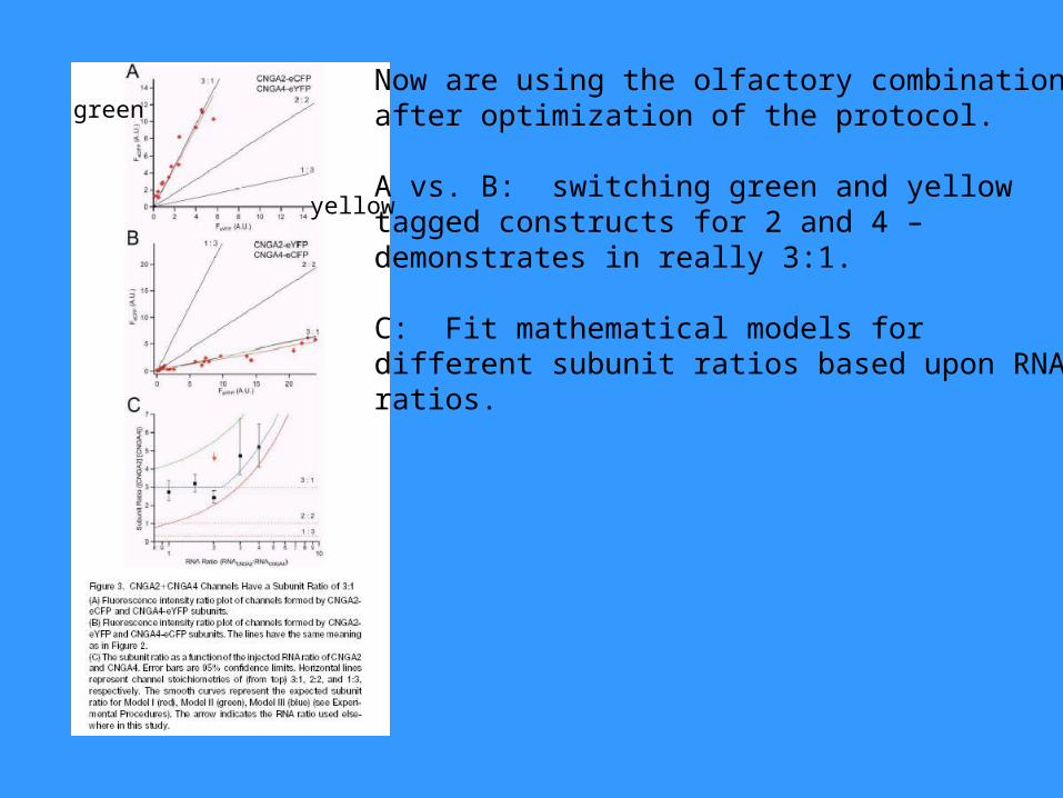
green
yellow
Now are using the olfactory combinationsafter optimization of the protocol.
A vs. B: switching green and yellowtagged constructs for 2 and 4 –demonstrates in really 3:1.
C: Fit mathematical models for different subunit ratios based upon RNAratios.
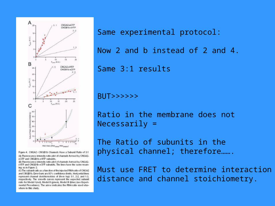
Same experimental protocol:
Now 2 and b instead of 2 and 4.
Same 3:1 results
BUT>>>>>>
Ratio in the membrane does not Necessarily =
The Ratio of subunits in the physical channel; therefore…….
Must use FRET to determine interactiondistance and channel stoichiometry.
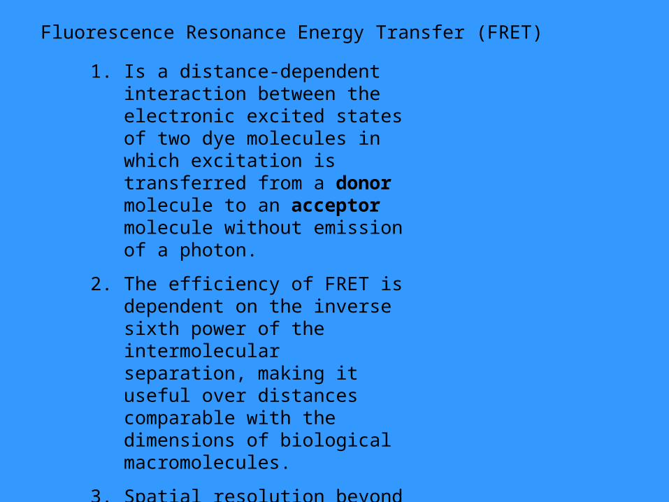
Fluorescence Resonance Energy Transfer (FRET)
1. Is a distance-dependent interaction between the electronic excited states of two dye molecules in which excitation is transferred from a donor molecule to an acceptor molecule without emission of a photon.
2. The efficiency of FRET is dependent on the inverse sixth power of the intermolecular separation, making it useful over distances comparable with the dimensions of biological macromolecules.
3. Spatial resolution beyond the limits of conventional optical microscopy.
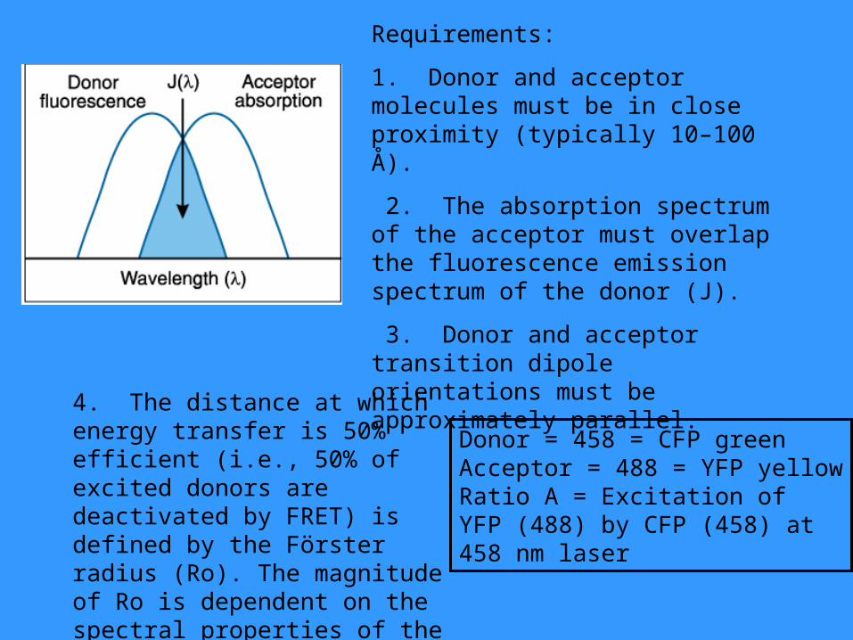
Requirements:
1. Donor and acceptor molecules must be in close proximity (typically 10–100 Å).
2. The absorption spectrum of the acceptor must overlap the fluorescence emission spectrum of the donor (J).
3. Donor and acceptor transition dipole orientations must be approximately parallel.
4. The distance at which energy transfer is 50% efficient (i.e., 50% of excited donors are deactivated by FRET) is defined by the Förster radius (Ro). The magnitude of Ro is dependent on the spectral properties of the donor and acceptor dyes
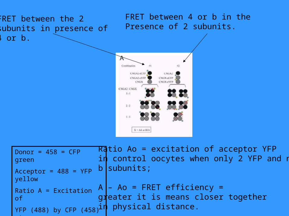
Donor = 458 = CFP greenAcceptor = 488 = YFP yellowRatio A = Excitation of YFP (488) by CFP (458) at 458 nm laser
Donor = 458 = CFP green
Acceptor = 488 = YFP yellow
Ratio A = Excitation of
YFP (488) by CFP (458) at
458 nm laser
FRET between the 2subunits in presence of 4 or b.
FRET between 4 or b in the Presence of 2 subunits.
Ratio Ao = excitation of acceptor YFPin control oocytes when only 2 YFP and no b subunits; A – Ao = FRET efficiency = greater it is means closer togetherin physical distance.
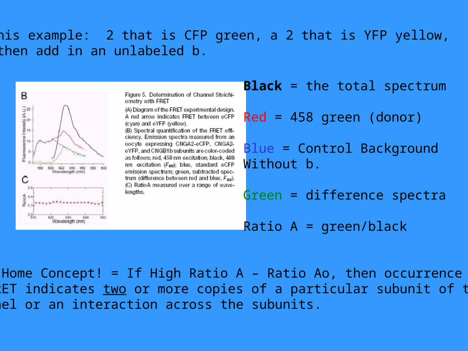
In this example: 2 that is CFP green, a 2 that is YFP yellow, and then add in an unlabeled b.
Black = the total spectrum
Red = 458 green (donor)
Blue = Control Background Without b.
Green = difference spectra
Ratio A = green/black
Take Home Concept! = If High Ratio A – Ratio Ao, then occurrenceof FRET indicates two or more copies of a particular subunit of thechannel or an interaction across the subunits.
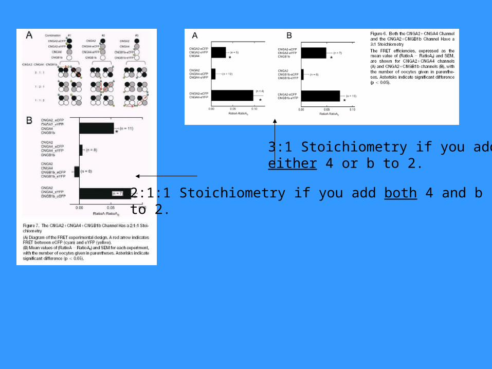
3:1 Stoichiometry if you addeither 4 or b to 2.
2:1:1 Stoichiometry if you add both 4 and bto 2.
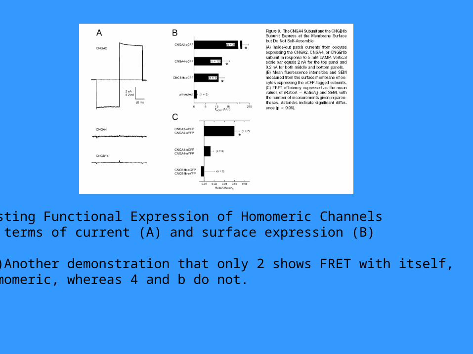
Testing Functional Expression of Homomeric ChannelsIn terms of current (A) and surface expression (B)
(C) Another demonstration that only 2 shows FRET with itself,homomeric, whereas 4 and b do not.
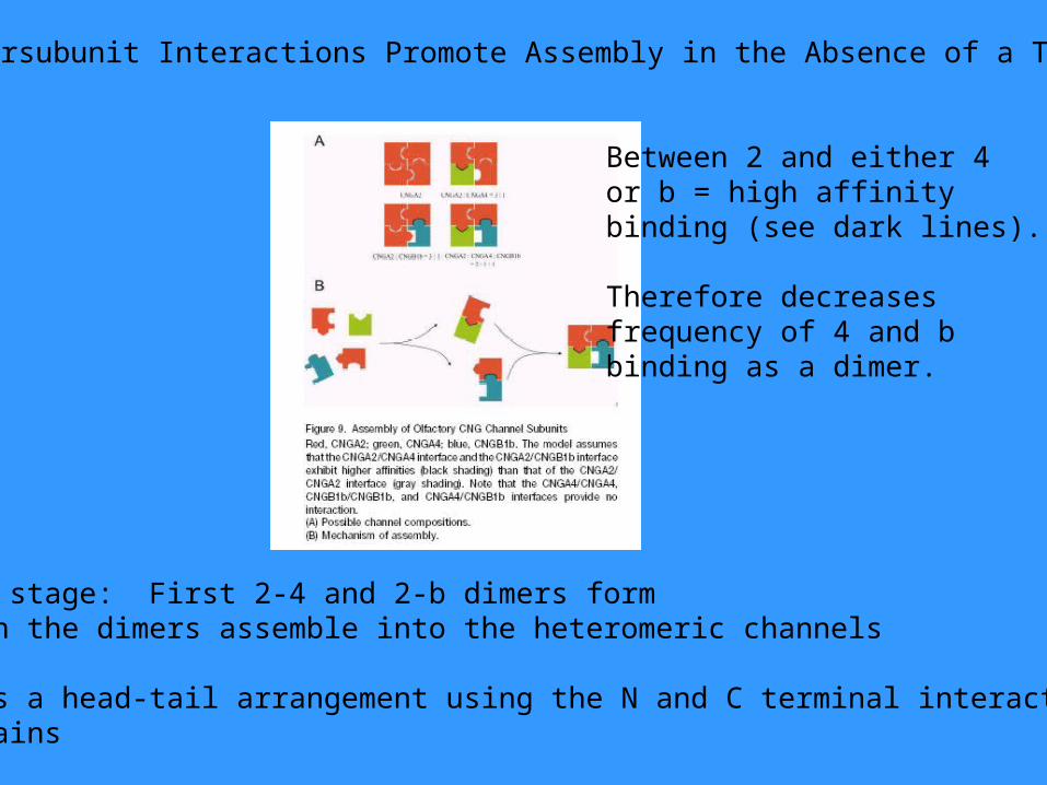
Intersubunit Interactions Promote Assembly in the Absence of a T1
Two stage: First 2-4 and 2-b dimers formThen the dimers assemble into the heteromeric channels
Uses a head-tail arrangement using the N and C terminal interactingdomains
Between 2 and either 4or b = high affinity binding (see dark lines).
Therefore decreasesfrequency of 4 and bbinding as a dimer.
Paper 2:Krajewski et al. 2003. Tyrosine Phosphory-Lation of Rod Cyclic Nucleotide-gatedChannels Switches off Ca/Cam Inhibition.JNS 23(31): 10100-10106.
Key Finding: Y498 in the CNGA1 is the phosphorylation Site responsible for Ca/Cam Inhibition.Secondary Finding: Y Phosphorylation of CNGA1 on the C-terminus can cause an uncoupling of the N-terminusof CNGB1 so that there is no Ca/Cam modulation.Conclusion: Y Phosphorylation decreases Propen whereas Dephosphorylation increases Propen.
Background:1. It was known that Ca/Cam binds with high affinity to the N-terminusof the CNGB1 subunit to weaken the intramolecular interaction Between the N and C termini of CNGB1 and CNGA1. 2. Y498 is on CNGA1 and Y1097 is on CNGB1; either mutation causesa decreased affinity for cGMP to decrease gating.3. IGF causes dephosphorylaton of these sites to increase 4. CNG sensitivity.
5. What the authors wish to address…..do phosphorylation and Ca/Cam act as separate, independent modulators of the channel or is there a common mechanism?
6. I-O patches of transfected oocytes, spontaneously dephosphorylatethe channels, so the Propen increases over a 5-10 minute period.7. During dePhos…… K1/2 decreases approximately 2x change, reflecting an increase in cGMP affinity.
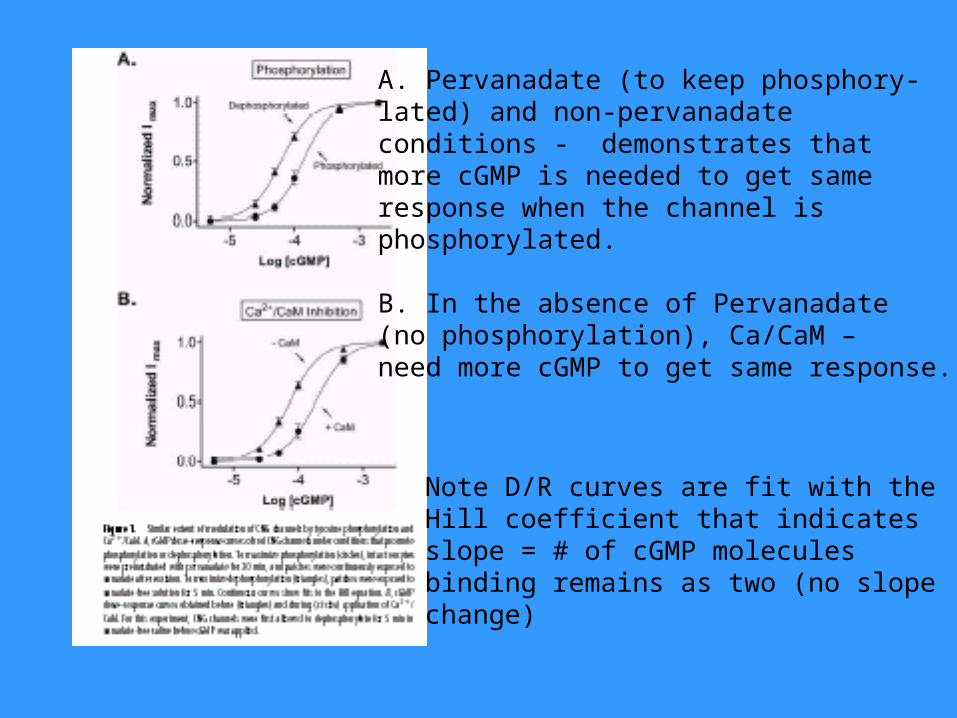
A. Pervanadate (to keep phosphory-lated) and non-pervanadate conditions - demonstrates that more cGMP is needed to get sameresponse when the channel isphosphorylated.
B. In the absence of Pervanadate (no phosphorylation), Ca/CaM – need more cGMP to get same response.
Note D/R curves are fit with the Hill coefficient that indicatesslope = # of cGMP molecules binding remains as two (no slopechange)
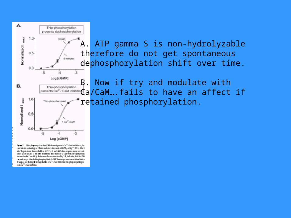
A. ATP gamma S is non-hydrolyzabletherefore do not get spontaneousdephosphorylation shift over time.
B. Now if try and modulate withCa/CaM….fails to have an affect ifretained phosphorylation.
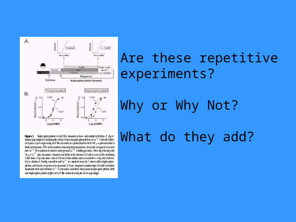
Are these repetitiveexperiments?
Why or Why Not?
What do they add?
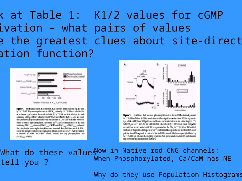
Look at Table 1: K1/2 values for cGMPactivation – what pairs of valuesgive the greatest clues about site-directedmutation function?
What do these valuestell you ?
Now in Native rod CNG channels:When Phosphorylated, Ca/CaM has NE
Why do they use Population Histograms?
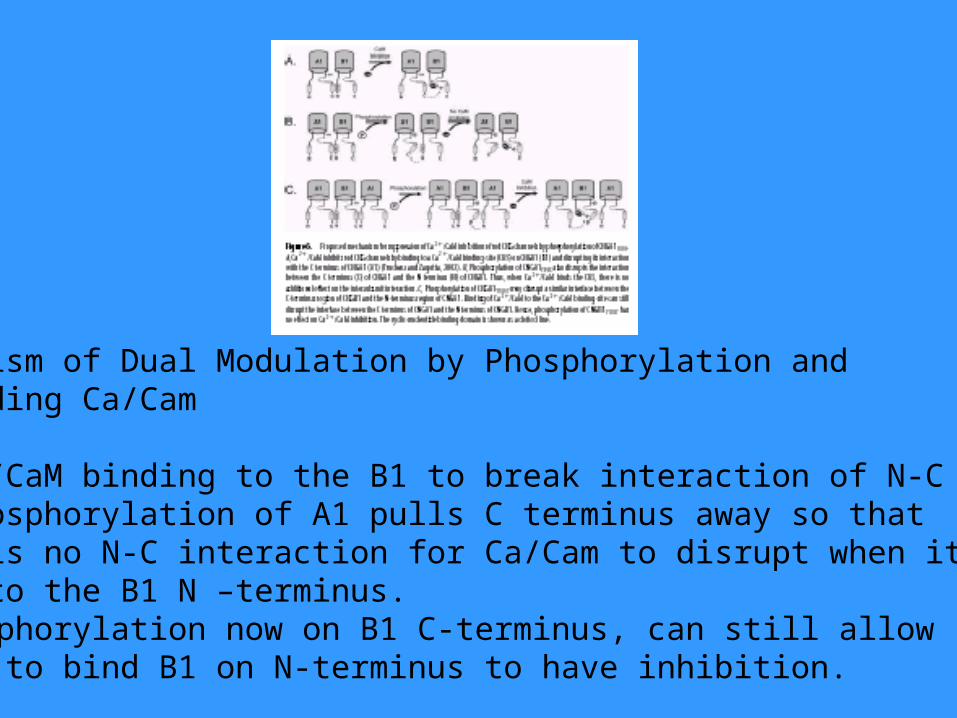
Mechanism of Dual Modulation by Phosphorylation and by binding Ca/Cam
A. Ca/CaM binding to the B1 to break interaction of N-CB. Phosphorylation of A1 pulls C terminus away so thatthere is no N-C interaction for Ca/Cam to disrupt when it binds to the B1 N –terminus.C. Phosphorylation now on B1 C-terminus, can still allowCa/CaM to bind B1 on N-terminus to have inhibition.
Paper 3:Brady et al., 2003. Functional Role of LipidRaft Microdomains in Cyclic Nucleotide-Gated Channel Activation. Mol. Pharm. 65:503-511.
Key Finding: Movement of CNGA2 into lipid raft domainscan change function of the channel by increasing affinity of cAMP. Secondary Finding: Heterologously expressed and nativeCNGA2 in olfactory tissue are expressed as a fraction in the lipid raft domains.Conclusion: Lipid lowering drugs could affect olfaction viafunctionally altering the biophysics of the CNGA2 (but theydo not have the correct stoichiometry…..?)
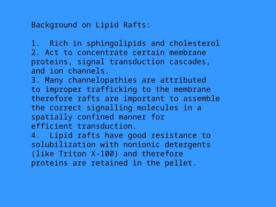
Background on Lipid Rafts:
1. Rich in sphingolipids and cholesterol 2. Act to concentrate certain membraneproteins, signal transduction cascades, and ion channels.3. Many channelopathies are attributedto improper trafficking to the membranetherefore rafts are important to assemblethe correct signalling molecules in a spatially confined manner for efficient transduction.4. Lipid rafts have good resistance tosolubilization with nonionic detergents(like Triton X-100) and therefore proteins are retained in the pellet.
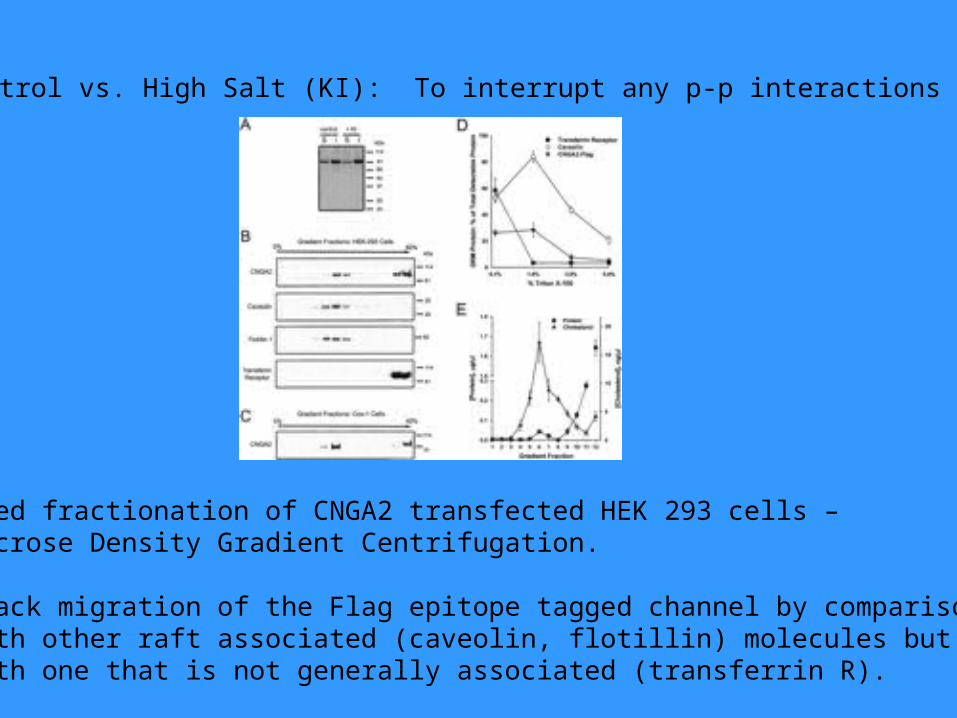
Control vs. High Salt (KI): To interrupt any p-p interactions
Used fractionation of CNGA2 transfected HEK 293 cells –Sucrose Density Gradient Centrifugation.
Track migration of the Flag epitope tagged channel by comparisonwith other raft associated (caveolin, flotillin) molecules but notwith one that is not generally associated (transferrin R).
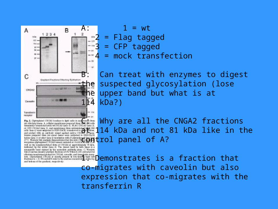
A: 1 = wt2 = Flag tagged3 = CFP tagged4 = mock transfection
B: Can treat with enzymes to digest the suspected glycosylation (losethe upper band but what is at 114 kDa?)
C: Why are all the CNGA2 fractionsat 114 kDa and not 81 kDa like in the control panel of A?
1. Demonstrates is a fraction that co-migrates with caveolin but also expression that co-migrates with thetransferrin R
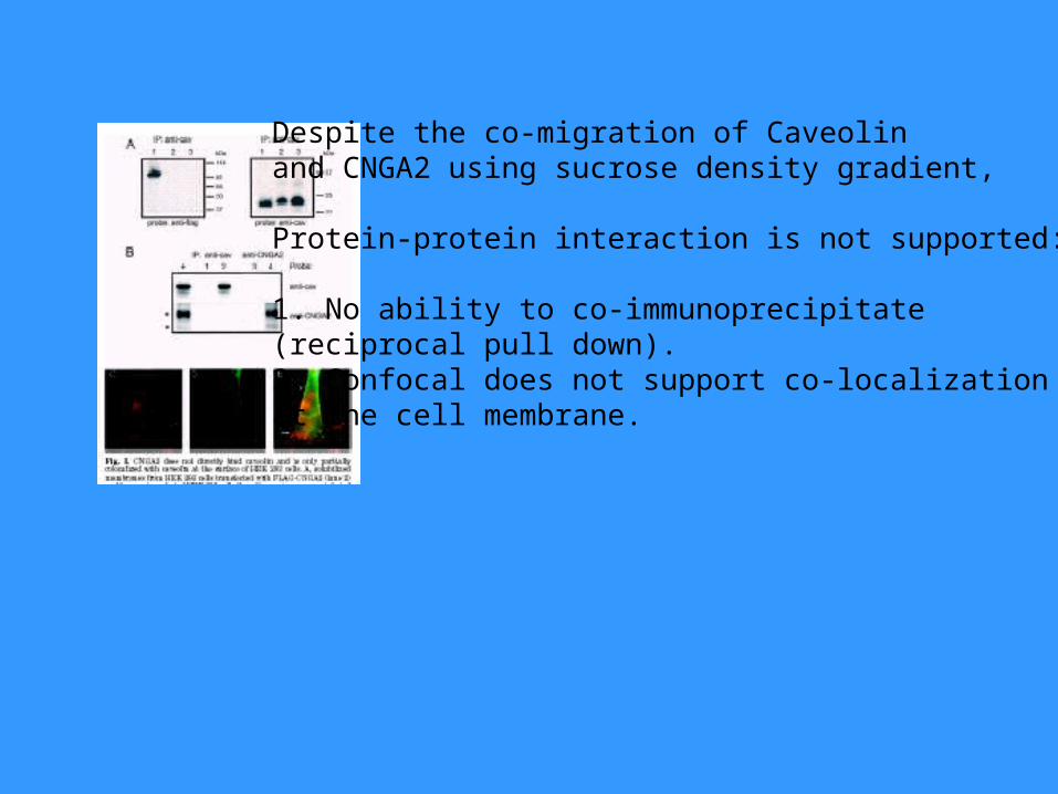
Despite the co-migration of Caveolinand CNGA2 using sucrose density gradient,
Protein-protein interaction is not supported:
1. No ability to co-immunoprecipitate (reciprocal pull down).2. Confocal does not support co-localizationat the cell membrane.
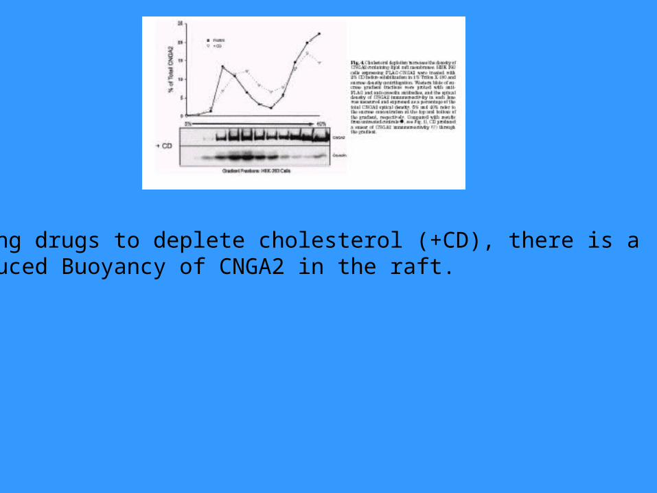
Using drugs to deplete cholesterol (+CD), there is a Reduced Buoyancy of CNGA2 in the raft.
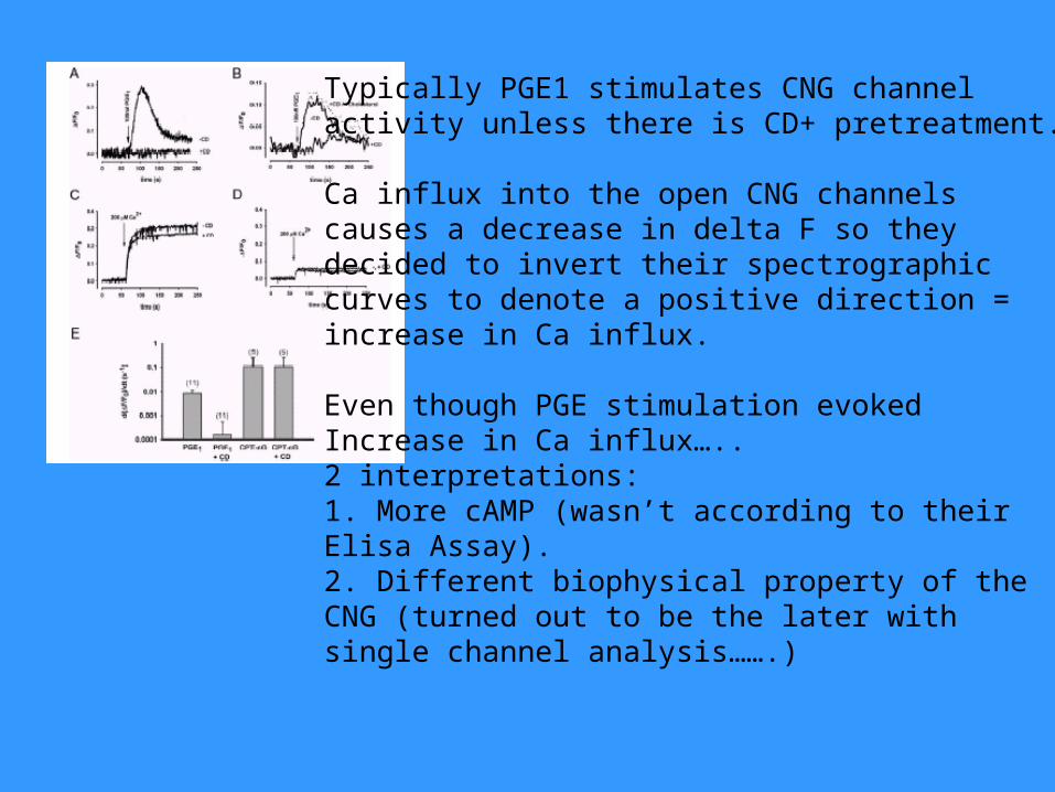
Typically PGE1 stimulates CNG channelactivity unless there is CD+ pretreatment.
Ca influx into the open CNG channelscauses a decrease in delta F so theydecided to invert their spectrographiccurves to denote a positive direction = increase in Ca influx.
Even though PGE stimulation evokedIncrease in Ca influx…..2 interpretations:1. More cAMP (wasn’t according to theirElisa Assay).2. Different biophysical property of theCNG (turned out to be the later withsingle channel analysis…….)
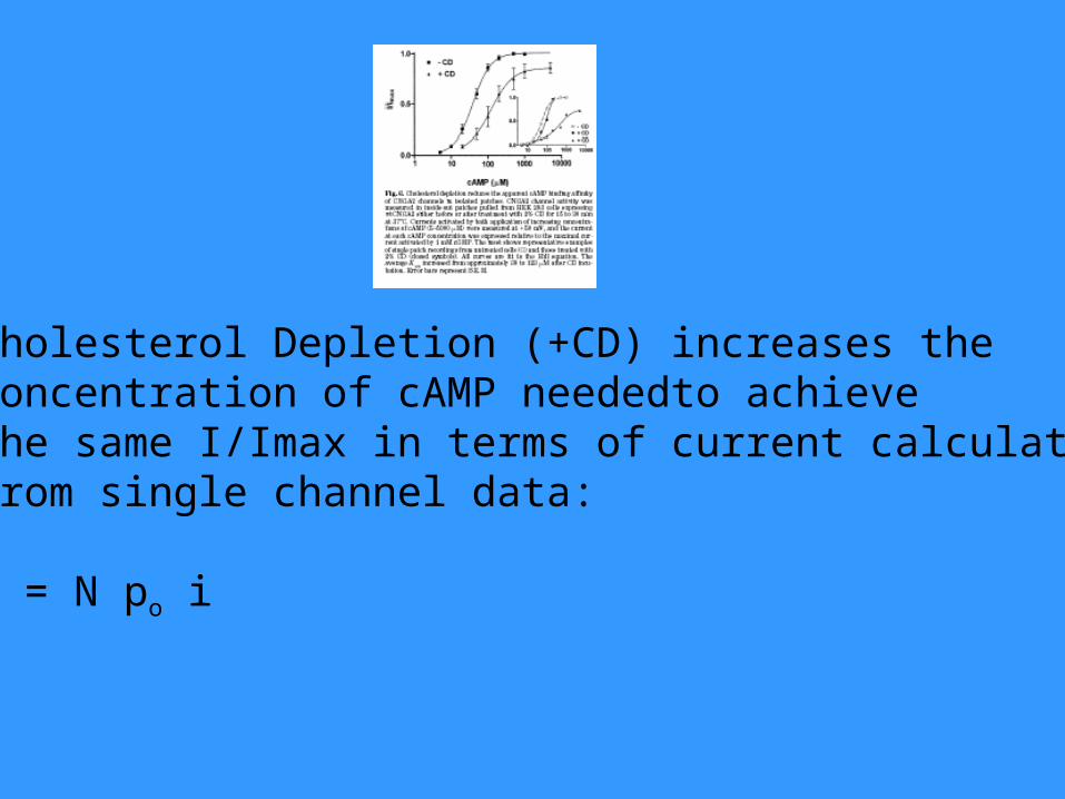
Cholesterol Depletion (+CD) increases the Concentration of cAMP neededto achieve the same I/Imax in terms of current calculated from single channel data:
I = N po i