Embed Size (px)
Citation preview
Association for Biology Laboratory Education (ABLE) ~ http://www.zoo.utoronto.ca/able 80
Chapter 4
Seafood Forensics: Crabby Proteins
Kathy Frame
National Association of Biology Teachers 11250 Roger Bacon Drive, #19
Reston, VA 20190-5202 800-406-0775
Kathy received her B.S. from Slippery Rock University and her M.S. from The Johns Hopkins University. Presently, she is the Director of Education for the National Association of Biology Teachers located in Reston, Virginia. She has taught a wide range of students from the preschool level through adult education, but her experience is predominantly at the high school level. Through grant work and in collaboration with scientific societies, industry, and other organizations, she has managed projects that have resulted in several biology supplementary curricula. She volunteers her time to the Virginia Junior Academy of Science Committee and serves as Virginia State Director for the Intel Talent Search Competition, President of her local chapter of Delta Kappa Society International, and President of the Virginia Association of Biology Teachers.
Adapted with permission from the National Association of Biology Teachers (NABT) publication Shoestring Biotechnology© (2000). Funded by the National Science Foundation ATE/DUE 9553720. Principal Investigator: Kathy Frame.
Reprinted From: Frame, K. 2000. Seafood forensics: Crabby proteins. Pages 80-101, in Tested studies for laboratory teaching, Volume 21 (S. J. Karcher, Editor). Proceedings of the 21st Workshop/Conference of the Association for Biology Laboratory Education (ABLE), 509 pages. - Copyright policy: http://www.zoo.utoronto.ca/able/volumes/copyright.htm Although the laboratory exercises in ABLE proceedings volumes have been tested and due consideration has been given to safety, individuals performing these exercises must assume all responsibility for risk. The Association for Biology Laboratory Education (ABLE) disclaims any liability with regards to safety in connection with the use of the exercises in its proceedings volumes.
Seafood Forensics
81
Contents
Introduction..................................................................................................................................81 Synopsis ...........................................................................................................................81 Objectives ........................................................................................................................81 Level ................................................................................................................................81 Length of the Lab.............................................................................................................82 Preparation Time Required ..............................................................................................82 Concept Information ........................................................................................................82
Materials ......................................................................................................................................83 Equipment and Materials Shared by the Class ................................................................83 Equipment and Materials for Each Team of Four Students.............................................84
Notes for Instructor ......................................................................................................................84 Directions for Setting Up the Lab....................................................................................84 Safety Procedures ............................................................................................................86 Teaching Tips...................................................................................................................87 Student Team Logistics....................................................................................................88 Pedagogical Information..................................................................................................88 Suggested Modifications for Students Who Are Exceptional .........................................88 Instructional Procedure ....................................................................................................89 Data Analysis and Interpretation ....................................................................................94 Hypothesis Generation for Further Investigation ............................................................96 Student Sample Investigation ..........................................................................................96 Test Questions..................................................................................................................97 Answers to Test Questions & Directions for Students Pages Questions/Analysis..........98
Student Outline ............................................................................................................................99 Acknowledgments........................................................................................................................100 Literature Cited ............................................................................................................................101
Introduction
Synopsis Students perform protein analysis of crab tissue to determine if species substitution has occurred. Samples are analyzed using horizontal agarose gel electrophoresis. Objectives At the end of this lab, students will be able to:
• Discuss a method for detecting seafood species substitution. • Describe a method for separating proteins by size using gel electrophoresis.
Level
• High school: Intermediate, Advanced • Two-year college: All levels • Four-year college: Freshman and Non-majors Biology
Length of Lab
• Day 1 (45 minutes): Practice and prepare gels.
Seafood Forensics
82
• Day 2 (45 minutes): Introduction and sample preparation. • Day 3 (45 minutes): Load and run gel; instructor stains and destains gels. • Day 4 (45 minutes): Interpret results.
Preparation Time Required
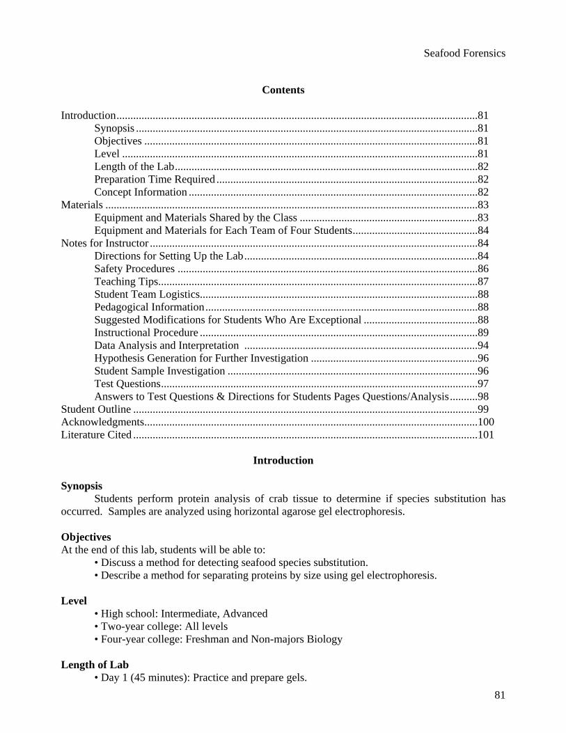
1.5 hours Concept Information Since more Americans are including seafood in their diets, the seafood industry has grown dramatically. An issue for seafood consumers is species substitution. Most people can distinguish imitation crab meat from the real thing readily by sight and taste. However, other species substitutions are occurring that fool the unwary consumer. Examples include bay scallops, which may be shark fins punched into scallop shapes and treated to look and taste like sea scallops, and canned or frozen breaded fish that are sometimes not the species claimed. In this lab activity, protein analysis of tissue from a variety of seafood will be examined to identify potential species substitutions. One way to analyze proteins is to use polyacrylamide gels with a vertical gel electrophoresis system. In most instances, this may pose a two-prong dilemma of safety and cost for the classroom. With respect to safety, the precursors of polyacrylamide, acrylamide and bis-acrylamide, are neurotoxic and possibly carcinogenic (Atkins 1991). When both monomers are polymerized completely, they are considered inert. However, Morgan et al. (1991) caution that “The degree of polymerization of the monomers into gels is variable unless the reaction conditions are rigorously controlled.” In the classroom, it may not be possible to control these conditions “rigorously.” The second dilemma for the classroom is the cost and availability of a vertical gel electrophoresis system. Horizontal electrophoresis systems are found more commonly in the classroom and are considerably less expensive than vertical gel systems. In 1995, Thorton, Daum & Case developed a protocol utilizing SeaKem™ gold agarose to analyze high-molecular mass protein complexes with agarose gel electrophoresis on a horizontal system. They concluded that agarose provided an excellent matrix for the resolution of high-molecular mass protein complexes with both horizontal and vertical electrophoresis systems. The protocol in this laboratory uses a protocol modeled after Thorton et al. (1995). A 2% agarose gel is used to separate proteins by horizontal gel electrophoresis. Seafood tissue is mashed and diluted with buffer, loaded onto a horizontal agarose gel containing sodium dodecyl sulfate (SDS), and subjected to electrophoresis. Proteins need to be denatured with SDS to migrate in the gel at a rate proportional to their molecular weight. The negatively-charged detergent, SDS binds to most proteins in an amount proportional to their mass, thus providing each protein with a standard charge to mass ratio. The denaturing process also forces most proteins into a semi-rigid rod shape that helps produce a clearer banding pattern. After the gel is stained with Coomassie® Blue, the protein banding pattern for each sample is used to determine if species substitution has occurred. The molecular weight of the unknown protein bands may be determined by comparison to protein standards of known molecular weight. These standards have been calibrated accurately so the log 10 of their molecular weights can be plotted versus their migration distance (Rf) as shown in Figure 4.1. These data will plot in a straight line, allowing the molecular weight of the unknown to be calculated from the standard curve.
Seafood Forensics
83
Rf = Distance migratedDistance of
Frontdistance
Proteinmigrationdistance
Figure 4.1. Protein band as seen in the stained gel.
Materials
Equipment and Materials Shared by the Class For the instructor-led protocol, supply the following equipment and materials as indicated for a class of 24 students. Equipment
• 1 1-liter flask • 1 500-ml graduated cylinder • 1 balance • 1 weigh boat • 1 hot plate • 1 pair of heat-proof gloves or pot holders • 1 50°C water bath • 1 95°C water bath • 1 tabletop centrifuge • 3 horizontal mini-gel electrophoresis chambers • 3 electrophoresis power supplies • 1 disposable Wiretrol® II Micropipet Set (Carolina Biological #21-1155) • 1 box micropipette tips (optional) • 1 micropipettor (optional) • 1 white light box
Materials
• 10.0 g agarose • 500.0 ml Laemmli buffer
Seafood Forensics
84
Equipment and Materials for Each Team of Four Students Equipment
• 1 51x75-cm (double-wide) slide • 1 1-mm thick, 6-8 well comb • 2 small bull dog clamps • 8 pennies • 1 8.5x5.5-cm piece of stiff plastic • 1 pump or bulb • 1 10-ml pipette • 1 balance • 1 scalpel or safety razor blade • 1 spatula • 2 weigh boats • 1 mortar and pestle • 2 plastic spoons • 1 waterproof marker • 2 15-ml centrifuge tubes • 2 1.5-ml microcentrifuge tubes • 2 1-ml plastic transfer pipettes • 1 dissecting needle • 1 floating microcentrifuge tube rack • 1 timer or clock with second hand • 1 1-20 µl micropipettor • 1 staining tray • 1 centimeter ruler
Materials
• 1.0 g each of fresh and imitation crab meat • 4.0 ml cold, deionized water • 6 ml Sample Preparation Buffer • 0.5 ml Unknown Sample • 5.0 µl High Protein Molecular Weight Standards, High Range (LTI #16001-018)
(optional)
• 10 ml Coomassie® Blue Staining Solution • 300 ml Destaining Solution • Kimwipes® or paper towels • 1 sheet of graph paper
Notes for the Instructor
Directions for Setting Up the Lab The instructor will need the following equipment and materials for preparing the lab: Equipment
• 1 balance
Seafood Forensics
85
• 7 weigh boats • 1 spatula • 1 2-liter flask • 1 20-ml pipette • 1 pump or bulb • 1 1-liter graduated cylinder • 2 2-liter plastic storage bottles with caps • 1 waterproof marking pen • 1 50-ml graduated cylinder • 1 glass stirring rod • 1 60-ml plastic storage bottle with cap • 1 refrigerator • 1 mortar and pestle • 1 15-ml centrifuge tube • 1 tabletop centrifuge • 1 transfer pipette • 1 1-ml pipette • 6 1.5-ml microcentrifuge tubes • 1 500-ml flask • 1 funnel • 1 coffee filter • 1 500-ml plastic storage bottle with cap
Materials
• 20.0 ml 10% sodium dodecyl sulfate (SDS) or lauryl sulfate, pH 8.5 • 22.0 g Tris base • 12.0 g boric acid • 3.5 l deionized water • 5.0 g sucrose • 0.05 g bromophenol blue • 1.0 g each of fresh and imitation crab meat • 0.62 g Coomassie® Blue • 200.0 ml methanol • 1.5 L white vinegar
Solution Preparation Laemmli buffer
1. Combine 20.0 ml 10% SDS, 22.0 g Tris base, and 12.0 g boric acid in a 2-liter flask. 2. Adjust total volume to 2.0 liters with deionized water. 3. Swirl until the components are dissolved completely. 4. Pour into a 2-liter plastic storage bottle. 5. Store tightly capped at room temperature.
Seafood Forensics
86
Sample Preparation Buffer 1. Add 5.0 g sucrose and 0.05 g bromophenol blue to 30.0 ml Laemmli buffer in a 50-ml
graduated cylinder. Stir until the components are dissolved. 2. Adjust the volume to 50.0 ml by adding Laemmli buffer. 3. Pour into a 60-ml plastic storage bottle. 4. Store tightly capped in the refrigerator.
"Unknown" Crab Meat
1. Grind the crab and imitation crab samples together with 4 ml deionized water using a mortar and pestle.
2. Add 6.0 ml Sample Preparation Buffer and mix thoroughly. 3. Pour the solution into a 15-ml centrifuge tube. Centrifuge the mixture for 5 minutes to
pellet the solids. 4. Collect the supernatant with a transfer pipette. 5. Aliquot 0.5 ml into a 1.5-ml microcentrifuge tube for each lab team. 6. Label the tubes "Unknown." 7. Store the samples in the freezer for up to 1 month.
Coomassie®Blue Staining Solution
1. Weigh 0.62 g Coomassie® Blue powder. Place the powder in a 500-ml flask. 2. Add 200.0 ml methanol, 25.0 ml white vinegar, and 275.0 ml deionized water to the
flask. Swirl the flask to combine the components. 3. Filter the solution through a coffee filter and collect the solution in a 500-ml plastic
storage bottle. 4. Cap the bottle tightly and store at room temperature.
Destaining Solution
1. Combine 1.0 L vinegar with 1.0 L deionized water in a 2-liter storage bottle. 2. Cap the bottle tightly and store it at room temperature.
Safety Procedures
All laboratory procedures should be conducted with gloves, goggles, and aprons.
Wash hands before and at the conclusion of the lab.
If the power is on, electrical shock may result from touching the buffer or electrophoresis equipment with wet hands.
Never leave the electrophoresis power unit on without supervision. There is a risk of fire if the buffer leaks out or if the buffer should evaporate completely during electrophoresis.
Seafood Forensics
87
Be sure that students are familiar with the operating instructions and safety precautions before they use any centrifuge. These machines can be very hazardous if handled carelessly.
Use caution with hot liquids and glassware. Wear heat-proof gloves to move hot glassware and metals. Teaching Tips
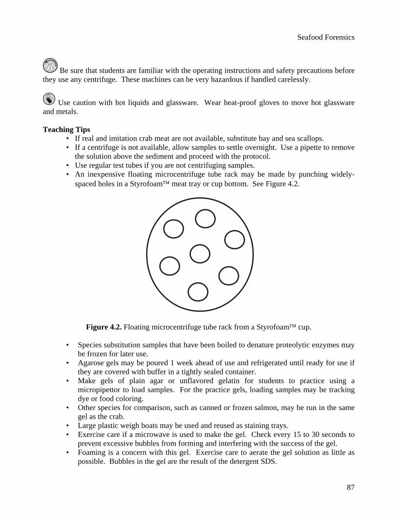
• If real and imitation crab meat are not available, substitute bay and sea scallops. • If a centrifuge is not available, allow samples to settle overnight. Use a pipette to remove
the solution above the sediment and proceed with the protocol. • Use regular test tubes if you are not centrifuging samples. • An inexpensive floating microcentrifuge tube rack may be made by punching widely-
spaced holes in a Styrofoam™ meat tray or cup bottom. See Figure 4.2.
Figure 4.2. Floating microcentrifuge tube rack from a Styrofoam™ cup.
• Species substitution samples that have been boiled to denature proteolytic enzymes may be frozen for later use.
• Agarose gels may be poured 1 week ahead of use and refrigerated until ready for use if they are covered with buffer in a tightly sealed container.
• Make gels of plain agar or unflavored gelatin for students to practice using a micropipettor to load samples. For the practice gels, loading samples may be tracking dye or food coloring.
• Other species for comparison, such as canned or frozen salmon, may be run in the same gel as the crab.
• Large plastic weigh boats may be used and reused as staining trays. • Exercise care if a microwave is used to make the gel. Check every 15 to 30 seconds to
prevent excessive bubbles from forming and interfering with the success of the gel. • Foaming is a concern with this gel. Exercise care to aerate the gel solution as little as
possible. Bubbles in the gel are the result of the detergent SDS.
Seafood Forensics
88
• SDS may be added after heating the agarose to help reduce the “suds” effect. Add at the ratio of 1 ml SDS to 100 ml Laemmli buffer.
• Speed the destaining process by - Placing gels in a 50°C incubator - Changing the destaining solution several times - Placing Kimwipes®, firm tissues, paper towels, foam rubber chunks, or an
aquarium charcoal filter in the destaining solution to absorb the stain. Replace materials as they become stained. Remove the aquarium filter and rinse it several times during the process. Aquarium filters may be dried and reused from year to year.
• Coomassie® Blue Staining Solution should be collected after staining gels. It may be reused 3 to 5 times.
• Coomassie® Blue Staining Solution may be purchased from any biological supply house.
• 5.0% glacial acetic acid may be substituted for vinegar. For a 5.0% solution, add 50.0 ml glacial acetic acid to 500.0 ml of deionized water. Bring to a volume of 1.0 L with deionized water. Always add acid to water.
• Construct a substitute white-light box by placing a piece of white paper on an overhead projector. Cover the paper with a transparency sheet. Place the gel directly on the transparency sheet to visualize it.
Student Team Logistics
Organize the class into teams of 3 or 4. Pedagogical Information
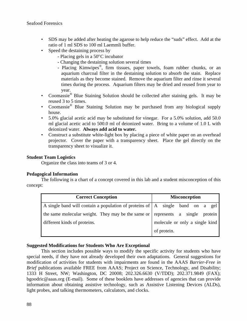
The following is a chart of a concept covered in this lab and a student misconception of this concept:
Correct Conception Misconception
A single band will contain a population of proteins of
the same molecular weight. They may be the same or
different kinds of proteins.
A single band on a gel
represents a single protein
molecule or only a single kind
of protein.
Suggested Modifications for Students Who Are Exceptional This section includes possible ways to modify the specific activity for students who have special needs, if they have not already developed their own adaptations. General suggestions for modification of activities for students with impairments are found in the AAAS Barrier-Free in Brief publications available FREE from AAAS; Project on Science, Technology, and Disability; 1333 H Street, NW; Washington, DC 20008; 202.326.6630 (V/TDD); 202.371.9849 (FAX); [email protected] (E-mail). Some of these booklets have addresses of agencies that can provide information about obtaining assistive technology, such as Assistive Listening Devices (ALDs), light probes, and talking thermometers, calculators, and clocks.
Seafood Forensics
89
Visually Impaired • Use plastic test tubes, beakers, graduated cylinders or culture dishes or paper containers,
where possible. • Use raised-line drawings of the lab setup. • Use measuring devices with raised lines. Puff paints or staples work well on rulers. • Provide lab assistance with techniques if needed. • Use sensory alarms where possible. • Use lab partners when running chemical tests, data recording, and interpretation.
Hearing Impaired
• Use visual alarms where possible. • Use a video camera to record the lab setup and results. • Use lab partners when running chemical tests, data recording, and interpretation.
Mobility Impaired
• For students with motor coordination difficulties, use a hand-held blender instead of a mortar and pestle to grind the tissue.
• Use a hand-held automatic pipettor or programmable repipettor with a digital read-out. • Use lab partners when running chemical tests, data recording, and interpretation.
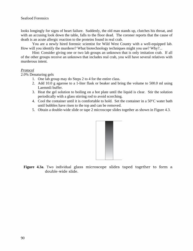
Instructional Procedure Practice making and loading microscope slide gels Technicians in the early days of biotechnology needed to have results quickly for screening purposes. They would tape two individual glass microscope slides side by side to form a double-wide slide as shown in Figure 3. Then agar was poured over the makeshift slide creating a very thin gel that could be run more quickly than a standard-sized gel. This technique may be used in the classroom to save time and expense. The results are reliable and quick. As with pouring and loading any gel, practice is important. Have students use a purchased double-wide slide or make their own double-wide slide, just as the early technologists did, by taping two microscope slides side by side. Since a minimal amount of agarose is used, have one lab group make the agarose for the entire class as described in Steps 2 to 4 of the Instructor-led Protocol that follows this section. Each lab group should pour their own gel. The gels may be stored in Laemmli buffer for approximately 1 week. Instructor-led Protocol Introduction to the Protocol
You may want to introduce the crab forensics activity with the following scenario: Great Uncle Edgar Earp, the family curmudgeon and third cousin to the famous Marshall Wyatt, lives in a decaying old mansion outside of Tombstone, Arizona. He is a wealthy miser who, over the years, has hinted to every one of his relatives that he or she alone will inherit his money when he dies. To their dismay, Uncle Edgar is now in his 90s and seems to be as healthy as ever. The family members are summoned to Tombstone for Uncle Edgar's annual pot luck birthday party. Everyone brings a crab dish, since this is the birthday boy's favorite food. However, they all know to use imitation crab because Uncle Edgar is dangerously allergic to the real thing. As the meal progresses he tries several bites of each offering while the rest of the family
Seafood Forensics
90
looks longingly for signs of heart failure. Suddenly, the old man stands up, clutches his throat, and with an accusing look down the table, falls to the floor dead. The coroner reports that the cause of death is an acute allergic reaction to the proteins found in real crab. You are a newly hired forensic scientist for Wild West County with a well-equipped lab. How will you identify the murderer? What biotechnology techniques might you use? Why?... Hint: Consider giving one or two lab groups an unknown that is only imitation crab. If all of the other groups receive an unknown that includes real crab, you will have several relatives with murderous intent. Protocol 2.0% Denaturing gels
1. One lab group may do Steps 2 to 4 for the entire class. 2. Add 10.0 g agarose to a 1-liter flask or beaker and bring the volume to 500.0 ml using
Laemmli buffer. 3. Heat the gel solution to boiling on a hot plate until the liquid is clear. Stir the solution
periodically with a glass stirring rod to avoid scorching. 4. Cool the container until it is comfortable to hold. Set the container in a 50°C water bath
until bubbles have risen to the top and can be removed. 5. Obtain a double-wide slide or tape 2 microscope slides together as shown in Figure 4.3.
Figure 4.3a. Two individual glass microscope slides taped together to form a double-wide slide.
Seafood Forensics
91
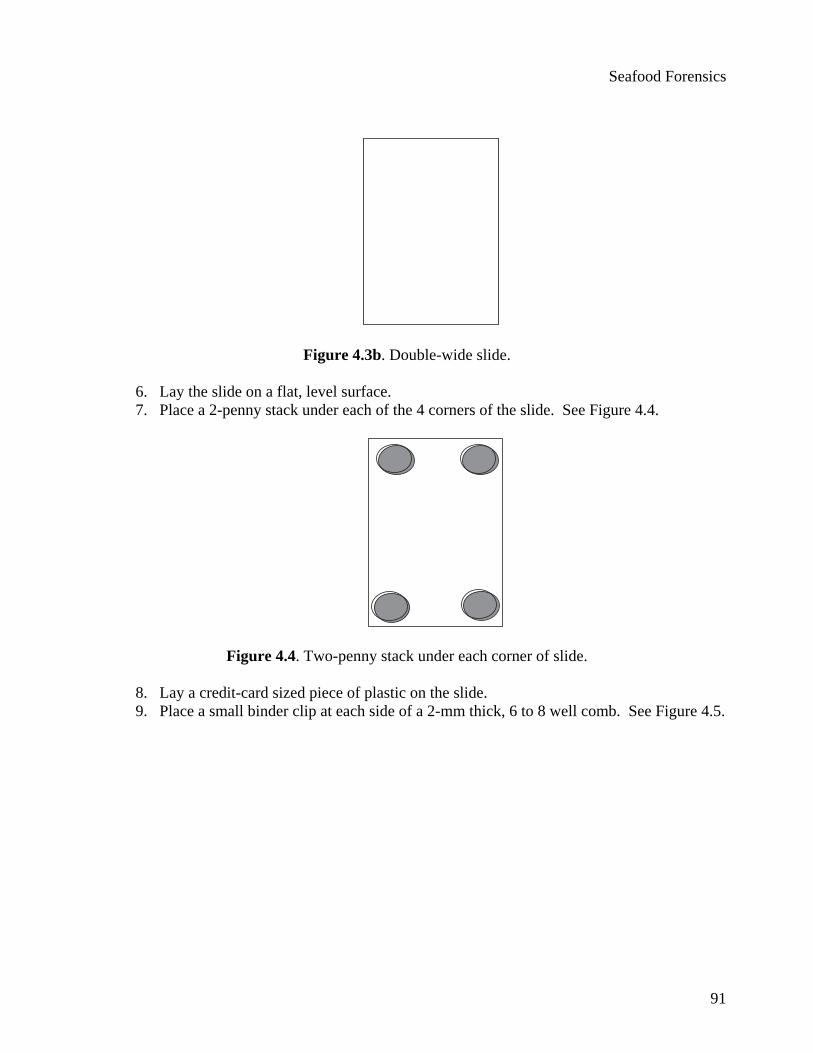
Figure 4.3b. Double-wide slide.
6. Lay the slide on a flat, level surface. 7. Place a 2-penny stack under each of the 4 corners of the slide. See Figure 4.4.
Figure 4.4. Two-penny stack under each corner of slide.
8. Lay a credit-card sized piece of plastic on the slide. 9. Place a small binder clip at each side of a 2-mm thick, 6 to 8 well comb. See Figure 4.5.
Seafood Forensics
92
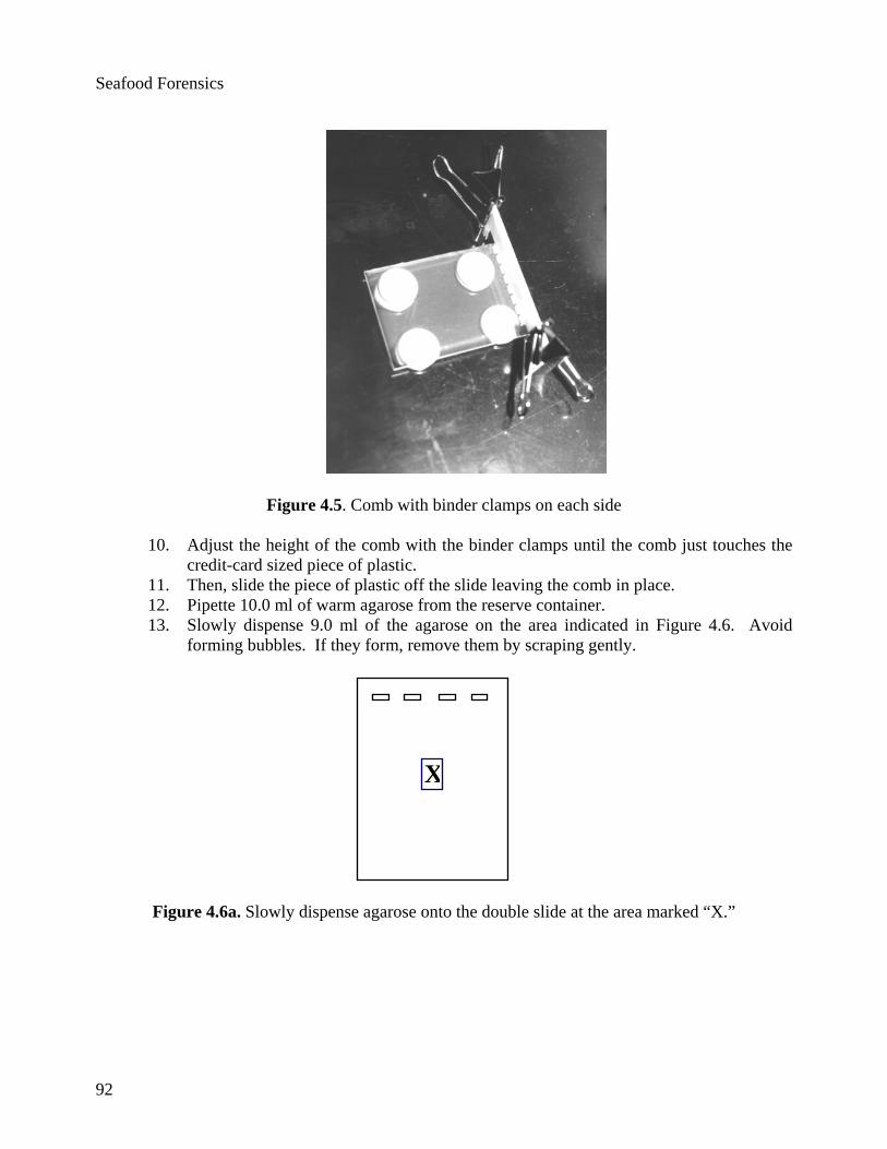
Figure 4.5. Comb with binder clamps on each side
10. Adjust the height of the comb with the binder clamps until the comb just touches the credit-card sized piece of plastic.
11. Then, slide the piece of plastic off the slide leaving the comb in place. 12. Pipette 10.0 ml of warm agarose from the reserve container. 13. Slowly dispense 9.0 ml of the agarose on the area indicated in Figure 4.6. Avoid
forming bubbles. If they form, remove them by scraping gently.
X
Figure 4.6a. Slowly dispense agarose onto the double slide at the area marked “X.”
Seafood Forensics
93
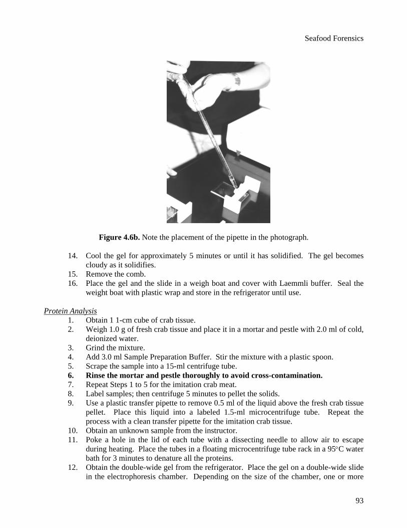
Figure 4.6b. Note the placement of the pipette in the photograph.
14. Cool the gel for approximately 5 minutes or until it has solidified. The gel becomes cloudy as it solidifies.
15. Remove the comb. 16. Place the gel and the slide in a weigh boat and cover with Laemmli buffer. Seal the
weight boat with plastic wrap and store in the refrigerator until use. Protein Analysis
1. Obtain 1 1-cm cube of crab tissue. 2. Weigh 1.0 g of fresh crab tissue and place it in a mortar and pestle with 2.0 ml of cold,
deionized water. 3. Grind the mixture. 4. Add 3.0 ml Sample Preparation Buffer. Stir the mixture with a plastic spoon. 5. Scrape the sample into a 15-ml centrifuge tube. 6. Rinse the mortar and pestle thoroughly to avoid cross-contamination. 7. Repeat Steps 1 to 5 for the imitation crab meat. 8. Label samples; then centrifuge 5 minutes to pellet the solids. 9. Use a plastic transfer pipette to remove 0.5 ml of the liquid above the fresh crab tissue
pellet. Place this liquid into a labeled 1.5-ml microcentrifuge tube. Repeat the process with a clean transfer pipette for the imitation crab tissue.
10. Obtain an unknown sample from the instructor. 11. Poke a hole in the lid of each tube with a dissecting needle to allow air to escape
during heating. Place the tubes in a floating microcentrifuge tube rack in a 95°C water bath for 3 minutes to denature all the proteins.
12. Obtain the double-wide gel from the refrigerator. Place the gel on a double-wide slide in the electrophoresis chamber. Depending on the size of the chamber, one or more
Seafood Forensics
94
gels may be placed in the chamber. Be sure the wells are at the negative end of the gel box.
13. Fill the electrophoresis chamber gently with Laemmli buffer until the top of the gel is just covered.
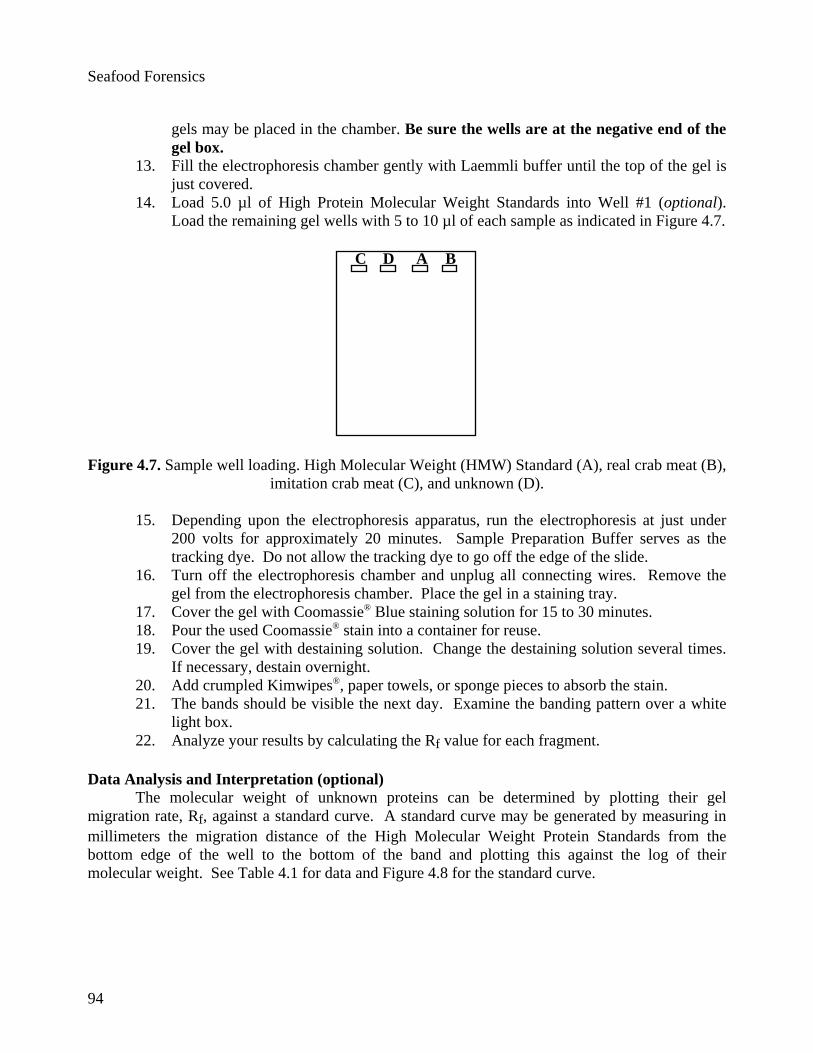
14. Load 5.0 µl of High Protein Molecular Weight Standards into Well #1 (optional). Load the remaining gel wells with 5 to 10 µl of each sample as indicated in Figure 4.7.
C A BD
Figure 4.7. Sample well loading. High Molecular Weight (HMW) Standard (A), real crab meat (B),
imitation crab meat (C), and unknown (D).
15. Depending upon the electrophoresis apparatus, run the electrophoresis at just under 200 volts for approximately 20 minutes. Sample Preparation Buffer serves as the tracking dye. Do not allow the tracking dye to go off the edge of the slide.
16. Turn off the electrophoresis chamber and unplug all connecting wires. Remove the gel from the electrophoresis chamber. Place the gel in a staining tray.
17. Cover the gel with Coomassie® Blue staining solution for 15 to 30 minutes. 18. Pour the used Coomassie® stain into a container for reuse. 19. Cover the gel with destaining solution. Change the destaining solution several times.
If necessary, destain overnight. 20. Add crumpled Kimwipes®, paper towels, or sponge pieces to absorb the stain. 21. The bands should be visible the next day. Examine the banding pattern over a white
light box. 22. Analyze your results by calculating the Rf value for each fragment.
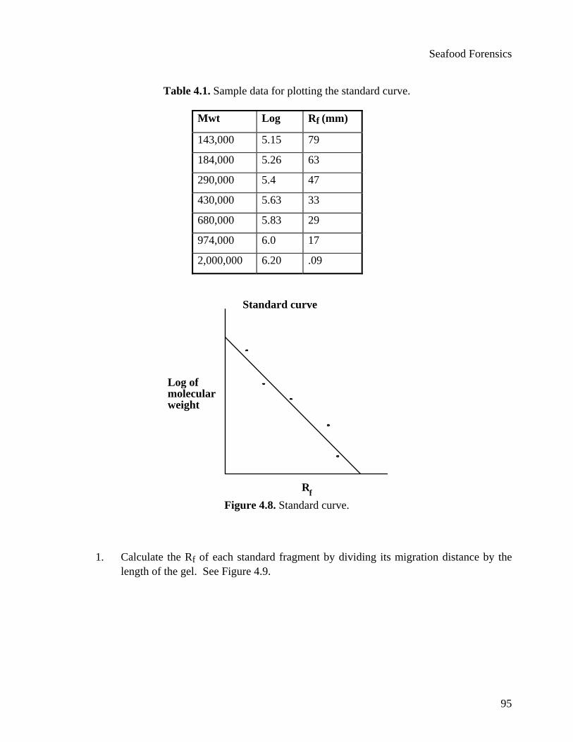
Data Analysis and Interpretation (optional) The molecular weight of unknown proteins can be determined by plotting their gel
migration rate, Rf, against a standard curve. A standard curve may be generated by measuring in millimeters the migration distance of the High Molecular Weight Protein Standards from the bottom edge of the well to the bottom of the band and plotting this against the log of their molecular weight. See Table 4.1 for data and Figure 4.8 for the standard curve.
Seafood Forensics
95
Table 4.1. Sample data for plotting the standard curve.
Mwt Log Rf (mm)
143,000 5.15 79
184,000 5.26 63
290,000 5.4 47
430,000 5.63 33
680,000 5.83 29
974,000 6.0 17
2,000,000 6.20 .09
Standard curve
Rf
Log ofmolecularweight
Figure 4.8. Standard curve.
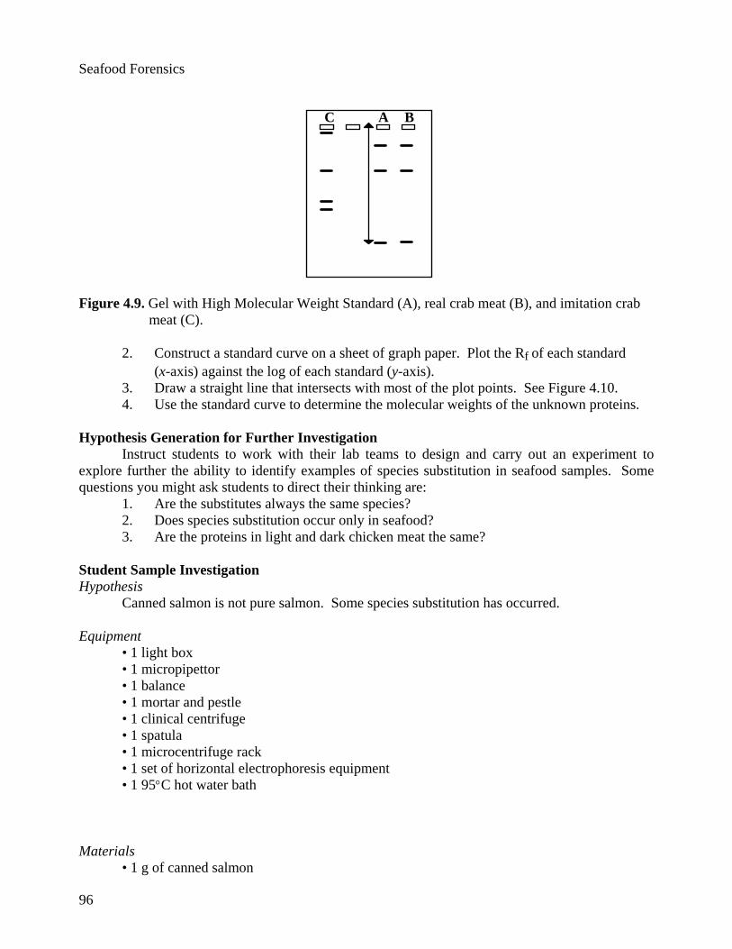
1. Calculate the Rf of each standard fragment by dividing its migration distance by the
length of the gel. See Figure 4.9.
Seafood Forensics
96
C A B
Figure 4.9. Gel with High Molecular Weight Standard (A), real crab meat (B), and imitation crab meat (C).
2. Construct a standard curve on a sheet of graph paper. Plot the Rf of each standard (x-axis) against the log of each standard (y-axis). 3. Draw a straight line that intersects with most of the plot points. See Figure 4.10. 4. Use the standard curve to determine the molecular weights of the unknown proteins.
Hypothesis Generation for Further Investigation
Instruct students to work with their lab teams to design and carry out an experiment to explore further the ability to identify examples of species substitution in seafood samples. Some questions you might ask students to direct their thinking are:
1. Are the substitutes always the same species? 2. Does species substitution occur only in seafood? 3. Are the proteins in light and dark chicken meat the same?
Student Sample Investigation Hypothesis
Canned salmon is not pure salmon. Some species substitution has occurred.
Equipment • 1 light box • 1 micropipettor • 1 balance • 1 mortar and pestle • 1 clinical centrifuge • 1 spatula • 1 microcentrifuge rack • 1 set of horizontal electrophoresis equipment • 1 95°C hot water bath
Materials
• 1 g of canned salmon
Seafood Forensics
97
• 1 g of fresh salmon • 1 g each of light and dark chicken meat • filter paper • transfer pipettes • 2 15-ml centrifuge tubes • Coomassie® Blue stain • 3 ml Preparation Buffer
Procedure
1. Weigh 1 g of fresh salmon tissue. 2. Place the tissue in a mortar. Add 2 ml of cold tap water and grind with the pestle. 3. Add 3 ml of the Preparation Buffer to the ground salmon sample. 4. Repeat Steps 1 to 3 for the canned salmon. 5. Spin the samples for 5 minutes in a clinical centrifuge. 6. Carefully, remove 1 ml of the liquid above the fresh salmon in the centrifuge tube and
place it into a labeled 1.5-ml microcentrifuge tube. 7. Repeat Step 6 for the canned salmon sample. 8. Place the microcentrifuge tubes into a 95˚C hot water bath. 9. Set up the electrophoresis gel with Laemmli buffer. 10. Prepare a 2% agarose gel. 11. Load 15 µl of each sample into the wells of the gel. 12. Run the gel until the tracking dye has migrated at least 75% of the distance down the
gel, approximately 1.5 hours. 13. Remove the gel from the chamber and stain it with Coomassie® Blue staining solution
for 1 to 2 hours. 14. Destain the gel with destaining solution. 15. Examine the gel on a light box and analyze the protein bands.
Test Questions
1. Recently, a high school student nearly died after eating a hamburger. The hamburger was tested for bacterial contamination and was found to be negative for bacteria. No one else became ill after eating hamburgers from the same batch. The student has a severe allergy to soybean products. Explain how a forensic scientist might analyze the hamburger meat to determine what caused the attack.
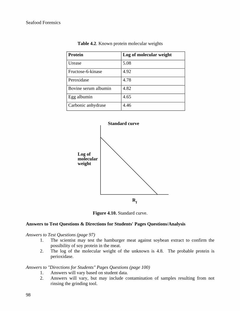
2. Your unknown protein had an Rf value of 0.18. Use the standard curve in the Figure 10 to determine the molecular weight of your protein. Use Table 4.2 to determine which protein your sample most resembles.
Seafood Forensics
98
Table 4.2. Known protein molecular weights
Protein Log of molecular weight
Urease 5.08
Fructose-6-kinase 4.92
Peroxidase 4.78
Bovine serum albumin 4.82
Egg albumin 4.65
Carbonic anhydrase 4.46
Standard curve
Rf
Log ofmolecularweight
Figure 4.10. Standard curve. Answers to Test Questions & Directions for Students' Pages Questions/Analysis Answers to Test Questions (page 97)
1. The scientist may test the hamburger meat against soybean extract to confirm the possibility of soy protein in the meat.
2. The log of the molecular weight of the unknown is 4.8. The probable protein is perioxidase.
Answers to "Directions for Students" Pages Questions (page 100)
1. Answers will vary based on student data. 2. Answers will vary, but may include contamination of samples resulting from not
rinsing the grinding tool.
Seafood Forensics
99
3. Another questions may be “Does the protein composition of imitation and fresh crab affect the taste?”
4. Marine and freshwater science. A question for ethics would be “Is it unethical to replace ‘fake’ tissue for the tissue the buyer believes he is buying?”
5. Species of crab. 6. One would need to verify data and results. This may not be a sophisticated enough
system to detect species substitution in a court of law.
Student Outline
Introduction More Americans are including seafood and freshwater fish in their diets, making the seafood business a fast-growing industry. One of the biggest issues in commercial seafood is species substitution. You might have eaten crab salad or "king crab legs" that were really pollock, an inexpensive white fish that has been treated and stained to look like crab meat. Another example of species substitution is bay "scallops" that are actually shark fins punched into scallop shapes and treated to look and taste like sea scallops. In breaded fish, "flounder" underneath the breading or batter is sometimes a cheaper species of fish. In this lab, you will determine if species substitution has occurred for crab meat using protein analysis. Your instructor will lead you through a protocol that analyzes proteins. Then you will be asked to design an experiment of your own using a modification of this protocol. Safety Notes
All laboratory procedures should be conducted with gloves, goggles, and aprons.
Wash your hands before and at the conclusion of the lab.
Use caution when working with electrophoresis. Never leave the power on without supervision. There is a risk of fire if the buffer leaks out or if it completely evaporates during electrophoresis. There is also a risk of electrical shock if your hands are wet or if the buffer is accidentally touched while the power is on.
Be familiar with operating instructions and safety precautions before using any equipment. Hypothesis or Prediction and Reasoning From the information you have on this topic, develop a hypothesis that could be tested in a controlled experiment that gathers quantitative data. Explain the reasoning behind your hypothesis. Based on class discussion and your library search of species substitutions in seafood, what question could your group investigate? Each group should select one question and form a hypothesis that it can support with reasoning. Some questions that may help you form a hypothesis are:
• Is there a possibility of species substitution in frozen or canned salmon?
Seafood Forensics
100
• Are bay and sea scallops related? • How high is the occurrence of species substitution in breaded or batter-dipped frozen
fish? Plan of Investigation Design an experiment to test your hypothesis. You may use the materials your instructor has available from the introductory protocol or devise your own. Make a list of steps to test your hypothesis that will yield quantitative data. Be sure to include positive and negative controls and to perform more than one trial so there are sufficient data to analyze. Develop a way to measure and analyze your data.
Obtain Instructor approval before doing this investigation. Questions/Analysis Answer the following questions after you have gathered your data:
1. How do these data relate to your hypothesis? 2. What caused errors in your experiment? 3. What other questions come from your results? 4. To what other biology topics is this lab related? Explain. 5. What variables may not have been controlled sufficiently in your experiment? How
would you redesign this experiment to help control for that variable? 6. Would you use this test for species substitution in a lawsuit? Explain.
Student Design of the Next Experiment After you have collected and analyzed data from your experiments and shared results and conclusions with the class, brainstorm ideas for experiments to do next. Think about questions that arose while you conducted your first experiments. What quantitative experiments could you do based on your observations? In your lab group, share ideas before writing your proposals. Questions you may consider include:
• Are hamburgers pure beef? • Are chicken nuggets made of dark or white meat? • What is in a hot dog? ham?
Acknowledgments
Special thanks to Ray Hadley, Margaret Burton, Jonathan Morrison, and Brian Shmaefsky
for developing, trouble-shooting, and perfecting this lab based on a template developed by Judy Brown. In addition, special thanks to the National Science Foundation, GIBCO BRL: Life Technologies, Inc., Genentech, Inc., and MediaSeek, Inc. who provided funding for this project.
Seafood Forensics
101
Literature Cited
Atkins, T. (1991). Protein electrophoresis in the biology classroom using "safe" gels. The American Biology Teacher, 53(8), 490-495.
FMC BioProducts. (1991). Separation of high molecular weight proteins in the ProSieve™ Gel
System. Resolutions™ 7(3), 1,3. Morgan, J.H., Curtis, F.P. & Nochumson, S. (1991). Protein recovery from the All-Agarose
ProSieve™ Gel System. BioTechniques, 11(2), 256-261. Thornton, J.R., Daum, H.A., III & Case, S.T. (1995). Agarose gel electrophoresis of high molecular
mass protein complexes. BioTechniques, 18(2), 324-327.