Embed Size (px)
Citation preview
Hindawi Publishing CorporationCase Reports in Infectious DiseasesVolume 2011, Article ID 787961, 3 pagesdoi:10.1155/2011/787961
Case Report
Scrotal Swelling and Testicular Atrophy due toSchistosomiasis in a 9-Year-Old Boy: A Case Report
Peter F. Rambau,1 Alphonce Chandika,2 Philipo L. Chalya,2 and Kahima Jackson3
1 Department of Pathology, Weill Bugando University College of Health Sciences, P.O. Box 1464, Mwanza, Tanzania2 Department of Surgery, Weill Bugando University College of Health Sciences, P.O. Box 1464, Mwanza, Tanzania3 Department of Pathology, Bugando Medical Center, P.O. Box 1370, Mwanza, Tanzania
Correspondence should be addressed to Peter F. Rambau, [email protected]
Received 27 May 2011; Accepted 16 June 2011
Academic Editors: M. de Gorgolas, M. Keita, F. Mansour-Ghanaei, and T. Mduluza
Copyright © 2011 Peter F. Rambau et al. This is an open access article distributed under the Creative Commons AttributionLicense, which permits unrestricted use, distribution, and reproduction in any medium, provided the original work is properlycited.
Schistosomiasis is a communicable disease which commonly involves urinary bladder causing hematuria, or large bowel causingbloody stool. The common species encountered in this lake region surrounding Lake Victoria in Tanzania are Schistosomahaematobium and Schistosoma mansoni. Complications can lead to portal hypertension due portal fibrosis in liver, and fibrosisin lung can lead to pulmonary hypertension; this commonly seen with S. mansoni. Major complications of S. maeametobium arechronic cystitis with squamous metaplasia with subsequent development of squamous cell carcinoma. Involvement of spinal cordcausing paraplegia has been observed in S. haematobium. Other unusual pathology of schistosomiasis has been described, suchas involvement of the appendix, ovary, prostate, and cervix. Here, we present a case of schistosomiasis in a 9-year-old boy whopresented with left scrotal pain for one year which was accompanied by scrotal swelling; surgical exploration was done, and thefinding was hydrocele and atrophic testes with nodules on the surface. Histological examination reveals atrophic testis and heavyactive granulomatous inflammation with schistosoma eggs consistent with Schistosoma haematobium in the tunica vaginalis.
1. Introduction
Schistosomiasis is endemic disease in Tanzania, and it is oneof the most important causes of morbidity with significantmortality. In Tanzania, the common encountered speciesare S. mansoni and S. haematobium, which causes intestinalschistosomiasis and urinary schistosomiasis, respectively.The common symptoms are blood in stool and hematuria.Significant morbidity is seen in those patients who developcomplications such as end-stage renal diseases, chronic liverdiseases with portal hypertension, and cancers associatedwith schistosomiasis [1–3]. Unusual presentations of schis-tosomiasis have been described in many organs in thebody, and commonly, these lesions are not clinically suspi-cious for schistosomiasis, and most of them are diagnosedhistologically as incidental findings. In our settings, suchlesions have been described in appendix causing perforationwith peritonitis [4], and schistosomal eggs was also seenwithin the prostate cancers [5]. Ovarian schistosomiasis
has been described causing chronic granulomatous inflam-mation producing ovarian pseudotumor, and it was alsoseen in fallopian tube in association with carcinoma [6, 7].Schistosomiasis of the cervix presents like other cervicalcondition such as cervicitis or cancer, and cervical cancerwith schistosoma eggs had been described in Tanzania[8].
Testicular schistosomiasis is not commonly reported, andsome cases have been reported due to S. mansoni. Testicularschistosomiasis can present as testicular nodule [9], andsometimes, it can mimic carcinoma leading to unnecessaryorchidectomy [10, 11]. In epididimis, schistosomal causesscrotal pain, and involvement of seminal vesicles has beendocumented [12, 13]. Schistosomiasis of the scrotum hasalso been described, causing hydrocele or causing chronicdermatitis [14, 15]. Here, we presents a case of a 9-year-oldboy presented with left scrotal swelling and pain for one yearwhich was later diagnosed histologically to be schistosomiasisafter exploration and orchidectomy.
2 Case Reports in Infectious Diseases
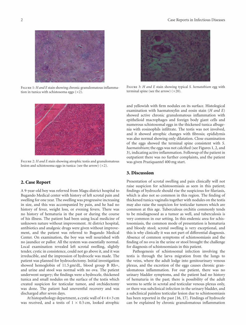
Figure 1: H and E stain showing chronic granulomatous inflamma-tion in tunica with schistosoma eggs (×2).
Figure 2: H and E stain showing atrophic testis and granulomatouslesion and schistosoma eggs in tunica (see the arrow) (×2).
2. Case Report
A 9-year-old boy was referred from Magu district hospital toBugando Medical center with history of left scrotal pain andswelling for one year. The swelling was progressive increasingin size, and this was accompanied by pain, and he had nohistory of fever, weight loss, or evening fevers. There wasno history of hematuria in the past or during the courseof his illness. The patient had been using local medicine ofunknown nature without improvement. At district hospital,antibiotics and analgesic drugs were given without improve-ment, and the patient was referred to Bugando MedicalCenter. On examination, the boy was well nourished withno jaundice or pallor. All the system was essentially normal.Local examination revealed left scrotal swelling, slightlytender, cystic in consistence, could not go above it, and it wasirreducible, and the impression of hydrocele was made. Thepatient was planned for hydrocelectomy. Initial investigationshowed hemoglobin of 11.5 gm/dL, blood group B Rh+,and urine and stool was normal with no ova. The patientunderwent surgery; the findings were a hydrocele, thickenedtunica and small nodules on the surface of the testis whichcreated suspicion for testicular tumor, and orchidectomywas done. The patient had uneventful recovery and wasdischarged after seven days.
At histopathology department, a cystic wall of 4×4×3 cmwas received, and a testis of 1 × 0.5 cm, looked atrophic
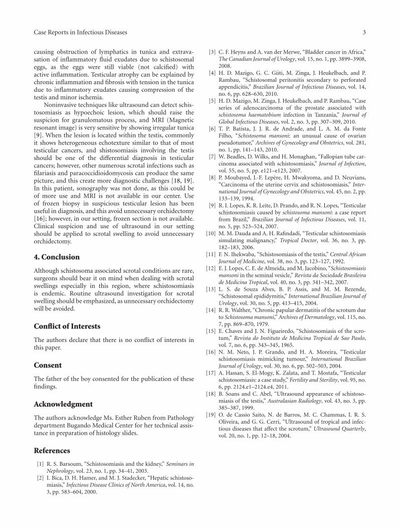
Figure 3: H and E stain showing typical S. hematobium egg withterminal spine (see the arrow) (×20).
and yellowish with firm nodules on its surface. Histologicalexamination with haematoxylin and eosin stain (H and E)showed active chronic granulomatous inflammation withepithelioid macrophages and foreign body giant cells andnumerous schistosomal eggs in the thickened tunica albuge-nia with eosinophils infiltrate. The testis was not involved,and it showed atrophic changes with fibrosis; epididymiswas also normal showing only dilatation. Close examinationof the eggs showed the terminal spine consistent with S.haematobium; the eggs was not calcified (see Figures 1, 2, and3), indicating active inflammation. Followup of the patient inoutpatient there was no further complaints, and the patientwas given Praziquantel 400 mg start.
3. Discussion
Presentation of scrotal swelling and pain clinically will notraise suspicion for schistosomiasis as seen in this patient;findings of hydrocele should rise the suspicious for filariasis,which is also not so common in this region. The finding ofthickened tunica vaginalis together with nodules on the testismay also raise the suspicion for testicular tumors which arecommon at this age. Tuberculous orchitis commonly tendsto be misdiagnosed as a tumor as well, and tuberculosis isvery common in our setting. In this endemic area for schis-tosomiasis, the common mode of presentation is hematuriaand bloody stool; scrotal swelling is very exceptional, andthis is why clinically it was not part of differential diagnosis.Absence of common symptoms of schistosomiasis and thefinding of no ova in the urine or stool brought the challengefor diagnosis of schistosomiasis in this patient.
Pathogenesis of schistosomal infection involving thetestis is through the larva migration from the lungs tothe veins, where the adult lodge into genitourinary venousplexus, and the excretion of the eggs causes chronic gran-ulomatous inflammation. For our patient, there was nourinary bladder symptoms, and the patient had no historyof hematuria in the past; there is possibility of the adultworms to settle in scrotal and testicular venous plexus only,or there was subclinical infection in the urinary bladder, anda subclinical painless testicular lesion due to schistosomiasishas been reported in the past [16, 17]. Findings of hydrocelecan be explained by chronic granulomatous inflammation
Case Reports in Infectious Diseases 3
causing obstruction of lymphatics in tunica and extrava-sation of inflammatory fluid exudates due to schistosomaleggs, as the eggs were still viable (not calcified) withactive inflammation. Testicular atrophy can be explained bychronic inflammation and fibrosis with tension in the tunicadue to inflammatory exudates causing compression of thetestis and minor ischemia.
Noninvasive techniques like ultrasound can detect schis-tosomiasis as hypoechoic lesion, which should raise thesuspicion for granulomatous process, and MRI (Magneticresonant image) is very sensitive by showing irregular tunica[9]. When the lesion is located within the testis, commonlyit shows heterogeneous echotexture similar to that of mosttesticular cancers, and shistosomiasis involving the testisshould be one of the differential diagnosis in testicularcancers; however, other numerous scrotal infections such asfilariasis and paracoccidioidomycosis can produce the samepicture, and this create more diagnostic challenges [18, 19].In this patient, sonography was not done, as this could beof more use and MRI is not available in our center. Useof frozen biopsy in suspicious testicular lesion has beenuseful in diagnosis, and this avoid unnecessary orchidectomy[16]; however, in our setting, frozen section is not available.Clinical suspicion and use of ultrasound in our settingshould be applied to scrotal swelling to avoid unnecessaryorchidectomy.
4. Conclusion
Although schistosoma associated scrotal conditions are rare,surgeons should bear it on mind when dealing with scrotalswellings especially in this region, where schistosomiasisis endemic. Routine ultrasound investigation for scrotalswelling should be emphasized, as unnecessary orchidectomywill be avoided.
Conflict of Interests
The authors declare that there is no conflict of interests inthis paper.
Consent
The father of the boy consented for the publication of thesefindings.
Acknowledgment
The authors acknowledge Ms. Esther Ruben from Pathologydepartment Bugando Medical Center for her technical assis-tance in preparation of histology slides.
References
[1] R. S. Barsoum, “Schistosomiasis and the kidney,” Seminars inNephrology, vol. 23, no. 1, pp. 34–41, 2003.
[2] I. Bica, D. H. Hamer, and M. J. Stadecker, “Hepatic schistoso-miasis,” Infectious Disease Clinics of North America, vol. 14, no.3, pp. 583–604, 2000.
[3] C. F. Heyns and A. van der Merwe, “Bladder cancer in Africa,”The Canadian Journal of Urology, vol. 15, no. 1, pp. 3899–3908,2008.
[4] H. D. Mazigo, G. C. Giiti, M. Zinga, J. Heukelbach, and P.Rambau, “Schistosomal peritonitis secondary to perforatedappendicitis,” Brazilian Journal of Infectious Diseases, vol. 14,no. 6, pp. 628–630, 2010.
[5] H. D. Mazigo, M. Zinga, J. Heukelbach, and P. Rambau, “Caseseries of adenocarcinoma of the prostate associated withschistosoma haematobium infection in Tanzania,” Journal ofGlobal Infectious Diseases, vol. 2, no. 3, pp. 307–309, 2010.
[6] T. P. Batista, J. J. R. de Andrade, and L. A. M. da FonteFilho, “Schistosoma mansoni: an unusual cause of ovarianpseudotumor,” Archives of Gynecology and Obstetrics, vol. 281,no. 1, pp. 141–143, 2010.
[7] W. Beadles, D. Wilks, and H. Monaghan, “Fallopian tube car-cinoma associated with schistosomiasis,” Journal of Infection,vol. 55, no. 5, pp. e121–e123, 2007.
[8] P. Moubayed, J.-F. Lepere, H. Mwakyoma, and D. Neuvians,“Carcinoma of the uterine cervix and schistosomiasis,” Inter-national Journal of Gynecology and Obstetrics, vol. 45, no. 2, pp.133–139, 1994.
[9] R. I. Lopes, K. R. Leite, D. Prando, and R. N. Lopes, “Testicularschistosomiasis caused by schistosoma mansoni: a case reportfrom Brazil,” Brazilian Journal of Infectious Diseases, vol. 11,no. 5, pp. 523–524, 2007.
[10] M. M. Dauda and A. H. Rafindadi, “Testicular schistosomiasissimulating malignancy,” Tropical Doctor, vol. 36, no. 3, pp.182–183, 2006.
[11] F. N. Ihekwaba, “Schistosomiasis of the testis,” Central AfricanJournal of Medicine, vol. 38, no. 3, pp. 123–127, 1992.
[12] E. J. Lopes, C. E. de Almeida, and M. Jacobino, “Schistosomiasismansoni in the seminal vesicle,” Revista da Sociedade Brasileirade Medicina Tropical, vol. 40, no. 3, pp. 341–342, 2007.
[13] L. S. de Souza Alves, B. P. Assis, and M. M. Rezende,“Schistosomal epididymitis,” International Brazilian Journal ofUrology, vol. 30, no. 5, pp. 413–415, 2004.
[14] R. R. Walther, “Chronic papular dermatitis of the scrotum dueto Schistosoma mansoni,” Archives of Dermatology, vol. 115, no.7, pp. 869–870, 1979.
[15] E. Chaves and J. N. Figueiredo, “Schistosomiasis of the scro-tum,” Revista do Instituto de Medicina Tropical de Sao Paulo,vol. 7, no. 6, pp. 343–345, 1965.
[16] N. M. Neto, J. P. Grando, and H. A. Moreira, “Testicularschistosomiasis mimicking tumour,” International BrazilianJournal of Urology, vol. 30, no. 6, pp. 502–503, 2004.
[17] A. Hassan, S. El-Mogy, K. Zalata, and T. Mostafa, “Testicularschistosomiasis: a case study,” Fertility and Sterility, vol. 95, no.6, pp. 2124.e1–2124.e4, 2011.
[18] B. Soans and C. Abel, “Ultrasound appearance of schistoso-miasis of the testis,” Australasian Radiology, vol. 43, no. 3, pp.385–387, 1999.
[19] O. de Cassio Saito, N. de Barros, M. C. Chammas, I. R. S.Oliveira, and G. G. Cerri, “Ultrasound of tropical and infec-tious diseases that affect the scrotum,” Ultrasound Quarterly,vol. 20, no. 1, pp. 12–18, 2004.

Submit your manuscripts athttp://www.hindawi.com
Stem CellsInternational
Hindawi Publishing Corporationhttp://www.hindawi.com Volume 2014
Hindawi Publishing Corporationhttp://www.hindawi.com Volume 2014
MEDIATORSINFLAMMATION
of
Hindawi Publishing Corporationhttp://www.hindawi.com Volume 2014
Behavioural Neurology
EndocrinologyInternational Journal of
Hindawi Publishing Corporationhttp://www.hindawi.com Volume 2014
Hindawi Publishing Corporationhttp://www.hindawi.com Volume 2014
Disease Markers
Hindawi Publishing Corporationhttp://www.hindawi.com Volume 2014
BioMed Research International
OncologyJournal of
Hindawi Publishing Corporationhttp://www.hindawi.com Volume 2014
Hindawi Publishing Corporationhttp://www.hindawi.com Volume 2014
Oxidative Medicine and Cellular Longevity
Hindawi Publishing Corporationhttp://www.hindawi.com Volume 2014
PPAR Research
The Scientific World JournalHindawi Publishing Corporation http://www.hindawi.com Volume 2014
Immunology ResearchHindawi Publishing Corporationhttp://www.hindawi.com Volume 2014
Journal of
ObesityJournal of
Hindawi Publishing Corporationhttp://www.hindawi.com Volume 2014
Hindawi Publishing Corporationhttp://www.hindawi.com Volume 2014
Computational and Mathematical Methods in Medicine
OphthalmologyJournal of
Hindawi Publishing Corporationhttp://www.hindawi.com Volume 2014
Diabetes ResearchJournal of
Hindawi Publishing Corporationhttp://www.hindawi.com Volume 2014
Hindawi Publishing Corporationhttp://www.hindawi.com Volume 2014
Research and TreatmentAIDS
Hindawi Publishing Corporationhttp://www.hindawi.com Volume 2014
Gastroenterology Research and Practice
Hindawi Publishing Corporationhttp://www.hindawi.com Volume 2014
Parkinson’s Disease
Evidence-Based Complementary and Alternative Medicine
Volume 2014Hindawi Publishing Corporationhttp://www.hindawi.com