Embed Size (px)
Citation preview

Screening of Protein Kinases by ATP-STD NMR SpectroscopyMark A. McCoy,* Mary M. Senior, and Daniel F. Wyss
Schering-Plough Research Institute, 2015 Galloping Hill Road, Kenilworth, New Jersey 07033Received December 9, 2004; E-mail: [email protected]
Protein kinases are key mediators of cellular function: deregula-tion of kinase activity has been related to a wide range of diseases.Searching for potent inhibitors that are selective against the morethen 500 potential human kinase targets is currently an active area ofdrug discovery.1 The identification of novel ATP-competitive kinaseinhibitors is nonetheless challenging. Not only are corporate librariesdeficient in compounds with kinase binding motifs, but also highthroughput kinase assays are susceptible to a high rate of false posi-tives.2 ATP typically binds to kinases withKd’s of 10-100 µM;purine replacements would be expected to have similar affinities.This affinity range is difficult for many assay formats but ideal forNMR screening methods. While SAR-by-NMR is the best-knownof these methods,3 ligand detected NMR screening methods (suchas STD-NMR) are an excellent choice to screen for novel kinasecores.4 Ligand detected methods typically do not reveal their targetbinding site, although paramagnetic relaxation enhancement (PRE)has been used to estimate distances between ligands that bind simul-taneously to two different sites.5 Some advantages of STD-NMR andrelated methods are reduced protein requirements, no restrictions ontarget size and no requirement of obtaining high quality proteinNMR spectra. We report the use of ATP as a site-specific markerin STD-NMR experiments that are designed to test the potency ofprotein kinase inhibitors. ATP-STD spectra detect NMR signalsthat originate from target-bound ATP. Reduction of the ATP-STD sig-nal by competitive inhibitor binding permits a direct measurementof the inhibitorKi with respect to the natural substrate: ATP. Weshow that by adding MnCl2, ATP is converted into a paramagenticprobe6 from which the proximity of non-ATP competitive inhibitorscan be inferred.
ATP-STD NMR requires the collection of 4 simple 1D protonNMR spectra typically consisting of two presaturation experiments,a 2-hr ATP-STD reference and a 2-hr ATP-STD competition ex-periment. There are 4 objectives to these experiments: quality control,Ki measurement, identification of non-ATP competitive compoundsand proximity determination of non-ATP competitive compounds.
(1) Quality control is monitored in two 1D spectra of the proteinkinase, ATP, and TSP, with and without inhibitor. Protein aggrega-tion and unfolding in response to compound addition can be directlyobserved for proteins tested at about 5µM concentrations. Com-pound & ATP aliquots are variable and their concentrations shouldbe measured directly relative to a known concentration of TSP (tri-methylsilylpropane sulfonate) as an internal reference. Compoundimpurities and degradation can also be detected. ATP hydrolysiscan be monitored by observing chemical shift changes in purine &sugar protons.
(2) Target-ligand interactions are detected and quantified by STDdetected competition.4 ATP and MgATP bind to most active andmany inactive kinases. In the absence of protein, magnesium has aKd of ∼38 µM for ATP.7 When ATP and MgATP bind the targetprotein kinase with different affinities, the MgCl2 concentration canbe adjusted (0-10 mM) to alter ATP-target affinity.8 We use theadjustability of the ATP-target affinity to optimize the ATP-STDsignal and to tune the affinity range of the ATP/inhibitor competition
experiment (See Supporting Information). A typical STD spectrumfrom target-bound ATP yields peaks from the purine H8 and H2protons and the sugar H1′ proton. Binding an ATP competitiveinhibitor reduces the ATP-STD signal (Figure 1). Simultaneousreduction of the ATP epitope and appearance of a compound STDepitope suggests ATP competitive binding from whichKi’s can becalculated.9 Active kinases can be tested with ATP (no Mg2+), non-hydrolyzable ATP analogues; AMP and ADP can also be used.
(3) The ATP site can be blocked with a high affinity inhibitorto distinguish competitive from non-ATP competitive inhibitors.Addition of staurosporine (which is ATP competitive and has alow nanomolarKd for most Ser/Thr kinases) eliminates the STDpeaks for ATP and for competitive inhibitors such as olomoucine.The STD signals of non-ATP competitive compounds are notreduced upon staurosporine addition.
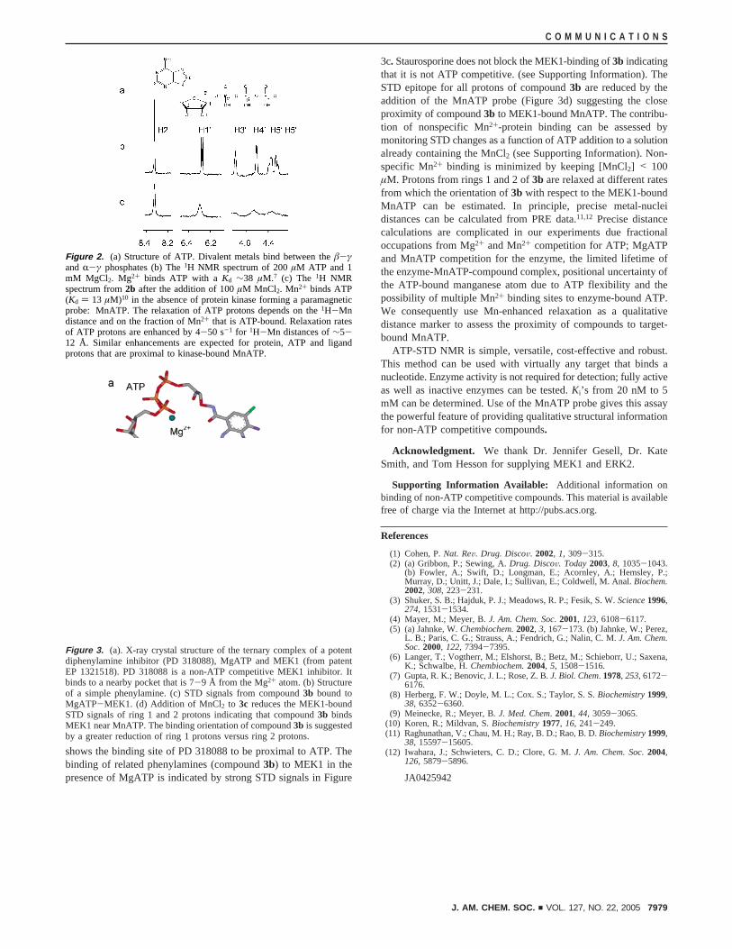
(4) A site-specific paramagnetic probe can be used to determinethe proximity of non-ATP competitive compounds. The MnATPprobe is formed by binding Mn2+ to ATP (Figure 2). The relaxationof ATP protons is strongly enhanced by Mn2+ binding even in theabsence of the protein kinase. Upon binding to the protein kinase,MnATP enhances the relaxation of protein and ligand protons inand around the ATP site. PRE of target-bound ligand protons isconveniently monitored in STD-NMR spectra. PRE is activethroughout the STD data collection. The protein-bound ligandprotons are, however, saturated in the on-resonance (transfer) datacollection. PRE therefore affects ligand protons primarily duringthe off-resonance (reference) data collection. The STD ligandepitope decreases due to PRE which has an r-6 dependence on theproton-Mn2+ distance.
We demonstrate the use of the MnATP proximity probe on thecatalytic domain of the Ser/Thr kinase MEK1. The crystal structureof the MEK1-MgATP-PD318088 ternary complex (Figure 3a)
Figure 1. (a) H2 ATP-STD peak for MgATP (200µM ATP; 1 mM MgCl2)bound to 5µM ERK2. Strong ATP-STD peaks for H8 (8.63 PPM) and H1′(6.26 PPM) are also present but not shown. (b) STD-NMR spectrum after170µM olomoucine is added to1a. Appearance of olomoucine STD peaksindicates that it binds to ERK2. Reduction of the ATP-STD peak indicatesthat olomoucine competes with MgATP for ERK2 with an estimatedKi of∼30 µM.
Published on Web 05/12/2005
7978 9 J. AM. CHEM. SOC. 2005 , 127, 7978-7979 10.1021/ja0425942 CCC: $30.25 © 2005 American Chemical Society
shows the binding site of PD 318088 to be proximal to ATP. Thebinding of related phenylamines (compound3b) to MEK1 in thepresence of MgATP is indicated by strong STD signals in Figure
3c. Staurosporine does not block the MEK1-binding of3b indicatingthat it is not ATP competitive. (see Supporting Information). TheSTD epitope for all protons of compound3b are reduced by theaddition of the MnATP probe (Figure 3d) suggesting the closeproximity of compound3b to MEK1-bound MnATP. The contribu-tion of nonspecific Mn2+-protein binding can be assessed bymonitoring STD changes as a function of ATP addition to a solutionalready containing the MnCl2 (see Supporting Information). Non-specific Mn2+ binding is minimized by keeping [MnCl2] < 100µM. Protons from rings 1 and 2 of3b are relaxed at different ratesfrom which the orientation of3b with respect to the MEK1-boundMnATP can be estimated. In principle, precise metal-nucleidistances can be calculated from PRE data.11,12 Precise distancecalculations are complicated in our experiments due fractionaloccupations from Mg2+ and Mn2+ competition for ATP; MgATPand MnATP competition for the enzyme, the limited lifetime ofthe enzyme-MnATP-compound complex, positional uncertainty ofthe ATP-bound manganese atom due to ATP flexibility and thepossibility of multiple Mn2+ binding sites to enzyme-bound ATP.We consequently use Mn-enhanced relaxation as a qualitativedistance marker to assess the proximity of compounds to target-bound MnATP.
ATP-STD NMR is simple, versatile, cost-effective and robust.This method can be used with virtually any target that binds anucleotide. Enzyme activity is not required for detection; fully activeas well as inactive enzymes can be tested.Ki’s from 20 nM to 5mM can be determined. Use of the MnATP probe gives this assaythe powerful feature of providing qualitative structural informationfor non-ATP competitive compounds.
Acknowledgment. We thank Dr. Jennifer Gesell, Dr. KateSmith, and Tom Hesson for supplying MEK1 and ERK2.
Supporting Information Available: Additional information onbinding of non-ATP competitive compounds. This material is availablefree of charge via the Internet at http://pubs.acs.org.
References
(1) Cohen, P.Nat. ReV. Drug. DiscoV. 2002, 1, 309-315.(2) (a) Gribbon, P.; Sewing, A.Drug. DiscoV. Today2003, 8, 1035-1043.
(b) Fowler, A.; Swift, D.; Longman, E.; Acornley, A.; Hemsley, P.;Murray, D.; Unitt, J.; Dale, I.; Sullivan, E.; Coldwell, M. Anal.Biochem.2002, 308, 223-231.
(3) Shuker, S. B.; Hajduk, P. J.; Meadows, R. P.; Fesik, S. W.Science1996,274, 1531-1534.
(4) Mayer, M.; Meyer, B.J. Am. Chem. Soc. 2001, 123, 6108-6117.(5) (a) Jahnke, W.Chembiochem.2002, 3, 167-173. (b) Jahnke, W.; Perez,
L. B.; Paris, C. G.; Strauss, A.; Fendrich, G.; Nalin, C. M.J. Am. Chem.Soc. 2000, 122, 7394-7395.
(6) Langer, T.; Vogtherr, M.; Elshorst, B.; Betz, M.; Schieborr, U.; Saxena,K.; Schwalbe, H.Chembiochem.2004, 5, 1508-1516.
(7) Gupta, R. K.; Benovic, J. L.; Rose, Z. B.J. Biol. Chem. 1978, 253, 6172-6176.
(8) Herberg, F. W.; Doyle, M. L.; Cox. S.; Taylor, S. S.Biochemistry1999,38, 6352-6360.
(9) Meinecke, R.; Meyer, B. J. Med. Chem. 2001, 44, 3059-3065.(10) Koren, R.; Mildvan, S.Biochemistry1977, 16, 241-249.(11) Raghunathan, V.; Chau, M. H.; Ray, B. D.; Rao, B. D.Biochemistry1999,
38, 15597-15605.(12) Iwahara, J.; Schwieters, C. D.; Clore, G. M.J. Am. Chem. Soc. 2004,
126, 5879-5896.
JA0425942
Figure 2. (a) Structure of ATP. Divalent metals bind between theâ-γand R-γ phosphates (b) The1H NMR spectrum of 200µM ATP and 1mM MgCl2. Mg2+ binds ATP with aKd ∼38 µM.7 (c) The 1H NMRspectrum from2b after the addition of 100µM MnCl2. Mn2+ binds ATP(Kd ) 13 µM)10 in the absence of protein kinase forming a paramagneticprobe: MnATP. The relaxation of ATP protons depends on the1H-Mndistance and on the fraction of Mn2+ that is ATP-bound. Relaxation ratesof ATP protons are enhanced by 4-50 s-1 for 1H-Mn distances of∼5-12 Å. Similar enhancements are expected for protein, ATP and ligandprotons that are proximal to kinase-bound MnATP.
Figure 3. (a). X-ray crystal structure of the ternary complex of a potentdiphenylamine inhibitor (PD 318088), MgATP and MEK1 (from patentEP 1321518). PD 318088 is a non-ATP competitive MEK1 inhibitor. Itbinds to a nearby pocket that is 7-9 Å from the Mg2+ atom. (b) Structureof a simple phenylamine. (c) STD signals from compound3b bound toMgATP-MEK1. (d) Addition of MnCl2 to 3c reduces the MEK1-boundSTD signals of ring 1 and 2 protons indicating that compound3b bindsMEK1 near MnATP. The binding orientation of compound3b is suggestedby a greater reduction of ring 1 protons versus ring 2 protons.
C O M M U N I C A T I O N S
J. AM. CHEM. SOC. 9 VOL. 127, NO. 22, 2005 7979


![Receptor-Like Kinases Sustain Symbiotic Scrutiny1[OPEN]...Update on Receptor-Like Kinases in Symbiosis Receptor-Like Kinases Sustain Symbiotic Scrutiny1[OPEN] Chai Hao Chiu,2 and Uta](https://img.pdfslide.us/doc/110x75/60aa214268722c0ce00ae5e7/receptor-like-kinases-sustain-symbiotic-scrutiny1open-update-on-receptor-like.jpg)


![Arabidopsis Casein Kinase1 Proteins CK1.3 and CK1.4 ... · code Active Casein Kinases. (A) In vitro kinase assays by [g-32P]ATP autoradiography indicated that recombinant CK1.3 and](https://img.pdfslide.us/doc/110x75/5fd572d92d5adf1c9e637682/arabidopsis-casein-kinase1-proteins-ck13-and-ck14-code-active-casein-kinases.jpg)