Embed Size (px)
Citation preview

Alma Mater Studiorum – Università di Bologna
DOTTORATO DI RICERCA IN
Scienze Veterinarie
Curriculum Sicurezza Alimentare
Ciclo XXIX
Settore Concorsuale di afferenza: 07/G1 – SCIENZE E TECNOLOGIE ANIMALI
Settore Scientifico disciplinare: AGR/19 – ZOOTECNIA SPECIALE
STUDIES ON ANTE- AND POST- MORTEM LESIONS AS
ANIMAL-BASED CRITERIA TO IMPROVE WELFARE AND
MEAT QUALITY IN SWINE
Presentata da: Marika Vitali
Coordinatore Dottorato Relatore
Chiar.mo Prof. Arcangelo Gentile Chiar.mo Prof. Luca Sardi
Correlatore
Dott. Luigi Faucitano
Esame finale anno 2017

DISSERTATION
STUDIES ON ANTE- AND POST- MORTEM LESIONS AS
ANIMAL-BASED CRITERIA TO IMPROVE WELFARE AND
MEAT QUALITY IN SWINE
By
Marika Vitali
Department of Veterinary Medical Sciences
Alma Mater Studiorum – University of Bologna
Ozzano Emilia, Bologna, Italy
Thesis submitted to the Doctoral Studies Office in partial fulfilment of the
requirements for the degree of Doctor of Philosophy
March 2017
© Marika Vitali, 2017

ABSTRACT
The study of lesions as animal-based criteria on pigs is of increasing interest at both research and
industry level. Lesions are important outcome measures, able to detect when animal welfare is
suboptimal. The presence of lesions is also correlated to a lower quality, and thus to a loss of
profits, in the derived products.
This manuscript is composed by two studies investigating the use of lesions as suitable indicators of
welfare level and meat quality at both farm and slaughter level.
The first study, performed in Italy, studied the effect of different environmental enrichment devices
provided to Italian heavy pigs (intended for the production of Parma Ham PDO) on the occurrence
of skin, tail and anatomopathological (e.g. oesophageal gastric lesion) lesions, on carcass traits,
meat quality and long-dried products. The tested devices were: hanging chains, wood-log inside a
metal racket, edible block inside a metal racket. Results showed an increased tail score in the
wooden enrichment, united to a higher F-o-M and a lower backfat thickness in the carcass, and to a
lower water holding capacity of the meat. The edible block has not presented changes in skin and
tail score, while an increased number of oesophageal lesion score was observed, which did not
affect carcass and derived products.
The second study was conducted in Canada. It aimed at assessing the age of the lesion on pig
carcass at slaughter through the use of a spectrophotometer and biological indicators (i.e. gene
expression, histochemistry, histology) on skin samples analyzed in the laboratory. Results
demonstrated that spectrophotometric color assessment is a suitable method which allows to
discriminate between fresh lesions (occurred pre-slaughter) and older lesions (on-farm). The results
were also comparable with what was obtained from the expression of some tested genes and from
inflammation scores assessed through histology.

RIASSUNTO
Lo studio delle lesioni nel suino è di crescente interesse nel panorama scientifico e industriale in
quanto la presenza di lesioni è un importante indicatore per evidenziare uno scarso livello di
benessere animale che si può tradurre in un deterioramento e in una perdita di valore dei prodotti
derivati.
L’elaborato si compone di studi che utilizzano le lesioni come indicatori di problematiche presenti
all’interno della filiera suinicola.
Il primo studio, svoltosi in Italia, considera come diversi tipi di arricchimenti ambientali impiegati
nel suino pesante italiano (destinato alla produzione di Prosciutto di Parma DOP) influenzino
l’insorgenza di lesioni cutanee, della coda e anatomopatologiche (con particolare riferimento alle
lesioni della pars oesophagea), nonché gli effetti sulla qualità delle carcasse, della carne ottenuta e
del prosciutto stagionato. Gli arricchimenti testati erano: catena, tondelli di legno inseriti in una
rastrelliera, substrato edibile inserito in una rastrelliera. I risultati hanno dimostrato che
l’arricchimento legnoso ha provocato un incremento di lesioni della coda e un tenore di carne magra
superiore e di spessore del lardo dorsale inferiore nella carcassa, nonché una minore capacità di
ritenzione idrica nella carne. Il substrato edibile non ha prodotto effetti indesiderati sulle lesioni
cutanee e della coda mentre è stato riscontrato un incremento di lesioni dello stomaco, che non ha
influenzato la qualità della carcassa e dei prodotti ottenuti.
Il secondo studio, svoltosi in Canada, si è occupato di determinare l’età delle lesioni cutanee
presenti sulla carcassa suina attraverso l’utilizzo di uno spettrofotometro in sede di macellazione e
di indicatori biologici (espressione genica, istochimica, istologia) sui campioni prelevati e analizzati
in laboratorio. I risultati hanno dimostrato che è possibile, attraverso l’utilizzo dello
spettrofotometro, differenziare tra lesioni recenti (pre-macellazione) o lesioni più vecchie in
allevamento. I risultati sono stati confermati da quanto ottenuto dall’espressione di alcuni geni
testati e dalla risposta infiammatoria.

“In a world full of people who couldn’t care less,
be the one who couldn’t care more.”
Anonymous

LIST OF CONTENTS
PREFACE 8
1. LITERATURE REVIEW 10
1.1. Animal-based measures 10
1.2. Lesion as animal-based measures in pigs 11
1.3. Impact of pre-slaughter lesions on carcass condemnation and poor meat quality 23
1.4. Consideration on the economic impact of lesions 25
1.5. The prospective of assessing lesions post-mortem at abattoer 28
Literature cited 31
PREFACE TO CHAPTER 2 39
2. EFFECTS OF DIFFERENT ENRICHMENT DEVICES ON ON-FARM AND POST-
MORTEM LESIONS AND MEAT QUALITY OF ITALIAN HEAVY PIGS 40
2.1. Introduction 41
2.2. Material and Methods 43
2.3. Results 49
2.4. Discussion 53
2.5. Conclusion 56
PREFACE TO CHAPTER 3 AND 4 61
3. CAN THE AGE OF LESIONS ON THE PIG BODY AND CARCASS BE DETERMINED
BY GENE EXPRESSION? A PRELIMINARY STUDY 62
3.1. Introduction 62
3.2. Materials and Methods 63
3.3. Results and Discussion 67
3.4. Conclusions 69
4. USE OF THE SPECTROPHOTOMETRIC COLOR METHOD FOR THE
DETERMINATION OF THE AGE OF SKIN LESIONS ON THE PIG CARCASS AND ITS
RELATIONSHIP WITH GENE EXPRESSION AND HISTOCHEMICAL AND
HISTOLOGICAL PARAMETERS 71
4.1. Introduction 72
4.2. Materials and Methods 73
4.3. Results and Discussion 82
4.4. Conclusions 91
5. GENERAL CONCLUSIONS 97
AKNOWLEDGEMENTS 98

8
PREFACE
Maintain livestock animals in a good animal welfare is a responsibility which involves the entire
human society. According to Broom (2016), despite a utilitarian approach, a deontological approach
is also necessary because in some cases a poor welfare cannot be justified by benefits to others. The
term sentient has become used in legislation about animals since the Treaty of Lisbon (European
Union, 2007) unless the concept that animals used by people should be protected from actions that
might cause suffering was widespread so far. So animal welfare becomes a scientific concept and
being able to assess how good the welfare is as well as to evaluate poor welfare one of the major
challenges during the last 30 years (Broom, 2016). Faster, animal welfare becomes an important
component in the sustainability of systems and the quality of animal products (Velarde et al., 2015),
indeed a part of consumers’ concern (Di Pasquale et al., 2014). Welfare outcome indicators have
been developed by many scientists, with the purpose to be used by veterinary, inspectors and,
generally, by those who work with animals. Animal-based measures are now considered best
reliable criteria to identify animals whose welfare is poor or to identify where welfare is
deteriorating in order to prevent the risks and to maximize benefits (EFSA Panel on Animal Health
and Welfare, 2012). An example of such indicator in pigs is tail lesion assessment. Although many
animal-based measures are simple and easy to use even under commercial conditions, in some cases
the measure may require further analysis or further studies to be correctly interpreted. A prospective
for those last ones, with continued technical developments, is to improve automatic recording and
precision livestock farming techniques, to let currently unfeasible animal-based measures become
suitable in the future (Wathes, 2009).
Talking about swine, among the others, the study of lesions in the pig skin, tail lesion and
anatomopathological lesions, appears to be interesting indicators which may not only provide
important information on the level of animal welfare and of a given pig or batch of pigs, it may
provide information on the critical points in pig marketing, both in rearing system and in pre-

9
slaughter practices. To a better understanding of the information and opportunities on the utilization
of this welfare indicators in risk assessment, deeper investigations both at basic and applied science
level are required, with the purpose to improve sustainability and quality of the entire production
system and derived products.
Literature cited
Broom, D. M. 2016. Considering animals’ feelings. Anim. Sentience.
EFSA Panel on Animal Health and Welfare. 2012. Statement on the use of animal-based measures
to assess the welfare of animals. EFSA J. 10:2767. doi:10.2903/j.efsa.2012.2767.
Di Pasquale, J., E. Nannoni, I. Del Duca, F. Adinolfi, F. Capitanio, L. Sardi, M. Vitali, and G.
Martelli. 2014. What foods are identified as animal friendly by Italian consumers? Ital. J. Anim.
Sci. 13: 782-789. doi:10.4081/ijas.2014.3582.
Pomar, C., and M. Marcoux. 2005. The accuracy of measuring backfat and loin muscle thickness on
pork carcasses by the Hennessy HGP2, Destron PG-100, CGM and ultrasound CVT grading
probes. Can. J. Anim. Sci. 85:481–492. doi:10.4141/A05-041.
Velarde, A., E. Fàbrega, I. Blanco-Penedo, and A. Dalmau. 2015. Animal welfare towards
sustainability in pork meat production. Meat Sci. 109:13–17. doi:10.1016/j.meatsci.2015.05.010.
Wathes, C. M. 2009. Precision livestock farming for animal health, welfare and production. In:
Sustainable animal production. p. 411–419.

10
1.
LITERATURE REVIEW
1.1. Animal-based measures
Animal Welfare can be assessed through wide variety of parameters which measure behavior,
physiological changes, clinical condition, productivity parameters and others. Nowadays, the trend
in writing animal welfare protocols are more focused on the adoption of animal-based measures
instead of considering resource-based or managed-based measure (Grandin, 2014). An example of
this protocols is the European Welfare Quality® (Welfare Quality
®, 2009) (Fig. 1.1).
Animal-based measure can be assessed directly on the animals themselves, regardless of their
keeping conditions (Pandolfi et al., 2017) and it considers how an individual will react, according to
its physiological characteristic, to the many factors which may affect animal welfare, such as
management practices, rearing conditions, environment etc.
These outcome measures make possible to assess and compare animal welfare across different
environments. Animal-based measures can be collected during all the phases of the production
Figure 1. 1 The 4 principles
and 12 animal-based criteria used
in the Welfare Quality® project.
Animal-based measures could predict
the risk of poor welfare if no change
or intervention will made and help to
identify hazards in a risk assessment
prospective.
(Source: EFSA, Panel on Animal
Health and Welfare, 2012).

11
marketing in farms or at the abattoir. Indeed, they can be obtained directly from the animals or
through the use of reporting systems (e.g. sanitary surveillance), recording production parameters,
or after laboratory analyses (EFSA Panel on Animal Health and Welfare, 2012).
Unless animal-based indicators are considered the most appropriate to effectively measure animal
welfare in animals, being able to select which measure or combinations of measures are the most
appropriated to monitor a given issue on animal welfare may become very challenging.
There are many factors which should be taken in account such as practical feasibility, economical
aspect, required technology if the output is easy to be interpreted or further study are required. For
this reason, the research of so-called iceberg-indicators, with the purpose to reduce the large amount
of existent outcomes through the use of multivariate analysis, seems very promising (Pandolfi et al.,
2017). Also, the developing of precision livestock-farming technologies for monitoring animal
welfare may be greatly supportive. It might allow checking routinely a lot of animals, through the
development of specific technology for monitoring specific animal welfare criteria or integrating
the existing technology with more specific data, helping to better integrate animal welfare in the
risk-assessment perspective (Wathes et al., 2008).
1.2. Lesion as animal-based measures in pigs
1.2.1. Skin lesions
Also if may be plenty of sources of skin lesions in pigs (Fig.1.2), the most frequent derive from
fighting (ITP, 1996; Varón-Álvarez et al., 2014).
Figure 1. 2 Shapes of frequent lesions detected on the carcasses.
1= comma; 2= rectangular; 3=linear; 4=diffuse; 5= rhomboidal (Modified from: Varón-Álvarez et al., 2014)

12
Fighting-type lesion is usually up to 10 cm length, linear and tramline, mostly concentrated in high
number in the front and hindquarter part (Teixeira and Boyle, 2014), presenting a typical comma
shape (Fig. 1.3). According to Stewart et al., (2008) fighting occurs between 2 pigs placed with the
body parallel or perpendicular to each other, ramming or pushing with the head and often biting, in
rapid succession, the opponent (Fig 1.3). The number of skin lesions in the upper-front region of
pigs’ body is positively correlated with the duration of the fights (Turner et al., 2006) unless,
according to the same author, pigs that refuse to fight presents more lesion located in the rear part of
the body.
The main risk factor for the expression of this aggressive behavior consists in when unfamiliar pigs
are mixed. Mixing is considered the major source of social stress in pigs (van de Weerd and Day,
2009) and it may occur many times during the pig marketing chain: on farm, at loading, in the truck
during transportation and at lairage. A study from Aaslyng et al. (2013) demonstrated that the
number of skin lesions increased significantly during all these phases (Fig 1.4.).
Particularly the duration of lairage has been largely investigated. In a study from (Geverink et al.,
1996), skin damage was found to increase with the increasing between 0 and 3h, and from 1 to 15h
(Guàrdia et al., 2009). A longer lairage appears to allow the pig to rest and then increase the fight
and thereby skin damage (Faucitano, 2010). Also keeping a pig in big groups during lairage and
increasing stocking density from 1.0 to 2.7 pig/m2 was found to increase the risk of fighting and
Figure 1. 3 Comma-shape lesions (b)
and long linear lesion (d) deriving from
biting during fighting (a) and mounting
behavior (c) respectively.
(Sources: ITP, 1996;
www.welfarequality.net; qpc.adm.slu.se.
Last access: 26/03/2017)

13
skin lesion appearance (Geverink et al., 1996; Rabaste et al., 2007).
Other factors may influence the incidence of fighting. One of them is imputable to the poor
environment, intended as a lack of space and to the absence of stimulus, due to a barren
environment or to the provision of ineffective environmental enrichment (Thomsen et al., 2012).
Also and increased incidence of fighting was found in groups composed of only male (Teixeira and
Boyle, 2014), and a partial correlation with faster-growing pig was noticed by Pitts et al., (2000).
Another common-type of lesions is the result of mounting behavior, which is a part of a normal
pigs’ behavior repertoire, consisting of placing hooves on the back of a standing pen-mate (Scott et
al., 2006). This behavior was found common in both sexes and in barrows, with and increased
incidence groups constituted only by entire males (Van Staaveren et al., 2015). Lesions, in this case,
are more than 10 cm length, linear and mostly placed in the back region (Teixeira and Boyle, 2014).
An increase occurrence of this behavior was also correlated with aggressive behaviors and to an
increased number of lesion assessed in finishing pigs prior to slaughter (Brandt et al., 2015).
Mounting behavior may also result in loin bruises (Teixeira and Boyle, 2014) (Fig. 1.5).
Another source of skin blemishes may be imputable to rough handling, due to the use of electric
Figure 1. 4 Example of a severe
damaged carcass. Lesions were mostly
due to fighting
Figure 1. 5 Loin bruising score for carcasses:
0= no evidence of bruising; 1= moderate loin bruising; 2=severe or extensive loin
bruising (Modified from: Teixeira and Boyle, 2014)
14
goads and sticks and as a consequence of a blunting trauma (Fig. 1.6) (Barington and Jensen, 2013).
The assessment of skin lesion may be done using pictorial standard (ITP, 1996) or counting the
lesion, per each region of the body as proposed from some protocols such as the Welfare Quality®
(Welfare Quality®
, 2009) and the Canadian Animal Care Assessment, and American Meat Institute
audit guide criteria (Rocha et al., 2016).
Skin lesion may be measured both at farm and at slaughter level (and in the last case they may be
measured on the animal at lairage, in the stunning cage, or post-mortem on the carcass). Studies
between the correspondence of lesions assessed ante- and post- mortem presented contrasting
results. Some researchers argue that carcass treatments (i.e. dehairing and singeing) could make
welfare-related carcass damage difficult to detect (Aaslyng et al., 2013). Conversely, Carroll et al.,
(2016) stated that skin lesion were more detectable after carcass deharing and singeing and Brandt
et al. (2015) have found that assessing lesion in the carcass was most correlated with aggressive
behaviors and welfare of pigs at farm, while (Van Staaveren et al., 2015) have found correlation
between skin lesions scores recorded at farm and in the carcass, declaring that both methods are
accurate indicators of pigs’ welfare. Overall, it is important to consider that on-farm assessment is
more dependent from the occurrence of some on-field conditions, such as a big group of animals,
lack of space, light conditions during the assessment and pigs’ body dirtiness. Also the presence of
bristles, especially in some pigs, may hide some lesions from the count. A correct choice about
Figure 1. 6 Examples of lesion inflicted
by humans: skin of a finishing pigs with
lesions due to the excessive use of a tatoo
hammer which has caused haemorragia
in the subcutaneous fat tissue (a);
skin of a finishing pig whit bruises due to
a blunt trauma (Source: Nielsen et al., 2014).

15
where is more appropriate to assess skin lesions needs to be evaluated in accordance with the
purpose of the study and to both farm and slaughter conditions.
1.2.2. Tail lesions
Tail biting is deeply important in swine production, due to the high prevalence of the pathology and
to the difficulty to prevent and to eradicate its occurrence in intensive barren husbandry systems
(EFSA, 2007). Hothersal et al., (2016) survey suggested that an intact tail indicates a good level
and quality of animal welfare related to pig’s husbandry and management. Moreover, reducing the
number and severity of tail biting behavior and so tail lesions has been associated with a higher
incidence of carcass condemnation (Harley et al., 2014).
Tail biting is an abnormal behavior, which arises when a pig (biter) bites the tail of another pig
(victim). Motivationals basis are mostly related to the frustration of the exploratory and oral needs
in pigs. In fact, in intensive husbandry, the poor environment conditions don’t allow them to
manifest rooting, chewing and manipulate behaviors (Studnitz et al., 2007).
A study from Schrøder-Petersen and Simonsen (2001) identified two phases on the development of
the pathology. The first one called “the pre-injury stage”, is characterized by a pig chewing lightly
the tail of another, which usually tolerates it. The second one, “the injury stage”, follows when the
tail is injured and bleeding. Usually, the first stage is not detectable from the farmer, and when the
second stage occurs, it is already difficult to solve the problem. The frequency and intensity of tail
biting may results in tail skin damage, bleeding, and even the complete loss of the tail.
Subsequently, infections, abscesses, paralysis, pyaemia and death may occur (Moinard et al., 2003).
When tail biting occurs, the sign that welfare is poor is clear in both victims and biters. The victim
will suffer pain and fear, being unable to escape from the attacks because of the limited space; the
taste of blood and other stimulations (e.g. an increased activity) make biters more excited, causing
an escalation of tail biting behavior, involving more pigs in the pen (EFSA, 2007).
Tail biting is considered as the main “iceberg indicator” of animal welfare in finishing pigs (D’Eath

16
et al., 2014). In fact, many factors may interact to increase the incidence of tail biting behavior (Fig.
1.7). Anyway, barren environment, lack of space, poor air quality, rationed feeding and mixing of
animals are considered the most important factors in increasing tail biting in growing-finishing pigs.
Environmental enrichments are proposed as one way to reduce tail lesion to furnish pigs with
rootable, chewing and destructible substrates.
Figure 1. 7 The scheme shows how many underlying processes may interact and outbreak as tail biting. In bold, factors connected by
solid arrows, while the others are suspected risk factors. The numbers in the arrows show how some of the risk factors might
influence each other or the process of tail biting. (Source: D’Eath et al., 2014)
Environmental enrichment for pigs is mandatory in the EU since the Directive 2008/120/EC
(European Council, 2008), and it usually consists in alternative-enriched husbandry systems, straw-
based housing systems or point-source-enrichments objects (van de Weerd and Day, 2009).
However, because of its feasibility with the slatted floor, which is the most common husbandry

17
system in EU (EFSA, 2007), the use of point-source enrichment objects is the most common, unless
it presents some critical points. In fact, providing pigs with enrichment devices was found to be
barely effective on the reduction of tail lesions , because most of the objects tested were found to be
less attractive to pigs, or were not able to maintain interest all along the rearing time (van de Weerd
and Day, 2009) . Other factors limiting the effectiveness of point-source enrichment objects may
have occurred where the toys provision alone was not enough to guarantee a good welfare status
(Nannoni et al., 2016). The provision of straw was found to considerably reduce tail lesion score in
pigs (EFSA, 2007a). This may be related to its attitude to reduce aggressive behavior because of its
being a highly manipulative substrate (EFSA, 2007a) as well as a functional nutrient: it increases
fiber content in high-rationed pigs’ diet (Di Martino et al., 2013) and, if correctly managed, it
improves the comfort of bedding and the quality of the air. Conversely, problems concerning its
application in slatted-floor husbandry systems, waste disposal and economic costs, have limited its
application in modern industrial farming (EFSA, 2007a).
Differently than skin lesions, tail lesions are less influenced by animal fighting or pre-slaughter
handling, unless mixing of animals and unstable group hierarchy increase the incidence of tail
lesions (Van Staaveren et al., 2015). Indeed, other factors were found to be effective in the
reduction of tail lesion such as: mixed-sex groups, weight homogeneity between pen-mates., and
keep pigs in small groups instead of large groups (Schrøder-Petersen and Simonsen, 2001;
Zonderland et al., 2008; D’Eath et al., 2014; A. Scollo et al., 2016)
The role of high stocking density in the development of tail biting cannot be underestimated: the
reduction in the available space per pig increases the social tension within the pen and compromises
pigs’ freedom to perform normal activities such as locomotion, resting synchronization, and
generally the ability to reach resources, increasing frustration (Schrøder-Petersen and Simonsen,
2001; Moinard et al., 2003; A Scollo et al., 2016). Moreover, some production systems may
increase the risk of high stock density. In fact, the current directive provides indication on space
allowance for pigs up to 110 kg (1.0 m2/pig), but in some productive system (i.e. Italian heavy-pig
18
production) pigs must reach a weight range of 160-170 kg to be used for PDO (protective
designation of origin) products, unless they are reared in the same design-environment common
throughout the EU (Scollo et al., 2016).
Additional risk factors for tail biting is the poor air quality, because sub-optimal microclimate
conditions may be a source of chronic stress (Hunter et al., 2001). Particularly, ammonia level was
considered as the primary noxious gas able to induce aversive behavior (Wathes et al., 2002).
However, it could also be assumed that an adverse combination of environmental parameters (e.g.
temperature, humidity, ammonia, carbon monoxide, dust) may influence the occurrence of tail
biting (Wathes et al., 2000).
Feeding practices are also very important. Studies have demonstrated that an appropriated design of
the feeder, e.g. number, types, position, length (Hunter et al., 2001; Moinard et al., 2003), and
constancy in feeding time (Scollo et al., 2016) are factors able to limit the incidence of tail biting.
Survey data showed that pellet or dry feed increased tail biting outbreaks, while liquid meal feeding
didn’t (Van Putten, 1969; Hunter et al., 2001; Moinard et al., 2003).
Despite the factors mentioned above, the main risk factor is still the pig’s tail, hence the main
“preventive” measure usually adopted by pig farmers: tail docking. Tail docking is now prohibited
in EU from the Council Directive 2008/120/EC because of welfare and ethical issues concerning the
acute and chronic pain deriving from the amputation (Nannoni et al., 2014), with some exception in
the case of effective needs. Despite that, tail-docking is still a widespread practice throughout the
Union (Nannoni et al., 2014). Indeed, studies on the effectiveness of this practice on preventing tail
biting are contrasting. Hunter et al. (2001) and Scollo et. al. (2016) observed that animals with
longer tails were the recipients of more tail-directed behaviors, showing an increased tail score;
while Moinard et al., (2003) observed that tail-docking increased the risk of tail biting. Differences
in the experimental design and tail lesion score method may have been the cause of this
discrepancy.
19
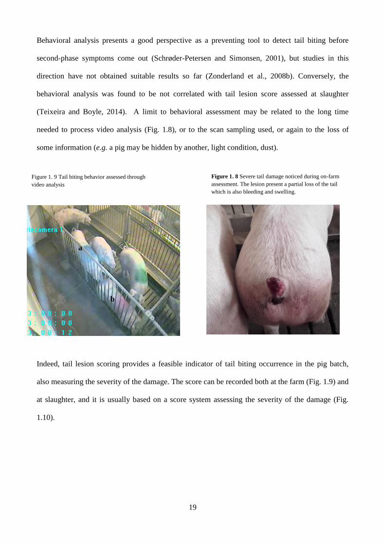
Figure 1. 8 Severe tail damage noticed during on-farm
assessment. The lesion present a partial loss of the tail
which is also bleeding and swelling.
Behavioral analysis presents a good perspective as a preventing tool to detect tail biting before
second-phase symptoms come out (Schrøder-Petersen and Simonsen, 2001), but studies in this
direction have not obtained suitable results so far (Zonderland et al., 2008b). Conversely, the
behavioral analysis was found to be not correlated with tail lesion score assessed at slaughter
(Teixeira and Boyle, 2014). A limit to behavioral assessment may be related to the long time
needed to process video analysis (Fig. 1.8), or to the scan sampling used, or again to the loss of
some information (e.g. a pig may be hidden by another, light condition, dust).
.
The letters a and b indicates the two pigs involved
(biter and victim, respectively).
Indeed, tail lesion scoring provides a feasible indicator of tail biting occurrence in the pig batch,
also measuring the severity of the damage. The score can be recorded both at the farm (Fig. 1.9) and
at slaughter, and it is usually based on a score system assessing the severity of the damage (Fig.
1.10).
a
b
Figure 1. 9 Tail biting behavior assessed through
video analysis

20
Figure 1. 10 Tail lesion score for the carcasses: 0= no evidence of tail biting; 1= healed or mild lesions; 2= evidence of chewing or
puncture wounds, but noevidence of swelling; 3=evidence of chewing or puncture wounds with swelling and signs of possible
infection; 4= partial or total loss of the tail. (Source: Teixeira and Boyle, 2014)
1.2.3. Oesophageal gastric lesions
Ulceration of the pars oesophagea is a common widespread condition in growing-finishing pigs
worldwide, reared in the intensive production system (EFSA Panel on Animal Health and Welfare,
2012). Surveys at the abattoir reveal that 20% of pigs present erosive lesion and 60% have pre-
ulcerative parakeratosis lesion (Friendship, 2004). The major concern associated with gastric
ulceration is represented by mortality. A survey from Melnichouk (2002) evidenced at necroscopy
that approximately 27% of pigs died because of severe blood losses, becoming OGU the first cause
of death, followed by pneumonia (19%).
The pars oesophagea is a non-glandular region of the stomach and does not secrete mucus. Is
covered with stratified squamous epithelium and this layer offers limited protection against low pH
in the lumen and gastric enzymes (Amory et al., 2006).
When the epithelium is damaged, hyperplasia results in thickening hyperkeratosis, and, if left to
spread, it prevents the nutrients to reach the cells. Subsequently, the weakening of the junctions
between the epithelial cells allows gastric juices into the underlying tissue, causing the development
of erosion and ulcers (Robertson et al., 2002). Oesophageal gastric ulceration (OGU) is a
multifactorial condition arising from one or more factors (Tab. 1.1), involving feeding, managing
and welfare practices.
Factors which are able to maintain firmness in the stomach instead of a rapid transit were
considered able to prevent OGU (Herskin et al., 2016). Because of that, the presence of liquid
content in the stomach was associated with an higher score in OGU assessment (Baustad and

21
Nafstad, 1969). Factors as providing the pigs with straw, also in small quantity (Nielsen and
Ingvartsen, 2000; Di Martino et al., 2013; Herskin et al., 2016), and a generally coarse ground diet
were found to be able to give partial or complete protection against OGU in relation to the
percentage of inclusion in pigs’ diet (Baustad and Nafstad, 1969; Bubenik et al., 1998).
Table 1. 1 Risk factors associated with oesophageal gastric ulceration in pigs
Nutricion Housing/Managemnt Other
Absence of straw providing 1,2
Confinement rearing 4 Concurrent disease
4
Ad libitum feeding 11
Feeding regiment 4 Genetic
3
Automated feeding system 11
Herd size 4 Helicobacter infection
4
Changing in diet 11
Holding and transport 4 Helicobacter spp.
8
Dam water 11
Lairage duration 5 Heredity
4
Fasting 6,7,9
Mixing pigs 4 Histamine
4
Feed particle size 4,5
Overcrowding 4 Parturition
4
Finely ground feed 3 Slatted floor
1 PCV2
4
Grinding vs. rolling 4 Starving
12 Season
4
Hammer-mill grain ground 3
Somatotrophin 4
Heat processing 4
Stress 3
Lack of fiber 4
Maize 10
Milling 4
Pellet feeding 1,11
Pelleting 4
Rancid fat 4
Type of grain 4
Vitamine E/Se deficiency 4
Withdrawal of feed 4
1 Amory et al., 2006; 2 Di Martino, 2013; 3 EFSA, 2007; 4 Friendship 2006 in: EFSA, 2007; 5 Friendship, 2004; 6 Lang et al., 1998; 7
Lawrence et al., 1998; 8 Maghalha and Miranda, 1996; 9 Makinde and Gous, 1998; 10 Nielsen and Ingvartsen, 2000; 11 Robertson,
2002; 12 Straw et al., 1994
Type and frequency of antibiotics’ administering was found to have no effect on OGU in a study
from Robertson et al. (2000). The same author observed that the quality and the source of the
drinking water provided might have an impact on gastric lesion development, due to pH values,
buffering abilities (e.g. the presence of bicarbonate anions) and microbiological quality. A study
from Wondra et al., (1995) reported that adding sodium bicarbonate and potassium bicarbonate to a
finely ground diet limited the incidence of ulcers. The role played by stress in the development of
22
gastric lesion was hypothesized in some studies (Bubenik et al., 1998; Robertson et al., 2002;
Herskin et al., 2016), but a lack of evidence is attested, due to the multifactorial source of the
pathology (Amory et al., 2006). Still, it appears logical that the presence of ulcers or erosions on the
pars oesophagea represent a source of pain for the animals, requiring to consider this indicator on
the overall evaluation of animal welfare of a given pig or batch of pigs.
The first step on OGU assessment should be done on living animals by detecting the clinical signs
of gastric ulceration. In fact, according to Friendship (2004), blood loss into the gastrointestinal
tract is the main clinical event in this condition and a source of anemia and melena. Severe anemia
results in a very pale, weak pig, with rapid breathing. The feces are scant, black and tarry. Death
may occur, also as a secondary consequence to another clinical problem (e.g. an outbreaking
respiratory disease, or anorexia by infectious disease). Nevertheless, in many cases symptoms are
subclinical and pigs may appear healthy if blood loss is minimal. In some cases, a reduction of
average daily gain was detected also in pigs with lesions of a medium level (Ayles et al., 1996).
Endoscopic examination may be useful to detect subclinical OGU. The technique is simple and
easy to master. The advantage is that erosions are easily detectable, while a disadvantage is that the
visualization needs an empty stomach and, because of starving, gastric acids and bile may worsen
damages on the proximal part of the stomach, increasing the risk of ulcer development (Ayles et al.,
1996). Moreover, endoscopic examination requires anesthesia, which increases the cost of the
practice. The usual method for monitoring the prevalence of oesophageal gastric lesion in a large
group of animals is to record gross lesions at slaughter (Ayles et al., 1996) (Fig. 1.11).
Monitoring the prevalence of OGU at slaughter may help to identify issues regarding the
management/feeding practices and to improve the quality of pig marketing, therefore preventing
OGU. It is important to consider that, because ulcer may occur rapidly (24 h in total for the entire
progression from normal pars oesophagea to complete ulceration) and heal quickly as well, in some
cases it is difficult to relate lesions at slaughter with farm-related issues (EFSA 2007b). From
another point of view, if serious occurrence of OGU is noticed (i.e. high mortality), a change in diet

23
to a coarsely ground ration, also from a short period, can quickly resolve the problem (Ayles et al.,
1996). Lastly, because many of the factors associated with increased risk of OGU development are
also associated with economic competitiveness (e.g. finely ground feed and fast-growing pigs), a
cost-benefits analysis applied to the prevention program is recommended for each farm, in order to
best balance economic and productive traits with animal welfare concerns (EFSA, 2007).
1.3. Impact of pre-slaughter lesions on carcass condemnation and poor meat quality
Consequences deriving from a poor animal welfare during all the pre-slaughter phases are actually
well recognized from both researchers involved in animal science, veterinaries and stakeholders
involved in pigs’ marketing (Grandin, 2014) so far.
For example, increasing animal welfare may have an impact on reducing carcass damages and
waste by reducing the occurrence of injuries; decreasing pre-slaughter mortality; improving the
quality of the meat by reducing stress in animals (Chambers and Grandin, 2001).
According to Guàrdia et al. (2009), skin damage affects the risk of obtaining PSE (pale soft and
Figure 1. 11 Examples of oesophageal
gastric lesion. (Source: Nielsen and Ingvartsen, 2000)

24
exudative) and DFD (dark firm and dry) pork meat. The risk of PSE occurrence doubled in skin
damaged carcasses than in unblemished carcasses (6.9% vs 3.3% respectively). Also, the incidence
of PSE was higher in pigs with the nn genotypes for the gene RYR1 and skin damages than in pigs
with the same genotype but with unblemished skin or in NN genotype pigs with skin lesions. Also,
DFD pork was found to be positively influenced by skin damage, and the risk was estimated to be
almost 4 times higher than the risk between the same categories of damage (11.7% vs 3.3%
respectively). The same result on the correlation between the incidence of DFD meat and skin
lesion was also obtained from Guàrdia et al. (2009). This result is interesting because as DFD pork
is more related to early stress (e.g. at loading) and PSE more to acute stress (e.g. at lairage) it
suggests that the skin damage score may be a valid indicator to better understand the effect that
some underlying source of stress may have on meat quality (Guàrdia et al., 2009). For example,
because mixing is the main source of skin lesion and results moslty in fighting-type lesions (as
described above), if there were a greater incidence of a higher pH24 value, these aggressive
behaviors were more related to on-farm practices, loading or transport practices (depending on the
length of the transport). Contrarily, if aggressive behavior and fighting take place just prior to
slaughtering (at unloading or lairage), then skin blemishes are expected to be more related to a very
low pH24 (Guàrdia et. al., 2009; Warriss and Brown, 1985).
Harley et al. (2014) have found that 16% of carcasses were affected by severe loin bruising, and
that among that, 2.5% were condemned mainly because of abscesses, and 3.3% were trimmed.
Skin lesions and bruises were also associated with pre-slaughter losses (Nannoni et al., 2016) and
carcass condemnation (Carroll et al., 2016). Also, along with the increasing number and severity of
skin damages, the average cold carcass weight decreased (Carrol et al., 2016).
Lee and Veary (1993) found that tail-biting caused 94% of cases of carcasses condemned because
of pyaemia. Pyemia was found to lead to partial or full condemnation of carcasses (Schrøder-
Petersen and Simonsen, 2001). Conversely, Harley et al., 2014 found that tail lesion was detectable
in 72% of pigs, but didn’t increase the risk of abscess, unless it was significantly associated with

25
carcass condemnation. Also, male pigs were found to have a higher risk of tail lesions and
subsequent carcass condemnation. According to Moinard et al. (2003) a higher tail score
corresponded to a reduced thickness of P2 back-fat in the carcass. This concords with the results
obtained by Harley et al. (2014), in which severe tail lesions were associated with an estimated 12
kg loss in carcass weight, while mild lesions were associated with a -1.2 kg loss.
Oesophageal gastric ulceration is also an important cause of pre-slaughter losses, as previously
described. Melnichouk (2002) assessed that 27% out of 147 pigs died because of hemorrhage due to
gastric ulceration in one month.
To conclude, information on partial or total carcass condemnation in pigs is still limited (Garcia-
Diez and Coelho, 2014), just like the effect of lesions in carcass trait and meat quality. It is worth
noting that investigations aiming at a deeper knowledge of the risk factors are strongly
recommended.
1.4. Consideration on the economic impact of lesions
The quantification of farm-to-fork losses in pig chain is an inaccurate science (Faucitano, 2010,
Harley et al., 2012). In fact, disease, injury or chronic stress may occur at numerous stages and
result in a loss of value by the production. Being able to determine the stage at which rejection of
the products occurs can define which stakeholders must bear such losses (Harley et al., 2012;
Grandin, 2014) (Tab 1.2.).
The main sources of loss of value are pre-slaughter losses during transport and lairage (Nannoni et
al., 2016), or carcass condemnation at meat inspection (Faucitano, 2010). Anyway, the financial
losses by producers are indicative, since the real value of meat depends on the carcass’s traits (e.g.
meat and fat content, carcass classification), anatomical location of the rejected parts, production
target and seasonal price variation (Faucitano, 2001)
Growth-retarded pigs are an underappreciated source of loss for the producer, and it is related to
poor welfare on farm. Martinez (2007) has estimated a loss of 30,000 € (according to the market

26
value) related to carcass condemnation in a 3-year survey conducted on 6017 pigs in an abattoir in
Spain, showing a correlation between carcass condemnations and grow-retarded pig carcasses.
Disease and welfare problems, such as tail biting, oesophageal gastric ulcers, pleurisy and other
disease conditions are correlated with decreased carcass weight (Martínez et al., 2007).
In a study from Harley et al. (2014), tail lesions were considered responsible for financial losses due
to carcass condemnation and trimming, and also to minor carcass weight. Losses from carcass
condemnation and trimmings were estimated at 1.10 €/pig, while potential reduction of carcass
weight was estimated to be 0.59 €/pig. On the whole, a loss of 43% of the profit margin per pig was
attributable to tail biting.
In addition, the efficiency of the slaughter line depends from the number of pigs processed daily.
According to Faucitano (2001), reducing the line speed for trimming or more detailed inspection
processes decreases the efficiency and profitability of the business. Moreover, cuts deriving from a
trimmed carcass were usually destined to lower value products with a lower margin of profit. With
regards to that, it is important to considered that some high-quality meat-derived products are
composed from an whole cut. The presence of blemished skin (usually correlated with damages on
the subcutaneous fat or muscular tissue) in these products will cause their exclusion from the
disciplinary with a considerable loss of value (Faucitano, 2001). One example of this kind of
products is represented by the Italian Parma Ham (PDO). According to the production disciplinary
(Consortium for Parma Ham, 1992), skin must be unblemished, otherwise it will cause the
exclusion from the PDO production.
According to Harley et al. (2014): “These findings illustrate the magnitude of the impact of the
presence of animal welfare for the profitability of the pig industry. They also emphasize the
potential contribution that the inclusion of welfare parameters at meat inspection could make to pig
producers in informing herd health and welfare management plans”.

27
Table 1. 2 Stages at which losses may occur in pig marketing chain (modified from: Harley et al., 2012)
Stage of
production
Reason for loss Resource in which
losses occur
Stage loss
is incurred
Stakeholder
incurring losses
Farm Mortality Carcass Farm Producer
Clinical illness Medicines Farm Producer
Subclinical illness Carcass Abattoir Producer
Injury Carcass Farm Producer
Transport/
unloading
Mortality (dead on arrival) Carcass Transit Producer unless very hight
numbers indicate transporter
responsible Injuries: fracture/bruise Carcass Abattoir
Stress Meat quality Retail
Dehydration Decreased carcass
weight
Abattoir Producer
Ante-mortem
inspection
Mortality (dead in lairage) Carcass Abattoir Abattoir
Injuries Carcass Abattoir Abattoir
Post-mortem
inspection
Desease conditions Carcass Abattoir Producer
Injuries Carcass Abattoir Producer
Welfare conditions Carcass Abattoir Producer
Factory Loss
(mangling/contamination)
Carcass Abattoir Abattoir
Carcass
granding
Weight too hight/low Penalty c/kg Abattoir Producer
Poor learn meat % Penalty c/kg Producer
Processor Trimmed cuts can't go
for premium products
Decreased value Retail Abattoir
Retailer Pale, soft and exudative Dark,
firm and dry
Trimmed cuts
Decreased retail
potential
Retail Retailer

28
1.5. The prospective of assessing lesions post-mortem at abattoer
The perspective of developing an animal-welfare surveillance at the slaughter plant is of concrete
interest for the industries due to the provision of valuable information about conditions all along the
pig marketing chain (Van Staaveren et al., 2015). Meat inspection in particular has the potential to
contribute to the surveillance of animal welfare, and currently meat inspection data are under-
utilized in the EU, even as a means to inform herd health programs (Harley et al., 2012).
Being animal-based, the welfare indicators may reflect conditions during on-farm, transport, lairage
and pre-slaughter. Moreover, since animal health is a component of animal welfare, meat inspection
data can be used to inform herd health programs, thereby reducing the risk of injury and disease and
improving production efficiency (Harley et al., 2012). Although certain measures on the carcass can
be reliably used to detect injuries and disease, for most indicators research is needed to validate
them as welfare indicators (Brandt et al., 2013, 2015).
Ante- and post-mortem carcass condemnation records could allow to create a database for swine
issues. This database could be used to monitor swine disease as well as to improve animal health,
food safety and animal welfare (Garcia-Diez and Coelho, 2014). A positive correlation was found
between the percentage of carcasses condemned and the percentage of pigs with skin or tail lesion
and loin bruising (Carroll et al., 2016).
Surveillance of the type and quantity of lesions on the carcass may be helpful to establish the degree
to which they are correlated to aggressive behaviors – for example, the relationship between tail
lesion at slaughter, and tail biting behavior at the farm is unknown (Van Staaveren et al., 2015).
Teixeira and Boyle (2014) have found that skin lesion assessment on the carcass was a more
sensitive indicator of aggressiveness and animal welfare assessment than those involving living
animals.
Many lesions (e.g. fractures, skin lesion, tail lesion, pulmonary lesions) detected at meat inspections
were considered a direct consequence of suboptimal production systems (Harley et al., 2012). Some
studies have demonstrated how adopting farm-level measures have decreased the incidence of this

29
disease (Martínez et al., 2009; Meyns et al., 2011). Likewise, Belk et al. (2002) argued how also the
prevalence of disease conditions which may occur during transportation and slaughter (e.g. Porcine
Stress Syndrome) may be reduced by changes at the farm-level. On the other hand, a recent study
from Rocha et al. (2016), evidenced that the debate is still been open.
As previously described, monitoring the presence of gastric lesion at slaughter is presently the best
method to notice hazards in farms, these being mostly related to feeding and on-farm management
practices.
Among meat inspection, there is a great variability on the utilization and recording of information
all along the European Union. Carcass condemnation, lesions and a classification of each area of the
body presenting lesions is recorded in countries such as Denmark, Netherlands, Norther Ireland and
United Kingdom. The Danish pig health scheme aims to identify farms with particularly high
prevalence of carcass condemnations and provide them with expert veterinary assistance, while in
Northern Ireland data from sanitary surveillance are directly uploaded and accessible for producers
via an online platform (Harley et al., 2012). Nevertheless, the EFSA opinion (EFSA, 2011) stated
that the potential for meat inspection all along Europe is “greatly underutilized”.
When developing diagnostic tools to be used at the abattoir, many factors should be taken into
account. Cleveland-Nielsen (Cleveland-Nielsen et al., 2004) said that meat inspection might be a
“cheap diagnostic tool” in herd welfare classification. Diagnostic tools should be created as to be
applicable in every abattoir, reducing the actual variation existing in the sensitivity, specificity and
prevalence of the inspection (EFSA, 2011). Post-mortem outputs need to be applicable under
dressing-line conditions, these being throughput, line-speed and working conditions. Outputs also
need to be compatible with compatible with the recording methods already in use in the abattoir
(Elbers et al., 1992). The variation and inconsistency of data should also be reduced by using a
standard terminology – variation in the description of identical conditions or a single cause of
condemnation, or recoding a single cause of condemnation when more conditions may exist is a
source of inconsistency in data capture (Harley et al., 2012). Validation of the results is also

30
essential to have reliable data on the prevalence of the output (Bonde et al., 2010). Finally, data
should be of practical value and must be made available to decision-makers (EFSA, 2011) and at
national or local level (Fig. 1.12, 1.13).
Figure 1. 12 Example of key steps for data utilization. OV= Official Veterinarian; PVP= Private Veterinary Practitioner. (Modified
from: Harley et al., 2012)
Figure 1. 13 Diagram of the information flow in the UK pig health schemes. The collection of electronic records of the gross
pathology in the abattoirs will result in the summary reports sent to producers and veterinary advisers. (Modified from: Harley et al.,
2012)
1 • The lesion is detected during meat inspection
2 • The data are captured ( using a checklist or a thouchscreen)
3 • The captured data is linked (to farm of origin/ individual transporters) to
enable traceability
4 • The captured data are analysed in such a way as to facilitate decision making
(by OV/ PVP)
5 • The results are communicated to relevant decision makers (PVP/ producer)
6 • The results are used by decision makers (PVP/ producer) to inform strategy
Abbattoir meat inspection
Central database
Meat inspection databank
National disease sourveillance
Research
Epidemiological analysis
Quarterly analysis
Private veterinary
practinoner
Real-time results
Pig farmers

31
Literature cited
Aaslyng, M. D., P. Brandt, and L. Blaabjerg. 2013. Assessment and Incidence of Skin Damage in Slaughter
Pigs. In: Proceedings of the 59th International Congress of Meat Science and Technology. p. 18–21.
Amory, J. R., A. M. Mackenzie, and G. P. Pearce. 2006. Factors in the housing environment of finisher pigs
associated with the development of gastric ulcers. Vet Rec. 158:260–264. doi:10.1136/vr.158.8.260.
Ayles, H. L., R. M. Friendship, and R. O. Ball. 1996. Effect of dietary particle size on gastric ulcers, assessed
by endoscopic examination, and relationship between ulcer severity and growth performance of
individually fed pigs. Swine Heal. Prod. 4:211–216.
Barington, K., and H. E. Jensen. 2013. Forensic cases of bruises in pigs. Vet. Rec. 173:526.
doi:10.1136/vr.101854.
Baustad, B., and I. Nafstad. 1969. Gastric Ulcers in Swine. 4. Effects of Dietary Particle Size and Crude
Fiber Contents on Ulceration. Vet. Pathol. 6:546–556. doi:10.1177/030098586900600608.
Belk, K. E., J. A. Scanga, , G. C. Smith . and T. Grandin. 2002. The Relationship Between Good
Handling/Stunning and Meat Quality in Beef, Pork, and Lamb. Kansas: American Meat Institute
Foundation, Animal Handling and Stunning Conference.
Bonde, M., N. Toft, P. T. Thomsen, and J. T. Sørensen. 2010. Evaluation of sensitivity and specificity of
routine meat inspection of Danish slaughter pigs using Latent Class Analysis. Prev. Vet. Med. 94:165–
169. doi:10.1016/j.prevetmed.2010.01.009.
Brandt, P., M. D. Aaslyng, T. Rousing, S. L. A. Schild, and M. S. Herskin. 2015. The relationship between
selected physiological post-mortem measures and an overall pig welfare assessment from farm to
slaughter. Livest. Sci. 180:194–202. doi:10.1016/j.livsci.2015.07.007.
Brandt, P., T. Rousing, M. S. Herskin, and M. D. Aaslyng. 2013. Identification of post-mortem indicators of
welfare of finishing pigs on the day of slaughter. Livest. Sci. 157:535–544.
32
doi:10.1016/j.livsci.2013.08.020.
Bubenik, G. A., H. L. Ayles, R. M. Friendship, G. M. Brown, and R. O. Ball. 1998. Relationship between
melatonin levels in plasma and gastrointestinal tissues and the incidence and severity of gastric ulcers in
pigs. J. Pineal Res. 24:62–66. doi:10.1111/j.1600-079X.1998.tb00367.x.
C. M. Wathes, C. M., J. B. J. B. Jones, H. H. H. H. Kristensen, E. K. M. E. K. M. Jones, and A. J. F. A. J. F.
Webster. 2002. Aversion of pigs and domestic fowl to atmospheric ammonia. Trans. ASAE. 45:1605.
doi:10.13031/2013.11067.
Carroll, G. A., L. A. Boyle, D. L. Teixeira, N. van Staaveren, A. Hanlon, and N. E. O’Connell. 2016. Effects
of scalding and dehairing of pig carcasses at abattoirs on the visibility of welfare-related lesions. animal.
10:460–467. doi:10.1017/S1751731115002037.
Chambers, P. G., and T. Grandin. 2001. Guidelines for humane handling, transport and slaughter of
livestock. RAP Publ. 1–94.
Cleveland-Nielsen, A., G. Christensen, and A. K. Ersbøll. 2004. Prevalences of welfare-related lesions at
post-mortem meat-inspection in Danish sows. Prev. Vet. Med. 64:123–131.
doi:10.1016/j.prevetmed.2004.05.003.
Consortium for Parma Ham (1992) Prosciutto di Parma (Parma Ham), Protected Designation of Origin.
Specifications and Dossier. Available at:
http://www.prosciuttodiparma.com/pdf/en_UK/Specifications.pdf (verified March 2017).
D’Eath, R. B., G. Arnott, S. P. Turner, T. Jensen, H. P. Lahrmann, M. E. Busch, J. K. Niemi, A. B.
Lawrence, and P. Sandøe. 2014. Injurious tail biting in pigs: how can it be controlled in existing systems
without tail docking? animal. 8:1479–1497. doi:10.1017/S1751731114001359.
Di Martino, G., K. Capello, A. Scollo, F. Gottardo, A. L. Stefani, F. Rampin, E. Schiavon, S. Marangon, and
L. Bonfanti. 2013. Continuous straw provision reduces prevalence of oesophago-gastric ulcer in pigs
slaughtered at 170kg (heavy pigs). Res. Vet. Sci. 95:1271–1273. doi:10.1016/j.rvsc.2013.08.012.
EFSA Panel on Animal Health and Welfare. 2012. Statement on the use of animal-based measures to assess

33
the welfare of animals. EFSA J. 10:2767. doi:10.2903/j.efsa.2012.2767.
EFSA. 2007a. Scientific Opinion of the Panel on Animal Health and Welfare on a request from Commission
on the risks associated with tail biting in pigs and possible means to reduce the need for tail docking
considering the different housing and husbandry systems. EFSA J. 611:1–109.
doi:10.2903/j.efsa.2007.611.
EFSA. 2007b. Scientific Opinion of the Panel on Animal Health and Welfare on a request from the
Commission on Animal health and welfare aspects of different housing and husbandry systems for adult
breeding boars, pregnant, farrowing sows and unweaned piglets. The EFSA Journal (2007) 572: 1-13.
doi: 10.2903/j.efsa.2007.572
EFSA. 2011. Scientific Opinion on the public health hazards to be covered by inspection of meat (swine).
EFSA J. 9:2351. doi:10.2903/j.efsa.2011.2351.
Elbers, A. R. W., M. J. M. Tielen, J. M. A. Snijders, W. A. J. Cromwijk, and W. A. Hunneman. 1992.
Epidemiological studies on lesions in finishing pigs in the Netherlands. I. Prevalence, seasonality and
interrelationship. Prev. Vet. Med. 14:217–231.doi:10.1016/0167-5877(92)90018-B.
EU Council Directive 2008/120/EC of 18 December 2008 laying down minimum standards for the
protection of pigs (Codified version). Available at:
<http://eurlex.europa.eu/LexUriServ/LexUriServ.do?uri=CELEX:32008L0120:en:NOT>(verified March
2017)
Faucitano, L. 2001. Causes of skin damage to pig carcasses. Can. J. Anim. Sci. 81:39–45. doi:10.4141/A00-
031.
Faucitano, L. 2010. Handling of Pigs Prior to Slaughter: Economical Impact of Good Practices. Pork Expo
2010 e V Fórum Internacional de Suinocultura, Brazil.
Friendship, R. M. 2004. Gastric ulceration in swine. Swione Heal. Prod. 12:34–35.
Garcia-Diez, J., and A. C. Coelho. 2014. Causes and factors related to pig carcass condemnation. Vet. Med.
(Praha). 59:194–201.

34
Geverink, N. A., B. Engel, E. Lambooij, and V. M. Wiegant. 1996. Observations on behaviour and skin
damage of slaughter pigs and treatment during lairage. Appl. Anim. Behav. Sci. 50:1–13.
doi:10.1016/0168-1591(96)01069-6.
Grandin, T. 2014. Livestock handling and transport.
Guàrdia, M. D., J. Estany, S. Balasch, M. A. Oliver, M. Gispert, and A. Diestre. 2009. Risk assessment of
skin damage due to pre-slaughter conditions and RYR1 gene in pigs. Meat Sci. 81:745–751.
doi:10.1016/j.meatsci.2008.11.020.
Harley, S., L. A. Boyle, N. E. O’Connell, S. J. More, D. L. Teixeira, and A. Hanlon. 2014. Docking the value
of pigmeat? Prevalence and financial implications of welfare lesions in Irish slaughter pigs. Anim. Welf.
doi:10.7120/09627286.23.3.275.
Harley, S., S. More, L. Boyle, N. O. Connell, and A. Hanlon. 2012. Good animal welfare makes economic
sense: potential of pig abattoir meat inspection as a welfare surveillance tool. Ir. Vet. J. 65:11.
doi:10.1186/2046-0481-65-11.
Herskin, M. S., H. E. Jensen, A. Jespersen, B. Forkman, M. B. Jensen, N. Canibe, and L. J. Pedersen. 2016.
Impact of the amount of straw provided to pigs kept in intensive production conditions on the occurrence
and severity of gastric ulceration at slaughter. Res. Vet. Sci. 104:200–206.
doi:10.1016/j.rvsc.2015.12.017.
Hothersal, B., L. Whistance, Z. Zedlacher, B. Algers, E. Andersson, M. Bracke, V. Courboulay, P. Ferrari,
C. Leeb, and S. Mullan. 2016. Standardising the assessment of environmental enrichment and tail-
docking legal requirements for finishing pigs in Europe. Eur. Anim. Welf. J. 25:499–509.
doi:10.7120/09627286.25.4.499.
Hunter, E. J., T. A. Jones, H. J. Guise, R. H. C. Penny, and S. Hoste. 2001. The Relationship between Tail
Biting in Pigs, Docking Procedure and Other Management Practices. Vet. J. doi:10.1053/tvjl.2000.0520,.
ITP. 1996. Notation des hématomes sur couenne - porcs vivant ou carcasse. Institute Technique du Porc,
Rennes, France.

35
Lee, H. W., C. M. Veary, P. Ingkaninun and P. Poomvises. 1993. A post slaughter investigation into tail
biting in pig carcasses from selected problem herds. In: Proceedings 11th International Symposium of the
World Association of Veterinary Food Hygienists . 128-131.
Martínez, J., B. Peris, E. A. Gómez, and J. M. Corpa. 2009. The relationship between infectious and non-
infectious herd factors with pneumonia at slaughter and productive parameters in fattening pigs. Vet. J.
179:240–246. doi:10.1016/j.tvjl.2007.10.006.
Martínez, J., P. J. Jaro, G. Aduriz, E. A. Gómez, B. Peris, and J. M. Corpa. 2007. Carcass condemnation
causes of growth retarded pigs at slaughter. Vet. J. 174:160–164. doi:10.1016/j.tvjl.2006.05.005.
Melnichouk, S. I. 2002. Mortality associated with gastric ulceration in swine. Can. Vet. J. 43:223–5.
Meyns, T., J. Van Steelant, E. Rolly, J. Dewulf, F. Haesebrouck, and D. Maes. 2011. A cross-sectional study
of risk factors associated with pulmonary lesions in pigs at slaughter. Vet. J. 187:388–392.
doi:10.1016/j.tvjl.2009.12.027.
Moinard, C., M. Mendl, C. J. Nicol, and L. E. Green. 2003. A case control study of on-farm risk factors for
tail biting in pigs. Appl. Anim. Behav. Sci. 81:333–355. doi:10.1016/S0168-1591(02)00276-9.
Nannoni E., G. Liuzzo, A. Serraino , F. Giacometti, G. Martelli , L. Sardi, M. Vitali, L. Romagnoli E.
Moscardini, F. Ostanello. 2016. Evaluation of pre-slaughter losses of Italian heavy pigs. Animal
Production Science. https://doi.org/10.1071/AN15893
Nannoni, E., L. Sardi, M. Vitali, E. Trevisi, A. Ferrari, F. Barone, M. L. Bacci, S. Barbieri, and G. Martelli.
2016. Effects of different enrichment devices on some welfare indicators of post-weaned undocked
piglets. Appl. Anim. Behav. Sci. 184:25–34. doi:10.1016/j.applanim.2016.08.004.
Nannoni, E., T. Valsami, L. Sardi, and G. Martelli. 2014. Tail docking in pigs: A review on its short- and
long-term consequences and effectiveness in preventing tail biting. Ital. J. Anim. Sci. 13:98–106.
doi:10.4081/ijas.2014.3095.
Nielsen, E. K., and K. L. Ingvartsen. 2000. Effects of Cereal Disintegration Method, Feeding Method and
Straw as Bedding on Stomach Characteristics including Ulcers and Performance in Growing Pigs. Acta

36
Agric. Scand. Sect. A — Anim. Sci. 50:30–38. doi:10.1080/090647000423906.
Pandolfi, F., K. Stoddart, N. Wainwright, I. Kyriazakis, and S. A. Edwards. 2017. The “Real Welfare”
scheme: benchmarking welfare outcomes for commercially farmed pigs. animal. 1–9.
doi:10.1017/S1751731117000246.
Pitts, A. D., D. M. Weary, E. A. Pajor, and D. Fraser. 2000. Mixing at young ages reduces fighting in
unacquainted domestic pigs. Appl. Anim. Behav. Sci. 68:191–197. doi:10.1016/S0168-1591(00)00104-0.
Rabaste, C., L. Faucitano, L. Saucier, P. Mormède, J. A. Correa, A. Giguère, and R. Bergeron. 2007. The
effects of handling and group size on welfare of pigs in lairage and their influence on stomach weight,
carcass microbial contamination and meat quality. Can. J. Anim. Sci. 87:3–12. doi:10.4141/A06-041.
Robertson, I. D., J. M. Accioly, K. M. Moore, S. J. Driesen, D. W. Pethick, and D. J. Hampson. 2002. Risk
factors for gastric ulcers in australian pigs at slaughter. Prev. Vet. Med. 53:293–303. doi:10.1016/S0167-
5877(01)00286-0.
Rocha, L. M., A. Velarde, A. Dalmau, L. Saucier, and L. Faucitano. 2016. Can the monitoring of animal
welfare parameters predict pork meat quality variation through the supply chain (From farm to
slaughter)? J. Anim. Sci. doi:10.2527/jas2015-9176.
Schrøder-Petersen, D. L., and H. B. Simonsen. 2001. Tail biting in pigs. Vet. J. 162:196–210.
doi:10.1053/tvjl.2001.0605.
Scollo, A., B. Contiero, and F. Gottardo. 2016. Frequency of tail lesions and risk factors for tail biting in
heavy pig production from weaning to 170 kg live weight. Vet. J. 207:92–98.
doi:10.1016/j.tvjl.2015.10.056.
Scott, K., D. J. Chennells, F. M. Campbell, B. Hunt, D. Armstrong, L. Taylor, B. P. Gill, and S. A. Edwards.
2006. The welfare of finishing pigs in two contrasting housing systems: Fully-slatted versus straw-bedded
accommodation. Livest. Sci. 103:104–115. doi:10.1016/j.livsci.2006.01.008.
Stewart, C. L., N. E. O’Connell, and L. Boyle. 2008. Influence of access to straw provided in racks on the
welfare of sows in large dynamic groups. Appl. Anim. Behav. Sci. 112:235–247.

37
doi:10.1016/j.applanim.2007.09.006.
Studnitz, M., M. B. Jensen, and L. J. Pedersen. 2007. Why do pigs root and in what will they root?. A review
on the exploratory behaviour of pigs in relation to environmental enrichment. Appl. Anim. Behav. Sci.
107:183–197. doi:10.1016/j.applanim.2006.11.013.
Teixeira, D. L., and L. A. Boyle. 2014. A comparison of the impact of behaviours performed by entire male
and female pigs prior to slaughter on skin lesion scores of the carcass. Livest. Sci. 170:142–149.
doi:10.1016/j.livsci.2014.09.026.
Thomsen, R., M. Bonde, A. G. Kongsted, and T. Rousing. 2012. Welfare of entire males and females in
organic pig production when reared in single-sex groups. Livest. Sci. 149:118–127.
doi:10.1016/j.livsci.2012.07.003.
Turner, S. P., M. J. Farnworth, I. M. S. White, S. Brotherstone, M. Mendl, P. Knap, P. Penny, and A. B.
Lawrence. 2006. The accumulation of skin lesions and their use as a predictor of individual
aggressiveness in pigs. Appl. Anim. Behav. Sci. 96:245–259. doi:10.1016/j.applanim.2005.06.009.
Van de Weerd, H. A., and J. E. L. Day. 2009. A review of environmental enrichment for pigs housed in
intensive housing systems. Appl. Anim. Behav. Sci. 116:1–20. doi:10.1016/j.applanim.2008.08.001.
Van Staaveren, N., D. L. Teixeira, A. Hanlon, and L. A. Boyle. 2015. The effect of mixing entire male pigs
prior to transport to slaughter on behaviour, welfare and carcass lesions. PLoS One. 10.
doi:10.1371/journal.pone.0122841.
Varón-Álvarez, L., M. Romero, and J. Sánchez. 2014. Caracterización de las contusiones cutáneas e
identificación de factores de riesgo durante el manejo presacrificio de cerdos comerciales. Arch. Med.
Vet. 46:93–101. doi:10.4067/S0301-732X2014000100013.
Warriss, P. D., and S. N. Brown. 1985. The physiological responses to fighting in pigs and the consequences
for meat quality. J. Sci. Food Agric. 36:87–92. doi:10.1002/jsfa.2740360207.
Wathes, C. M., H. H. Kristensen, J. M. Aerts, and D. Berckmans. 2008. Is precision livestock farming an
engineer’s daydream or nightmare, an animal’s friend or foe, and a farmer’s panacea or pitfall? Comput.

38
Electron. Agric. 64:2–10. doi:10.1016/j.compag.2008.05.005.
Welfare Quality® (2009). Welfare Quality
® assessment protocol fot pigs (sow and piglets, growing and
finishing pigs). Welfare Quality® Consortium, Lelystad, Netherlands.
Wondra, K. J., J. D. Hancock, K. C. Behnke, R. H. Hines, and C. R. Stark. 1995. Effects of particle size and
pelleting on growth performance, nutrient digestibility, and stomach morphology in finishing pigs. J.
Anim. Sci. 73:757. doi:10.2527/1995.733757x.
Zonderland, J. J., M. Wolthuis-Fillerup, C. G. van Reenen, M. B. M. Bracke, B. Kemp, L. A. den Hartog,
and H. A. M. Spoolder. 2008a. Prevention and treatment of tail biting in weaned piglets. Appl. Anim.
Behav. Sci. 110:269–281. doi:10.1016/j.applanim.2007.04.005.
Zonderland, J. J., M. Wolthuis-Fillerup, C. G. van Reenen, M. B. M. Bracke, B. Kemp, L. A. den Hartog, H.
A. M. Spoolder, and H. Hopster. 2008b. Prevention and treatment of tail biting in weaned piglets. Appl.
Anim. Behav. Sci. 110:269–281. doi:10.1016/j.applanim.2007.04.005.

39
PREFACE TO CHAPTER 2
The presence of environmental enrichment for pigs is mandatory in Europe, but still exist an open
debate on their effectiveness, mostly on the prevention of aggressive behavior which results in
increasing body lesions, and a lack of knowledge on their potentially effect on carcass traits and
meat quality.
The study aims to evaluate the effect of providing pigs with three different environmental
enrichment devices on skin, tail and anatomopathological lesions occurrence, and their influence on
meat quality.
This study was carried out in the piggery and laboratory at the Department of Veterinary Medical
Science of the University of Bologna during a two-year period.
Preliminary results based on this study were presented at the WAFL 2014 conference in Clermont-
Ferrand (FR):
Nannoni E., Vitali M., Bassi P., Sardi L., Militerno G., Barbieri S., Martelli G. 2014. Study on gastric ulcers
in heavy pigs receiving different environmental enrichment materials, in: Proceedings of the 6th
International Conference on the Assessment of Animal Welfare at Farm and Group Level, Wageningen,
Wageningen Academic Publishers, 2014, pp. 105 - 105 [Poster].

40
2.
EFFECTS OF DIFFERENT ENRICHMENT DEVICES ON ON-FARM
AND POST- MORTEM LESIONS AND MEAT QUALITY
OF ITALIAN HEAVY PIGS
M. Vitalia, E Nannoni
a*, L Sardi
a, P. Bassi
a, G. Militerno
a, G Martelli
a
a Department of Veterinary Medical Sciences, University of Bologna, Via Tolara di Sopra 50,
40064 Ozzano Emilia (BO), Italy
Abstract: the presence of environmental enrichment devices in pig production is mandatory in
Europe. Although straw is considered as the gold enrichment for pigs, it is mostly unsuitable in
slatted floor rearing systems. Hanging chains are presently the most widespread enrichment device
but they are considered not sufficient to satisfy pig’s needs and to prevent adverse behaviors. Two
separate and independent trials were carried out in order to test the influence of two innovative
environmental devices on the prevalence of skin and tail lesions on-farm, on oesophago-gastric
lesions (OGL) and other anatomopathological lesions (pleuritis, pericarditis, pneumonia, white
spots on the liver) and on carcass and meat quality traits of Italian heavy pig. A total of 80
undocked barrows was used. In Trial 1, one group received a metal chain (C1) as a positive control,
and the other group received a wooden log placed inside a metal rack (WL). In Trial 2, the control
group was provided with hanging chain (C2), and the other group with an edible block placed inside
the metal rack (EB). In both trials, no differences were observed in the incidence of skin lesions and
anatomopathological lesion. Besides, most of the carcass and meat quality traits were not affected
by the type of enrichment provided. In Trial 1, WL group presented a higher overall tail lesion
score, higher F-o-M and low backfat thickness compared to C1 (P < 0.05 for all the parameters). In
Trial 2, EB group presented a higher incidence of severe OGL (25% vs 0%, P < 0.001) and a lower
loin thickness (P < 0.05) when compared to C2 group. Our results indicate that wood logs are a less

41
effective device than hanging chains to prevent tail biting and, considering the urge to reduce this
issue in pigs, it may not be suitable as environmental enrichment device.
Conversely, edible blocks seem to be suitable for pigs and do not alter meat quality and carcass
traits, but further studies are required in order to better understand the reason for the higher OGL
prevalence in this group.
Highlights:
Enrichment devices (wood-logs, edible blocks, hanging chains) were tested in pigs;
Wood-logs increased tail lesions and affected carcass quality;
Edible blocks increased the risk of gastric lesions;
Further studies on block formulation are deemed necessary.
Keywords: Animal welfare, environmental enrichment, gastric ulcers, intensive pig husbandry,
skin lesions, tail biting.
Funding: This research was supported by Progetto AGER, grant number 2011-0280.
2.1. Introduction
Environmental enrichment has been defined as “an improvement in the biological functioning of
captive animals resulting from modifications to their environment” (Newberry, 1995). In swine, the
emphasis is put on the satisfaction of some species-specific behaviors, such as positive social
interaction, exploration and rooting, and on the prevention of abnormal behaviors, mostly redirected
towards penmates, which results in an increased risk of lesions on pigs’ body (EFSA, 2007).
Since 2013, in the European Union the presence of environmental enrichment is mandatory by the
Directive 2008/120/EC (European Council, 2008). Although the legislation provides some
indication of the characteristics of manipulable materials, the final choice is on the farmer and a
lack of ability to identify the effectiveness of an enrichment was evidenced at both farm and

42
inspection level, as reported from Hothersal et al. (2016). Nowadays in Europe, metal chains are
widespread in intensive production, but their use is a source of concern from the scientist because
they are deemed to be incapable of satisfying rooting, manipulating and exploring behavioural
needs in pigs (Bracke, 2006).
Despite a large body of literature defining straw as the gold standard enrichment for pigs (Beattie et
al., 2000) its use is unsuitable when pigs are raised on slatted floors (Zonderland et al., 2008). Other
devices were largely investigated but with conflicting results (Bracke et al., 2006; Studnitz et al.,
2007).
Among the manipulable materials, wood materials and edible enrichments are relatively less
investigated, although they meet some important requirements for enrichment devices, i.e., they are
rootable and chewable (Bracke et al., 2006), and, in the case of edible substrates, they may be able
to sustain interest all along the rearing time (van de Weerd and Day, 2009). One of the main welfare
outcome measures in pigs is the lesion assessment (EFSA, 2012).
Effective enrichment devices were proved to help preventing biting lesions, particularly in tails and
ears, and they may reduce overall aggressiveness in pigs and decrease the incidence of lesions on
the surface of pigs’ body (Studnitz et al., 2007). Nowadays, one of the most important welfare
challenges in pig production is the reduction of tail biting, and the abandon of the tail docking
practice, which is actually regulated in Europe from the Directive 2008/120/EC but is still a current
practice across the EU (Nannoni et al., 2014). Tail biting was also positively correlated with the
number of lesions on the surface of pigs and with carcass downgrading and condemnations (Carroll
et al., 2016). Moreover, poor rearing conditions may be responsible for acute or chronic stress,
which may results in an augmented incidence of gastric lesions, particularly involving the pars
oesophagea, which may cause losses in the growth ratio and be a source of pain, poor welfare and
death of the animals (Di Martino et al., 2013; Melnichouk, 2002; Robertson et al., 2002).
Moreover, another important aspect to consider when introducing an enrichment device is whether
it affects production traits (Weerd et al., 2006). Studies investigating the effects on carcass and meat

43
quality of an enrichment object have found contrasting results (Beattie et al., 2000; Foury et al.,
2011; Hill et al., 1998; Klont et al., 2001). This study assessed the impact of two innovative
environmental enrichments (wood logs and a block of edible material), compared to a positive
control group (provided with hanging chains).
The enrichment objects were tested for their effect on on-farm lesion (skin and tail lesion) and post-
mortem lesions (OGL and anatomopathological lesions) and on the main carcass and meat quality
traits of Italian heavy pigs. It is important to mention that these pigs are intended for the production
PDO (protected designation of origin) dry-cured ham (Parma Ham). The product specification
require pigs at least nine months old and weighing approximately 160 kg at slaughter and reared
under specific feeding strategies (Consortium for Parma Ham, 1992). Because of all the reasons
above, these pigs are more exposed to stress factors such as frustration for hunger, poor
environment, space limitation and competition for the resources.
2.2. Material and Methods
The study was carried out at the experimental farm of the Department of Veterinary Medical
Sciences (DIMEVET) of the University of Bologna, Italy, in compliance with the Italian legislation
implementing European Council Directive 2008/120 on swine protection. The experimental
protocol was approved by the institutional Ethics Committee of the University of Bologna
(Authorization Prot. n. 2-IX/9 – 27.02.2012). In accordance with the mentioned Directive, and so to
mimic farm conditions (i.e., to provide environmental enrichment materials to all categories of
pigs), the experimental protocol did not include a negative control (i.e., without enrichment) group.
2.2.1. Animals
A total of 80 barrows (Large White/Landrace crossbred) were used in two independent and separate
trials (40 animals per trial). The trials started when animals were 80 days old the trials started when
animals were 80 days old, at an average body weight (BW) of 27.1 kg (24.2 kg [±3.2kg] and 29.9

44
kg [± 3.9kg] respectively in Trial 1 and 2) and lasted until they reached the average BW of 163.7kg
(159.0 kg [±12.1 kg] and 157.72 [±14.0 kg], respectively), for a total period of 205 days in Trial 1
and 200 days in Trial 2.Tails were left undocked. Sanitary status of the animals was constantly
monitored. Pigs in collective pens (5 pigs/pen, 4 replications per group), balanced for body weight.
Animals were allowed a floor space of 1.3 m2 per pig and kept on a slatted floor. Groups were
maintained the same during all the trial period (pigs were never regrouped or mixed during the
trials). Each pen was provided with a nipple drinker (installed on the opposite side than the
enrichment device) and a metal trough feeder. Feed was provided in a liquid form (feed to water
ratio = 1:3), and distributed twice a day. The temperature in the room was kept at 22°C and
humidity at 65% during the whole study, thanks to a forced-air ventilation system.
2.2.2. Enrichment devices
TRIAL 1:
In the first trial the two experimental groups were provided with the following enrichment devices:
Chain (C1), (control) consist of a steel metal chain, hanging from a side of the pen;
Wood Logs (WL), 3 poplar logs (10 cm in diameter, 1 m long) were put horizontally inside
a metal rack. The rack was attached to the pen wall approximately 10 cm above the ground,
and its overall height was approximately 70 cm as illustrated in Fig. 2.1. Pigs were able to
rotate or chew the poplar logs between the metal bars of the rack.
TRIAL 2:
The two groups were provided with the following enrichment tools:
Chain (C2) group (control), as previously described in trial 1;
Edible Block (EB) group: these pens were enriched by providing an edible block
(approximately 10 cm thick, 1m long and 30 cm wide) inside the same metal rack described
above (Fig.2.1). The block was specifically formulated for the study and consisted of a
mixture of corn feed, alfalfa meal, sugar beet molasses, and minerals.

45
Animals were checked at least once a day, and if necessary the enrichment devices were topped up
in order to ensure that the manipulable materials were always available to the animals.
Figure 2. 1 Enrichment devices used in the study.
C= hanging chain; WL= wood logs placed inside a metal rack; ED= edible block placed inside a metal rack.
2.2.3. On-farm lesion assessment
The presence of skin and tail biting lesions was assessed according to the Welfare Quality®
protocol
for growing and finishing pigs (Welfare Quality®
, 2009), with some slight modification. Skin
lesions were assessed on both sides of the body and the result was evaluated following the scale
described in the Welfare Quality®
protocol. In the protocol, the body was divided into 5 regions
(ears, front, middle, hindquarters, and legs) and scored as “a” (up to 4 lesions), “b” (5 to 10 lesions)
or “c” (11 to 15 lesions). Each pig was then scored using a 0-to-2 scale, where 0 corresponded to a
pig having the full body classified as “a” and 2 to a pig having at least two body regions or more
classified as “c”, or at least one body region with more than 15 lesions.
Tail biting was assessed according to the following scale: 0 (intact tail, no evidence of tail biting), 1
(superficial biting but no evidence of fresh blood or swelling) and 2 (fresh blood, evidence of
swelling or infection, or tissue missing with the formation of a crust).
2.2.4. Post- mortem lesion assessment

46
Pericarditis, pleurisy, pneumonia and white spots on the liver were assessed in the abattoir during
the veterinary inspection. The presence of pericarditis and white spots on the liver was scored with
a yes/no scoring system (presence/absence). Pneumonia was scored on each of the lung lobes
according to a scale proposed by Madec and Kobish (1982). The scale considers the percentage in
which each lobe was damaged, and it can be summarized as follows: 0 (absence of lesions), 1
(<25% of the lobe damaged), 2 (25-50%), 3 (51-75%), 4 (75-100%). The score for each lobe was
summed up. Pleuritis was scored as 0 (absence), 1 (visceral focal lesion), 2 (dorso-caudal focal
lesion), 3 (deep lesions and adherence of the lung to the rib cage - lung partially ripped), 4 (diffuse
inflammation and seriously ripped lung) according to Madec and Kobish (1982).
Figure 2. 2 Examples of oesophago-gastric lesions (OGL) observed during the study:(the numbers in the lower left corner of each
picture indicate the score attributed to the lesion)
0 = intact epithelium;
1 = small degree of hyperkeratosis;
2 = distinct hyperkeratosis
3 = distinct hyperkeratosis and erosion at an early stage
4 = hyperkeratosis, mucus and one erosion
5 = hyperkeratosis plus more than five erosions and/or erosions > 2.5 cm in diameter
6a = hyperkeratosis and ulcers
6b = chronic ulcer
Stomachs were collected at slaughter and transferred to the Department of Veterinary Medical
Sciences at the University of Bologna. There the stomachs were opened along the greater curvature,
emptied and gently washed removing stomach content, in order to evaluate the lesion on the pars
oesophagea (Fig.2.2). A picture of each stomach was taken, and lesions on the pars oesophagea

47
were scored according to Hessing et al. (1992, in Amory et al., 2006) using the following scale: 0
(intact epithelium); 1 (small degree of hyperkeratosis occupying <50 % of total surface); 2 (distinct
hyperkeratosis at stage 1: > 50 % of total surface but thickness <1mm); 3 (distinct hyperkeratosis at
stage 2: > 50 % of total surface but > 1mm thickness); 4 ( hyperkeratosis plus less than five erosions
< 2.5 cm in diameter); 5 (hyperkeratosis plus more than five erosions and/or erosions > 2.5 cm in
diameter); 6 (hyperkeratosis plus more than 10 erosions and/or erosions > 5 cm in diameter, and/or
ulcers, with or without bleeding, or stenosis of the esophagus towards the stomach). Scores were
then regrouped in 3 classes of damage: absent or slight (0-1), medium (2-3), severe (5-6).
2.2.5. Meat Quality
Pigs were slaughtered at the average weight of 158 kg (±14 kg), according to the required body
weight for Parma Ham production (Consortium for Parma Ham, 1992). The weight of each carcass
was recorded. The lean meat percentage and back-fat thickness per each carcass were measured by
means of a Fat-o-Meater (FOM-SFK, Copenhagen, Denmark). The pH was measured using a
pHmeter (model 250A; Orion Research, Boston, MA) at 45 minutes post-mortem in the
semimembranosus muscle. Then the carcasses were dissected and the weights of the main
commercial cuts (thigh, loin and shoulder) were recorded. Dressing out percentage and the yield of
the main commercial cuts was calculated from the carcass weight. At 24h post-mortem, pH was
measured for the second time in the semimembranosus muscle and the color of the lean portion of
the thighs (Biceps femoris muscle) was assessed using a Minolta Chromameter CR-400 (Konica
Minolta optics INC., Japan), set with the D65 illuminant, and according to the CIELab (L*, a*, b*)
color space (CIE, 1976). A sample was taken from the Longissimus dorsi (LD) muscle of each pig
and used to assess meat quality. Drip and cooking loss analysis were carried out on the LD samples,
according to the method proposed by Honikel (1998). Shear Force was measured on six cores from
the cooked samples using an Instron Universal Testing Machine, model 1011 (Instron Ltd.,

48
England) fitted with a Warner-Bratzler (WB) device at a cross-head speed of 200 mm/min (Fig.
2.3). Hams were followed during the entire dry-curing process. They were weighted after dissection
from the carcass, and at the end of the dry-curing period (18 months), then weight losses were
calculated.
2.2.6. Statistical Analysis
Statistical analysis was carried out for each trial separately using the STATISTICA 10 software
(StatSoft, 2011). For all the on-farm and post-mortem lesions the Mann-Whitney test was used.
Chi-squared test was used to evaluate the distribution of skin lesion and tail lesions in the severity
classes. Data from post-mortem lesions were analyzed using the Mann-Whithney test. Carcass traits
(F-o-M and weight of the main carcass cuts) and meat quality (drip and cooking loss, shear force,
pH and color) were analyzed using the Kolmogorov–Smirnov test, in order to assess their normal
distribution, then submitted to one-way ANOVA using the type of environmental enrichment as the
main effect. The significance level was set at P < 0.05 for all the statistical tests.
a b
c
Figure 2. 3 Determination of meat quality.
a= drip loss; b= cooking loss; c= shear force.

49
2.3. Results
On- farm lesions
In both trials. skin lesion score presented no statistically significant differences between the
experimental groups (Table 2.1). In Trial 1, tail lesions were significantly more severe in WL than
in C1 group (P<0.01). In Trial 2, no differences were found between the experimental groups for
tail lesions.
Table 2. 1 Skin and tail lesion assessment of pigs receiving different environmental enrichment materials.
(C1 and C2 = hanging chains; WL = Wood Log; EB = Edible Block)
Trial 1 Trial 2
C1
WL
C2
EB
Pigs n 20 20 20 20
1st assessment
Skin lesions1
pt 0.38 0.43 0.40 0.35
Tail lesions2
pt 2.00 1.75 1.37 1.50
2nd assessment
Skin lesions pt 0.41 0.39 0.40 0.60
Tail lesions pt 1.14 1.41 1.31 1.32
3rd assessment
Skin lesions pt 0.64 0.66 0.55 0.80
Tail lesions pt 1.11 1.12 1.10 1.21
Overall skin score distribution
pt 0.50 0.50 0.58 0.64
None or slight lesion (0) % 53.1 52.9 44.54 39.50
Moderate lesion (1) % 45.8 46.0 52.94 57.14
Severe lesion (2) % 1.1 1.1 2.52 3.36
Overall tail score distribution
2 pt 1.11
A 1.24
B 1.26 1.27
Intact tail % 79.2 73.2 76.72 76.27
Moderate tail lesion % 15.5 22.0 20.69 20.34
Severe tail damage % 5.4 4.8 2.59 3.39
Values with different superscripts within the same row are significantly different (A, B= P < 0.01) 1 Skin scoring system: 0=a pig having the full body regions with up to 4 lesions; 1=all body region with at least 5 lesion and up to
one region with more than 11 lesion; 2 = a pig having two body regions with more than 11 lesions or at least 15 lesions in one on the
considered body regions (Welfare Quality ®, 2009). 2 Tail scoring system: 0= intact tail, no evidence of tail biting; 1=superficial biting but no evidence of fresh blood or swelling;
2=fresh blood, evidence of swelling or infection, or tissue missing with the formation of a crust ( Welfare Quality ®, 2009)

50
Post- mortem lesions
The incidence of pneumonia, pleurisy, pericarditis and white spot on the liver were almost absent in
both trials and has shown no statistical differences between the tested groups (P>0.05, data not
shown). Results from the pars oesophagea are presented in Table 2.2. In Trial 1, the C1 group
presented more severe lesions than WL group (P < 0.01), although WL had the 60% of the lesions
in the middle class of severity, compared to the 35% in the C1 group (P < 0.01). Anyway, the
average score did not statistically differ between the two groups (P > 0.05). In trial 2 the overall
score of gastric lesions is significantly different (P < 0.001) between the 2 groups, with a lower
average score in C2 than in EB. The class distribution also confirms that EB group presented an
increased incidence of severely damaged stomachs, stated at 25% versus 0% of the C2 group (P <
0.001) and an opposite pattern in the absent-slight damage class (P < 0.001).
Table 2. 2 Oesophago-gastric lesion (OGL) score of pigs receiving different environmental enrichment
materials. (C1 and C2 = hanging chains; WL = Wood Log; EB = Edible block)
Trial 1 Trial 2
C1 WL C2 EB
Pigs n 20 20 20 20
OGL score1
pt 3.0 3.4 2.7 3.6
Severity
Absent-slights (0-1) % 35a 20
b 25
A 0
B
Medium (2-4) % 35a 60
b 75 75
Severe (5-6) % 30a 20
b 0
A 25
B
Values with different superscripts within the same row are significantly different (a, b= P < 0.01; A,B= P < 0.001) 1 Oesophago- gastric lesion scoring system (From Hessing et al. (1992, in Amory et al., 2006)):
0-1= intact epithelium or small degree of hyperkeratosis occupying <50 % of total surface;
2-4= distinct hyperkeratosis and up to five erosions < 2.5 cm in diameter;
5-6= more than five erosions and/or erosions > 2.5 cm in diameter and/or ulcers.
Carcass trait and meat quality
Results from carcass and meat quality traits are shown in Table 2.3. In Trial 1, WL group presented
higher F-o-M value and lower backfat thickness, compared to C1 group (P < 0.05). In Trial 2,
carcasses from EB group had lower loin thickness than C2 group (P < 0.05). All the other carcass

51
traits did not differ between the experimental groups. Meat quality parameters showed differences
in trial one for the parameter drip loss which was increased in WL if compared to C1 (P < 0.05). In
Trial 2, meat quality parameters did not differ between the experimental groups. In both trials,
instrumental color and hams weight losses during dry-curing showed no differences between the
experimental groups (Tables 2.4 and 2.5).
Table 2. 3 Carcass traits and meat quality variations of pigs receiving different environmental enrichment
materials. (C1 and C2 = hanging chains; WL = Wood Log; EB = Edible block)
Trial 1 Trial 2
C1 WL RMSE1
C2 EB RMSE
Pigs n 20 20 20 20
Live weight kg 164.90 165.00 4.08 167.34 169.58 7,52
Cold carcass weight
(CW)
kg 140.58 140.68 3.95 143.14 145.37 6,33
Dressing out % 85.24 85.24 1.09 85.55 85.73 1,06
Fat-o-Meater % 49.86a 51.06
b 1.41 51.27 49.54 3,07
Backfat thickness mm 25.15a 22.55
b 2.10 23.25 24.85 4,98
Loin thickness mm 63.15 62.55 2.63 64.50a 59.20
b 7,62
Shoulder %CW 13.48 13.39 0.75 13.86 13.97 1,17
Loin %CW 24.09 24.97 1.05 22.93 23.23 0,77
Thigh %CW 24.10 24.04 0.96 23.64 23.50 0,83
Lean cuts %CW 61.67 62.40 1.50 60.43 60.70 1,87
pH1h Loin 6.54 6.46 0.34 6.54 6.43 0,24
pH1h Ham 6.53 6.35 0.67 6.43 6.46 0,24
pH24h Ham 5.59 5.66 0.22 5.73 5.79 0,14
Drip loss % 1.00a 1.23
b 0.31 1.55 1.52 0,30
Cooking loss % 23.13 22.08 1.91 23.92 24.67 2,54
Shear force kg/cm2 5.61 4.84 0.89 3.52 4.02 1,06
Values with different superscripts within the same row are significantly different (a, b= P < 0.05). 1Root mean square error

52
Table 2. 4 Meat color of pigs receiving different environmental enrichment materials.
(C1 and C2 = hanging chains; WL = Wood Log; EB = Edible block)
Trial 1 Trial 2
C1 WB RMSE1
C2 EB RMSE
Pigs (n) 20 20 - 20 20 -
Color (Longissimus dorsi muscle)
L* 48.05 49.86 2.43 49.11 50.43 6,35
a* 9.35 9.77 2.01 10.25 11.20 2,98
b* 7.91 8.30 1.28 7.10 8.00 1,97
Hue 0.73 0.72 0.27 34.80 35.84 5,66
Croma 12.39 12.92 1.91 12.51 13.84 3,35
Color (Biceps femoris muscle)
L* 46.44 47.56 2.07 45.76 45.02 3,50
a* 6.11 6.76 1.17 7.85 8.39 1,73
b* 5.92 7.49 1.07 4.90 4.76 1,39
Hue 43.89 47.21 5.35 31.56 29.58 4,94
Croma 8.73 9.93 1.26 9.29 9.67 2,06
Data analysis evidenced no statistically significant difference (P > 0.05) between the experimental groups.
1Root mean square error
Table 2. 5 Hams weight losses during dry-curing (C1 and C2 = hanging chains; WL = Wood Log; EB =
Edible block)
Trial 1 Trial 2
C1 WL RMSE1
C2 EB RMSE
Pigs n 20 20 - 20 20 -
Pre-trimming weight kg 16.87 16.77 - 17.05 17.19 -
Final weight after 18 months kg 9.08 8.89 - 9.49 9.66 -
Weight loss after 18 months % 35.74 36.86 1.40 34.58 33.94 1.49
Data analysis evidenced no statistically significant difference (P > 0.05) between the experimental groups. 1Root mean square error

53
2.4. Discussion
No sanitary problems occurred during the experimental trials. Behavioral results from the
companion paper (Nannoni et al., 2017, submitted) on the same trials have showed that pigs
receiving WL enrichment, interacted the 0.64% of the time with it, while C1 group spent 1.84% of
the time with chain (P < 0.01). In Trial 2, the time spent in the interaction was the 2.85% in EB
group and 1.12% in C2 group (P < 0.0001).
On-farm lesions
No differences were found in the lesion score parameter. Furthermore, the incidence of severe skin
lesions was very low during all the rearing period in all the experimental groups. Also, the
behavioral analysis (Nannoni et al., submitted) saw that the incidence of aggressive behaviors was
very low (below 0.60% of the diurnal hours) and showed no differences between the experimental
groups . In this trial, pigs were grouped at the beginning of the trial (according to their weight) and
never mixed again. Because mixing unfamiliar pigs was found to be the main risk factor for skin
lesions in pigs (Aaslyng et al., 2013; Beattie et al., 2000; Fàbrega et al., 2013; Olsson et al., 1999;
Turner et al., 2006; Van Staaveren et al., 2015), this result is not surprising. Instead, tail lesions
presented an increase incidence only in the WL group, and this result is due to the higher number of
lesions classified as moderate in that group. It may suggest that wood logs placed in the rack were
not enough accessible and, therefore, rootable for the pigs. According to Moinard et al. (2003),
providing an enrichment which is not manipulable may increase the incidence of tail biting.
Behavioral observation in the companion study has shown no differences in the frequency of tail
biting behavior between the groups, but it may be due to the scan sampling method used for video
analysis, which may not have been representative in this case. Indeed an increased amount of time
spent in lateral recumbency and a reduced time spent walking and interacting with the environment
was observed in WL group. According to Schrøder-Petersen and Simonsen (2001) pigs which spend
time laying down on the floor are likely to be either victims of tail biting or biters. Overall, the

54
presence of severe damages on the tail was low in all the groups. It is important to consider also that
the tail was left undocked, and other factors may have helped preventing tail lesions, such as the
low number of pigs per pen (5 pigs/pen), the liquid meal, the presence of environmental
enrichments and the stability of the groups, according with other studies in this field (Moinard et al.,
2003; Scollo et al., 2016; Taylor et al., 2010).
Post-mortem lesions
Results from Trial 2 showed a higher incidence of severe lesions of the pars oesophagea in EB
compared to C2 group. However, the worse average gastric lesion score didn’t affect the average
daily gain (ADG) and feed conversion ratio (FCR) (Nannoni et al., submitted), and did not result in
disease signs. This is in contrast with a study from Ayles et al. (1996) which observed a lower ADG
in pigs with at least moderate damages in the pars oesophagea (assessed through endoscopic
examination in alive animals). Moreover, none of the pigs has showed clinical signs of oesophageal
gastric ulceration (OGU), described in Friendship (2004). OGU is a serious multifactorial pathology
in intensive pig farming and it is a source of animal welfare and economic concern worldwide
(Amory et al., 2006b; EFSA, 2007; Melnichouk, 2002). Many factors were found to play a role in
the incidence and severity of OGU such as feeding practices, fineness of feed particles, feed
nutrient content, infections with bacteria and stress (Robertson et al., 2002). Perhaps the difference
between the 2 groups in Trial 2 may be explained by the composition of the edible block. Many
studies evidenced that small particle-size (Ayles et al., 1996; Nielsen and Ingvartsen, 2000); low
content of fiber (Amory et al., 2006a; Di Martino et al., 2013; Fuller, Dale E., and Boenker, 1968;
Herskin et al., 2016); maize (Nielsen and Ingvartsen, 2000) and a high content of sugar (Fuller,
Dale E., and Boenker, 1968) have increased the incidence of OGU incidence in pigs. Question is if
the intake of the enrichment from the animals was really able to affect gastric physiology or if other
unknown factors may have occurred. In fact, according to the companion study (Nannoni et al.,
submitted), pigs from the EB group have spent 2.85% of their time interacting with the environment

55
and, based on piggery recording, the rack was refilled with a new block on average once a month
during the 6 months of the trial. Other factors that should be investigated are the stress or frustration
derived from reaching the enrichment material, the possible competition to access the material
(Docking et al., 2008), the individual pig intake, and bacterial infection, especially from
Helicobacter spp. (Magalha et al., 1996).
Carcass traits and meat quality
In Trial 1, results showed that pigs which received the wood-log device had lower backfat
thickness, higher value at the F-O-M, and higher drip loss if compared with the control group with
hanging chains. A study from Moinard et al. (2003) which considered the risk factors for tail biting
on 92 pig farms in England, observed that farms which low backfat were more likely to report tail
biting, which happened also in this study. An increased drip loss was evidenced also from Klont et
al. (2001), in pigs kept in a barren environment, while Beattie et al. (2000), evidenced also an
increased cooking loss (but not drip loss) and reduced backfat thickness in pigs kept in a barren
environment compared to pigs with a fully enriched environment (extra space, plus peat and straw
in a rack). Similarly, Lebret et al. (2006) measured lower backfat thickness, higher F-o-M value and
increased drip loss in pigs kept in a barren environment compared to pigs kept in an enriched
environment (access to outdoor space).
In Trial 2, the group EB presented a lower loin thickness. However, the parameters backfat
thickness and F-o-M did not differ between the two groups. To the author’s knowledge, no studies
on environmental enrichment provision have considered this parameter before.
General remarks
Surprisingly, the results obtained from WL group in both this and the companion study, suggest that
the level of enrichment provided by wood log placed inside a rack, is more similar to a barren
environment than to an enriched one, and also that its effectiveness is lower than what has been

56
obtained in the present study by supplying pigs with plain hanging chains as enrichment. In fact, to
resume, pigs in WL group showed less interaction with the environment, less activity and more time
spent in lateral recumbency (Nannoni et al., submitted) united to higher tail lesion score and
differences in backfat content, F-o-M and drip loss. The reason might be searched in the design of
the enrichment. In fact, WL was a mostly static and less rootable enrichment. Also if the design was
similar to what provided for the edible block, we believe that the block, conversely to the wood
logs, might compensate with flavor and odor stimulus and might also create a positive
reinforcement, which motivates pigs to reach and manipulate it, according to Van De Weerd et al.,
(2003) and van de Weerd and Day (2009).
2.5. Conclusion
The results obtained from the present research assessed that point-source environmental device may
have a slight effect on tail lesion, OGL and carcass and meat quality variation. Particularly, wood-
logs resulted to have lower efficacy than hanging chain enrichments, due to the higher prevalence of
tail biting in the pigs of the wood-log group and to a worse water-holding capacity of the meat.
Edible blocks appear to be a promising device for pigs, although they presents a critical point, due
to the increased incidence of OGL in the group, which may suggest further studies to increase the
fiber content in the formulation.
Literature cited
Aaslyng, M.D., Brandt, P., Blaabjerg, L., 2013. Assessment and Incidence of Skin Damage in Slaughter
Pigs. Proceedings of the 59th International Congress of Meat Science and Technology, pp. 18–21.
Amory, J.R., Mackenzie, A.M., Pearce, G.P., 2006a. Factors in the housing environment of finisher pigs
associated with the development of gastric ulcers. Vet Rec 158, 260–264. doi:10.1136/vr.158.8.260
Amory, J.R., Mackenzie, A.M., Pearce, G.P., 2006b. Factors in the housing environment of finisher pigs
associated with the development of gastric ulcers. Vet. Rec. Vet. Rec. 158, 260–264.

57
doi:10.1136/vr.158.8.260
Ayles, H.L., Friendship, R.M., Ball, R.O., 1996. Effect of dietary particle size on gastric ulcers, assessed by
endoscopic examination, and relationship between ulcer severity and growth performance of individually
fed pigs. Swine Heal. Prod. 4, 211–216.
Beattie, V.. E., O’Connell, N.., Moss, B.. W., O’Connell, N.E., Moss, B.. W., 2000. Influence of
environmental enrichment on the behaviour, performance and meat quality of domestic pigs. Livest. Prod.
Sci. 65, 71–79. doi:10.1016/S0301-6226(99)00179-7
Bracke, M.B.M., 2006. Expert opinion regarding environmental enrichment materials for pigs. Anim. Welf.
15, 67–70.
Bracke, M.B.M., Zonderland, J.J., Lenskens, P., Schouten, W.G.P., Vermeer, H., Spoolder, H.A.M.,
Hendriks, H.J.M., Hopster, H., 2006. Formalised review of environmental enrichment for pigs in relation
to political decision making. Appl. Anim. Behav. Sci. 98, 165-182. doi:10.1016/j.applanim.2005.08.021
Carroll, G.A., Boyle, L.A., Teixeira, D.L., van Staaveren, N., Hanlon, A., O’Connell, N.E., 2016. Effects of
scalding and dehairing of pig carcasses at abattoirs on the visibility of welfare-related lesions. animal 10,
460–467. doi:10.1017/S1751731115002037
CIE, Commission Internationale de l’Eclairage (1976): Colorimetry. Pubbl. n. 15, Bureau Central de la CIE,
Wien, Austria.
Consortium for Parma Ham (1992) Prosciutto di Parma (Parma Ham), Protected Designation of Origin.
Specifications and Dossier. Available at:
http://www.prosciuttodiparma.com/pdf/en_UK/Specifications.pdf (verified March 2017).
Di Martino, G., Capello, K., Scollo, A., Gottardo, F., Stefani, A.L., Rampin, F., Schiavon, E., Marangon, S.,
Bonfanti, L., 2013. Continuous straw provision reduces prevalence of oesophago-gastric ulcer in pigs
slaughtered at 170kg (heavy pigs). Res. Vet. Sci. 95, 1271–1273. doi:10.1016/j.rvsc.2013.08.012
Docking, C.M., Van De Weerd, H.A., Day, J.E.L., Edwards, S.A., 2008. The influence of age on the use of
potential enrichment objects and synchronisation of behaviour of pigs. Appl. Anim. Behav. Sci. 110,
244–257. doi:10.1016/j.applanim.2007.05.004
EFSA Panel on Animal Health and Welfare, 2012. Statement on the use of animal-based measures to assess
the welfare of animals. EFSA J. 10, 2767. doi:10.2903/j.efsa.2012.2767
EFSA, 2007. Opinion of the Scientific Panel on Animal Health and Welfare on a request from the
Commission related to animal health and welfare in fattening pigs in relation to housing and husbandry.
EFSA J. 5, 564. doi:10.2903/j.efsa.2007.564
EU Council Directive 2008/120/EC of 18 December 2008 laying down minimum standards for the

58
protection of pigs (Codified version). Available at: <http://eur-
lex.europa.eu/LexUriServ/LexUriServ.do?uri=CELEX:32008L0120:en:NOT>(verified March 2017)
Fàbrega, E., Puigvert, X., Soler, J., Tibau, J., Dalmau, A., 2013. Effect of on farm mixing and slaughter
strategy on behaviour, welfare and productivity in Duroc finished entire male pigs. Appl. Anim. Behav.
Sci. 143, 31–39. doi:10.1016/j.applanim.2012.11.006
Foury, A., Lebret, B., Chevillon, P., Vautier, A., Terlouw, C., Mormede, P., 2011. Alternative rearing
systems in pigs: consequences on stress indicators at slaughter and meat quality. Animal 5, 1620–1625.
doi:10.1017/s1751731111000784
Friendship, R.M., 2004. Gastric ulceration in swine. Swione Heal. Prod. 12, 34–35.
Fuller, Dale E., and Boenker, D.E., 1968. Gastric Ulcers in Swine. Jama 204, 714–716.
doi:10.1001/jama.1968.03140210070016
Herskin, M.S., Jensen, H.E., Jespersen, A., Forkman, B., Jensen, M.B., Canibe, N., Pedersen, L.J., 2016.
Impact of the amount of straw provided to pigs kept in intensive production conditions on the occurrence
and severity of gastric ulceration at slaughter. Res. Vet. Sci. 104, 200–206.
doi:10.1016/j.rvsc.2015.12.017
Hill, J.D., McGlone, J.J., Fullwood, S.D., Miller, M.F., 1998. Environmental enrichment influences on pig
behavior, performance and meat quality. Appl. Anim. Behav. Sci. 57, 51–68. doi:10.1016/S0168-
1591(97)00116-0
Honikel K.O. (1998): Reference methods for the assessment of physical characteristics of meat. Meat
Science 49: 447–457.
Hothersal, B., Whistance, L., Zedlacher, Z., Algers, B., Andersson, E., Bracke, M., Courboulay, V., Ferrari,
P., Leeb, C., Mullan, S., 2016. Standardising the assessment of environmental enrichment and tail-
docking legal requirements for finishing pigs in Europe. Eur. Anim. Welf. J. 25, 499–509.
doi:10.7120/09627286.25.4.499
Klont, R.E., Hulsegge, B., Gerritzen, M.A., Kurt, E., Jong, I.C. De, Kranen, R.W., 2001. Relationships
between behavioral and meat quality characteristics of pigs raised under barren and enriched housing
conditions The online version of this article , along with updated information and services , is located on
the World Wide Web at : Relation. J. Anim. Sci 79, 2835–2843.
Lebret, B., Meunier-Salaü, M.C., Foury, A., Mormè De, P., Dransfield, E., Dourmad, J.Y., 2006. Influence
of rearing conditions on performance, behavioral, and physiological responses of pigs to preslaughter
handling, carcass traits, and meat quality. J. Anim. Sci 84, 2436–2447. doi:10.2527/jas.2005-689
Madec, F., Kobish, M.E., 1982. A survey of pulmonary lesions in bacon pigs (Observations made at the

59
slaughterhouse). Ann. Zootech. 31, 341.
Magalha, Dulciene Maria Dees Queiroz, Gifone Aguiar Rocha, E.N., Mendes, Si Moura, ´Lvia Beleza De
Oliveira, Andreia Maria Rocha De Miranda, A.D., 1996. Association Between Helicobacter and Gastric
Ulcer Disease of the Pars Esopagea in Swine. Gastroenterology 19–27.
Melnichouk, S.I., 2002. Mortality associated with gastric ulceration in swine. Can. Vet. J. 43, 223–5.
Moinard, C., Mendl, M., Nicol, C.J., Green, L.E., 2003. A case control study of on-farm risk factors for tail
biting in pigs. Appl. Anim. Behav. Sci. 81, 333–355. doi:10.1016/S0168-1591(02)00276-9
Nannoni, E., Valsami, T., Sardi, L., Martelli, G., 2014. Tail docking in pigs: A review on its short- and long-
term consequences and effectiveness in preventing tail biting. Ital. J. Anim. Sci. 13, 98-106.
doi:10.4081/ijas.2014.3095
Newberry, R.C., 1995. Environmental enrichment: Increasing the biological relevance of captive
environments. Appl. Anim. Behav. Sci. 44, 229–243. doi:10.1016/0168-1591(95)00616-Z
Nielsen, E.K., Ingvartsen, K.L., 2000. Effects of Cereal Disintegration Method, Feeding Method and Straw
as Bedding on Stomach Characteristics including Ulcers and Performance in Growing Pigs. Acta Agric.
Scand. Sect. A — Anim. Sci. 50, 30–38. doi:10.1080/090647000423906
Olsson, I.A.S., De Jonge, F.H., Schuurman, T., Helmond, F.A., 1999. Poor rearing conditions and social
stress in pigs: repeated social challenge and the effect on behavioural and physiological responses to
stressors. Behav. Processes 46, 201–215.
Robertson, I.D., Accioly, J.M., Moore, K.M., Driesen, S.J., Pethick, D.W., Hampson, D.J., 2002. Risk
factors for gastric ulcers in australian pigs at slaughter. Prev. Vet. Med. 53, 293–303. doi:10.1016/S0167-
5877(01)00286-0
Schrøder-Petersen, D.L., Simonsen, H.B., 2001. Tail biting in pigs. Vet. J. 162, 196-210.
doi:10.1053/tvjl.2001.0605
Scollo, A., Contiero, B., Gottardo, F., 2016. Frequency of tail lesions and risk factors for tail biting in heavy
pig production from weaning to 170 kg live weight. Vet. J. 207, 92–98. doi:10.1016/j.tvjl.2015.10.056
Studnitz, M., Jensen, M.B., Pedersen, L.J., 2007. Why do pigs root and in what will they root?. A review on
the exploratory behaviour of pigs in relation to environmental enrichment. Appl. Anim. Behav. Sci. 107,
183-197. doi:10.1016/j.applanim.2006.11.013
Swaby, H., Gregory, N.G., 2012. A note on the frequency of gastric ulcers detected during post-mortem
examination at a pig abattoir. Meat Sci. 90, 269–271. doi:10.1016/j.meatsci.2011.06.015
Taylor, N.R., Main, D.C.J., Mendl, M., Edwards, S.A., 2010. Tail-biting: A new perspective. Vet. J. 186,

60
137-147. doi:10.1016/j.tvjl.2009.08.028
Turner, S.P., Farnworth, M.J., White, I.M.S., Brotherstone, S., Mendl, M., Knap, P., Penny, P., Lawrence,
A.B., 2006. The accumulation of skin lesions and their use as a predictor of individual aggressiveness in
pigs. Appl. Anim. Behav. Sci. 96, 245–259. doi:10.1016/j.applanim.2005.06.009
Van de Weerd, H.A., Day, J.E.L., 2009. A review of environmental enrichment for pigs housed in intensive
housing systems. Appl. Anim. Behav. Sci. 116, 1-20. doi:10.1016/j.applanim.2008.08.001
Van De Weerd, H.A., Docking, C.M., Day, J.E.L., Avery, P.J., Edwards, S.A., 2003. A systematic approach
towards developing environmental enrichment for pigs. Appl. Anim. Behav. Sci. 84, 101–118.
doi:10.1016/S0168-1591(03)00150-3
Van Staaveren, N., Teixeira, D.L., Hanlon, A., Boyle, L.A., 2015. The effect of mixing entire male pigs prior
to transport to slaughter on behaviour, welfare and carcass lesions. PLoS One 10.
doi:10.1371/journal.pone.0122841
Weerd, H.A. Van de, Docking, C.M., Day, J.E.L., Breuer, K., Edwards, S.A., 2006. Effects of species-
relevant environmental enrichment on the behaviour and productivity of finishing pigs. Appl. Anim.
Behav. Sci. 99, 230–247. doi:10.1016/j.applanim.2005.10.014
Welfare Quality® (2009). Welfare Quality
® assessment protocol fot pigs (sow and piglets, growing and
finishing pigs). Welfare Quality® Consortium, Lelystad, Netherlands.
Zonderland, J.J., Wolthuis-Fillerup, M., van Reenen, C.G., Bracke, M.B.M., Kemp, B., Hartog, L.A. den,
Spoolder, H.A.M., 2008. Prevention and treatment of tail biting in weaned piglets. Appl. Anim. Behav.
Sci. 110, 269–281. doi:10.1016/j.applanim.2007.04.005

61
PREFACE TO CHAPTER 3 AND 4
The perspective of developing a diagnostic tool suitable at slaughter which allows assessing the age
of the lesion in the carcass is of importance today because it will respond to both industries and
animal welfare needs in term of risk assessment. Being able to estimate the age of lesions in the
carcass can allow knowing at which point of the pig marketing problems may occur, and therefore
to prevent it.
This study tested the use of spectrophotometry as a method for skin age lesion assessment and
compared it with biological indicators such are gene expression, histology and histochemistry.
The study has been anticipated from a preliminary study in where potential genes indicators of the
time of injury were selected.
The research was carried out in the facilities of Agriculture and Agri-food Canada, Research and
Development Centre in Sherbrooke (Canada).
Preliminary results based on this study were presented at the Joint annual meeting ASAS 2016
conference in Salt Lake City (UT):
Vitali M., Conte S., Lessard M., Martelli G., Guay F., Faucitano L. 2016. Assessment of the age of
lesions on the pig carcass at the abattoir through spectrophotometric color assessment and gene
expression analysis. Journal Of Animal Science, 94, pp. 845 - 845 [oral presentation].

62
3.
CAN THE AGE OF LESIONS ON THE PIG BODY AND CARCASS
BE DETERMINED BY GENE EXPRESSION? A PRELIMINARY STUDY
Vitali M.1, Conte S.
2, Martelli G.
1, Lessard M.
2, Faucitano L.
2
1Department of Veterinary Medical Science, University of Bologna, Ozzano dell’emilia, 40064,
Italy
2Agriculture and Agri-food Canada, Research and Development Centre, Sherbrooke, J1M 0C8,
Canada
3.1. Introduction
Besides being an economic issue as it downgrades the carcass value, the presence of skin damages
on the carcass is an indicator of poor animal welfare (Faucitano, 2001). In pigs, the most frequently
observed lesions (71.8%) are the coma-shaped (2 to 10 cm long; Varón-Álvarez et al., 2014), which
are mostly located on the head, neck and shoulder, because they are caused by biting during
aggressive acts (Turner et al., 2006).
The second most common lesion is the mounting-type (> 10 cm long), which is usually located on
the back (Teixeira and Boyle, 2014). However, to better monitor the welfare of animals from the
farm to slaughter and identify the critical areas through the preslaughter process, the assessment of
the age of skin lesions is also required as it provides useful information on the time of infliction
allowing the identification of the cause (Aaslyng et al., 2013). According to Epstein et al. (1999)
and Vanezis (2001), in the assessment of the age of a skin lesion it would be appropriate to consider
the inflammatory, proliferation and remodeling phases, and related biochemical changes occurring
in the affected skin. Objective methods for this assessment, such as the analysis of the expression of
genes involved in inflammation and tissue repair in a skin wound, have been studied in human and
animal models (Gallant-Behm and Hart, 2006; Kubo et al., 2014; Wang et al., 2015), but few

63
studies were done in pigs (Barington and Jensen, 2013). Moreover, promising method, such as
quantitative PCR analysis of cytokines, chemokines growth factors and other genes involved in
tissue repair (Cecchi, 2010), have been generally used for determining the age of deep wounds
(Sato et al., 1999; Wang et al., 2015; Wang et al., 2016) and thus they must be validated in
superficial lesions such as those commonly observed on the pig carcass at the slaughterhouse.
The objectives of this preliminary trial have been to determine which genes are the most suitable for
the assessment of the lesion age in pigs and to evaluate the effects of post-mortem carcass handling
(i.e. dehairing and singeing) on the gene expression in skin samples collected on the slaughter line
in order to identify the most reliable genes to use for the validation of on-line objective methods
(i.e. spectrophotometric color evaluation).
3.2. Materials and Methods
All experimental procedures performed in this study were approved by the institutional animal care
committee at the Agriculture and Agri-Food Canada Research and Development Centre in
Sherbrooke (AAFC, QC, Canada), based on the current guidelines of the Canadian Council on
Animal Care (2009).
3.2.1. Animals
For this study, 10 crossbred pigs (7 gilts and 3 boars; Large White x Landrace x Piétrain) weighing
100 kg (±10 kg) on average were raised individually at the AAFC swine unit in Sherbrooke (QC,
Canada). After one week of habituation, a total 5 lesions (or scratches) were inflicted on each pig’s
shoulders using a piglet’s lower jaw. Scratches were 4 mm large and 2 cm long and separated by 3
cm (Fig. 3.1). On each lesion 5 successive skin biopsies were done using a 4 mm biopsy punch
(Integra™ Miltex®, York, PA) at 1, 4, 8, 24 and 48 h after the infliction of the lesion (1 time = 1
lesion). A biopsy of the non-lesioned skin (control) was also done in the area surrounding the
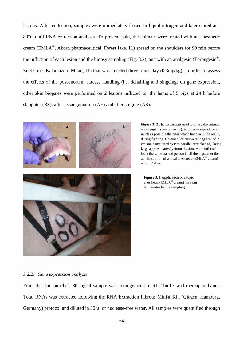
64
Figure 3. 2 The instrument used to injury the animals
was a piglet’s lower jaw (a), in order to reproduce as
much as possible the bites which happen in the reality
during fighting. Obtained lesions were long around 2
cm and constituted by two parallel scratches (b), being
large approximatively 4mm. Lesions were inflicted
from the same trained person in all the pigs, after the
administration of a local anesthetic (EMLA® cream)
on pigs’ skin.
lesions. After collection, samples were immediately frozen in liquid nitrogen and later stored at -
80°C until RNA extraction analysis. To prevent pain, the animals were treated with an anesthetic
cream (EMLA®, Akorn pharmaceutical, Forest lake, IL) spread on the shoulders for 90 min before
the infliction of each lesion and the biopsy sampling (Fig. 3.2), and with an analgesic (Torbugesic®
,
Zoetis inc. Kalamazoo, Milan, IT) that was injected three times/day (0.3mg/kg). In order to assess
the effects of the post-mortem carcass handling (i.e. dehairing and singeing) on gene expression,
other skin biopsies were performed on 2 lesions inflicted on the hams of 5 pigs at 24 h before
slaughter (BS), after exsanguination (AE) and after singing (AS).
3.2.2. Gene expression analysis
From the skin punches, 30 mg of sample was homogenized in RLT buffer and mercaptoethanol.
Total RNAs was extracted following the RNA Extraction Fibrous Mini® Kit, (Qiagen, Hamburg,
Germany) protocol and diluted in 30 µl of nuclease-free water. All samples were quantified through
Figure 3. 1 Application of a topic
anesthetic (EMLA® cream) in a pig
90 minutes before sampling
a b

65
a NanoDrop Spectrophotometer (NanoDrop Technologies Inc., Wilmington, DE) at a wavelength of
260 nm; the accepted value was ≥ 62 ng/µl. RNA quality was assessed through bioanalyzer with
Agilent RNA 6000 Nano Kit (Agilent technologies, Germany). Obtained values of RIN >8
confirmed that the RNA quality was adequate for proceeding with Retro-transcription PCR. cDNA
synthesis was performed from 1µg of RNA using the RT2 First Strand Kit (Qiagen, Hamburg,
Germany) obtaining 20 µl of cDNA solution than diluted to a concentration of 1:15 µl in nuclease-
free water. Real-time qPCR was performed using Step One Plus Real-Time PCR System (Applied
Biosystems, MA). The qPCR analysis was initially performed using the Pig Wound Healing RT2
Profiler PCR Array by Qiagen (Qiagen, Hamburg, Germany), in 5 pigs for the following categories
of samples: controls; 1h, 4h, 8h, 24h and 48h (age categories); BS; AE (40 samples in total). The
genes tested in this phase are listed in Fig. 3.3.
Later, the same procedure was used to test 13 genes selected based on the literature and whose
primers were designed (Table 3.1a). Genes from the Kit and from other studies whose expression
was not influenced by the exsanguination process and which presented a significant fold change
(threshold was set at ±1.5 fold change) were selected to be tested on 10 animals in the 5 age classes
and in the controls. Second-time designed primers are listed in the Table 3.1b. In all cases qPCR
amplification was started with an initial 10 min of denaturation stage at 95°C, followed by 40 cycles
of 15 s at 95°C of denaturation and 1 min at 60°C of annealing and elongation. For the genes home-
Figure 3. 3 Genes tested using the Pig Wound Healing
RT2 Profiler PCR Array (Qiagen, Hamburg, Germany).
Modified from: https://www.qiagen.com (last access:
March 12, 2017).

66
designed, the fold change in the expression was calculated using the 2-ΔΔ
CT method (Livak and
Schmittgen, 2001), using B2M, HPRT, RPS18 as housekeeping genes (with the exception of the
genes in the kit which have already their housekeeping genes) and non-lesioned skin samples as a
control. When the effect of exsanguination was tested, the control was represented by BS skin
samples.
Table 3. 1 List of genes and primers used in the study.
Gene Primer Dilution (nM) Sequence number
a
IFN F AGGTTCCTAAATGGTAGCTCTGGG 300 NM_213948.1
R AGTTCACTGATGGCTTTGCGCT 300
IL12 sub p35 F GGCTGCCAGAGAGACCTCTTTAAT 900 NM_213993.1
R ATACTGCTAAGGCACAGGGTTGTC 900
IL12 sub p40 F GCCAAGGTTACATGCCACAA 300 NM_214013.1
R TAGAACCTAATTGCAGGACACAGATG 900
IL17 F CCAGACGGCCCTCAGATTAC 300 NM_001005729.1
R CACTTGGCCTCCCAGATCAC 300
IL6 F GGAAATGTCGAGGCTGTGCAGATT 300 NM_214399.1
R GGTGGTGGCTTTGTCTGGATTCTT 300
IL2 F TGGAGCCATTGCTGCTGGATTTAC 900 NM_213861
R TTCTGTAGCCTGCTTGGGCATGTA 900
IL4 F GTGACGGACGTCTTTGCTGC 900 NM_214123.1
R TGCTGCTCAGGTTCCTGTCAAG 900
IL8 F AGAACTGAGAAGCAACAACAACAG 300 NM_213867.1
R CACAGGAATGAGGCATAGATGTAG 300
CCL5 F GAAGGTCTCCACCGCTGCCC 50 NM_001129946.1
R ACAGCAGGGTGTGGTGTCCGA 300
ANXA1 F GCAGTGAGCCCCTATCCTACCTTC 300 NM_001163998
R GCAAAGCCAAAGCAACTTCCTC 300
TIMP1 F ACCAAGATGTTCAAAGGGTTCAAT 300 NM_213857
R TCCGCAGACGCTCTCCAT 300
MMP14 F GCCTACTGACAAGATTGATGCTGC 300 NM_214239
R CATCGCTGCCCATGAATGACCC 300
HPRT1 F TTGTGGTAGGCTATGCCCTTGACT 300 NM_001032376.2
R CTCAACTTGAACTCTCCTCTTAGG 300
RPS18 F CATGTGGTGTTGAGGAAAGCA 300 NM_213940.1
R TTGGCGAGGATTCTGCATAAT 150
B2M F TCACTCCTAACGCTGTGGATCAGT 300 NM_213978.1
R TGATGCCGGTTAGTGGTCTCGAT 300
b
CCL2 F CTGCACCCAGGTCCTTGCCC 300 NM_214214
R CTGCTGGTGACTCTTCTGTAG 300
COX2 F AAGCGAGGACCAGCTTTCACCAAA 300 NM_214321.1
R GCGCAGTTTATGCTGTCTCTCCAA 300
EGF F GAGGCAGTTCTCGTATTTGTTCCTG 50 NM_214020
R CACCAAAAAGGGACATTGCAAACAC 900
HBEGF F CTGGACTTGGAAGAGGCAGAC 300 NM_214299
R CACGTACTTGCACTCTCCGTGG 300
IL10 F GATATCAAGGAGCACGTGAACTC 300 NM_214041.1

67
R GAGCTTGCTAAAGGCACTCTTC 300
ITGA6 F TTCTTGCCAGCAAGGTGTAGC 300 XM_005657544
R CTCCACCAACTTCATAAGGCCC 300
MMP1 F TGCCAAATGGACTTCAAGCTGC 300 NM_001166229
R GATCTGTGGATGTCCTTGGGG 300
MMP3 F TGGAGCCAGGTTTTCCCAAGC 300 NM_001166308
R GGGTCAAACTCGAACTGCGAAG 300
SERPINE1 F CCACCCCCGACGGCCATTAC 300 NM_213910.1
R TGGTGAGGGCGGAGAGAGGC 300
TNF F CACTGACCACCACCAAGAATTGGA 300 NM_214022.1
R CATTCCAGATGTCCCAGGTTGCAT 300
3.2.3. Statistical analysis
For all analyses, the experimental unit was the lesion. The fold change value for each cytokine was
log transformed and analyzed using the MIXED procedure of SAS (SAS Inst. Inc., Cary, NC) with
the age categories in the model. Multiple comparisons were made using the Tukey-Kramer
adjustment. Back-transformed values are reported as means with lower and upper limits in squared
brackets.
A probability level of P < 0.05 was chosen as the limit for statistical significance in all tests,
whereas probability levels of P ≤ 0.10 were considered to be a tendency.
3.3. Results and Discussion
All AS samples presented a very low content of RNA extracted (6.6 ±6.2 ng/l), which is likely due
to the high temperature carcass skin was exposed to during singeing resulting in RNA denaturation.
No difference was found in the RNA content between BS and AE samples (106.6 ± 26.3 ng/l and
126.9 ± 29.5 ng/l, respectively; results not shown), which confirms that collection of skin samples
immediately after exsanguination ensures the availability of good quality samples for the analysis.
Of the 98 tested genes, 14 (ANXA1, CCL2, CCL5, COX2, IL2, IL6, IL8, IL10, ITGA6, MMP1,
MMP14, SERPINE1, TIMP1 and TNF) did not present a different expression after exsanguination
and were also differently expressed between lesions and controls. As showed in Fig. 3.4, the
expression pattern of CCL2, COX2, IL2, IL6, IL8, MMP1, SERPINE1 and TIMP1 varied with the

68
time of the lesion (P<0.05). Particularly, a greater (P < 0.01) expression was found for COX2 in 1
h old lesions (1 h = 6.2 [3.2-12.1], 4 h = 1.4 [0.7-2.6], 8 h = 1.7 [0.9-3.4], 24 h = 1.6 [0.8-3.1], 48 h
= 1.1 [0.6-2.2], for SERPINE1 may allow classifying lesions in 3 groups: 1h, 4-8h and 24-48h (1h=
6.70 [4.15-10.81], 4h= 3.05 [1.89-4.92], 8h= 1.92 [1.19-3.09], 24h = 0.77 [0.48-1.24], 48h= 0.80
[0.5-1.3], P < 0.05), for MMP1 in 4-8 h old lesions (1 h = 1.9 [0.7-5.5], 4 h = 35.6 [12.4-102.3], 8 h
= 19.2 [6.7-55.3], 24 h = 4.0 [1.4-11.4], 48 h = 1.3 [0.4-3.6]; P < 0.001) and for CCL2 and IL8 in <
or > 8 h old lesions (1h= 3.49 [2.45-4.97], 4h= 2.71 [1.90-3.86], 8h= 2.17 [1.52-3.10], 24h= 1.05
[0.74-1.50], 48h= .95 [0.67-1.36], P < 0.05).
Figure 3. 4 Fold change in gene expression for 14 genes. The legend on the top of the graphs indicates the 5 age categories of the
skin lesions. Means within a row with different superscripts differ (P <0.05). The error bars represents the maximum and minimum
values in fold change.

69
3.4. Conclusions
The analysis of the expression of genes involved in inflammation and tissue repair is useful in the
determination of the age of skin lesions. However, this study showed that sampling time should be
considered to ensure the suitability of samples for the analysis and the reliability of the results.
Based on the expression of the genes tested in this study, COX2 and SERPINE1 were the best
indicators of 1 h old lesions, while the MMP1 gene is able to identify 4-8 h old lesions. To confirm
the usefulness of CCL2, IL2, IL6, IL8 and TIMP1 genes for the assessment of the age of lesions in
pigs, further larger scale studies are needed.
Literature cited
Aaslyng, M. D., P. Brandt, and L. Blaabjerg. 2013. Assessment and Incidence of Skin Damage in Slaughter
Pigs. In: Proceedings of the 59th International Congress of Meat Science and Technology. p. 18–21.
Barington, K., and H. E. Jensen. 2013. Forensic cases of bruises in pigs. Vet. Rec. 173:526.
doi:10.1136/vr.101854.
Cecchi, R. 2010. Estimating wound age: looking into the future. Int. J. Legal Med. 124:523–536.
doi:10.1007/s00414-010-0505-x.
Epstein, F. H., A. J. Singer, and R. A. F. Clark. 1999. Cutaneous Wound Healing. N. Engl. J. Med. 341:738–
746. doi:10.1056/NEJM199909023411006.
Faucitano, L. 2001. Causes of skin damage to pig carcasses. Can. J. Anim. Sci. 81:39–45. doi:10.4141/A00-
031.
Gallant-Behm, C. L., and D. A. Hart. 2006. Genetic analysis of skin wound healing and scarring in a porcine
model. Wound Repair Regen. 14:46–54. doi:10.1111/j.1524-475X.2005.00087.x.
Kubo, H., T. Hayashi, K. Ago, M. Ago, T. Kanekura, and M. Ogata. 2014. Temporal expression of wound
healing-related genes in skin burn injury. Leg. Med. 16:8–13. doi:10.1016/j.legalmed.2013.10.002.
Livak, K. J., and T. D. Schmittgen. 2001. Analysis of Relative Gene Expression Data Using Real-Time
Quantitative PCR and the 2−ΔΔCT Method. Methods. 25:402–408. doi:10.1006/meth.2001.1262.
Sato, Y., T. Ohshima, and T. Kondo. 1999. Regulatory role of endogenous interleukin-10 in cutaneous
70
inflammatory response of murine wound healing. Biochem Biophys Res Commun. 265:194–199.
doi:10.1006/bbrc.1999.1455.
Teixeira, D. L., and L. A. Boyle. 2014. A comparison of the impact of behaviours performed by entire male
and female pigs prior to slaughter on skin lesion scores of the carcass. Livest. Sci. 170:142–149.
doi:10.1016/j.livsci.2014.09.026.
Turner, S. P., M. J. Farnworth, I. M. S. White, S. Brotherstone, M. Mendl, P. Knap, P. Penny, and A. B.
Lawrence. 2006. The accumulation of skin lesions and their use as a predictor of individual
aggressiveness in pigs. Appl. Anim. Behav. Sci. 96:245–259. doi:10.1016/j.applanim.2005.06.009.
Vanezis, P. 2001. Interpreting bruises at necropsy. J. Clin. Pathol. 54:348–355. doi:10.1136/jcp.54.5.348.
Varón-Álvarez, L., M. Romero, and J. Sánchez. 2014. Caracterización de las contusiones cutáneas e
identificación de factores de riesgo durante el manejo presacrificio de cerdos comerciales. Arch. Med.
Vet. 46:93–101. doi:10.4067/S0301-732X2014000100013.
Wang, L. L., R. Zhao, C. S. Liu, M. Liu, S. S. Li, J. Y. Li, S. K. Jiang, M. Zhang, Z. L. Tian, M. Wang, M.
Z. Zhang, and D. W. Guan. 2016. A fundamental study on the dynamics of multiple biomarkers in mouse
excisional wounds for wound age estimation. J. Forensic Leg. Med. 39:138–146.
doi:10.1016/j.jflm.2016.01.027.
Wang, Y., Y. Yamamoto, Y. Kuninaka, T. Kondo, and F. Furukawa. 2015. Forensic Potential of MMPs and
CC Chemokines for Wound Age Determination. J. Forensic Sci. 60:1511–1515. doi:10.1111/1556-
4029.12831.

71
4.
USE OF THE SPECTROPHOTOMETRIC COLOR METHOD
FOR THE DETERMINATION OF THE AGE OF SKIN LESIONS ON
THE PIG CARCASS AND ITS RELATIONSHIP WITH GENE EXPRESSION
AND HISTOCHEMICAL AND HISTOLOGICAL PARAMETERS
M. Vitali,* S. Conte,† M. Lessard,† K. Deschêne,† M.O. Benoit-Biancamano,‡ C. Celeste,§ G.
Martelli,* L. Sardi,* F. Guay,# and L. Faucitano†
*Department of Veterinary Medical Sciences, University of Bologna, Ozzano Emilia, 40064, Italy
†Agriculture and Agri-Food Canada, Sherbrooke Research and Development Centre, Sherbrooke,
J1M 0C8, Canada
‡Department of Pathology and Microbiology, Faculty of Veterinary Medicine, University of
Montréal, Saint-Hyacinthe, J2S 2M2, Canada
§Department of Veterinary Biomedecine, Faculty of Veterinary Medicine, University of Montréal,
Saint-Hyacinthe, J2S 2M2, Canada
#Department of Animal Science, Université Laval, Québec, G1V 0A6, Canada
ABSTRACT: The presence of lesions on the pig carcass is both an indicator of poor animal welfare
and economical issue as it downgrades the carcass value. The assessment of the age of lesions on
the carcass may help identify and prevent the risk factors. The aim of this study was to assess the
age of lesions on the pig carcass through spectrophotometric color evaluation and to relate the
results with gene expression and histochemical and histological parameters. A total of 96 barrows
were mixed 4 times during 3 d before slaughter and 80 lesions were selected after skin lesion
observations to define 4 age categories: < 7 h (T1), 7-25 h (T2), 25-30 h (T3) and 49-54 h (T4). A
non-lesioned skin area was used as a control. At slaughter, 3 biopsies per lesion and control skin
were taken immediately after bleeding for analyses of gene expression (CCL2, COX2, IL6, IL8,

72
IL10, ITGA3, MMP1, TNF, TIMP1, SERPINE1), skin histological characteristics (inflammation,
erosion or ulceration and necrosis) and enzyme activity (alkaline phosphatase and adenosine
triphosphatase). The number of lesions was counted on each carcass and the color was assessed
visually by a pictorial chart and instrumentally through a spectrophotometer. Delta values () were
calculated as the difference between the value of the lesion and the value of the control for all
measures, except for the histological analysis. Results indicated that visual color observation was
not sufficiently accurate to discriminate lesions by time of infliction (P > 0.10), while the
spectrophotometer L* and a* values variation allowed identify < 7 h or > 25 h old lesions (P <
0.05). Similarly, the expression of CCL2, IL6, ITGA3, MMP1 and SERPINE1 genes increased in <
7 h old lesions (P < 0.05), while TIMP1 gene expression was greater (P < 0.05) in < 25 h old
lesions. As for the histological analysis, only the inflammation parameter was able to detect a
difference between < 7 h and > 25 h old lesions (P < 0.05). To conclude, the spectrophotometric
color assessment of the carcass lesions at slaughter appears to be a reliable method to discriminate
between fresh and older lesions at the abattoir.
Key words: carcass, color assessment, enzyme histochemistry, gene expression, histology, lesion
age, pig, skin
4.1. Introduction
The presence of skin lesions is both an animal-based criterion in the animal welfare assessment
(Welfare Quality®
, 2009; EFSA, 2012) and an economical issue related to the loss of profits due to
carcass downgrading (MLC, 1985) and additional handling for carcass inspection (Faucitano,
2001). Common causes of skin lesions on the pig carcass are poor housing, mixing of unfamiliar
pigs and rough handling (Faucitano, 2001). Lesions are commonly assessed on the carcass using
pictorial standards (MLC, 1985) or by giving a score based on their number and type (ITP, 1996;
Welfare Quality®
, 2009). However, no reliable technique exists for the determination of the time of
infliction of a lesion in pigs. As injuries can occur at any moment during the marketing process

73
(Dalla Costa et al., 2007; Aaslying et al., 2013), knowing the time of infliction may be very helpful
to prevent their occurrence by limiting the impact of risk factors. The most common method to
assess lesion age at the abattoir is the visual color assessment (Rocha et al., 2013), which, however,
may be biased by human error (Scafide, 2012). Objective evaluation of lesion color using
spectrophotometry, molecular biology, histochemical and histological techniques has been studied
in different livestock species (Hamdy et al., 1957; Strappini et al., 2009; Munro and Munro, 2013)
and in forensic medicine using human and animal models (Wang et al., 2001, Gallant-Behm and
Hart, 2006; Kondo and Ishida, 2010; Barington and Jensen, 2016a,b). However, despite the need for
a reliable assessment of the age of lesions on the pig carcass at slaughter (Barington and Jensen,
2013), to our knowledge, an objective technique for this determination on the pig carcass under
marketing conditions is still missing.
The objective of the study was to provide a reliable determination of the age of the lesion on the pig
carcass at the abattoir through objective color assessment and to relate the results with biological
indicators, such as expression of genes, enzyme histochemistry and histological parameters.
4.2. Materials and Methods
All experimental procedures performed in this study were approved by the institutional animal care
committee at the Agriculture and Agri-Food Canada (AAFC) Sherbrooke Research and
Development Centre (Sherbrooke, QC, Canada) based on the current guidelines of the Canadian
Council on Animal Care (2009).
4.2.1. Animals and Handling
A total of 96 crossbred barrows (Large White x Landrace x Piétrain) were distributed into 8 groups
of 12 pigs (4 replicates of 2 groups; 124.2 ± 1.7 kg body weight) at the AAFC swine unit in
Sherbrooke (QC). Pigs from 2 groups were mixed 3 times over 3 d before slaughter: twice at the
farm (2 d before slaughter and the day before slaughter) and once at loading. The first mixing

74
consisted in grouping 6 pigs from one pen with 6 pigs from the second pen to create two new
groups of 12 pigs. The second mixing involved the remaining 6 pigs left in each pen and that had
never met before. For the 3rd
mixing, all pigs (24) were grouped at loading and in lairage (Fig. 4.1).
Figure 4. 1 Scheme of the experimental protocol.
Before and after each mixing event, skin lesions were scored according to the Welfare Quality®
Protocol for Pigs (Welfare Quality®
, 2009) by a trained observer. At the same time, lesions on all
pigs were mapped on both sides of a pig body image printed on a paper and marked with different
colors according to the observational times (6) in order to trace-back the exact time of the lesion’s
appearance. A video of each pig body was also recorded to validate the visual observation (Fig.
4.2). Biting- and mounting-type lesions, as defined by the comma short or long scratch shape,
respectively, were only considered in this study as they are the most commonly observed on the pig
carcass at slaughter (Varón-Álvarez et al., 2014). Lesions on the head, ears, abdominal part, legs
and tail were not considered because of the risk of being affected by the post-mortem carcass
handling procedures. Two parallel scratches of at least 2.5 cm length and 4 mm width were
considered as one lesion. Just before slaughter, the lesions were selected and distributed into 4 age
categories of 20 lesions each, based on the time of their appearance on the pig body and slaughter
time (time 0), as follows: T1 (< 7 h old), T2 (7-25 h old), T3 (25-30 h old) and T4 (49-54 h old).
The T1 lesions corresponded to fresh lesions inflicted during loading, transport and/or lairage, while
the other lesions (T2, T3, and T4) corresponded to lesions inflicted during mixing at the farm.
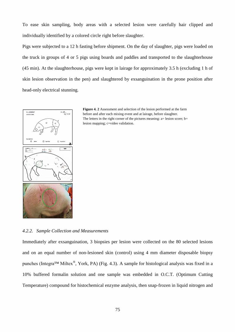
75
To ease skin sampling, body areas with a selected lesion were carefully hair clipped and
individually identified by a colored circle right before slaughter.
Pigs were subjected to a 12 h fasting before shipment. On the day of slaughter, pigs were loaded on
the truck in groups of 4 or 5 pigs using boards and paddles and transported to the slaughterhouse
(45 min). At the slaughterhouse, pigs were kept in lairage for approximately 3.5 h (excluding 1 h of
skin lesion observation in the pen) and slaughtered by exsanguination in the prone position after
head-only electrical stunning.
4.2.2. Sample Collection and Measurements
Immediately after exsanguination, 3 biopsies per lesion were collected on the 80 selected lesions
and on an equal number of non-lesioned skin (control) using 4 mm diameter disposable biopsy
punches (Integra™ Miltex®, York, PA) (Fig. 4.3). A sample for histological analysis was fixed in a
10% buffered formalin solution and one sample was embedded in O.C.T. (Optimum Cutting
Temperature) compound for histochemical enzyme analysis, then snap-frozen in liquid nitrogen and
a
b
Figure 4. 2 Assessment and selection of the lesion performed at the farm
before and after each mixing event and at lairage, before slaughter.
The letters in the right corner of the pictures meaning: a= lesion score; b=
lesion mapping; c=video validation.

76
later stored at -80°C. A third sample was snap-frozen in liquid nitrogen and stored at -80°C pending
Real-Time PCR mRNA analysis.
In the cooler, the number of lesions was counted on each carcass using an adapted version of the
Welfare Quality®
Protocol for Pigs (Welfare Quality®, 2009), excluding the ears from the count as
they were removed during the carcass processing. Skin damages that may have been caused by
carcass processing (i.e. dehairing) were also excluded from the count. Carcasses were classified
using a scale from 0 (up to 4 lesions on the whole body) to 2 (> 11 lesions in two body regions or at
least 15 lesions in one body region) according to the Welfare Quality®
Protocol (Welfare Quality®
,
2009). The lesions and non-lesioned skin color was first assessed visually using a scale of hue and
brightness values (Fig. 4.4) and a picture of each lesion was taken for further validation. Color was
then measured 3 times at 3 different positions along each lesion and on the non-lesioned skin using
a CM 700d Spectrophotometer (Konica Minolta Sensing Inc., Osaka, Japan). The measure were
taken using a 8° viewing angle, a 2° observer angle, D65 illuminant, SCI (specular component
included) mode, with an illumination measurement area of 3 mm in diameter. CIE L*a*b* color
coordinates were used (Fig. 4.5).
Figure 4. 4 Skin sampling at the abattoir. Biopsies were taken
immediately after exsanguination.
Figure 4. 3 Color assessment,
performed in the cooler, after carcass
deharing and singing.

77
4.2.3. Histological and Histochemical Analysis
Histology. Samples were cut into two, routinely processed and embedded in paraffin (TissuePrep™
2 Micron, Fisher Scientific). Sections of 4 μm thickness were deparaffinized and then stained with
hematoxylin-eosin-phloxin-saffron (HEPS).
Alkaline phosphatase (ALP). Samples were cut into 4 μm sections in a Leica CM3050S research
cryostat (Leica Biosystems, Concord, Canada) at -24°C. Alkaline Phosphatase Staining (Red) Kit
(Cell Biolabs Inc., San Diego, CA) was used. The specimens were stained with StemTAGTM AP
Staining solution and incubated at room temperature for 15 to 30 min, protected from light.
Alkaline phosphatase activity appeared as a reddish stain.
Adenosine triphosphatase (ATP). Samples were cut into 4 μm sections in cryostat at -24°C and
stained at pH 9.4 following the method described by Bancroft (1975). Slides were counter-stained
with Harris hematoxylin for 25 s, then rinsed with distilled water. Dehydration was performed as
follows: 80% alcohol for 2 min, 95% alcohol for 2 min, 100% alcohol for 2 min, and toluol for 5
min. Slides were mounted with a Leica CV 5000 automated coverslipper (Leica Biosystems,
Concord, Canada).
Microscopic examination. Microscopic examination for HEPS and histochemistry stains was
performed with an Olympus BX41 optical microscope (Olympus America Inc., Melville, NY).
Figure 4. 5 Visual color
assessment scale used to describe
the color of both lesion and intact
skin (control)
78
Magnification of 25 to 400 X was used as appropriate. For inflammation, hemostasis and enzyme
histochemistry, the following grading system was used to describe lesions: 0 = absent, 1 = minimal
to mild, 2 = from moderate to severe. For epidermal erosion/ulceration and necrosis, this grading
was modified and expressed as the percentage of epidermal surface of the sample section being
affected: 0 = 0% of the epidermis, 1 = < 25% - < 50%, 2 = 50 -100%.
4.2.4. Gene Expression Analysis
Thirty (30) mg of sample was collected from the thawed skin punches and homogenized in RLT
buffer and mercaptoethanol. Total RNAs was extracted following the RNA Extraction Fibrous
Mini® Kit (Qiagen, Hamburg, Germany) protocol and eluate in 30-µl of nuclease-free water. All
samples were quantified through a NanoDrop Spectrophotometer (NanoDrop Technologies Inc.,
Wilmington, DE) at a wavelength of 260 nm; the accepted value to proceed with Retro-
Transcription was ≥ 62 ng/µl. The quality of RNA was assessed through bioanalyzer with Agilent
RNA quality 6000 Nano Kit (Agilent Technologies, Waldbronn, Germany). Obtained values of
RNA integrity number (RIN) > 8 confirmed that the RNA quality was adequate for proceeding with
the real-time PCR analysis. The synthesis of cDNA was performed from 1 µg of RNA using the
RT2 First Strand Kit (Qiagen, Hamburg, Germany) obtaining 20 µl of cDNA solution, which was
later diluted to a concentration of 1:15 µl in nuclease-free water. Real-time PCR was performed
using Step One Plus Real-Time PCR System (Applied Biosystems, Framingham, MA) to evaluate
the mRNA modulation for the 12 genes listed in Table 1. Of these genes, 10 were the target genes
(CCL2, IL6, ITGA3, SERPINE1, MMP1, T1MP1, TNF, IL8, IL10 and COX2) that were
previously selected out of 94 genes analysed for their efficiency in skin lesion age determination in
a preliminary study on live pigs (unpublished results). Primers were designed using known pig
sequences for the genes of interest (Table 4.1). Real-time PCR amplification was started with an
initial 10-min denaturation stage at 95°C, followed by 40 cycles of 15 s denaturation at 95°C and 1
min annealing and elongation at 60°C. Each sample was analysed in triplicate. The fold change in

79
the expression of the 10 target genes was calculated using the 2-ΔΔ
CT method (Livak and
Schmittgen, 2001), using B2M and RPS18 as housekeeping genes and non-lesioned skin samples as
a control.
Table 4. 1 PCR primer sequences of genes used in this study
Gene Primer Dilution, nM Sequence number
CCL2 F CTGCACCCAGGTCCTTGCCC 300 NM_214214
R CTGCTGGTGACTCTTCTGTAG 300
IL6 F GGAAATGTCGAGGCTGTGCAGATT 300 NM_214399.1
R GGTGGTGGCTTTGTCTGGATTCTT 300
COX2 F AAGCGAGGACCAGCTTTCACCAAA 300 NM_214321.1
R GCGCAGTTTATGCTGTCTCTCCAA 300
ITGA3 F ATCATTTGCGAGCTGGGGAAC 50 XM_005668880
R CAGCTGCGCCTGGAGTTC 50
MMP1 F TGCCAAATGGACTTCAAGCTGC 300 NM_001166229
R GATCTGTGGATGTCCTTGGGG 300
TIMP1 F ACCAAGATGTTCAAAGGGTTCAAT 300 NM_213857
R TCCGCAGACGCTCTCCAT 300
IL8 F AGAACTGAGAAGCAACAACAACAG 300 NM_213867.1
R CACAGGAATGAGGCATAGATGTAG 300
TNFα F CACTGACCACCACCAAGAATTGGA 300 NM_214022.1
R CATTCCAGATGTCCCAGGTTGCAT 300
SERPINE1 F CCACCCCCGACGGCCATTAC 300 NM_213910.1
R TGGTGAGGGCGGAGAGAGGC 300
IL10 F GATATCAAGGAGCACGTGAACTC 300 NM_214041.1
R GAGCTTGCTAAAGGCACTCTTC 300
B2M F GGATCAGTATAGCTGCCGCG 300 NM_213978.1
R TCTGTGATGCCGGTTAGTGG 300
RPS18 F CATGTGGTGTTGAGGAAAGCA 300 NM_213940.1
R TTGGCGAGGATTCTGCATAAT 150
80
4.2.5. Statistical Analysis
Visual color assessment. Delta values (Δhue and Δbrightness) were calculated as the difference
between the value of the lesion and the value of the control skin. Data were analyzed using Kruskal-
Wallis test including the four age categories in the model. Values are reported as median (lower and
upper quartiles in brackets).
Spectrophotometric color assessment. The three spectrophotometer L*,a*,b* color values of each
lesion were averaged and a delta value (ΔL*, Δa* and Δb*) was obtained by calculating the
difference between the color measure taken on the lesion and taken on the control skin. Delta values
were analyzed using the MIXED procedure of SAS (SAS Inst. Inc., Cary, NC) including the lesion
age categories in the model. Multiple comparisons were made using the Tukey adjustment. Data are
presented as LSM ± SEM. Moreover, the accuracy of the spectrophotometric measures for the
determination of the age of a lesion was evaluated as described by Parikh et al. (2008). Briefly, this
method consisted in comparing the classification of the lesion age obtained with a measure (e.g.
ΔL*) with the real age of the lesion (based on direct observations of the lesion appearance on live
pigs after each mixing event). To select the best threshold value above or below which the lesion
would be correctly classified as fresh or old, sensitivity, specificity and the positive predictive value
(PPV: the probability that the lesion is actually fresh when the indicator is above a certain value)
were calculated for several threshold values. The more the sum of sensitivity, specificity and PPV
was high, the more accurate a threshold was for the lesion age classification. The negative
predictive value (NPV: the probability that the lesion is old when the indicator is below the
threshold value) of the indicators was also calculated (Parikh et al., 2008).
Histology. The relationship between necrosis, erosion, inflammation and hemorrhage score and age
categories was assessed using the Cochran–Mantel–Haenszel test of association for ordinal

81
categories (row mean score statistic). In the case of a significant association, multiple comparisons
(6 comparisons) were made using a Bonferroni adjusted P-value (P < 0.008).
Enzyme histochemistry. Delta values were calculated as the difference between the scores of the
lesion and of the control. Delta values were then re-categorised into score 0 if the delta was 0, score
1 if the delta was 1 or 2, and score 2 if the delta was 3 or 4. The relationship between the score for
each enzyme and the age categories was assessed using the Cochran–Mantel–Haenszel test of
association for ordinal categories (row mean score statistic).
Gene expression. For technical reasons, gene expression data from one lesion in the T3 age
category were missing. The fold change value for each cytokine was log-transformed and analyzed
using the MIXED procedure of SAS including the lesion age categories in the model. Multiple
comparisons were made using the Tukey-Kramer adjustment. Back-transformed values are reported
as means with lower and upper limits in squared brackets.
Multivariate analysis. A Factorial Analysis of Mixed Data (FAMD) was performed with the
PRINCOMP procedure of SAS on selected variables related to the lesion age categories in order to
analyze relationships among these variables and the lesion age category. The categorical variables
were transformed into a complete disjunctive table and included in the PCA analysis along with the
centered and scaled matrix of numerical variables.
For all analyses, the experimental unit was the skin lesion (n = 80). A probability level of P < 0.05
was chosen as the limit for statistical significance in all tests, whereas probability levels of P ≤ 0.10
were considered to be a tendency.

82
4.3. Results and Discussion
4.3.1. Lesion Score
According to Hamdy et al. (1957), the number of lesions on the body can influence the speed of
physiological responses in the healing process. This was not the case in this study as all carcasses
that were selected for the lesion assessment presented the same high score (score 2) for the number
of lesions according to the Welfare Quality®
Protocol (2009), meaning that in this study this factor
influenced the healing process of the lesions in all pigs indifferently and did not bias the results.
4.3.2. Color Assessment
In this study, overall the visual color assessment showed that the lesions were darker (- ∆brightness;
P|t| < 0.001) with a redder hue (-∆hue; P|t| < 0.001) than the control/non-lesioned skin (Table 4.2).
Table 4. 2 Effect of the age categories on the delta color values (difference between the skin lesion and the
control) as assessed by visual and spectrophotometric color methods
Age category1
Color assessment T1 T2 T3 T4 se P-value
Visual
Δ brightness -4.50
(-6.50, -3.00)
-4.00
(-5.00, -3.00)
-3.00
(-4.00, -2.50)
-4.00
(-5.00, -3.00)
- 0.34
Δ hue
-1.50
(-2.00, -1.00)
-1.00
(-2.00, -1.00)
-1.00
(-2.00, -1.00)
-1.00
(-2.00, -1.00)
-
0.87
Spectrophotometric
∆L* -13.16 aA
-9.05 abB
-8.22 b -6.63
b 1.141 0.001
∆a* 15.11aA
10.62 abB
9.34b 8.43
b 1.317 0.003
∆b* 5.85 4.99 5.38 5.04 0.589 0.71
1Age categories: T1= < 7 h; T2= 7-25 h; T3=25-30 h; T4= 49-54 h a–c Means within a row with different superscripts differ (P < 0.05) A–B Means within a row with different superscripts tend to differ (P < 0.10)

83
However, the visual assessment failed to find differences in these color parameters between age
categories (P > 0.10) confirming the inaccuracy of this method for lesion age determination as
already reported in previous studies (Hughes et al., 2004a; Barington and Jensen, 2013).
The spectrophotometric color assessment also showed that overall the lesions assessed in this study
were darker (-∆L* value; P|t| < 0.001), redder (+∆a* value; P|t| < 0.001) and yellower (+∆b* value;
P|t| < 0.001) than non-lesioned skin (Table 2). However, differently for the visual assessment, the
objective color evaluation could detect differences between lesion age categories. Based on the ∆L*
and ∆a* color values, T1 lesions (< 7 h old) were darker and redder than T3 lesions (P = 0.01 for
both color values) and T4 lesions (P < 0.001 and P = 0.003 for ∆L* and ∆a* values, respectively).
The same trend was observed for ∆L* and ∆a* color values between T1 and T2 (P=0.06 and
P=0.08, respectively). No time-dependent difference was found for ∆b*, indicator of the yellowness
of the lesion (P > 0.10). This latter result is not surprising as the yellow component, which results
from the degradation of hemoglobin into bilirubin during the late phases of healing (Hughes et al.,
2004b; Mimasaka et al., 2010), is well known as an indicator of lesions older than those assessed in
this study. Indeed, the yellow color was observed up to 72 h in human adult bruises (Langlois,
2007) or from 5 d to 3 wks after the injury in children bruises (Mimasaka et al., 2010).
Thus, the spectrophotometric color assessment appears to be a useful method to discriminate
between fresh and older lesion (< 7 h vs. > 25 h old) as previously reported in a number of studies
on humans (Hughes and Langlois, 2010; Mimasaka et al., 2010; Grossman et al., 2011).
In this study, the efficiency of the use of ΔL* or Δa* parameters for the determination of the age of
lesions was validated through a diagnostic test. The best threshold value identified for the ΔL* was
-15. When a ΔL* value higher than the threshold was considered as an indicator of lesions older
than 7 h, the diagnostic test presented a sensitivity of 0.40, a specificity of 0.95, a PPV of 0.73 and a
NPV of 0.83. The best threshold value identified for the Δa* was 15. When a Δa* value lower than
the threshold was considered as an indicator of lesions older than 7 h, the diagnostic test presented a

84
sensitivity of 0.50, a specificity of 0.88, a PPV of 0.59 and a NPV of 0.84. Based on this test, it may
be concluded that there was 73% of chance that a lesion was old if the ΔL* value was higher than -
15 and 59% of chance that the lesion was old if the Δa* value was less than 15. However, for both
color parameters, the sensitivity values were low, indicating that the test was not sensitive enough
to discriminate between fresh and old lesions consistently. Differences in the depth of the lesion and
its location on the carcass may limit the efficiency of the spectrophotometric assessment as the
thickness of the subcutaneous fat tissue may alter the changes in the bruise color and the speed of
the healing process (Mimasaka et al., 2010). The individual variation in the degradation of
hemoglobin over time should be also taken into account in the interpretation of the results
(Grossman et al., 2011).
4.3.3. Histology and Enzyme Histochemistry
No difference was found in the necrosis and hemostasis scores between lesion age categories (P >
0.10; Table 3). Whereas, a relationship was found between inflammation score and the age of the
lesion (P < 0.001) in this study, with a higher proportion of lesions with the highest score (score 2)
being found in T3 and T4 lesions (> 25 h old) compared with T1 lesions (P = 0.001 and P < 0.001,
respectively). A similar inflammation rate has been also reported in 33-49 h old humans wounds
(Takamiya et al., 2008), and in mice, calves and lambs skin bruises between 24 and 71 h after the
injury (McCausland and Dougherty, 1978; Takamiya et al., 2005). Erosion/ulceration score also
tended to be higher (P = 0.07) in T3 and T4 than in T1 lesions (Table 4.3).
Within the lesion age categories, no difference in the epidermis ATP and epidermis and derma ALP
scores was found between the lesioned and the control skin (ΔATPE, ΔALPE and ΔALPD,
respectively) in this study (P > 0.10; Table 3), suggesting that the use of enzyme histochemical
techniques may not be appropriate for the evaluation of the age of a skin lesion, which is likely due
to the rather inconsistent appearance of ATP and ALP in the stains (Betz, 1994; Cecchi, 2010).

85
Table 4. 3 Variation of the histological scores and the delta values (difference between the skin lesion and
the control) for the enzymatic activities according to lesion age category
Age
category2
Score1
N 0 1 2 P-value
Histology
Necrosis 78 T1 25.00 60.00 15.00 0.16
T2 10.00 50.00 40.00
T3 15.00 55.00 30.00
T4 11.11 44.44 44.44
Erosion/ulceration 77 T1 50.00 35.00 15.00 0.07
T2 15.00 50.00 35.00
T3 26.32 42.11 31.58
T4 16.67 44.44 38.89
Inflammation 78 T1 45.00 55.00 0.00 <0.001
T2 20.00 50.00 30.00
T3 10.00 55.00 35.00
T4 5.56 50.00 44.44
Hemostasis 78 T1 15.00 75.00 10.00 0.50
T2 25.00 65.00 10.00
T3 0.00 95.00 5.00
T4 11.11 72.22 16.67
Enzyme histochemistry
ΔALPE3 79 T1 50.00 50.00 - 0.16
T2 84.21 15.79 -
T3 60.00 40.00 -
T4 60.00 40.00 -
Δ ALPD4 79 T1 40.00 45.00 15.00 0.76
T2 57.89 21.05 21.05
T3 40.00 55.00 5.00
T4 35.00 45.00 20.00
Δ ATPE5 T1 85.00 15.00 - 0.73
T2 84.21 15.79 -
T3 94.74 5.26 -
T4 85.00 15.00 -
Values represent the percentage of lesions in each age categories according to the score system for the different analyses (100% in
row). The P-value corresponds to the Cochran-Mantel-Haenszel test (row mean score). 1 Scoring system: 0 = absent; 1 = minimal to mild; 2 = moderate to severe lesions for inflammation and hemostasis. For Necrosis and
Erosion, the system was modified as: 0 = 0%; 1 = < 25% - <50%; 2 = 50-100% of the epidermis. For the enzyme histochemistry, 0 =
delta value of 0; 1 = delta value of 1 or 2; 2 = delta value of 3 or 4. 2 Age categories: T1 = < 7 h; T2 = 7-25 h; T3 = 25-30 h; T4 = 49-54 h 3 ALPE= Epidermis Alkaline Phosphatase 4 ALPD= Derma Alkaline Phosphatase 5 ATPE= Epidermis Adenine Triphosphatase

86
4.3.4. Gene Expression
Of the 10 tested genes, 6 (CCL2, IL6, ITGA3, SERPINE1, MMP1 and T1MP1) presented a
significant time-dependent expression pattern in this study, with the fold change attaining the
highest level in T1 lesions and then decrease in older lesions in this study (Fig. 4.5). Whereas, the
other 4 genes (TNF, IL8, IL10 and COX2) were equally over-expressed, regardless of the lesion
age category, resulting in no variation in their expression over time (P > 0.10).
When compared with T2 lesions, T1 lesions presented a higher fold change for CCL2 (P = 0.008),
ITGA3 (P = 0.01) and SERPINE1 (P = 0.01) genes. Furthermore, when compared with T3 and T4
lesions, a higher fold change was found in the T1 lesions for the CCL2 (P < 0.001 for both), IL6 (P
< 0.001 for both), ITGA3 (P < 0.001 for both), MMP1 (P < 0.001 for both) and SERPINE1 (P <
0.001 for both) genes. No difference was found in the fold change of these genes between T3 and
T4 lesion categories (P > 0.10). A different expression pattern was found for TIMP1 gene when
compared to the other genes, with its fold change being greater (P = 0.01) in T1 and T2 lesions
compared with T4 lesions. No difference was found in this gene expression between T1 and T2
lesions (P > 0.10).
Except for CCL2, IL6 and MMP1, to our knowledge, the expression of the genes tested in this
study was never assessed for the determination of lesion age in any species, including humans,
before. CCL2 is a chemokine mainly involved in the inflammatory response, controlling the
recruitment of monocytes/macrophages (Werner and Grose, 2003; Kondo and Ishida, 2010). The
increased CCL2 gene expression found in fresh lesions (T1) in this study is in agreement with other
studies assessing skin wound healing in mice (Kondo and Ishida, 2010; Wang et al., 2015). The
expression observed in this study of IL6, which is proinflammatory cytokine playing an essential
role to start the healing response (Lin et al., 2003; Werner and Grose, 2003), agrees with what
reported by Sato and Ohshima (2000) in mice wound healing, but is in contrast with Wang et al.
(2015) who reported a highest expression of this gene 1 d after wounding in humans and mice.
MMP1, which is associated with inflammation, control of matrix degradation, cells migration

87
(Rohani and Parks, 2015), tissue remodelling and angiogenesis (Gallant-Behm and Hart, 2006;
Wang et al., 2015), was the most expressed gene in this study. Madlener et al. (1998) also reported
an increased expression of this gene in mice wounds, but only starting from 1 d of age. This study
provides the first evidence that the expression of this gene increases in more recent lesions (< 7 h
post-injury).
Before this study, there was only evidence of an effect of ITGA3 gene deficiency in delaying the
wound healing process in mice (Reynolds et al., 2008), of a high quantity of SERPINE proteases in
the exudates of human wounds (Eming et al., 2010) and of T1MP1 expression in cutaneous wounds
in mice (Madlener et al., 1998). To our knowledge, the expression of these genes was never studied
for the determination of the lesion age before. This study is worthy given the involvement of these
genes in the biological process of wound healing (Li et al., 2003; Behm et al., 2012), with
SERPINE1 stimulting MMPs and integrins, both mostly expressed in T1 lesions. Integrins, in turn,
stimulate TIMPs which is an inhibitor of MMPs (Gill and Parks, 2008; Behm et al., 2012) and
whose expression achieved the greatest increase in T3 lesions in this study.
The lack of variation in the expression of TNF, IL8, IL10 and COX2 genes, all involved in the
inflammatory response and often in the regulatory process of the other phases of wound healing
(Werner and Grose, 2003; Takamiya et al., 2008; Gilroy et al., 1999; Cecchi, 2010), in the present
study is in contrast with the results from other studies. Studies on pigs and mice (Wang et al., 2001;
Wang et al., 2015; Wang et al., 2016) reported a great expression of TNF in excisional wounds
between 1 and 14 d of age. A similar variation in the expression was expected from IL10, whose
expression is promoted by TNF (Wanidworanun and Strober, 1993). However, its expression level
is inconsistently reported in the literature with peaks observed at 60 min (Ohshima and Sato, 1998),
or at 3 h and 3 d after the infliction of the lesion (Sato et al., 1999). The expression of IL8 and
COX2 has been reported until 4 d after wounding in humans (Engelhardt et al., 1998) and from 1 h
to 3 days in incised skin wound in rabbits (Bai et al., 2008), respectively.

88
0
5
10
15
COX2
Fo
ld c
han
ge
a
ab
b b
0
50
100
150
IL6
Fo
ld c
han
ge
0
2
4
6
8
10
IL10
Fo
ld c
han
ge
a
b
b b
0
1
2
3
4
5
ITGA3
Fo
ld c
han
ge
a
ab
b b
0
100
200
300
400
500
600
700
MMP1
Fo
ld c
han
ge
a
b
b b
02468
101214161820
SERPINE1
Fo
ld c
han
ge
a a
ac
bc
0
5
10
15
20
25
TIMP1
Fo
ld c
han
ge
0
1
2
3
4
TNFa
Fo
ld c
han
ge
a
b bc
c
0
5
10
15
20
CCL2
Fo
ld c
han
ge
T1 T2 T3 T4
0
50
100
150
200
250
300
350
IL8
Fo
ld c
han
ge
Figure 4. 6 Fold change in gene expression for the four age categories of skin lesions. Means within a row with different superscripts
differ (P < 0.05). The error bars represents the maximum and minimum values in fold change. T1= < 7h; T2= 7-25 h; T3=25-30 h;
T4= 49-54 h.

89
Overall, the combined assessment of MMP1, SERPINE1, IL6 and CCL2 genes expression may be
useful to discriminate between fresh and older lesions (< 7 h vs. 25 h), while the analysis of the
TIMP1 expression can help distinguish between < 25 h and > 49 h old lesions on a pig carcass.
Our results confirm the efficiency of the RT-PCR mRNA analysis for the assessment of the age of
lesions reported in a previous study (Kondo and Ishida, 2010). However, this method may have
some limitations under commercial conditions because it is laborious, time-consuming and
expensive (Takamiya et al., 2008).
4.3.5. Multivariate Analysis
An approach based on the simultaneous analysis of many factors is considered as the best way to
develop a reliable method on dating lesions (Cecchi, 2010). Factors used in the FAMD were
selected from gene expression analysis (TIMP1, IL6, CCL2, SERPINE1, ITGA3 and MMP1), the
spectrophotometric color analysis (Δa* and ΔL* color values) and from the histological parameters
(i.e. inflammation and erosion), based on their relationship with lesion age categories. The
multivariate analysis of the selected parameters shows that the variance in the lesion age can be
explained by 49.9% by the combination of Principal 1 (Prin 1) and Principal 2 (Prin 2) components
(Fig. 4.7), which may be attributed to the process of lesion healing. The other 50.1% of the variance
may be related to other factors, such as the seriousness of the lesion (Barington and Jensen, 2016b),
the individual physiological response, the animal body condition (i.e. body weight and fat
thickness) and the anatomical region where the lesion is located (Cecchi, 2010; Mimasaka et al.,
2010; Grossman et al., 2011). The position of variables on Prin 1 and 2 in the graphic (Fig. 4.7)
showed that the lesion age categories are negatively associated with the gene expressions and the
spectrophotometric Δa* color value, which indicates that the older is the lesion, the lower is the
gene expression and the Δa* color value. In this study, fresh lesions were actually redder and
presented a greater gene expression level. The opposite position of the ΔL* color vector in relation
to the Δa* colour value and to all genes vectors may be explained by negative L* color value, which

90
increases as long as the lesion color gets dark. The 0 and 2 inflammation scores (absence and
moderate to severe, respectively) were negatively and positively, respectively, associated with the
age categories, indicating that the inflammation response in the skin tissue increased with the age of
the lesions. No relationship was found between minimal inflammation rate (score 1) and the age of
the lesions, which confirms the lack of variation in the percentage of lesions presenting a minimal
or mild inflammation rate in the lesion age categories comparison (Table 4.3). This result suggest
that this degree of inflammation may be more related to factors, such as the seriousness of the lesion
or individual physiological response, as previously observed by Barington and Jensen (2016b) in
pig bruises.
In summary, the results of the multivariate analysis show the positive association between TIMP1,
IL6 and CCL2 gene expression and the Δa* color value and confirm the potential of
spectrophotometric color method for the reliable assessment of the age of a lesion.
Figure 4. 7 Factorial Analysis of Mixed Data for the selected parameters. The scores 0, 1 and 2 in the Erosion and Inflammation
parameters represent each category of the healing condition of the lesion. The age categories parameter consists of the numerical
categories (1= < 7h; 2 = 7-25 h; 3=25-30 h; 4 = 49-54 h). Delta () values indicate the differences between the lesions and controls
for the L* (lightness) and a* (redness) color parameters.

91
4.4. Conclusions
The need to monitor handling and critical areas preslaughter in order to improve animal welfare and
pork quality is resulting in increasing interest of the pork sector towards the development of a
practical and accurate technique for the determination of the age of lesions on the carcass at
slaughter.
Based on the assessment of preslaughter carcass lesions in this study, the spectrophotometric color
assessment of the lesion on the carcass appears to be a suitable and rapid technique to discriminate
between fresh lesions (< 7 h) occurring between loading and slaughter and old lesions (> 25 h old)
originating from the farm. However, the high variability in color results, likely due to pig individual
variation in body composition (i.e. subcutaneous fat thickness), prevented from obtaining a color
threshold allowing an accurate discrimination between lesion age categories in this study. Further
investigations are needed to assess the influence of this factor on the spectrophotometric lesion
color measurement.
The analysis of MMP1, SERPINE1, IL6, ITGA3, TIMP1 and CCL2 gene expression, and the
inflammation response in the skin lesion at slaughter supported the spectrophotometric assessment
of the lesion color in this study. This relationship either may indicate the potential of these
techniques for the validation of on-line objective determination of the lesion age or their usefulness
for this type of assessment using the pig as a model for scientific purposes in human studies and for
forensic investigation.
Acknowledgements
This research project was funded by Swine Innovation Porc within the Swine Cluster 2: Driving
Results Through Innovation research program. Funding was provided by Agriculture and Agri‐
Food Canada through the Agri Innovation Program, provincial producer organizations and industry
partners. The authors thank Janaina Braga, Davide Carbonari, Ariane de Castro, Taciana Diesel and

92
Fidèle Z. Kabemba for their valuable contribution in the data collection at the farm and abattoir.
Many thanks go to Catherine Thibault for her contribution to the gene analysis, Steve Méthot for his
assistance in the statistical analysis and to the staff of the AAFC swine unit for the care of animals
with expertise and professionalism. We are grateful to the COOP Fédérée for supplying pigs and
feed, and to the Rousseau abattoir staff for their collaboration during the slaughter trials. Authors
declare no conflict of interest.
Literature Cited
Aaslyng, M., P. Brandt, L. Blaabjerg, and S. Støier. 2013. Assessment and incidence of skin damage in
slaughter pigs. Proc. 59th Intern. Congr. Meat Sci. Techn., Izmir, Turkey.
Bai, R., L. Wan, and M. Shi. 2008. The time-dependent expressions of IL-1β, COX-2, MCP-1 mRNA in skin
wounds of rabbits. Forensic Sci. Int. 175 :193-197. doi:10.1016/j.forsciint.2007.07.006.
Bancroft, J. D. 1975. Histochemical techniques. Butterworths, London, UK.
Barington, K., and H. E. Jensen. 2013. Forensic cases of bruises in pigs. Vet. Rec. 173:526.
doi:10.1136/vr.101854.
Barington, K., and H. E. Jensen. 2016a. A novel, comprehensive, and reproducible porcine model for
determining the timing of bruises in forensic pathology. Forensic Sci. Med. Pathol. 12:58–67.
doi:10.1007/s12024-016-9744-6.
Barington, K., and H. E. Jensen. 2016b. The impact of force on the timing of bruises evaluated in a porcine
model. J. Forensic Leg. Med. 40:61–66. doi:10.1016/j.jflm.2016.03.005.
Behm, B., P. Babilas, M. Landthaler, and S. Schreml. 2012. Cytokines, chemokines and growth factors in
wound healing. J. Eur. Acad. Derm. Venereol. 26:812–820. doi:10.1111/j.1468-3083.2011.04415.x.
Betz, P. 1994. Histological and enzyme histochemical parameters for the age estimation of human skin
wounds. Int. J. Legal Med. 107:60–68. doi:10.1007/BF01225491.
Cecchi, R. 2010. Estimating wound age: looking into the future. Int. J. Legal Med. 124:523–536.
doi:10.1007/s00414-010-0505-x.
Dalla Costa, O. A., L. Faucitano, A. Coldebella, J. V. Ludke, J. V. Peloso, D. Dalla Roza, and M. J. R.
Paranhos da Costa. 2007. Effects of the season of the year, truck type and location on truck on skin
bruises and meat quality in pigs. Livest. Sci. 107:29-36. doi:10.1016/j.livsci.2006.08.015.

93
EFSA Panel on Animal Health and Welfare. 2012. Statement on the use of animal-based measures to assess
the welfare of animals. EFSA J. 10(6):2767 29 pp. doi:10.2903/j.efsa.2012.2767.
Eming, S. A., M. Koch, A. Krieger, B. Brachvogel, S. Kreft, L. Bruckner-Tuderman, T. Krieg, J. D.
Shannon, and J. W. Fox. 2010. Differential proteomic analysis distinguishes tissue repair biomarker
signatures in wound exudates obtained from normal healing and chronic wounds. J. Proteome Res.
9:4758-4766 doi:10.1021/pr100456d.
Engelhardt, E., A. Toksoy, M. Goebeler, S. Debus, E.-B. Bröcker, and R. Gillitzer. 1998. Chemokines IL-8,
GROα, MCP-1, IP-10, and Mig are sequentially and differentially expressed during phase-specific
infiltration of leukocyte subsets in human wound healing. Am. J. Pathol. 153:1849–1860.
doi:10.1016/S0002-9440(10)65699-4.
Faucitano, L. 2001. Causes of skin damage to pig carcasses. Can. J. Anim. Sci. 81:39–45. doi:10.4141/A00-
031.
Gallant-Behm, C. L., and D. A. Hart. 2006. Genetic analysis of skin wound healing and scarring in a porcine
model. Wound Repair Regen. 14:46–54. doi:10.1111/j.1524-475X.2005.00087.x.
Gill, S. E., and W. C. Parks. 2008. Metalloproteinases and their inhibitors: Regulators of wound healing. Int.
J. Biochem. Cell Biol. 40:1334-1347 doi:10.1016/j.biocel.2007.10.024.
Gilroy, D. W., P. R. Colville-Nash, D. Willis, J. Chivers, M. J. Paul-Clark, and D. A. Willoughby. 1999.
Inducible cyclooxygenase may have anti-inflammatory properties. Nat. Med. 5:698–701.
doi:10.1038/9550.
Grossman, S. E., A. Johnston, P. Vanezis, M., and D. Perrett. 2011. Can we assess the age of bruises? An
attempt to develop an objective technique. Med. Sci. Law 51:170–176. doi:10.1258/msl.2011.010135.
Hamdy, M. K., L. E. Kunkle, and F. E. Deatherage. 1957. Bruised Tissue. II. Determination of the age of a
bruise. J. Anim. Sci. 16:490-495. doi:10.2527/jas1957.162490x.
Hughes, V. K., P. S. Ellis, T. Burt, E. I. Langlois, and N. Langlois. 2004a. The practical application of
reflectance spectrophotometry for the demonstration of haemoglobin and its degradation in bruises. J.
Clin. Pathol. 57:355–359. doi:10.1136/jcp.2003.011445.
Hughes, V. K., P. S. Ellis, and N. E. I. Langlois. 2004b. The perception of yellow in bruises. J. Clin. Forensic
Med. 11:257–259. doi:10.1016/j.jcfm.2004.01.007.
Hughes, V. K., and N. E. I. Langlois. 2010. Use of reflectance spectrophotometry and colorimetry in a
general linear model for the determination of the age of bruises. Forensic Sci. Med. Pathol. 6:275-281
doi:10.1007/s12024-010-9171-z.

94
ITP. 1996. Notation des hématomes sur couenne - porcs vivant ou carcasse. Institute Technique du Porc,
Rennes, France.
Kondo, T., and Y. Ishida. 2010. Molecular pathology of wound healing. Forensic Sci. Int. 206:93-98
doi:10.1016/j.forsciint.2010.07.004.
Langlois, N. E. I. 2007. The science behind the quest to determine the age of bruises: a review of the English
language literature. Forensic Sci. Med. Pathol. 3:241–251. doi:10.1007/s.
Li, W.-Y., S. S. N. Chong, E. Y. Huang, and T.-L. Tuan. 2003. Plasminogen activator/plasmin system: A
major player in wound healing? Wound Repair Regen. 11:239–247. doi:10.1046/j.1524-
475X.2003.11402.x.
Lin, Z.-Q., T. Kondo, Y. Ishida, T. Takayasu, and N. Mukaida. 2003. Essential involvement of IL-6 in the
skin wound-healing process as evidenced by delayed wound healing in IL-6-deficient mice. J. Leukoc.
Biol. 73:713–21. doi:10.1189/JLB.0802397.
Livak, K. J., and T. D. Schmittgen. 2001. Analysis of relative gene expression data using Real-Time
Quantitative PCR and the 2−ΔΔCT method. Methods 25:402–408. doi:10.1006/meth.2001.1262.
Madlener, M., W. C. Parks, and S. Werner. 1998. Matrix Metalloproteinases (MMPs) and their physiological
inhibitors (TIMPs) are differentially expressed during excisional skin wound repair. Exp. Cell Res.
242:201–210. doi:10.1006/excr.1998.4049.
McCausland, I. P., and R. Dougherty. 1978. Histological ageing of bruises in lambs and calves. Aust. Vet. J.
54:525-527. doi:10.1111/j.1751-0813.1978.tb00322.x.
Mimasaka, S., M. Ohtani, N. Kuroda, and S. Tsunenari. 2010. Evaluating the age of bruises in children
spectrophotometric evaluation of the age of bruises in children: measuring changes in bruise color as an
indicator of child physical abuse. Tohoku J. Exp. Med. 220:171–175. doi:10.1620/tjem.220.171.
MLC. 1985. Concern at rindside damage in pigs. Meat and Marketing Technical Notes No. 4, Milton
Keynes, Meat and Livestock Commission, Bletchley, UK, 14-16.
Munro, R., and H. M. C. Munro. 2013. Some challenges in forensic veterinary pathology: A review. J.
Comp. Pathol. 149:57-73 doi:10.1016/j.jcpa.2012.10.001.
Ohshima, T., and Y. Sato. 1998. Time-dependent expression of interleukin-10 (IL-10) mRNA during the
early phase of skin wound healing as a possible indicator of wound vitality. Int. J. Legal Med. 111:251–
255. doi:10.1007/s004140050163.
Parikh, R., A. Mathai, S. Parikh, G. Chandra Sekhar, and R. Thomas. 2008. Understanding and using
sensitivity, specificity and predictive values. Indian J. Ophthalmol. 56:45–50.

95
Reynolds, L. E., F. J. Conti, R. Silva, S. D. Robinson, V. Iyer, R. Rudling, B. Cross, E. Nye, I. R. Hart, C. M.
Dipersio, and K. M. Hodivala-Dilke. 2008. Alpha3beta1 integrin-controlled Smad7 regulates
reepithelialization during wound healing in mice. J. Clin. Invest. 118:965–974. doi:10.1172/JCI33538.
Rocha, L. M., A. M. Bridi, P. Mormède, A. V. Weschenfelder,, N. Devillers, W. Bertoloni, and L. Faucitano.
2013. Effects of ractopamine administration and castration method on the response to pre-slaughter stress
and carcass and meat quality in pigs of two Pietrain genotypes. J. Anim. Sci. 91:3965-3977. doi:
10.2527/jas.2012-6058.
Rohani, M. G., and W. C. Parks. 2015. Matrix remodeling by MMPs during wound repair. Matrix Biol.
44:113-121 doi:10.1016/j.matbio.2015.03.002.
Sato, Y., T. Ohshima, and T. Kondo. 1999. Regulatory role of endogenous interleukin-10 in cutaneous
inflammatory response of murine wound healing. Biochem. Biophys. Res. Commun. 265:194–199.
doi:10.1006/bbrc.1999.1455.
Sato, Y., and T. Ohshima. 2000. The expression of mRNA of proinflammatory cytokines during skin wound
healing in mice: a preliminary study for forensic wound age estimation (II). Int. J. Legal Med. 113:140–
145. doi:10.1007/s004140050285.
Scafide, K. R. N. 2012. Determining the relationship between skin color, sex, and subcutaneous fat and the
change in bruise color over time. Doctoral Thesis, The Johns Hopkins University, Baltimore, MD.
Strappini, A. C., J. H. M. Metz, C. B. Gallo, and B. Kemp. 2009. Origin and assessment of bruises in beef
cattle at slaughter. Anim. 3:728–736. doi:10.1017/S1751731109004091.
Takamiya, M., K. Saigusa, R. Kumagai, N. Nakayashiki, and Y. Aoki. 2005. Studies on mRNA expression
of tissue-type plasminogen activator in bruises for wound age estimation. Int. J. Legal Med. 119: 16-21.
doi:10.1007/s00414-004-0453-4.
Takamiya, M., S. Fujita, K. Saigusa, and Y. Aoki. 2008. Simultaneous detection of eight cytokines in human
dermal wounds with a multiplex bead-based immunoassay for wound age estimation. Int. J. Legal Med.
122:143-148. doi:10.1007/s00414-007-0183-5.
Varón-Álvarez, L., M. Romero, and J. Sánchez. 2014. Caracterización de las contusiones cutáneas e
identificación de factores de riesgo durante el manejo presacrificio de cerdos comerciales. Arch. Med.
Vet. 46:93–101. doi:10.4067/S0301-732X2014000100013.
Wang, J. F., M. E. Olson, C. R. Reno, J. B. Wright, and D. A. Hart. 2001. The pig as a model for excisional
skin wound healing: characterization of the molecular and cellular biology, and bacteriology of the
healing process. Comp. Med. 51:341–348.
Wang, Y., Y. Yamamoto, Y. Kuninaka, T. Kondo, and F. Furukawa. 2015. Forensic potential of MMPs and

96
CC chemokines for wound age determination. J. Forensic Sci. 60:1511-1515. doi:10.1111/1556-
4029.12831.
Wang, L. L., R. Zhao, C. S. Liu, M. Liu, S. S. Li, J. Y. Li, S. K. Jiang, M. Zhang, Z. L. Tian, M. Wang, M.
Z. Zhang, and D. W. Guan. 2016. A fundamental study on the dynamics of multiple biomarkers in mouse
excisional wounds for wound age estimation. J. Forensic Leg. Med. 39:138-146.
doi:10.1016/j.jflm.2016.01.027.
Wanidworanun, C., and W. Strober. 1993. Predominant role of tumor necrosis factor-alpha in human
monocyte IL-10 synthesis. J. Immunol. 151:6853–61.
Welfare Quality® 2009. Welfare Quality
® assessment protocol for pigs (sows and piglets, growing and
finishing pigs). Welfare Quality® Consortium, Lelystad, Netherlands.
Werner, S., and R. Grose. 2003. Regulation of wound healing by growth factors and cytokines. Physiol. Rev.
83:835–870. doi:10.1152/physrev.00031.2002.

97
5.
GENERAL CONCLUSIONS
The presented results allow to underline the importance of lesions as indicators of animal welfare in
growing-finishing pigs.
In the study presented in chapter 2, the assessment of lesions was paramount to enlighten the
efficacy of different environmental enrichment devices for Italian Heavy Pigs. In particular, the
presence of tail biting was linked to a lack of effectiveness in one device, affecting also carcass
traits (backfat thickness, F-o-M) and water-holding capacity of the meat. Moreover, the results
suggest to be careful in choosing edible enrichment devices, because the vantages may be effective
in keeping interest during all the growing-finishing stage; but the occurrence of adverse effects
related to their consumption (e.g. the prevalence of gastric lesion at slaughter) should also be
considered.
In chapter 3, results demonstrated that, through the use of a spectrophotometer, it is possible to see
differences in lesion color according to the age of the lesion, and so to discriminating between
young lesions occurred pre-slaughter at lairage or older lesion occurred at farm level. Results were
confirmed also from histological and molecular indicators. Given the actual findings, to develop a
reliable diagnostic test, further research is required in order to better understand the role played by
the region where the lesion is located, the thickness of subcutaneous fat tissue, the force of the
trauma and the individual response in the lesion healing response. Overall, the method was found
suitable at abattoir, and should be considered for being applied to automated devices, thus becoming
part of the meat inspection at abattoir.

98
AKNOWLEDGEMENTS
These three years were deeply formatives and challenging, both professionally and personally. It
would never have happened if I was not in an invaluable guidance and company.
First, I would like to thanks my tutor Prof. Luca Sardi, and Prof. Giovanna Martelli. They were
insuperable for humanity and support and was a great privilege to work with them.
Many thanks to Dr. Luigi Faucitano for his welcoming in his team. Thank you for the trust they
reposed in me, and to push me in new situations, I have learned a lot about my limits and capacities.
I am very grateful to Dr. Eleonora Nannoni and Dr. Sabine Conte. They encourage me and teach me
with incredible patience, professionality and competence during the everyday working life.
Many thanks to Nicole Vitali and Marco Petrelli for the graphical and the linguistic revisions of the
manuscript, for their kindness and availability all the time I needed.
Thank you to my family, which through its imperfection gave me skills and braveness to the
everyday challenges, and to support my decisions also if sometimes they do not completely
understand.
Many thanks to Enrico, to be always on my side. For his unconditional support also through
distance.
I am glad to my colleagues at University of Bologna and Agriculture and Agri-food Canada. Your
friendship is a precious gift and I will keep the memory of this time forever with me.
Thank you to all my friends from everywhere, who have shared the road with me, really thank you
for make me smile.
Especially Federica and Elisa deserve a special mention for their support, patience, grace during this
manuscript writing and for have been always present. Thank you to Agata, which always remember
me that almost everything you want you can reach if you are ready to work hard for that.

99

LIST OF SCIENTIFIC PRODUCTION DURING THE PHD
AND NOT INCLUDED IN THE MANUSCRIPT
Scientific Papers
Nannoni E., Liuzzo G., Serraino A., Giacometti F., Martelli G., Sardi L., Vitali M., Romagnoli L., Moscardini E.,
Ostanello F. (2016). Evaluation of pre-slaughter losses of Italian heavy pigs. Animal Producton Science. p.1-10.
Nannoni, E., Sardi, L., Vitali, M., Trevisi, E., Ferrari, A., Barone, F., Bacci M.L., Barbieri S., Martelli, G. (2016).
Effects of different enrichment devices on some welfare indicators of post-weaned undocked piglets. Applied
Animal Behaviour Science, 184, 25-34.
Di Pasquale J., Nannoni E., Adinolfi F., Del Duca I., Capitanio F., Sardi L., Vitali M., Martelli G. (2016).
A case-study on profiling Italian consumers of animal-friendly foods. Italian Journal of Animal Science, 1-9.
Martelli G., Nannoni E., Grandi M., Bonaldo A., Zaghini G., Vitali M., Biagi G., Sardi L. (2015). Growth
parameters, behavior, and meat and ham quality of heavy pigs subjected to photoperiods of different duration.
Journal of Animal Science, 93(2), 758-766.
Martelli G., Nannoni E., Grandi M., Bonaldo A., Zaghini G., Vitali M., Biagi G., Sardi L. (2015). Growth
parameters, behavior, and meat and ham quality of heavy pigs subjected to photoperiods of different duration.
Journal of Animal Science, 93(2), 758-766.
Contribution to conference proceedings
Nannoni E., Di Pasquale J., Vitali M., Sardi L., Martelli G. (2016). The sensibility of European consumers towards
animal-friendly foods, in: PsiAnimal, European Congress Animal Welfare & Behavioural Medicine - AWBM,
Lisbona, Europress, 2016, pp. 81 - 81 in: European Congress Animal Welfare & Behavioural Medicine - AWBM,
Cascais (Portugal), 20-22 October 2016. [Poster].
Nannoni E., Martelli G., Vitali M., Sardi L. (2016). Welfare challenges for Italian heavy pigs, in: PsiAnimal,
European Congress Animal Welfare & Behavioural Medicine - AWBM, Lisbona, Europress, 2016, pp. 80 - 80 in:
European Congress Animal Welfare & Behavioural Medicine - AWBM, Cascais (Portugal), 20-22 October 2016.
[Poster].
Vitali M., Conte S., Lessard M., Martelli G., Guay F., Faucitano L. (2016). Assessment of the age of lesions on the
Di Pasquale J., Nannoni E., Del Duca I., Adinolfi F., Capitanio F., Sardi L., Vitali M., Martelli G. (2014). What
foods are identified as animal friendly by Italian consumers? Italian Journal of Animal Science, 13(4).
pig carcass at the abattoir through spectrophotometric color assessment and gene expression analysis. in: 2016 JAM
(Joint Annual Meeting), Salt Lake City, UT, USA, Volume: J. Anim. Sci Vol. 94, E-Suppl. 5/J. Dairy Sci. Vol. 99, E-
Suppl. 1. [Oral Presentation].
Barone F., Ventrella D., Elmi A., Nannoni E., Vitali M., Maya Vetencourt J.F., Martelli G., Bacci M.L. (2016).
Objective and subjective vision assessment in iodoacetic acid model of swine retinal degeneration. in: XIII FELASA
congress, Brussels, abstract book, 2016, Conference proceedings pp. 107 - 108. [Poster].
Nannoni E, Martelli G, Vitali M., Sardi L (2015) The effects of two floor space allowances on growth parameters,
meat and ham quality of Italian heavy pigs intended for Parma Ham production. VIIIème Congrès Mondial du
Jambon Sec, Toulouse, 25-26 juin 2015. Conference proceedings, pp. 1-5. [Short Presentation].
Nannoni E., Martelli G., Di Pasquale J., Vitali M., Sardi L. (2015). Behaviour and production traits of Italian heavy
pigs as affected by two floor space allowances. 2015 DCAW - Danish Minister of Food, Agriculture and Fisheries
Copenhagen (DK), April 29-30, 2015. [Poster].
Martelli G., Nannoni E., Grandi M., Bonaldo A., Zaghini G., Vitali M., Biagi G., Sardi L. (2015). Effetti di
fotoperiodi di diversa durata su parametri produttivi, comportamento, qualità di carni e prosciutti di suini pesanti
italiani. XLI meeting annuale SIPAS, Montichiari (BS), Italia, March 19-20, 2015. [Short Paper].
Nannoni E., Vitali M., Bassi P., Sardi L., Militerno G., Barbieri S., Martelli G. (2014). Study on gastric
ulcers in heavy pigs receiving different environmental enrichment materials, in: Proceedings of the 6th
International Conference on the Assessment of Animal Welfare at Farm and Group Level, Wageningen,
Wageningen Academic Publishers, 2014, pp. 105 - 105 in: WAFL 2014 - 6th International Conference on the
Assessment of Animal Welfare at Farm and Group Level, Clermont-Ferrand, France, September 3-5, 2014.
[Poster].