Embed Size (px)
Citation preview

October 2015 Volume XXXIII No 3
Scientifi c Journal of
MEDICAL & VISION RESEARCH FOUNDATIONS
insight
www.sankaranethralaya.org Editor: Parthopratim Dutta Majumder

Sankara Nethralaya – The Temple of the Eye.
It was in 1976 when addressing a group of doctors, His Holiness Sri Jayendra Saraswathi, the Sankaracharya of the Kanchi Kamakoti Peetam spoke of the need to create a hospital with a missionary spirit. His words marked the beginning of a long journey to do God’s own work. On the command of His Holiness, Dr. Sengamedu Srinivasa Badrinath, along with a group of philanthropists founded a charitable not-for-profi t eye hospital.
Sankara Nethralaya today has grown into a super specialty institution for ophthalmic care and receives patients from all over the country and abroad. It has gained international excellence and is acclaimed for its quality care and compassion. The Sankara Nethralaya family today has over 1400 individuals with one vision – to propagate the Nethralaya philosophy; the place of our work is an Alaya and Work will be our worship, which we shall do with sincerity, dedication and utmost love with a missionary spirit.
insight
Scientifi c Journal of Medical & Vision Research Foundations
Year: 2015
Issue: Vol. XXXIII | No. 3 | October 2015 | Pages 115–150
Typeset at: Techset Composition India (P) Ltd., Chennai, India
Printed at: Gnanodaya Press, Chennai, India
©Medical and Vision Research Foundations
ContentsGuest Editorial: Not Childs’ Play! 115Sumita Agarkar
Recent advances: New and complex strabismus surgeries: exciting times! 116Shruti Nishanth and Srikanth Ramasubramanian
Major Review: Progressive myopia: an update 122Meenakshi Swaminathan
Major Review: Infantile cataract: where are we now? 126Praveen Kumar KV and Sumita Agarkar
Short Review: Amblyopia: what else beyond patching and critical period? 132Jameel Rizwana Hussaindeen, Archayeeta Rakshit and Kalpa Negiloni
Short Review: Surgical management of small angle strabismus 135V Akila Ramkumar and Ketaki S Subhedar
Short Review: An approach to Nystagmus management 138N Kavitha Kalaivani
Update: Pediatric low vision care services: a new era 141Sarika Gopalakrishnan and Sumita Agarkar
Alumni Corner: Cortical visual impairment 148Preeti Patil Chhablani
Inquiries or comments may be mailed to the editor at [email protected]
Not Childs’ Play!
Sumita Agarkar
I started my career as a fellowshiptrained pediatric ophthalmologistat the cusp of the new millen-nium in 1999. Then there wereonly very few like me and thequestion which was frequentlyasked was what is there in
Pediatric ophthalmology? Fifteen years down theline, I can confidently report that probably none ofmy junior colleagues have had to face that question.
In this millennium, Pediatric Ophthalmology hasevolved in India as a standalone speciality encom-passing both comprehensive eye care in children aswell as adult strabismus. Interest in the specialityhas grown with more institutes offering long termfellowships and a matching increase in applications.But it still falls way short of requirements of ourcountry with 407 million children under 16 years ofage. In a survey, Murthy et al. found that less thana third of respondent hospitals provided Pediatriceye care. Many centers providing services were
hobbled by the absence of key components ofPediatric eye care like equipment, anesthesia facil-ities and support team consisting of optometrists,anesthetists and counselors. However, it is hearten-ing to see that advanced eye care hospitals didmuch better and provided services on par with thedeveloped world. In this millennium, I have greathopes that we will bring down the disparity in thePediatric eye care between cities and smallercenters. The solution perhaps lies in having moregeneral ophthalmologists with interest in Pediatriceye care along with specialists who can take careof more complex problems.
This issue reflects millennial changes in think-ing and approach to some common problemswhich have generated interest and controversyamong Ophthalmologists. Last words on theseissues have probably not been said as yet butSally Brown got it right in 1965 when she said“With nothing more than a simple eye patch wehave brought amblyopia to its knees”.
How to cite this article Sumita Agarkar. Not Childs’ Play! Sci J Med & Vis Res Foun 2015;XXXIII:115.
Correspondence to:Dr. Sumita Agarkar,Deputy Director – PediatricOphthalmology Department,Sankara NethralayaMedical Research Foundation18, College Road,Chennai - 600 006email: [email protected]
Sci J Med & Vis Res Foun October 2015 | volume XXXIII | number 3 | 115
Guest editorial

New and complex strabismus surgeries: exciting times!
Dr Shruti Nishanth and Dr Srikanth Ramasubramanian
Strabismus surgeries have, in recent times, pro-gressed at a rapid rate, with the introduction of amultitude of remarkable techniques that have defiedthe rules of classic surgery. With the advent ofnewer imaging modalities, and a better understand-ing of disease pathophysiology, surgical techniquesare more targeted toward the specific musclesinvolved and its pathology. This article endeavors tooutline some of the newer complex surgeries, theireffectiveness and the basis behind them.
Surgeries for “Heavy Eye Syndrome”“Heavy eye syndrome” or “myopic strabismus fixusconvergence”, is an acquired restrictive strabismusoccurring in patients with pathologic myopia.1
Patients often manifest extreme esotropia, hypotro-pia and restricted ocular motility. Simple recessionsand resections have a limited role in this entity.
What’s new ?After the advent of orbital imaging, MRI studiesshowed supero-temporal prolapse of the elongatedposterior portion of the eyeball, leading to aninferior shift of the lateral rectus (LR) and nasalshift of the superior rectus (SR) (Figure 1).2,3
Based on this new-found explanation, therewas a paradigm shift in surgical techniques,which aimed more at vector mechanics, ratherthat simple muscle strengthening/weakening.
Loop myopexyIn 2001, Yokoyama4 first proposed a new surgicalidea, when he suggested to union the LR and SRtogether, and use it as a muscle pulley to push theherniated eyeball back into the muscle cone.
TechniqueThis technique is performed by isolating both theSR and LR muscles through either a superotem-poral fornix based conjunctival incision or a lim-balperitomy. Once the muscles are isolated, a 5-0non-absorbable suture is passed through the lateralone-half of the SR and the superior one-half of theLR at ∼14–16 mm posterior to the limbus andsecurely tied. Scleral fixation is not necessary. Thiseliminates the risk of scleral perforation. This tech-nique can be done either with muscle splitting (e.g.partial Jensen’s)5–7 or without muscle splitting.8–10
Other variants of this surgery include scleral fix-ation11 anduse of type 240 silicone band withWatzke sleeve12 as a tethering aid instead of suturematerial. If preoperative forced duction testing(FDT) is positive or if the esotropia is long standingwith likely contracture of the medial rectus (MR),
recession with the possible use of an adjustablesuture can improve outcomes.10,13 However, in theabsence of these findings, MR recession may bestaged for a second surgery.
Surgical success of <20 prism diopters (PD)postoperatively is seen in 73%, according toShenoy et al.,14 with mean improvement in eso-tropia of 60 PD and hypotropia of 9 PD.
Muscle transplantationTrue muscle transplantation has been proposed forvery large angle strabismus patients like heavy eyesyndrome, especially when only one eye isplanned to be operated upon.15–17 This is of valueif the FDT shows a tight MR in cases of long-standing esotropia, and can be combined withloop myopexy. Successful outcomes have beenreported with >120 PD of esotropia and 40 PD ofhypotropia getting corrected to <20 PD esotropiaand 20 PD hypotropia after this procedure.18
Technique18
First the MR is dissected and separated via afornix incision in the lower nasal quadrant. Anon-absorbable 6‐0 prolene suture is tied at itsmuscle insertion. The muscle is then incised fromits insertion. Next the LR is hooked. Two 6‐0 vicrylsutures are placed away from the insertion as isdone in a routine resection of rectus muscle andanother pair of 6‐0 vicrylis placed at the insertion.The muscle is then incised from its insertion andthe posteriorly (distally) placed 6‐0 vicryl suturesare passed through the original insertion as in aroutine rectus muscle resection. The stump is thencut and placed at the MR site and the distal end ofthis stump is sutured with the proximal end of MRwith the 6‐0 prolene already placed on the MR. Thenow elongated muscle is sutured from the originalinsertion site of MR as is done in a routine rectusmuscle recession.
Paralytic strabismusComplete muscle palsies, where there is no residualmuscle function, do not respond to regularstrengthening procedures. Recently, a whole gamutof surgeries has been advocated, with the basis oftransposing the other available functioning musclesto serve the purpose of the palsied muscle.
Complete sixth nerve palsyVertical rectus transpositionsVertical rectus transpositions (VRT), coupled withthe antagonist muscle weakening, has showedpromising results. The main purpose of a VRT is
Department of PediatricOphthalmology,Sankara Nethralaya,Chennai, India
Correspondence:Dr Srikanth Ramasubramanian,Department of PediatricOphthalmology,Sankara Nethralaya,Chennai, India.Email: [email protected]
116 Sci J Med & Vis Res Foun October 2015 | volume XXXIII | number 3 |
Recent Advances

to allow better rotation of the eye into the field ofthe palsied muscle by creating tone through thetransposed muscles in primary position.19
They also increase the area of binocular visionand shift the binocular field toward the palsiedgaze. The transposed muscles do not have activeinnervation in the field of gaze of the palsy,thereby allowing the antagonist weakening pro-cedure to have better effect.20
The most commonly performed VRT is trans-position of the SR and IR to the palsied LR.Rosenbaum describes reattachment of the tem-poral border of the transposed muscle adjacent tothe LR.21 The nasal border is reattached followingthe spiral of Tillaux.
ResultsVRT of both vertical recti without antagonistrecession or posterior fixation can correct onaverage 32 PD of esotropia.22 The binocular fieldincreases from 25° to 41–51°.
Augmentation of VRT:
1. The partially transposed muscle can be resectedsymmetrically prior to transposition.24–26 Thisaugments the surgery by another 10 PD.25
2. Foster’s posterior fixation suture:As originally described by Foster,27 a single-armed permanent suture can be placed in thesclera 16 mm from the limbus at superiorborder of the LR. This suture is then passed8 mm from the insertion of the SR and IRmuscle incorporating ∼25% of the transposedmuscle. FDT at the conclusion of the transpos-ition with posterior fixation should be free.This can correct an average of 40–55 PD ofesotropia in primary and increases the degreesof binocular field to 71°.27,28
3. Mehendale29 describes a loop myopexy toclose the gap in the transposition procedure inplace of the posterior fixation suture. Thiscorrects ∼30–35 PD of esotropia for only theSR.
4. Botulinumtoxin:Injection of botulinum toxin into the ipsilateralMR at the conclusion of VRT or in the immedi-ate postoperative period30,31 can increase thesurgical effect to 30–50 PD.
5. To prevent induced torsion:The 12 o’clock and 6 o’clock positions on thecornea are marked preoperatively.32 Assessingthe markings on table following transpositionmay reveal a torsional shift, which may accom-pany an induced vertical deviation. Posteriorfixation sutures can then be loosen edintrao-peratively to relieve the torsion created andprevent vertical misalignment.
Nishida’s partial tendon transpositionprocedureThis is a ciliary vessel sparing surgery, where a 6-0polypropylene monofilament fiber suture or a 5-0polyester braided fiber suture is inserted throughthe temporal muscular margin of each verticalrectus muscle at a distance of 8–10 mm behind themuscle insertion, at approximately one-third of thewidth from the edge. The sutures should avoid theciliary vessels in their bite. The scleral bite is thentaken at a distance of 10–12 mm behind the limbusat the superotemporal or inferotemporal quadrant,thustransposing the vertical muscles without tenot-omy or muscle splitting.The surgical correction bymuscle transposition alone ranged from 24 to 36PD, and that by muscle transposition and recessionof the MR muscle ranged from 50 to 62 PD 33
(Figure 2a and b).
Figure 1: Shows the inferior shift of LR and nasal shift of SR, increasing the angle between them to. 180° (normal angle: 103°).
Sci J Med & Vis Res Foun October 2015 | volume XXXIII | number 3 | 117
Recent Advances
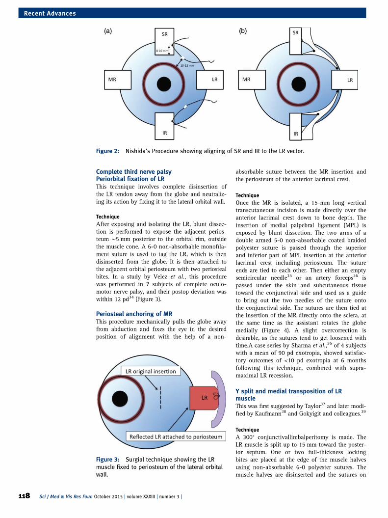
Complete third nerve palsyPeriorbital fixation of LRThis technique involves complete disinsertion ofthe LR tendon away from the globe and neutraliz-ing its action by fixing it to the lateral orbital wall.
TechniqueAfter exposing and isolating the LR, blunt dissec-tion is performed to expose the adjacent perios-teum ∼5 mm posterior to the orbital rim, outsidethe muscle cone. A 6-0 non-absorbable monofila-ment suture is used to tag the LR, which is thendisinserted from the globe. It is then attached tothe adjacent orbital periosteum with two periostealbites. In a study by Velez et al., this procedurewas performed in 7 subjects of complete oculo-motor nerve palsy, and their postop deviation waswithin 12 pd34 (Figure 3).
Periosteal anchoring of MRThis procedure mechanically pulls the globe awayfrom abduction and fixes the eye in the desiredposition of alignment with the help of a non-
absorbable suture between the MR insertion andthe periosteum of the anterior lacrimal crest.
TechniqueOnce the MR is isolated, a 15-mm long verticaltranscutaneous incision is made directly over theanterior lacrimal crest down to bone depth. Theinsertion of medial palpebral ligament (MPL) isexposed by blunt dissection. The two arms of adouble armed 5-0 non-absorbable coated braidedpolyester suture is passed through the superiorand inferior part of MPL insertion at the anteriorlacrimal crest including periosteum. The sutureends are tied to each other. Then either an emptysemicircular needle35 or an artery forceps36 ispassed under the skin and subcutaneous tissuetoward the conjunctival side and used as a guideto bring out the two needles of the suture ontothe conjunctival side. The sutures are then tied atthe insertion of the MR directly onto the sclera, atthe same time as the assistant rotates the globemedially (Figure 4). A slight overcorrection isdesirable, as the sutures tend to get loosened withtime.A case series by Sharma et al.,36 of 4 subjectswith a mean of 90 pd exotropia, showed satisfac-tory outcomes of <10 pd exotropia at 6 monthsfollowing this technique, combined with supra-maximal LR recession.
Y split and medial transposition of LRmuscleThis was first suggested by Taylor37 and later modi-fied by Kaufmann38 and Gokyigit and colleagues.39
TechniqueA 300° conjunctivallimbalperitomy is made. TheLR muscle is split up to 15 mm toward the poster-ior septum. One or two full-thickness lockingbites are placed at the edge of the muscle halvesusing non-absorbable 6-0 polyester sutures. Themuscle halves are disinserted and the sutures on
Figure 2: Nishida’s Procedure showing aligning of SR and IR to the LR vector.
Figure 3: Surgial technique showing the LRmuscle fixed to periosteum of the lateral orbitalwall.
118 Sci J Med & Vis Res Foun October 2015 | volume XXXIII | number 3 |
Recent Advances
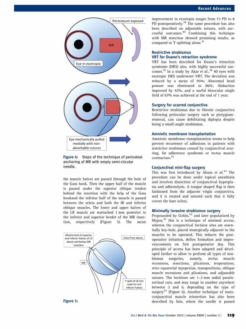
the muscle halves are passed through the hole ofthe Gass hook. Then the upper half of the muscleis passed under the superior oblique tendonbehind the insertion with the help of the Gasshookand the inferior half of the muscle is passedbetween the sclera and both the IR and inferioroblique muscles. The lower and upper halves ofthe LR muscle are reattached 1 mm posterior tothe inferior and superior border of the MR inser-tion, respectively (Figure 5). The mean
improvement in exotropia ranges from 73 PD to 8PD postoperatively.39 The same procedure has alsobeen described on adjustable sutures, with suc-cessful outcomes.40 Combining this techniquewith MR resection showed promising results, ascompared to Y-splitting alone.41
Restrictive strabismusVRT for Duane’s retraction syndromeVRT has been described for Duane’s retractionsyndrome (DRS) also, with highly successful out-comes.26 In a study by Akar et al.,28 40 eyes withesotropic DRS underwent VRT. The deviation wasreduced by a mean of 95%; Abnormal headposture was eliminated in 86%; Abductionimproved by 42%, and a useful binocular singlefield of 67% was achieved at the end of 1 year.
Surgery for scarred conjunctivaRestrictive strabismus due to fibrotic conjunctivafollowing periocular surgery such as pterygium-removal, can cause debilitating diplopia despitebeing a small-angle strabismus.
Amniotic membrane transplantationAmniotic membrane transplantation seems to helpprevent recurrence of adhesions in patients withrestrictive strabismus caused by conjunctival scar-ring, fat adherence syndrome or rectus musclecontracture.42
Conjunctival mini-flap surgeryThis was first introduced by Akura et al.43 Theprocedure can be done under topical anesthesiaand involves dissection of conjunctival hyperpla-sia and adhesiolysis. A tongue shaped flap is thenfashioned from the adjacent virgin conjunctiva,and it is rotated and sutured such that it fullycovers the bare sclera.
Minimally invasive strabismus surgeryPropounded by Gobin,44 and later popularized byMojon,45 this is a technique of minimal access,wherein the conjunctival incision sites are essen-tially key-hole, placed strategically adjacent to themuscles to be operated. This reduces the post-operative irritation, dellen formation and impro-vescosmesis on first postoperative day. Thisprinciple of access has been adapted and devel-oped further to allow to perform all types of stra-bismus surgeries, namely, rectus musclerecessions, resections, plications, reoperations,retro equatorial myopexias, transpositions, obliquemuscle recessions and plications, and adjustablesutures. The incisions are 1–2 mm radial parain-sertinal cuts, and may range in number anywherebetween 2 and 6, depending on the type ofsurgery45 (Figure 6). Another technique of trans-conjunctival muscle reinsertion has also beendescribed by him, where the needle is passed
Figure 4: Steps of the technique of periostealanchoring of MR with empty semi-circularneedle.
Figure 5:
Sci J Med & Vis Res Foun October 2015 | volume XXXIII | number 3 | 119
Recent Advances
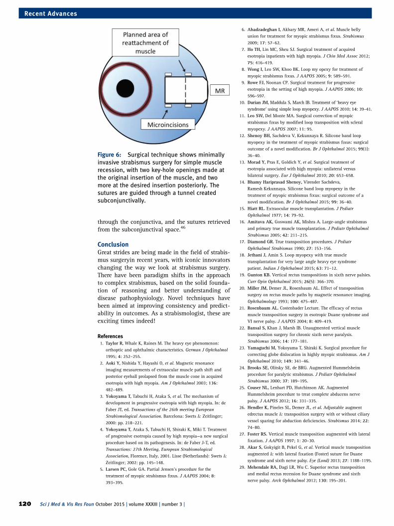
through the conjunctiva, and the sutures retrievedfrom the subconjunctival space.46
ConclusionGreat strides are being made in the field of strabis-mus surgeryin recent years, with iconic innovatorschanging the way we look at strabismus surgery.There have been paradigm shifts in the approachto complex strabismus, based on the solid founda-tion of reasoning and better understanding ofdisease pathophysiology. Novel techniques havebeen aimed at improving consistency and predict-ability in outcomes. As a strabismologist, these areexciting times indeed!
References1. Taylor R, Whale K, Raines M. The heavy eye phenomenon:
orthoptic and ophthalmic characteristics. German J Ophthalmol1995; 4: 252–255.
2. Aoki Y, Nishida Y, Hayashi O, et al. Magnetic resonanceimaging measurements of extraocular muscle path shift andposterior eyeball prolapsed from the muscle cone in acquiredesotropia with high myopia. Am J Ophthalmol 2003; 136:482–489.
3. Yokoyama T, Tabuchi H, Ataka S, et al. The mechanism ofdevelopment in progressive esotropia with high myopia. In: deFaber JT, ed. Transactions of the 26th meeting EuropeanStrabismological Association. Barcelona: Swets & Zeitlinger;2000: pp. 218–221.
4. Yokoyama T, Ataka S, Tabuchi H, Shiraki K, Miki T. Treatmentof progressive esotropia caused by high myopia—a new surgicalprocedure based on its pathogenesis. In: de Faber J-T, ed.Transactions: 27th Meeting, European StrabismologicalAssociation, Florence, Italy, 2001. Lisse (Netherlands): Swets &Zeitlinger; 2002: pp. 145–148.
5. Larsen PC, Gole GA. Partial Jensen’s procedure for thetreatment of myopic strabismus fixus. J AAPOS 2004; 8:393–395.
6. Ahadzadeghan I, Akbary MR, Ameri A, et al. Muscle bellyunion for treatment for myopic strabismus fixus. Strabismus2009; 17: 57–62.
7. Ho TH, Lin MC, Sheu SJ. Surgical treatment of acquiredesotropia inpatients with high myopia. J Chin Med Assoc 2012;75: 416–419.
8. Wong I, Leo SW, Khoo BK. Loop my opexy for treatment ofmyopic strabismus fixus. J AAPOS 2005; 9: 589–591.
9. Rowe FJ, Noonan CP. Surgical treatment for progressiveesotropia in the setting of high myopia. J AAPOS 2006; 10:596–597.
10. Durian JM, Maddula S, March IB. Treatment of ‘heavy eyesyndrome’ using simple loop myopexy. J AAPOS 2010; 14: 39–41.
11. Leo SW, Del Monte MA. Surgical correction of myopicstrabismus fixus by modified loop transposition with scleralmyopexy. J AAPOS 2007; 11: 95.
12. Shenoy BH, Sachdeva V, Kekunnaya R. Silicone band loopmyopexy in the treatment of myopic strabismus fixus: surgicaloutcome of a novel modification. Br J Ophthalmol 2015; 99(1):36–40.
13. Morad Y, Pras E, Goldich Y, et al. Surgical treatment ofesotropia associated with high myopia: unilateral versusbilateral surgery. Eur J Ophthalmol 2010; 20: 653–658.
14. Bhamy Hariprasad Shenoy, Virender Sachdeva,Ramesh Kekunnaya. Silicone band loop myopexy in thetreatment of myopic strabismus fixus: surgical outcome of anovel modification. Br J Ophthalmol 2015; 99: 36–40.
15. Hiatt RL. Extraocular muscle transplantation. J PediatrOphthalmol 1977; 14: 79–92.
16. Amitava AK, Goswami AK, Mishra A. Large‐angle strabismusand primary true muscle transplantation. J Pediatr OphthalmolStrabismus 2005; 42: 211–215.
17. Diamond GR. True transposition procedures. J PediatrOphthalmol Strabismus 1990; 27: 153–156.
18. Jethani J, Amin S. Loop myopexy with true muscletransplantation for very large angle heavy eye syndromepatient. Indian J Ophthalmol 2015; 63: 71–12.
19. Gunton KB. Vertical rectus transpositions in sixth nerve palsies.Curr Opin Ophthalmol 2015; 26(5): 366–370.
20. Miller JM, Demer JL, Rosenbaum AL. Effect of transpositionsurgery on rectus muscle paths by magnetic resonance imaging.Ophthalmology 1993; 100: 475–487.
21. Rosenbaum AL. Costenbader Lecture. The efficacy of rectusmuscle transposition surgery in esotropic Duane syndrome andVI nerve palsy. J AAPOS 2004; 8: 409–419.
22. Bansal S, Khan J, Marsh IB. Unaugmented vertical muscletransposition surgery for chronic sixth nerve paralysis.Strabismus 2006; 14: 177–181.
23. Yamaguchi M, Yokoyama T, Shiraki K. Surgical procedure forcorrecting globe dislocation in highly myopic strabismus. Am JOphthalmol 2010; 149: 341–46.
24. Brooks SE, Olitsky SE, de BRG. Augmented Hummelsheimprocedure for paralytic strabismus. J Pediatr OphthalmolStrabismus 2000; 37: 189–195.
25. Couser NL, Lenhart PD, Hutchinson AK. AugmentedHummelsheim procedure to treat complete abducens nervepalsy. J AAPOS 2012; 16: 331–335.
26. Hendler K, Pineles SL, Demer JL, et al. Adjustable augmentedrectus muscle & transposition surgery with or without ciliaryvessel sparing for abduction deficiencies. Strabismus 2014; 22:74–80.
27. Foster RS. Vertical muscle transposition augmented with lateralfixation. J AAPOS 1997; 1: 20–30.
28. Akar S, Gokyigit B, Pekel G, et al. Vertical muscle transpositionaugmented & with lateral fixation (Foster) suture for Duanesyndrome and sixth nerve palsy. Eye (Lond) 2013; 27: 1188–1195.
29. Mehendale RA, Dagi LR, Wu C. Superior rectus transpositionand medial rectus recession for Duane syndrome and sixthnerve palsy. Arch Ophthalmol 2012; 130: 195–201.
Figure 6: Surgical technique shows minimallyinvasive strabismus surgery for simple musclerecession, with two key-hole openings made atthe original insertion of the muscle, and twomore at the desired insertion posteriorly. Thesutures are guided through a tunnel createdsubconjunctivally.
120 Sci J Med & Vis Res Foun October 2015 | volume XXXIII | number 3 |
Recent Advances

30. Flanders M, Qahtani F, Gans M, Beneish R. Vertical rectusmuscle transposition and botulinum toxin for complete sixthnerve palsy. Can J Ophthalmol 2001; 36: 18–25.
31. Rosenbaum AL, Kushner BJ, Kirschen D. Vertical rectusmuscle transposition and botulinum toxin (Oculinum) tomedial rectus for abducens palsy. Arch Ophthalmol 1989; 107:820–23.
32. Holmes JM, Hatt SR, Leske DA. Intraoperative monitoring oftorsion to prevent vertical deviations during augmented verticalrectus transposition surgery. J AAPOS 2012; 16: 136–140.
33. Muraki S, Nishida Y, Ohji M. Surgical results of a muscletransposition procedure for abducens palsy without tenotomyand muscle splitting. Am J Ophthalmol 2013; 156(4): 819–824.
34. Federico G Velez, Neepa Thacker, Michelle T Britt,Deborah Alcorn, R Scott Foster, Arthur L Rosenbaum. Rectusmuscle orbital wall fixation: a reversible profound weakeningprocedure. J AAPOS 2004; 8: 473–480.
35. Kuldeep Kumar Srivastava, Kannan Sundaresh,Perumalsamy Vijayalakshmi. A new surgical technique forocular fixation in congenital third nerve palsy. J AAPOS 200484.
36. Pradeep Sharma MD, Madhurjya Gogoi MD, Sachin Kedar MD,Rahul Bhola MD. Periosteal fixation in third-nerve palsy. JAAPOS 2006; 10: 324–327.
37. Taylor JN. Surgical management of oculomotor nerve palsywith lateral rectus transplantation to the medial side of theglobe. Aust N Z J Ophthalmol 1989; 17: 27–31.
38. Kaufmann H. “Lateralis splitting” in total oculomotor paralysiswith trochlear nerve paralysis. Fortschr Ophthalmol 1991; 88:314–16.
39. Gokyigit B, Akar S, Satana B, Demirok A, Yilmaz OF. Medialtransposition of a split lateral rectus muscle for completeoculomotor nerve palsy. J AAPOS 2013; 17: 402–410.
40. Ankoor S Shah, Sanjay P Prabhu, Mohammad Ali A Sadiq,Iason S Mantagos, David G. Adjustable nasal transposition ofsplit lateral rectus musclefor third nerve palsy. JAMAOphthalmol 2014; 132(8): 963–969.
41. Jaspreet Sukhija, Savleen Kaur, Usha Singh. Nasal lateral rectustransposition combined with medial rectus surgery for completeoculomotor nerve palsy. J AAPOS 2014; 18: 395–396.
42. Yi Ning J Strube, Francisco Conte, Claudia Faria, Samuel Yiu,Kenneth W Wright. Amniotic membrane transplantation forrestrictive strabismus. Ophthalmology 2011; 118: 1175–1179.
43. Akura J, Kaneda S, Matsuura K, Setogawa A, Takeda K,Honda S. Measures for preventing recurrence after pterygiumsurgery. Cornea 2001; 20(7): 703–707.
44. Gobin MH, Bierlaagh JJM. Chirurgie horizontal eetcyclo verticalesimultanée du strabisme. Centrum voorStrabologie: Anvers,Belgium; 1994.
45. DS Mojon; Review: minimally invasive strabismus surgery. Eye2015; 29: 225–233.
46. Mojon DS. A new transconjunctival muscle reinsertiontechnique for minimally invasive strabismus surgery. J PediatrOphthalmol Strabismus 2010; 47: 292–296.
How to cite this article Nishanth S, Ramasubramanian S. New and complex strabismus surgeries: exciting times! SciJ Med & Vis Res Foun 2015;XXXIII:116–121.
Sci J Med & Vis Res Foun October 2015 | volume XXXIII | number 3 | 121
Recent Advances

Progressive myopia: an update
Meenakshi Swaminathan
Progressive myopia is a major concern to eyecareprofessionals all over the world. A simple searchon popular search engines for “myopia progres-sion” yields over 380,000 results. A Pubmedsearch yields. Myopia is a global health problemassociated with not only vision impairment butalso blinding complications. It is a significant eco-nomic burden too.1 In Singapore mean annualcost of myopia for a child 7–9 years of age is $148. InUS annual direct cost of correcting distance visionimpairment due to refractive errors is between US$3.9 and US$7.2 billion.2
Myopia also poses a significant medical burdenwith increased incidence of glaucoma and catar-acts in those myopic individuals.3 There is also ahigher incidence of blinding retinal complicationsdue to choroidal neovascular membranes, not tomention the impaired quality of life.
PrevalenceStudies in adults have found myopia prevalenceranging from 19.4% in Taiwan to 41.8% in Japanwith figures from Singapore and China falling inbetween.4–9 The population-based studies in chil-dren have found myopia prevalence ranging from1.2% in Nepal to 42.4% in China.10,11 Studiesfrom the early 2000s from India have quotedprevalence figures of 7.4% by Murthy et al. and4.1% by Dandona et al.12,13
A recent study by Saxena et al. looked atprevalence of myopia in Delhi. Amongst a total of9884 school children screened the prevalence ofmyopia was 13.1% with only one-fourth of thosewearing appropriate spectacles.14 A look at figuresfrom Asian countries have shown a sharp increasein prevalence in the last 20 years.
Risk factors for myopia progressionIn this section, various risk factors that have beenstudied will be discussed.
Outdoor activitiesRose et al. in an Australian study found that chil-dren with low outdoor time and high near workwere two to three times more likely to be myopiccompared to those performing low near work andhigh outdoor activities.15 Dirani et al. found a sig-nificant negative association between myopia andoutdoor activity.16 For each hour increase inoutdoor activity per day, spherical equivalent (SE)increased by 0.17 D and axial length (AL) decreasedby 0.06 mm. The Orinda Longitudinal Study ofMyopia (OLSM) found that children who becamemyopic by the eighth grade spent less time in sports
and outdoor activity (hours per week) at the thirdgrade compared to those who did not becomemyopic.17 More recently, the Guanzhou Outdooractivity longitudinal study results provided proof ofprinciple that increasing the amount of time chil-dren spent outdoors through the school system candecrease the number of children who becomemyopic.18
Researchers have also found that it is not thesports but the exposure to the outdoors that appearsto be protective. Chicks reared in less ambient lightbecame myopic.19 Higher light intensity outdoorscould make the depth of field greater and reduceimage blur. Studies have also shown that there isrelease of dopamine from the retina when stimu-lated by light and dopamine is known to inhibit eyegrowth.20 Spectral composition of the light ratherthan the intensity seems to be a more importantfactor.21
Near workThe summary of findings regarding near work andmyopia increase as found in The Sydney MyopiaStudy (SMS) and the Singapore Cohort Study forthe risk factors for myopia (SCORM) are asfollows:22,23
Children who read continuously for more than30 min had a higher incidence of myopia.Children who performed near work at <30 cmdistance were 2.5 times likely to be moremyopic. Children who read more than twobooks per week were also three times likely tohave higher myopia. Children who read morethan 2 h per day were 1.5 times likely to havehigher myopia. For every book read per weekthe AL elongation was more by 0.04 mm.However, several other studies failed to findany significant correlation between near workand myopia
EducationHigher educational level, higher achievement andearlier schooling were also found to be associatedwith an increase in myopia prevalence.24–26 It isunclear if this can be considered as implying thatit is the near work that was more important thanthe education itself.
Parental myopiaThe SMS reported that children with one or bothparents myopic had to two and eight times higherrisk of developing myopia compared with childrenwho had no myopic parents.23 SCORM cohortshowed that having one and two myopic parents
Dr Meenakshi Swaminathan,Director Academics,Senior Consultant,Pediatric Ophthalmology,SN-ORBIS POLTC,Sankara Nethralaya, Chennai,India
Correspondence:Dr Meenakshi Swaminathan,Department of Pediatrics,Pediatric Ophthalmology,Sankara Nethralaya, Chennai,India.Email: [email protected]
122 Sci J Med & Vis Res Foun October 2015 | volume XXXIII | number 3 |
Major Review
was associated with an increase in AL of 0.14 and0.32 mm, respectively, compared with no myopicparents.27
Peripheral refractionTheories of peripheral refraction state that due tothe oblong shape of the eyeball the peripheral raysof light do not focus on the retina. This resultanthyperopic defocus serves as a stimulus for growthof the eyeball. While this process may be import-ant in emmetropization, it also appears to contrib-ute to eyeball elongation.28,29 OLSM assessedperipheral refractive error in 822 children aged 5–14 years. This study indicated that myopic chil-dren had greater relative hyperopia in the periph-ery, compared to emmetropes and hyperopes.30
Control of myopia progressionSpectacles
1. The rationale for the use of bifocals or pro-gressive addition lenses (PAL) is that theyoptimize accommodative accuracy for neartasks and minimize the retinal blur. Bifocalsseem to slow the myopia progression in chil-dren with nearpoint esophoria but not neces-sarily in children with exophoria.31,32 TheCorrection of Myopia Evaluation Trial(COMET) was a multicenter randomized studyinvolving 469 ethnically diverse subjectswhere participants were randomized towearing single vision lenses (SVL) or PALS.Mean 3-year increases in myopia were −1.28D in the PAL group and −1.48 in the SVLgroup, which was statistically significant.Mean myopic progression in the PAL groupwith nearpoint esophoria was −1.18 D com-pared to −1.39D in SVL group.33
2. COMET 2 Trial looked at a separate cohort ofchildren with near esophoria and high accom-modative lag and placed them in PALs. Theoverall reduction in myopia over 3 years wasonly 0.28 D.34
3. A meta-analysis by Li et al. showed that PALsslowed myopia progression by 0.25 D/yearand reduced AL by 0.12 mm/year. Patientsthat appeared to benefit the most from PALswere those with moderate myopia, Asianancestry, Near point esophoria and highaccommodative lag.35
4. Altering peripheral defocus was the focus ofwork by Sankaridurg et al. In their study, 210Chinese children aged 6–16 years were fittedwith one of three novel spectacle lens forms.After 12 months, no statistically significantdifference between SE refraction or AL in thecontrol group wearing SVL versus childrenwearing the novel spectacle lenses was found.In younger children (6–12 years old), with a
parental history of myopia 30% less myopiaprogression in children wearing one of thelens designs than in children wearing controlSVD lenses was found. These novel lenseshowever are limited in their availability.36
Contact lensesExtending the idea of peripheral refraction tocontact lenses, the Dual Focus contact lenses werestudied and found to show effectiveness similar tospectacle dual focus lenses.37
OrthokeratologyOrthokeratology involves temporarily reshapingthe cornea with rigid gas permeable lenses. Itinvolves wearing the lenses overnight. Whilemeta-analysis of all studies using Orthokeratologyshowed a definite slowing of progression, the useof these lenses is also associated with a slightincrease in incidence of epithelial defects andcorneal infections. There is also a higher reboundincrease in myopia progression after cessation oflens wear.38 These lenses are of limited availabilityin India.
Pharmacological therapyMuscarinic receptor antagonists Atropine andPirenzpine have been studied for myopia control,the former extensively. The rationale behind theuse of these agents is the theory that excessiveaccommodation leads to myopia. The othermechanisms postulated are affecting the release ofneurotransimitter dopamine and synthesis of gly-cosaminoglycans in the sclera and thereby redu-cing AL elongation.
The ATOM (Atropine treatment of Myopia) 1and 2 studies have studied the daily application ofAtropine in various concentrations from 1 to0.01% through various phases. Atropine 1% wasthe first to be studied. There were concerns aboutthese children needing PALs due to the accommo-dative difficulties, UV toxicity and rebound pro-gression after cessation of the treatment.Typically, the atropine was administered for 2years and stopped. The children were followed fora washout period of 1 year following which theatropine was reinstituted for 2 more years. BothAL elongation and cycloplegic refraction wereclosely followed as indicators of myopia progres-sion. Children were also monitored for side effectssuch as photophobia and difficulties with accom-modation. The recently published 5 year results ofthe ATOM 2 study shows the efficacy of Atropine0.01% in the retardation of myopia progressionand its safety.40
• Pirenzipine which is a selective M1 receptorantagonist was also studied in a randomizedcontrol trial in the US Pirenzipine study. 174
Sci J Med & Vis Res Foun October 2015 | volume XXXIII | number 3 | 123
Major Review

children, aged from 8 to 12 years (mixed ethni-city/73% were Caucasian) were enrolled in thestudy. The mean increase in myopia was −0.26D/year in the pirenzepine group versus −0.53D/year in the control group.
Myths and traditional therapiesUndercorrection of myopia and giving plus glassesand witholding minus lenses are harmful practiceswith no evidence.
Chinese eye Exercise, Acupuncture, Qi-gongocular exercise, Electrophotomagnitostimulation,eye massage and Drishti Dosha, Triphala, SaptamritaLauha, Amla, Fennel, Licorice, Yoga eye excer-cises, one dose one globule Rhus Tox 1M andsplashing water, while mentioned in various arti-cles and websites, do not have a place in scientificliterature.
Clinical practiceThe ideal age to start treatment is between 6 and 8years. Children with one or two myopic parents,whose myopia started early, who live in an urbanenvironment, with less chance for outdoor play, withmore near work, intense school curriculum, perhapsfemales, shorter working distance with prolongedreading, with near esophoria and/or accommodativelag maybe good candidates for intervention.
If progression of more than 0.5 D SE in last 1year as per a cycloplegic autorefraction, then thesepatients may be considered for intervention. Abaseline testing for near esophoria and accommo-dative lag, a cycloplegic autorefraction and ALmeasurement using a non-contact method such asan IOL master are basic requirements prior to initi-ation of therapy.
Choice of therapy has been covered in detailearlier. It may be guided by the availability ofcertain therapies too. Six monthly monitoring isneeded. Increase in outdoor activity may beachieved by public health measures and interven-tions by school authorities through education ofteachers and parents.
In conclusion, progression of myopia is a majorpublic health burden.41 The control of progressioncontinues to be an area of debate and research.
References1. Lim MC, Gazzard G, Sim EL, Tong L, Saw SM. Direct costs of
myopia in Singapore. Eye 2009; 23: 1086–1089.2. Vitale S, Cotch MF, Sperduto R, Ellwein L. Costs of refractive
correction of distance vision impairment in the United States,1999–2002. Ophthalmology 2006; 113: 2163–2170.
3. Saw SM, Gazzard G, Shih-Yen EC, Chua WH. Myopia andassociated pathological complications. Ophthalmic Physiol Opt2005; 25: 381–391.
4. Xu L, Li J, Cui T, et al. Refractive error in urban and rural adultChinese in Beijing. Ophthalmology 2005; 112: 1676–1683.
5. Cheng CY, Hsu WM, Liu JH, Tsai SY, Chou P. Refractive errorsin an elderly Chinese population in Taiwan: the Shihpai EyeStudy. Invest Ophthalmol Vis Sci 2003; 44: 24.4630–4638.
6. Sawada A, Tomidokoro A, Araie M, Iwase A, Yamamoto T.Refractive errors in an elderly Japanese population: the 25.Tajimi study. Ophthalmology 2008; 115: 363–370.e3.
7. Wong TY, Foster PJ, Hee J, et al. Prevalence and risk factors forrefractive errors in adult Chinese in Singapore. InvestOphthalmol Vis Sci 2000; 41: 2486–2494.
8. Saw SM, Chan YH, Wong WL, et al. Prevalence and risk factorsfor refractive errors in the Singapore Malay Eye Survey.Ophthalmology 2008; 115: 1713–1719.
9. Pan CW, Wong TY, Lavanya R, et al. Prevalence and risk factorsfor refractive errors in Indians: the Singapore Indian Eye Study(SINDI). Invest Ophthalmol Vis Sci 2011; 52: 3166–3173.
10. Pokharel GP, Negrel AD, Munoz SR, Ellwein LB. Refractive errorstudy in children: results from Mechi Zone, Nepal. Am JOphthalmol 2000; 129: 436–444.
11. He M, Zeng J, Liu Y, Xu J, Pokharel GP, Ellwein LB. Refractiveerror and visual impairment in urban children in southernchina. Invest Ophthalmol Vis Sci 2004; 45: 793–799.
12. Dandona R, Dandona L, Srinivas M, et al. Refractive error inchildren in a rural population in India. Invest Ophthalmol VisSci 2002; 43: 615–622.
13. Murthy GV, Gupta SK, Ellwein LB, et al. Refractive error inchildren in an urban population in New Delhi. InvestOphthalmol Vis Sci 2002; 43: 623–631.
14. Saxena R, Vashist P, et al. Prevalence of myopia and its riskfactors in urban school children in Delhi: the North IndiaMyopia Study. PloS One 2015; 26: 10(2)e0117349.
15. Rose KA, Morgan IG, Ip J, et al. Outdoor activity reduces 69.The prevalence of myopia in children. Ophthalmology 2008;115: 1279–1285.
16. Dirani M, Tong L, Gazzard G, et al. Outdoor activity andmyopia in Singapore teenage children. Br J Ophthalmol 702009; 93: 997–1000.
17. Jones LA, Sinnott LT, Mutti DO, Mitchell GL, Moeschberger ML,Zadnik K. Parental history of myopia, sports and outdooractivities, and future myopia. Invest Ophthalmol Vis Sci 2007;48: 3524–3532.
18. He M, et al. Effect of time spent outdoor at school on thedevelopment of myopia among children in China; a randomizedclinical trial. AMA. 2015; 314(11): 1142–1148.
19. Ashby RS, Schaeffel F. The effect of bright light on lenscompensation in chicks. Invest Ophthalmol Vis Sci 2010; 51:5247–5253.
20. Rose KA, Morgan IG, Ip J, et al. Outdoor activity reduces theprevalence of myopia in children. Ophthalmology 2008; 115:1279–1285.
21. Mehdizadeh M, Nowroozzadeh MH. Outdoor activity andmyopia. Ophthalmology 2009; 116: 1229–1230; author reply.
22. Ip JM, Saw SM, Rose KA, et al. Role of near work in myopia:findings in a sample of Australian school children. InvestOphthalmol Vis Sci 2008; 49: 2903–2910.
23. Saw SM, Carkeet A, Chia KS, Stone RA, Tan DT. Componentdependent risk factors for ocular parameters in SingaporeChinese children. Ophthalmology 2002; 109: 2065–2071.
24. Tay MT, Au Eong KG, Ng CY, Lim MK. Myopia and educationalattainment in 421,116 young Singaporean males. Ann AcadMed Singapore 1992; 21: 785–791.
25. Teasdale TW, Goldschmidt E. Myopia and its relationship toeducation, intelligence and height. Preliminary results from anon-going study of Danish draftees. Acta Ophthalmol Suppl1988; 185: 41–43.
26. Rose KA, Morgan IG, Smith W, Burlutsky G, Mitchell P,Saw SM. Myopia, lifestyle, and schooling in students of Chineseethnicity in Singapore and Sydney. Arch Ophthalmol 2008;126: 527–530.
27. Ip JM, Huynh SC, Robaei D, et al. Ethnic differences in theimpact of parental myopia: findings from a population-basedstudy of 12-year-old Australian children. Invest Ophthalmol VisSci 2007; 48: 2520–2528.
124 Sci J Med & Vis Res Foun October 2015 | volume XXXIII | number 3 |
Major Review
28. Mutti D, Hayes J, Mitchell L, et al. Refractive error, axiallength, and relative peripheral refractive error before and afterthe onset of myopia. Invest Ophthalmol Vis Sci 2007; 48:2510–2519.
29. Sng CC, Lin XY, Gazzard G, et al. Change in peripheralrefraction over time in Singapore Chinese children. InvestOphthalmol Vis Sci 2011; 52: 7880–7887.
30. Mutti D, Sholtz R, Friedman N, Zadnik K. Peripheral refractionand ocular shape in children. Invest Ophthalmol Vis Sci 2000;41: 1022–1030.
31. Fulk GW, Cyert LA, Parker DE. A randomized trial of the effectof single-vision vs. bifocal lenses on myopia progression inchildren with esophoria. Optom Vis Sci 2000; 77(8): 395–401.
32. Goss DA, Jyesugi EF. Effectiveness of bifocal control ofchildhood myopia progression as a function of near pointphoria and binocular cross cylinder. J Optom Vis Devel 1995;26: 12–17.
33. Gwiazda J, Hyman L, Hussein M, et al. A randomized clinicaltrial of progressive addition lenses versus single vision lenses onthe progression of myopia in children. Invest Ophthalmol VisSci 2003; 44(4): 1492–1500.
34. Correction of Myopia Evaluation Trial 2 Study Group for thePediatric Eye Disease Investigator Group. Progressive-additionlenses versus single-vision lenses for slowing progression ofmyopia in children with high accommodative lag and nearesophoria. Invest Ophthalmol Vis Sci 2011; 52(5): 2749–57.
35. Li SM, et al. Multifocal vs single vision lenses intervention toslow progression of myopia in school-age children: ameta-analysis. Surv Ophthalmol 2011; 56(5): 451–460.
36. Sankaridurg P, et al. Spectacle lens designed to reduceprogression of myopia: 12 month results. Optom Vis Sci 2010;87(9): 631–641.
37. Sankaridurg P, Holden B, Smith E 3rd, Naduvilath T, Chen X,de la Jara PL, Martinez A, Kwan J, Ho A, Frick K, Ge J.Decrease in rate of myopia progression with a contact lensdesigned to reduce relative peripheral hyperopia: one-yearresults. Invest Ophthalmol Vis Sci 2011;52(13): 9362–9367.
38. Li SM, Kang MT, Wu SS, Liu LR, Li H, Chen Z, Wang N.Efficacy, safety and acceptability of orthokeratology on slowingaxial elongation in myopic children by meta-analysis. Curr EyeRes 2015: 1–9.
39. Chia A, Lu QS, Tan D. Five-year clinical trial on atropine for thetreatment of myopia 2: myopia control with atropine 0.01%eyedrops. Ophthalmology 2015.
40. Siatkowski RM, Cotter SA, Crockett RS, Miller JM, Novack GD,Zadnik K; U.S. Pirenzepine Study Group. Two-year multicenter,randomized, double-masked, placebo-controlled, parallel safetyand efficacy study of 2% pirenzepine ophthalmic gel in childrenwith myopia. J AAPOS 2008; 12(4): 332–329.
41. Chen-Wei Pan, Dharani R, Saw SM. Worldwide prevalence andrisk factors for myopia. Ophthalmic Physiol Opt 2012; 32(1):3–16.
How to cite this article Swaminathan M. Progressive myopia: an update, Sci J Med & Vis Res Foun 2015;XXXIII:122–125.
Sci J Med & Vis Res Foun October 2015 | volume XXXIII | number 3 | 125
Major Review
Infantile cataract: where are we now?
Praveen Kumar KV and Sumita Agarkar
IntroductionPediatric cataract is one of the major causes of pre-ventable childhood blindness affecting approximately200,000 children worldwide.1 In developing countries,the prevalence of blindness from cataractC is higher,about one to four per 10,000 children. Early diag-nosis and treatment WWWis essential to preventthe development of stimulus deprivation ambly-opia in these children. Cataract surgery in infantsposes greater challenges compared to young chil-dren. Primary implantation of an intraocular lensremains controversial for infants, and the selec-tion of an appropriate IOL power is difficult. Themanagement of infantile cataract has changedover the last decade. In this study, we present anoverview of the changing concepts of cataracts ininfants and its management.
Etiology of childhood cataractThe common causes of congenital cataract aregenetic, metabolic disorders, prematurity and intra-uterine infections. Almost 60% of cases of congeni-tal cataract in developed countries are idiopathic.2
One-third of cases of congenital cataract are her-editary without any known associated systemicdisease.3 The various causes of congenital cataractare
1. Heredity: These cataracts are usually auto-somal dominant but can be autosomal reces-sive and x linked.
2. Associated with genetic disorders: Seen indowns and turners syndrome.
3. Metabolic disorders: Galactosemia, Hypocalcemia.
4. Intrauterine infections: Toxoplasmosis, Rubella,Cytomegalovirus, Herpes, Varicella and Syphilis.
5. Associated Ocular conditions: Aniridia, Iriscoloboma, lens coloboma, Lenticonus,Lentiglobus, Persistent fetal vasculature.
Next generation DNA sequencing shown prom-ising results in identifying mutations leading tometabolic abnormalities and cataracts. This newtechnology has immense potential in identifyingmetabolic abnormalities and correct them withappropriate intervention. In future, this technologyis likely to become cheaper to allow testing in chil-dren with non-syndromic bilateral cataracts withsystemic manifestation and delayed development.4
Morphology of pediatric cataractMorphology of pediatric cataract can not onlyhelp in the diagnosis of specific associated
disorder but also helps in planning the manage-ment. Based on morphology, pediatric cataractscan be classified into cataracts involving theentire lens, central cataracts, anterior cataracts,posterior cataracts, punctate lens opacities, coral-line cataracts, sutural cataract, wedge shaped cata-ract and cataracts associated with PFV.
Preoperative evaluationHistory taking is an integral part in the evaluationof an infant with congenital cataract. The historyshould include
Family history of congenital or developmentalcataract,
1. Antenatal history of maternal drug intake andfever with rash.
2. Birth history should be specifically looked foras bilateral congenital cataract is morecommon in preterm, low birthweight, smallfor gestational age children.5
3. Developmental mile stones should be carefullyassessed.
4. History of visual interaction of the child withthe family members should also be inquiredas it helps in determining the severity ofvisual dysfunction.
5. History of the onset of the opacities, progres-sion and laterality is also important.Unilateral cataracts are isolated but areusually associated with other ocular abnor-malities like persistent fetal vasculature, lenti-conus, lentiglobus.6,7
Examination of the childDetailed ocular examination of the child can bedone either in an outpatient setting if the child iscooperative or under general anesthesia when thechild is being taken up for surgery. Quantificationof visual acuity of the child as far as possibleshould be done. In infants, fixation behavior, fix-ation preference and resistance to occlusion givesus a clue to the visual acuity. In young infantswith poorly developed fixation, an undilateddistant direct ophthalmoscopy can indicate whetherthe opacity is visually significant or not. Densecentral opacities larger than 3 mm in diameterusually need surgical removal.8
Examination of both the eyes has to be done todetermine whether the cataract is unilateral orbilateral. Unilateral cataract, even if mild cancause irreversible deep amblyopia if not treated.9
Correspondence to:Dr. Sumita Agarkar,Deputy Director – PediatricOphthalmology Department,Sankara NethralayaMedical Research Foundation18, College Road,Chennai - 600 006email: [email protected]
126 Sci J Med & Vis Res Foun October 2015 | volume XXXIII | number 3 |
Major Review

Often the first symptom is a white or partiallywhite reflex noted by the parents. Strabismus andnystagmus should be specifically looked for inthese children and sometimes these may be thepresenting signs. Strabismus is usually seen inchildren with unilateral cataracts and developswhen an irreparable visual loss has alreadyoccurred. The presence of manifest nystagmus atage of 2–3 months or elder generally indicates avery poor prognosis.10 The presence of either stra-bismus or nystagmus indicates that cataract isvisually significant.
Slit lamp biomciroscopy should be done toassess the size, location, density of the opacity.Corneal diameters and intraocular pressure haveto be measured with a tonopen or Perkins handheld applanation tonometer. Indirect ophthalmos-copy can reveal persistent fetal vasculature or otherposterior segment abnormalities that may affect thevisual outcome. In cases where the media opacityprecludes examination of the fundus, a B scan ultra-sonography has to be performed to rule out otherposterior segment pathologies that mimic congenitalcataract. These conditions include retinoblastoma,persistent hyperplastic primary vitreous, coatsdisease, ROP with retrolental fibroplasia, orgainzedvitreous hemorrhage, congenital falciform fold,ocular toxocariasis and retinal hamartomas.Performing cataract surgery in these conditions isdisastrous and can lead one into medicolegal pro-blems. These children should be evaluated by apediatrician to exclude systemic disorders or meta-bolic causes causing cataract.
Laboratory workupMost children with congenital cataract do notneed systemic work up. Unilateral, familial, iso-lated cataracts with no systemic association donot need any systemic investigations. Traumashould be ruled out in all cases of unilateral cata-ract. A child with peculiar facies or systemic mal-formations like microcephaly, deafness, cardiacabnormalities, developmental delayneed systemicworkup. The work up usually includes fastingblood sugar, urine for reducing substances forgalactosemia, aminoacids for Lowes syndrome.Plasma phosphorous, calcium levels, RBC transfer-ase and galactokinase levels have to be assessed.TORCH titers have to be done to rule out infec-tious causes of cataract.11,12
ManagementIndication for cataract surgery in infants dependson the extent of its effect on the visual function.Mere presence of a lenticular opacity does notwarrant surgical removal. Cataract which is incom-plete at birth, peripheral lens opacities, punctateopacities with intervening clear zones, opacities<3 mm in diameter can be kept under close followup.13 Associated amblyopia in these children
should be treated by appropriate glasses and patch-ing. Small opacities can be managed by prescribingmydriatric agents to achieve a larger area of clearvisual axis.
Timing of surgeryExtraction of unilateral congenital cataracts by 4–6 weeks and bilateral cataracts within 6–8 weeksof life can prevent the development of strabismus,nystagmus and amblyopia.14
How does an infant eye differ from the adult eye?A child’s eye is unique and is different from anadult eye. The eyes are smaller in size at birth andhave steeper corneas. The normal new born eye hasa mean axial length of 16.6–17 mm.15 It reaches amean adult value of 23.6 mm at 15 years age. Morethan half of this growth in axial length occursbefore 1 year age and most axial elongation occursduring the first 2 years of life. The corneal curva-ture reduces from 51.2 D in new borns to 43.5 D inadults.16 The sclera is thin and less rigid, the lenscapsule is more elastic, and there is a risk of severepostoperative inflammatory response.
Biometry in infantsWith advances in surgical techniques and instru-mentation, several surgeons are implanting IOLs ininfants. Refractive growth after IOL implantation ininfants cannot be predicted accurately and currentIOL formulae vary in their predictive outcomes. If rtarget postoperative emmetropia, amblyopia treat-ment is easier but this strategy results in highmyopia in later life. If we aim for hyperopia, ambly-opia therapy and refractive correction in initialphase is difficult but thisstrategy has the advantageof potentially achieving either emmetropia or lowmyopia later in adulthood. The amount of hyper-opia will vary depending on the age of the child atthe time of surgery. Most surgeons prefer to leaveinfants with hyperopia as it’s easy to titrate as chil-dren grow. IATS recommended an hyperopiaranging from +6 D to +8 D depending on the age ofinfant at the time of surgery. It is important tocounsel parents regarding need for glasses post-operatively as well as perhaps through the life.
Three important things to be considered whendetermining the IOL power to be implanted ininfants are
1. Anticipated refractive shift.
2. Age of the patient.
3. Target refraction in the immediate postoperativeperiod.
Children who are younger at the time ofsurgery, have a significantly greater myopic shiftand greater variance in predictive refractivechange than older children. Crouch et al. in astudy of 52 eyes undergoing cataract surgery with
Sci J Med & Vis Res Foun October 2015 | volume XXXIII | number 3 | 127
Major Review
IOL implantation found a mean myopic shift of3.66 D in children operated on at 3–4 weeks agewhich reduced to 0.38 D in children operated onat 15–18 years age.17 Most pseudophakic eyesgrow normally and so a significant shift after IOLimplantation is expected in these children.18
Postoperative refractive goal in infantsIn infants, implantation of IOL still remains con-troversial and several surgeons prefer to leave theinfants aphakic after cataract surgery.19 In theInfant Aphakia Treatment Study, the targetrefractive error after IOL implantation was +8 forinfants 4–6 weeks of age and +6 for infantsbetween 6 weeks to 6 months age.20
Keratometry and axial length measurements inchildren are usually less accurate compared toadults. These measurements are often obtainedunder anesthesia in infants who do not cooperatefor fixation. Mittelviefhaus et al. in their studyhave shown that lack of fixation in children undergeneral anesthesia can result in inaccurate kerato-metry measurements.21 However, the reliabilitycan be increased by averaging several readings pereye.
Axial length is a more significant source oferror in IOL power calculation. Inaccurate axiallength measurement can account for 4–14 diop-ters for each millimeter difference in IOL power.22
Errors are often magnified because of shorteraxial length. Immersion biometry is more predict-able than contact method for IOL power calcula-tion in infants.23 But the limitation of theimmersion scan is that it cannot be used in smalleyes and globe with shallow anterior chamber andother ocular anomalies as in infants. Partialcoherence interferometry can be used to measureaxial length in cooperative children with reliabilityand accuracy.24 Advantages over conventionalultrasound include high reproducibility, contactfree measurements, observer independence of themeasurements. The disadvantage is that it cannotbe used in total cataracts which are more oftenencountered in children.
IOL formulaFurthermore, no time tested formula exists for cal-culation of IOL power in infants. The accuracy ofeach formula depends on optimized values andmeasures of the formula components, includingfactors such as actual anterior chamber depth,lens thickness, vertex distance, and use of a perso-nalized surgeon factor or A-constant. The anteriorsegment of an infant eye is significantly smaller,eyes with congenital cataract may have greateranatomic variation in anterior segment structuresand the anterior segment of an infant is propor-tionally larger to the posterior segment comparedto an adult eye. The capsular bag of an infant eyeis smaller and will contract earlier, which may
result in greater posterior IOL displacement.Implantation of high-power IOLs in these eyes,can increase the measurement and calculationerrors as well as the errors induced by changesin IOL position. Nihalani and Vanderveen in aretrospective study of 135 pediatric eyes that under-went cataract surgery and primary IOL implantationfound mean predictability of four formulae wascomparable, with 57% of infants having a predic-tion error of more than 0.5 diopters.25 Greater pre-diction errors were seen in children <2 years, axiallength <22 mm and mean keratometry readings>43.5. The SRK II, SRK T and Holladay formulaetended to overcorrect whereas Hoffer Q had anequal number of undercorrection and overcorrec-tions. Kekunnaya et al. in their study on IOLpower calculation in children <2 years age foundprediction errors were larger for all formulas butSRK II had the least prediction error.26 In theInfant Aphakia treatment study to determine thepredictability of IOL power calculation formulae ininfants eyes, overall median absolute predictionerror values appeared to be similar for theHolladay 1, Holladay 2 and SRK/T formulae (1.2D, 1.4 D and 1.3 D, respectively), and in pairedcomparisons of SRK/T versus other formulae, themedian paired differences in absolute predictionerror was more than zero, indicating greater accur-acy for the SRK/T formula. The study concludedthat Holladay I and SRK T formula gave goodcomparable results and have the best predictivevalue for infant eyes. The greatest predictionerrors in their study were seen in eyes with axiallength of 18 mm or less.27
Type of IOL to be implantedThere is a large debate regarding the type of IOLto be implanted in infants. IOL implantation duringchildhood may be associated with better visualoutcomes but in IOL implantation in infancy,these potential advantages are offset by a higherincidence of intraoperative and postoperativeadverse events. Additional intraocular surgeriesare often required to treat these adverse eventswhich are associated with risks, costs and parentalstress. Although it is agreed that cataract surgeryduring early infancy is associated with the bestvisual outcomes, it remains undetermined whetherprimary IOL implantation is advisable in this agegroup. Ram et al. compared outcomes of hydro-phobic acrylic and PMMA lenses in children <1year age and reported that complication rates werecomparable in both the groups. PMMA lenses mayrequire early surgical intervention for PCO.28 Thesingle piece acrylic hydrophobic IOL is a soft IOLand can be implanted in the smaller capsular bagas in infants with relative ease. It also has theadvantage of requiring a smaller incision therebyallowing corneal incision leaving conjunctivaintact. However, single piece IOL cannot be placed
128 Sci J Med & Vis Res Foun October 2015 | volume XXXIII | number 3 |
Major Review
in sulcus. Silicon IOLs have also be implanted inyoung children, but are associated with more cap-sular contraction.29
Are IOLs good for infants?This question has been debated by Pediatricophthalmologists for several years. There was littleevidence to support the claim either way. Theinfant aphakia study was designed to answer thisquestion. This was a prospective randomized mul-ticentric trial comparing infants who underwentcataract surgery for unilateral cataract with eitherIOL implantation or were left aphakic and werefitted with a contact lens. The main outcome vari-able was visual acuity at 1 year and 4 ½ years ofage. The investigators also looked at complica-tions, resurgery rates and strabismus and stereop-sis and compliance to occlusion. IATS found thatthere was no difference in visual acuity at either 1year of age or at 4 ½ years between the twogroups. But alarmingly adverse events like mem-brane proliferation into the visual axis, corectopiawere almost 10 times more common in infantswith IOL implantation compared to aphakicinfants.30 In aphakic eyes, the margins of theanterior and posterior capsular bag usually fusetogether preventing lens material from migratingout of the Sommerring ring into the pupillaryspace. Whereas in pseudophakic eyes, lens epithe-lial cells are able to migrate into the pupillaryspace because the IOL interfers with the fusion ofthe lens capsular remnanats. Hence, not surpris-ingly, the commonest indication for resurgery inIOL group was to clear the visual axis. Additionalintraocular surgeries were 3 ½ times more inpseudophakic infants compared to aphakicinfants.31 The risk of glaucoma was same in boththe groups. The development of stereopsis did notdiffer depending on the type of optical rehabilita-tion. In conclusion, the study did not demonstrateany visual benefit from implanting an IOL at thetime of unilateral cataract surgery in infants <7months of age and the children who had IOLimplantation had more adverse events andrequired more reoperations to clear visual axisopacities. The investigators concluded by saying“When operating on an infant younger than 7months of age with a unilateral cataract, we rec-ommend leaving the eye aphakic and focusing theeye with a contact lens. Primary IOL implantationshould be reserved for those infants where, in theopinion of the surgeon, the cost and handling of acontact lens would be some burdensome as toresult in significant periods of uncorrectedaphakia.”32 In the context of our country,however the IATS conclusions need to be inter-preted in a slightly different light. Monocular cat-aracts in infants, where only way of visualrehabilitation is contact lens, is often problematicin developing countries because of poor hygiene,
socioeconomic factors and non-availability ofcontact lenses in smaller towns. So these infantswill probably do better with IOL. Only rider is thatlens be placed in the bag with appropriate capsu-lar management and anterior vitrectomy to ensureclear visual axis. Other important factor is patientselection and we do recommend IOL in infantswho have otherwise anatomically normal eye withno anterior segment dysgenesis or other anomal-ies. Bilateral cataracts in infants however aphakiacan be easily managed with aphakic glasses aswell as contact lenses. Attending surgeon is bestplaced to take that decision customising it accord-ing to patient’s profile and his/her skills. IATS hascertainly provided information for better informeddecisions.
Complications following surgeryPostoperative inflammatory response in childrencan result in fibrinous and pigment deposits onthe IOL. Inflammatory response can be reallyexaggerated in infants with Rubella syndrome.
Posterior capsular opacification is the mostcommon complication after pediatric cataractsurgery. Primary posterior capsulotomy with anter-ior vitrectomy combined with hydrophobic acrylicIOL in the bag, can prevent or delay the occurrenceof VAO. PCO if develops can be treated by NdYaglaser capsulotomy or membraectomy depending onthe child’s cooperation and the thickness of PCO.
Secondary glaucoma is the most feared compli-cation of infantile cataract surgery. IATS showedthat IOL implantation does not seem to protectagainst the development of glaucoma.33 Mataftsihavereported that glaucoma after pediatric cataractsurgery is associated with surgery with in first 1month of life and additional surgical proceduresbut not with primary IOL implantation.32 Parentsmust be counselled regarding glaucoma and needfor regular follow up. Rapid myopic shift andincreased axial length points towards glaucomaand high index of suspicion should be maintainedin these children.
Retinal detachment is a rare late postoperativecomplication of pediatric cataract surgery.
Postoperative visual rehabilitationVisual rehabilitation in children after surgery canbe achieved by aphakic glasses, contact lenses,IOL implantation. Aphakic glasses are efficientmethod of visual rehabilitation in infants espe-cially in bilateral cataracts. Contact lenses are par-ticularly useful in unilateral cataract. Silcon softlenses or rigid gas permeable lenses are commonlyused. Speegschatz et al. in their study of 157aphakic subjects found that initial rehabilitationwith aphakic glasses and secondary IOL implant-ation at a later date has the advantage of predictablepostoperative refraction and fewer complications.34
Visual rehabilitation can be done in the immediate
Sci J Med & Vis Res Foun October 2015 | volume XXXIII | number 3 | 129
Major Review

postoperative period by aphakic glasses in bilateralcases and contact lenses in unilateral cases. Whenfitting an infant with aphakia with contact lens theproblem of appropriate power of the contact lensarises. Silicon elastomer contact lens is the preferredcontact lens for the treatment of aphakia in infants.It is easy to fit and can be used as an extended wearcontact lens. Rigid gas permeable lens is also anoption given the advantage of cost and good oxy-genation for cornea. The preoperative axial lengthcan be used to determine the contact lens power tobe used. Martin et al. in their study reported thepower of the contact lens depending on the pre-operative axial length as: 0–6 months, +29 D; 7–17months, +26 D; 18–28 months, +23 D and 29–34months, +18 D.5 Moore noted that the mean spher-ical equivalent refractive error for these patients was+28.5 D at 6 months, +26.5 D at 12 months, +23 Dat 24 months and +21.5 D at 36 months.35 Trivediet al. recommends 32-D CL when the preoperativeAL is <17 mm, a 29-D Cl when the preoperative ALis between 17 and 18.5 mm, a 26-D CL when thepreoperative AL is 18.5–19.5 D, a 23-D CL when thepreoperative AL is between 19.5 and 20 mm (21 mm)and a 20-D CL for an AL of 20–21 mm (20 D for>21 mm).36 Secondary IOL implantation can bereserved as an option for later visual rehabilitationin these children. Nihalani et al. in their study of sec-ondary IOL implantation in children left aphakicduring initial cataract surgery found satisfactoryvisual and refractory outcomes in children receivingsecondary IOL implantation. However, immediatepostoperative inflammation and corneal edema wasmore in eyes with sulcus implanted IOL compared toin the bag IOL.37
Last but not the least, several of these infantswill need continued monitoring for amblyopiaand patching and strabismus. They will needappropriate refractive correction moving from asingle vision glass to a bifocal or progressive astheir visual needs change. It is important not tomiss these unglamorous factors in follow up visitsotherwise amblyopia can trump a beautifully donesurgery in a matter of few months.
ConclusionCataract surgery in infants is a specially challen-ging subset among all cataract surgeries in chil-dren in terms of surgical technique, formula to beused, biometry and postoperative visual rehabilita-tion. But it is rewarding, if we are able to rehabili-tate them either with glasses or primary IOLimplantation or contact lenses. Surgeon has theonus of deciding on the best course looking aheadnot few years but few decades.
References1. Holmes JM, Leske DA, Burke JP, Hodge DO. Birth prevalence of
visually significant infantile cataract in a defined U.S.population. Ophthalmic Epidemiol 2003; 10: 67–74.
2. Lim Z, Rubab S, Chan YH, Levin AV. Pediatric cataract: theToronto experience-etiology. Am J Ophthalmol 2010; 149:887–892.
3. Merin S, Crawford JS. The etiology of congenital cataracts. Asurvey of 386 cases. Can J Ophthalmol 1971; 6: 178–182.
4. Gillespie RL, Sullivan J, Ashworth J, Bhaskar S, Williams S,Biswas S, Kehdi E, Ramsden SC, Clayton-Smith J, Black GC,Lloyd IC. Personalized diagnosis and management of congenitalcataract by next-generation sequencing. Ophthalmology 2014;12: 2124–2137.
5. Haargaard B, Wohlfahrt J, Rosenberg T, Fledelius HC,Melbye M. Risk factors for idiopathic congenital/infantilecataract. Invest Ophthalmol Vis Sci 2005; 46: 3067–3073.
6. Amaya L, Taylor D, Russell-Eggitti , Nischal KK, Lengyel D. Themorphology and natural history of childhood cataracts. SurvOphthalmol 2003; 48: 125–144.
7. Haargaard B, Wohlfahrt J, Fledelius HC, Rosenberg T,Melbye M. A nationwide Danish study of 1027 cases ofcongenital/infantile cataracts: etiological and clinicalclassifications. Ophthalmology 2004; 111: 2292–2298.
8. Arkin M, Azar D, Fraioli A. Infantile cataracts. Int OphthalmolClin 1992; 32: 107–120.
9. Zetterström C, Lundvall A, Kugelberg M. Cataracts in children.J Cataract Refract Surg 2005; 31: 824–840.
10. Gelbart SS, Hoyt CS, Jastrebski EM. Long term visual results inbilateral congenital cataracts. Am J Ophthalmol 1982; 93:615–621.
11. Raghu H, Subhan S, Jose RJ, Gangopadhyay N, Bhende J,Sharma S. Herpes simplex virus-1—associated congenitalcataract. Am J Ophthalmol 2004;138:313–314.
12. Vijayalakshmi P, Kakkar G, Samprathi A, Banushree R. Ocularmanifestations of congenital rubella syndrome in a developingcountry. Indian J Ophthalmol 2002; 50: 307–311.
13. Choi J, Kim JH, Kim SJ, Yu YS. Clinical characteristics, course,and visual prognosis of partial cataracts that seem to be visuallyinsignificant in children. J AAPOS 2012; 16: 161–167.
14. Birch EE, Cheng C, Stager Dr, Weakley Dr. The critical periodfor surgical treatment of dense congenital bilateral cataracts.J AAPOS 2009; 13: 67–71.
15. Swan KC, Wilkins JH. Extraocular muscle surgery in earlyinfancy—anatomicalfactors. J Pediatr Ophthalmol Strabismus1984; 21: 44–49.
16. Eibschitz-Tsimhoni M, Archer SM, Del Monte MA. Intraocularlens power calculation in children. Surv Ophthalmol 2007; 52:474–482.
17. Crouch ER, Crouch ER Jr, Pressman SH. Prospective analysis ofpediatricpseudophakia: myopic shift and postoperativeoutcomes. J AAPOS 2002; 6: 277–282.
18. Hutchinson AK, Drews-Botsch C, Lambert SR. Myopic shiftafter intraocular lens implantation during childhood.Ophthalmology 1997; 104: 1752–1757.
19. Lambert SR, Lynn M, Drews-Botsch C, et al. Intraocularlensimplantation during infancy: perceptions of parents and theAmerican Association for Pediatric Ophthalmology andStrabismus members. J AAPOS 2003; 7: 400–405.
20. Eibschitz-Tsimhoni M, Archer SM, Del Monte MA. Intraocularlens power calculation in children. Surv Ophthalmol 2007; 52:474–482.
21. Mittelviefhaus H, Gentner C. Errors in keratometry forintraocularlen simplantation in infants. Ophthalmology 2000;97: 186–188.
22. Eibschitz-Tsimhoni M, Tsimhoni O, Archer SM, Delmonte MA.Effect of axial length and keratometry measurement error onintraocular lens implant power prediction formula in pediatricpatients. J AAPOS 2008; 12: 173–176.
23. Trivedi RH, Wilson ME. Prediction error after pediatric cataractsurgery with intraocular lens implantation: contact versusimmersion A-scan biometry. J Cataract Refract Surg 2011; 37:501–505.
130 Sci J Med & Vis Res Foun October 2015 | volume XXXIII | number 3 |
Major Review

24. Hussin HM, Spry PGD, Majid MA, et al. Reliability andvalidity of the partial coherence interferometry formeasurement of ocular axial length in children. Eye 2005; 20:1021–1024.
25. Nihalani BR, VanderVeen DK. Comparison of intraocular lenspower calculation formulae in pediatric eyes. Ophthalmology2010; 117: 1493–1499.
26. Kekunnaya R, Gupta A, Sachdeva V, Rao HL, Vaddavalli PK,Om Prakash V. Accuracy of intraocular lens power calculationformulae in children less than two years. Am J Ophthalmol2012; 154: 13–19.
27. VanderVeen DK, Nizam A, Lynn MJ, Bothun ED,McClatchey SK, Weakley DR, DuBois LG, Lambert SR; InfantAphakia Treatment Study Group. Predictability of intraocularlens calculation and early refractive status: the InfantAphakia Treatment Study. Arch Ophthalmol 2012; 130:293–299.
28. Ram J, Jain VK, Agarwal A, Kumar J. Hydrophobic acrylicversus polymethylmethacrylate intraocular lens implantationfollowing cataract surgery in the first year of life. Graefes ArchClin Exp Ophthalmol 2014; 252: 1443–1449.
29. Pavlovic S, Jacobi FK, Graef M, Jacobi KW. Silicone intraocularlens implantation in children: preliminary results. J CataractRefract Surg 2000; 26: 88–95.
30. Plager DA, Lynn MJ, Buckley EG, Wilson ME, Lambert SR.Complications, adverse events, and additional intraocular
surgery 1 year after cataract surgery in the Infant AphakiaTreatment Study. Ophthalmology 2011; 118: 2330–2334.
31. Carrigan AK, DuBois LG, Becker ER, Lambert SR. Cost ofintraocular lens versus contact lens treatment after unilateralcongenital cataract surgery: retrospective analysis at age 1 year.Ophthalmology 2013; 120: 14–19.
32. Mataftsi A, Haidich AB, Kokkali S, et al. Postoperativeglaucoma following infantile cataract surgery: an individualpatient data meta-analysis. JAMA Ophthalmol 2014; 132:1059–1067.
33. Trivedi RH, Wilson ME Jr, Golub RL. Incidence and risk factorsfor glaucoma after pediatric cataract surgery with and withoutintraocular lens implantation. J AAPOS 2006; 10: 117–123.
34. Speeg-Schatz C, Flament J, Weissrock M. Congenital cataractextraction withprimary aphakia and secondary intraocular lensimplantation in the ciliarysulcus. J Cataract Refract Surg 2005;750–756.
35. Martin NF, Kracher GP, Stark WJ, Maumenee AE.Extended-wear soft contact lenses for aphakic correction. ArchOphthalmol 1983; 101: 39–41.
36. Trivedi R, Wilson E. Selection of an initial contact lens powerfor infantile cataract surgery without primary intraocular lensimplantation. Ophthalmology 2013; 120: 1973–1976.
37. Nihalani BR, Vanderveen DK. Secondary intraocular lensimplantation after pediatric aphakia. J AAPOS 2011; 15:435–440.
How to cite this article Praveen Kumar KV, Agarkar S. Infantile cataract: where are we now? Sci J Med & Vis Res Foun2015;XXXIII:126–131.
Sci J Med & Vis Res Foun October 2015 | volume XXXIII | number 3 | 131
Major Review
Amblyopia: what else beyond patching and critical period?
Jameel Rizwana Hussaindeen1,2,3, Archayeeta Rakshit1,2,3 andKalpa Negiloni1,2
Amblyopia has been conventionally defined as “aunilateral or bilateral decrease of visual acuity(VA) caused by pattern deprivation or abnormalbinocular interaction, for which no cause could bedetected by the physical examination of the eyeand which, in some cases, could be reversed bytherapeutic measures”.1 Recent understandingemphasizes that, amblyopia could be redefined asa syndrome, “a visual cacophony of deficits incontrast sensitivity, spatial localization, fixation,ocular motility, accommodation, crowding, atten-tion, motion perception and temporal processingin addition to VA loss”.2
This article aims to review the understandingof amblyopia from the developments in literaturein the view of amblyopia mechanisms, treatmentand future directions.
Amblyopia is the major cause of defectivevision in the young with large population studiesshowing an amblyopia prevalence of 1.6–3.6%with higher rated in medically underserved popu-lation.3 Amblyopia has traditionally been classi-fied as strabismic (SA), anisometropic (AA),refractive or deprivational according to theaccompanying conditions thought to be respon-sible for the acuity loss.4 There is converging evi-dence in literature that suggests that amblyopiamight be more correctly classified in terms of thevisual and oculomotor disturbances noted.5
Amblyopia can be defined as a developmentalabnormality of the visual cortex.6,7 There may bea loss of connections (under-sampling) or a distor-tion or rearrangement of connections (neural dis-array) within the visual cortex, and this dependson the etiology that caused the amblyopia and thecentral nervous system compensates to the loss bymeans of the above-mentioned mechanisms.8 Thebinocular interactions and the associated deficitsare more apparent in the central visual field thanthe periphery9,10 and the inhibitory mechanismsare active under binocular viewing.11
The art of amblyopia treatment lies in balan-cing the conflicting demands of monocular andbinocular vision. During the past 20 years, differ-ent critical periods have been demonstrated fordifferent visual functions during the developmentof the visual system.12 Critical period is that sensi-tive period in the life span of an organism wherethe skills acquired are indispensable. It is believedthat, visual functions processed at higher anatom-ical levels within the system have a later criticalperiod than functions processed at lower levels.13
Binocular function and stereopsis, if disruptedwithin this sensitive period, are difficult to retrieveat later age. This general principle suggests thatthe treatment for amblyopia should follow alogical sequence, with treatment for each visualfunction starting before its critical period is over.8
Three periods in the development of the visualsystem in human infants have been postulated byDaw.8 These include a pre-stereoptic period (0–4months), onset of stereopsis (4–6 months) andpost-stereoptic period (6 months–2years). Thesefunctions are all plastic for a period after they firstdevelop.
Based on the understanding of critical period,it could be predicted that anomalous binocularvisual experience during early infancy severelydisrupts stereopsis. Fawcett et al.14 used a randomdot stereo test to determine the critical period forstereopsis in infantile and accommodative strabis-mus in children <5 years of age and found thatthe critical period for susceptibility of stereopsisextends through late infancy and early childhoodand continues to at least 4.6 years of age. In morethan 200 years, there is better understanding ofthe neurophysiology and neuropathology ofamblyopia than the past, though the treatment hasnot changed significantly.15 The mainstay of treat-ment still consists of forcing the use of the ambly-opic eye, most often by occlusion of the soundeye3 and is probably the oldest treatment modal-ity. Either Saint-Yves in 172216 or de Buffon(1743) is credited with the first documentation ofocclusion treatment for amblyopia. There has beenadequate research on standardizing the occlusionregimens that ranges from a few minutes of patch-ing a day to all waking hours, often continuingfor many months.17 More successful treatmentstrategies are being opted currently through wellcontrolled experiments and neurophysiologicresearch, and recent randomized controlled trialshave tried to answer the basic questions, “whichof the two eyes should be occluded, and withwhat, and for how long?”18,19
There is a variety of evidence from the litera-ture that the eye contralateral to the amblyopiceye, referred to as the good eye has subtle butmeasurable deficits in a wide variety of visualfunctions.3 In this context, we (Varadharajan &Hussaindeen, 2012)20 tried to understand thedevelopment of VA in the better eye of unilateralamblyopes. A total of 112 children with ambly-opia were included (SA, 14; AA, 51; combined
1Elite School of Optometry,Srimathi Sundari SubramanianDepartment of VisualPsychophysics2Sankara Nethralaya, PediatricOphthalmology and BinocularVision Department, Units ofMedical Research FoundationChennai3Birla Institute of Technology &Science, Pilani, Rajasthan
Correspondence:Jameel Rizwana Hussaindeen,Elite School of Optometry,Srimathi Sundari SubramanianDepartment of VisualPsychophysics.Email: [email protected]
132 Sci J Med & Vis Res Foun October 2015 | volume XXXIII | number 3 |
Short Review

mechanism (CMA), 47) in this study. BaselineVA in the fellow eye of these children differed sig-nificantly from that of age-matched controls up to8 years of age. Average logMAR acuity reached0.0 at age 5 years in controls versus age 9 years inpatients. Although the mean VA of the fellow eyesimproved during treatment, 21% developed tem-porary occlusion amblyopia. Full-time patchinghad no additional benefit when compared withpart-time patching. We concluded through thisstudy that VA in the fellow eye of children with uni-lateral amblyopia is reduced at baseline and maturesmore slowly than in healthy control patients.
Moving on to visual plasticity, it was believedthat adult amblyopia is irreversible beyond thesensitive period of development but newer studiessuggests that matured amblyopic brain retains asubstantial degree of plasticity.21 The primarysensory cortex also remains plastic in adulthood22
and perceptual learning (PL) could be of benefit inrestoring the visual functions of the adult ambly-opic system. PL refers to experience-inducedchanges in the way perceivers extract informationand plays a larger role in complex cognitive tasks.A new approach of binocularly based treatmentmodality that provides measurement and treat-ment of suppressive imbalance as a first step isDichoptic training.23,24 This leads to restoration ofstereoscopic function along with reduction in themonocular acuity deficit.23 Ultimately, it strength-ens the binocular vision by eventually combiningbinocular information under natural viewing con-ditions (stimuli of the same contrast in eacheye).24 However, PL as well as Dichoptic trainingneeds a controlled environmental set up as well aslarge number of trials.
An alternate or a simpler strategy to thisapproach would be Action Videogames (AVG)that can be used as a home-based therapy in anyenvironmental setup. It provides visually enrichedenvironment with extraordinary speed, constantmonitoring of the periphery, tracking of fastmoving objects, perceptual, cognitive and/ormotor skills, ignoring distracters and aiming smallmoving targets.25 For training amblyopic eyes, weneed a task that trains many visual functions at atime. Video games might be able to fit into such acriterion. AVG exercise has shown to improve VA,potential acuity, spatial attention and stereoacuityin adults with amblyopia.26
We tried to understand the efficacy of AVGs inimproving the visual functions in adult ambly-opia. This study carried out at Sankara Nethralayahad two main goals: to assess whether AVGs canimprove visual performance in adult and juvenileamblyopes; to assess the sustainability (retention)of visual functions after cessation of the exercise.A total of 36 subjects (age: 13–31 years) wererecruited and allocated into two interventiongroups: AVG group (n = 33) and Conventional
Patching group (n = 8). Crowded VA, uncrowdedVA, stereo acuity, contrast sensitivity and degreeof suppression were measured in each visit. Visualfunctions were reassessed after 20th and 40thhour of AVG exercise. Retention of these visualfunctions was assessed after 1, 5 and 7 months ofcessation of the exercise. After 80 h of occlusion,no significant improvement of visual functionswas noted in conventional patching group (P >0.05). Twenty-one subjects who completed 40 h ofAVG exercise showed substantial improvement ofVA (median improvement: 0.16 log MAR;uncrowded: 41.02% and crowded: 27.6%), contrastsensitivity (maximum improvement at four cycles/degree: 113.2%), stereo acuity and depth of sup-pression. The AA and CMA groups showedspeeder recovery of VA as compared to the SAgroup. Improvement of VA not only sustainedfully, but also improved further after 5–7 monthsof cessation.
These results suggest that AVGs can be admi-nistered for active vision training in adult andjuvenile amblyopes after the critical period. Oursis the first study to report regarding sustainabilityof the improvements obtained. Improvement ofVA not only sustained well in mild and moderateamblyopes, but also revealed further improvement.Overall improvements in CS almost fully retainedfor 1 and 5 months post cessation at maximumspatial frequencies and was above 76% after 7months of cessation. These results show a lot ofpromise toward newer treatment strategies such asvideo games and dichoptic training for amblyopiabeyond patching and beyond the critical period.Dichoptic training seems to be the future ofamblyopia treatment considering the advantagesthat it renders to both the monocular and binocu-lar visual system in amblyopia. Hence, it is timefor a well-planned randomized controlled trialassessing the efficacy of these modern treatmentapproaches in improving the visual functions inadult and juvenile amblyopes compared to con-ventional treatment strategies.
References1. Noorden GKV. Binocular vision and ocular motility; theory and
management of strabismus; 1996.2. Asper L, Crewther D, Crewther SG. Strabismic amblyopia: Part
1: psychophysics. Clin Exp Optom 2000; 83: 49–58.3. Simons K. Amblyopia characterization, treatment, and
prophylaxis. Surv Ophthalmol 2005; 50: 123–166. Review.4. American Academy of Ophthalmology Basic & Clinical Science
Course (1999–2000) Section VI: Binocular Vision and OcularMotility. San Francisco: American Academy of Ophthalmology.
5. McKee SP, Schor CM, et al. The classification of amblyopia onthe basis of visual and oculomotor performance. Trans AmOphthalmol Soc 1992; 90: 123–148.
6. Simmers AJ, Ledgeway T, et al. The extent of the dorsalextra-striate deficit in amblyopia. Vis Res 2006; 46: 2571–2580.
7. Li X, Dumoulin SO, et al. Cortical deficits in human amblyopia:their regional distribution and their relationship to the contrast
Sci J Med & Vis Res Foun October 2015 | volume XXXIII | number 3 | 133
Short Review

detection deficit. Invest Ophthalmol Vis Sci 2007; 48:1575–1591.
8. Daw NW. Critical periods and amblyopia. Arch Ophthalmol1998; 116: 502–505. Review.
9. Levi DM, Yap YL, et al. Is amblyopia an anomaly ofphotopicvision? Clin Vision Sci 1988; 3: 243–254.
10. Philipp W, Mayer W, et al. Investigation of visual field defectsin strabismic and anisometropicamblyopes with the OctopusProgram G1. Graefes Arch Clin Exp Ophthalmol 1989; 227:448–454.
11. Pardhan S, Gilchrist J. Binocular contrast summation andinhibition in amblyopia. The influence of the interoculardifference on binocular contrast sensitivity. Doc Ophthalmol1992; 82: 239–248.
12. Kiorpes L. Critical periods in visual development: implicationsfor amblyopia. J Vis 2004; 4: 31.
13. Levi DM, Carkeet A. Amblyopia: A Consequence of AbnormalVisual Development. In: Simons K (ed.), Early VisualDevelopment, Normal and Abnormal, New York: OxfordUniversity Press, Inc.: 1993; 391–408.
14. Fawcett SL, Wang Y-Z, et al. The critical period of susceptibilityof human stereopsis. Invest Ophthalmol Vis Sci 2005; 46:521–525.
15. Tulvatana W. Amblyopia treatment reevaluated. Chula Med J2004; 48: 657–658.
16. Garzia RP. Efficacy of vision therapy in amblyopia: a literaturereview. Am J Optom Physiol Opt 1987; 64: 393–404.
17. Hiscox FN, Strong N, et al. Occlusion for amblyopia: acomprehensive survey of outcome. Eye 1992; 6: 300–304.
18. The Pediatric Eye Disease Investigator Group (PEDIG). Arandomized trial of prescribed patching regimens for treatmentof severe amblyopia in children. Ophthalmology 2003; 110:2075–2087.
19. The Pediatric Eye Disease Investigator Group (PEDIG). Arandomized trial of patching regimens for treatment ofmoderate amblyopia in children. Arch Ophthalmol 2003; 121:603–611.
20. Varadharajan S, Hussaindeen JR. Visual acuity deficits in thefellow eyes of children with unilateral amblyopia. J AAPOS2012; 16: 41–45.
21. Levi DM, Polat U. Neural plasticity in adults with amblyopia.Proc Natl Acad Sci USA 1996; 93: 6380–6834.
22. Gilbert CD, Sigman M, Crist R. The neural basis of perceptuallearning. Neuron 2001; 31: 681–697.
23. Hess RF, Mansouri B, Thompson B. A binocular approach totreating amblyopia: antisuppression therapy. Optom Vis Sci2010; 87: 697–704.
24. Hess R, Mansouri B, Thompson B. Restoration of binocularvision in amblyopia. Strabismus 2011; 19: 110–118.
25. Achtman RL, Green CS, Bavelier D. Video games as a tool totrain visual skills. Restor Neurol Neurosci 2008; 26: 435–446.
26. Li RW, Ngo C, Nguyen J, Levi DM. Video-game play inducesplasticity in the visual system of adults with amblyopia. PLoSBiol 2011; 9: 1793.
How to cite this article Hussaindeen JR, Rakshit A, Negiloni K. Amblyopia: what else beyond patching and criticalperiod? Sci J Med & Vis Res Foun 2015;XXXIII:132–134.
Sketch: Dr Debamalya Das
134 Sci J Med & Vis Res Foun October 2015 | volume XXXIII | number 3 |
Short Review
Surgical management of small angle strabismus
Dr V. Akila Ramkumar and Dr Ketaki S. Subhedar
Small angle deviation refers to deviations <15prism diopters (PD). Standard rectus muscle reces-sion–resection is designed to correct moderate tolarge angle strabismus >10 PD. Small angle eso-deviations and vertical deviations cause astheno-pic symptoms and diplopia which may be frustrat-ing for the patient and surgeons alike. This can betrue for primary deviations and unfortunatelyeven in postoperative patients. Several non-surgical management options to overcome thediplopia include prisms, botuliniuminjections,Bangarters filters and the last resort of self-guidedcoping mechanism. If these fail or any of theseare non-desirable, then the alternative solutionwould be the surgical intervention. Unfortunately,small angle strabismus surgery is prone for overcorrection when using standard recess–resect pro-cedure. The reason leading to a list of alternativesurgical approaches which include
1. Partial thickness myotomies—marginalmyotomy—Z plasty.
2. Graded partial tenotomy of vertical rectus forhypertropia Mini-tenotomy.
3. Mini-plication.
4. Single muscle strabismus surgery.
5. Adjustable fadens/combined recession andresection of a rectus muscle.
Partial thickness myotomyThis technique has been in use over the past 150years. The common procedure today is marginalmyotomy, often performed as ‘Z’ plasty acrossone-third to ½ the tendon width. It is used toweaken the already dramatically recessed muscle,when recession is not prudent. It is also indicatedin patients with thin sclera or to weaken a rectusmuscle that has at or near its insertion animplant, exoplant or encircling element used inretinal detachment or for glaucoma filtration.
Partial tenotomy/graded tenotomyIn 2000, Alan Scott1 described a rectus muscletenotomy procedure called graded rectus muscletenotomy which he performed under local anes-thesia to treat small degrees of vertical strabismus.Biglan2 and co-workers did the same in 2004 onvertical muscles only. Partial tenotomy can bedone as a primary procedure or in reoperations forsmall angle strabismus to improve diplopia. Theprocedure was done under sub conjunctival injec-tion of lignocaine, conjunctival incision was made
to expose the tendon and making successive smallcuts in the rectus muscle at the insertion until thedesired effect is achieved. Over half the tendonwas removed starting at one pole, leaving onetendon pole attached to sclera, resulting in the cuttendon slanting back at an angle of 45°. A 60–70% tenotomy, or removing 6–7 mm of tendon,corrects ∼4Δ of strabismus.
The slanted tenotomy works by effectivelymoving the insertion, thus changing the vector ofmuscle force and potentially inducing incomi-tance. A vertical deviation could be induced if anupper tenotomy of one medial rectus muscle wasperformed along with a lower tenotomy on thecontralateral medial rectus muscle. Likewise, an Apattern could theoretically be induced, or a Vpattern treated, if the upper poles of both medialrectus muscles were removed, effectively movingthe insertions down. This induced incomitancecaused by the slanted pole tenotomy was substan-tiated by van der Muelen-Schot et al.7
The advantages of this procedure include the easeof the procedure, adjustments can be made intra-operatively, less bleeding, sutureless and the temporalincision reduces the risk of infection. Graded partialtenotomy of vertical rectus muscle may have applica-tions beyond that has been described.
Mini tenotomyThis is usually considered for the treatment of verti-cal deviation of ≤6 PD, and horizontal deviations<16 PD. Kenneth W3 described mini-tenotomy, acentral tenotomy of 3–mm of tendon insertion, pro-ducing a correcting effect of ∼2–3Δ of strabismus.A correction of up to 4–5Δ can be obtained if bin-ocular surgery was performed. For example, a righthypertropia of 4Δ could be corrected by a rightsuperior rectus tenotomy and left inferior rectustenotomy. An esotropia of 4–6Δ can be treated withbilateral medial rectus tenotomies. This mini-tenotomy could be adjusted by remeasuring thedeviation after operating on one muscle and deter-mine if additional surgery is needed. The size of thetenotomy can be enlarged by resnipping additionaltendon fibers for residual deviation.
Surgical techniqueUnder topical anesthesia following steps can beperformed (Figure 1)
(a) Grasp tendon of the rectus muscle centrallywith Wright 0.75-tooth tenotomy forcepsthrough the intact conjunctiva.
Department of PediatricOphthalmology, SankaraNethralaya, Chennai, India
Correspondence:Dr V. Akila Ramakumar,Associate Consultant, PaediatricOphthalmology and Strabismus.Email: [email protected]
Sci J Med & Vis Res Foun October 2015 | volume XXXIII | number 3 | 135
Short Review
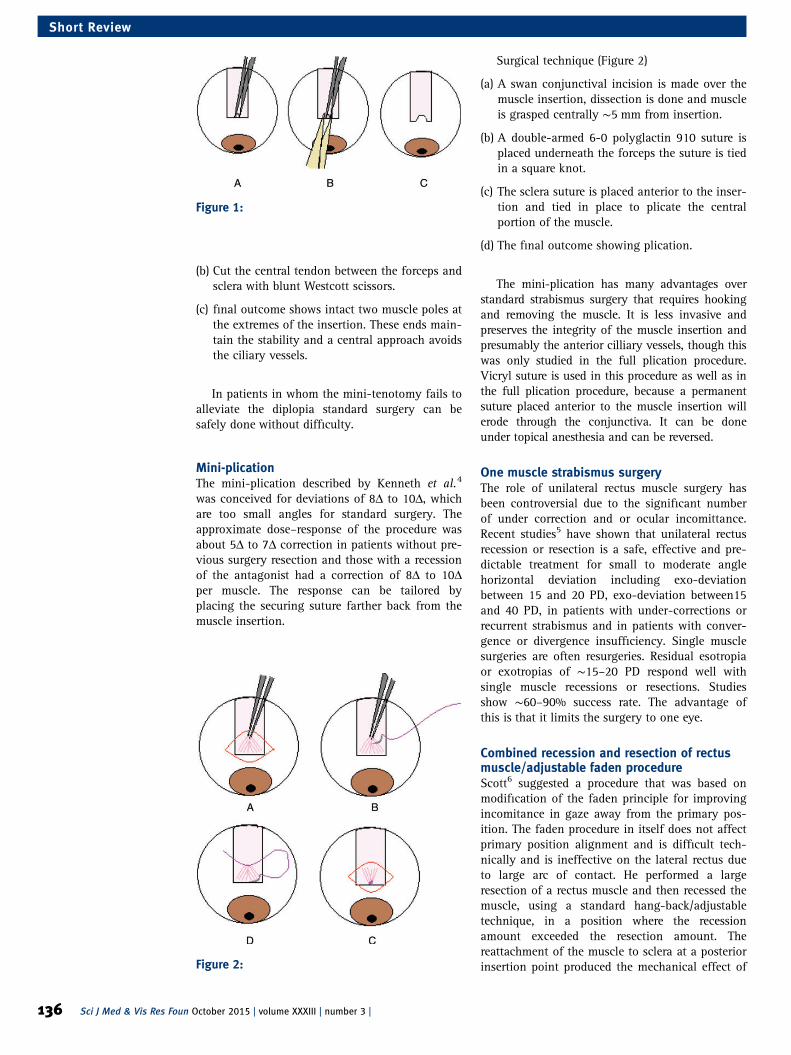
(b) Cut the central tendon between the forceps andsclera with blunt Westcott scissors.
(c) final outcome shows intact two muscle poles atthe extremes of the insertion. These ends main-tain the stability and a central approach avoidsthe ciliary vessels.
In patients in whom the mini-tenotomy fails toalleviate the diplopia standard surgery can besafely done without difficulty.
Mini-plicationThe mini-plication described by Kenneth et al.4
was conceived for deviations of 8Δ to 10Δ, whichare too small angles for standard surgery. Theapproximate dose–response of the procedure wasabout 5Δ to 7Δ correction in patients without pre-vious surgery resection and those with a recessionof the antagonist had a correction of 8Δ to 10Δper muscle. The response can be tailored byplacing the securing suture farther back from themuscle insertion.
Surgical technique (Figure 2)
(a) A swan conjunctival incision is made over themuscle insertion, dissection is done and muscleis grasped centrally ∼5 mm from insertion.
(b) A double-armed 6-0 polyglactin 910 suture isplaced underneath the forceps the suture is tiedin a square knot.
(c) The sclera suture is placed anterior to the inser-tion and tied in place to plicate the centralportion of the muscle.
(d) The final outcome showing plication.
The mini-plication has many advantages overstandard strabismus surgery that requires hookingand removing the muscle. It is less invasive andpreserves the integrity of the muscle insertion andpresumably the anterior cilliary vessels, though thiswas only studied in the full plication procedure.Vicryl suture is used in this procedure as well as inthe full plication procedure, because a permanentsuture placed anterior to the muscle insertion willerode through the conjunctiva. It can be doneunder topical anesthesia and can be reversed.
One muscle strabismus surgeryThe role of unilateral rectus muscle surgery hasbeen controversial due to the significant numberof under correction and or ocular incomittance.Recent studies5 have shown that unilateral rectusrecession or resection is a safe, effective and pre-dictable treatment for small to moderate anglehorizontal deviation including exo-deviationbetween 15 and 20 PD, exo-deviation between15and 40 PD, in patients with under-corrections orrecurrent strabismus and in patients with conver-gence or divergence insufficiency. Single musclesurgeries are often resurgeries. Residual esotropiaor exotropias of ∼15–20 PD respond well withsingle muscle recessions or resections. Studiesshow ∼60–90% success rate. The advantage ofthis is that it limits the surgery to one eye.
Combined recession and resection of rectusmuscle/adjustable faden procedureScott6 suggested a procedure that was based onmodification of the faden principle for improvingincomitance in gaze away from the primary pos-ition. The faden procedure in itself does not affectprimary position alignment and is difficult tech-nically and is ineffective on the lateral rectus dueto large arc of contact. He performed a largeresection of a rectus muscle and then recessed themuscle, using a standard hang-back/adjustabletechnique, in a position where the recessionamount exceeded the resection amount. Thereattachment of the muscle to sclera at a posteriorinsertion point produced the mechanical effect of
Figure 1:
Figure 2:
136 Sci J Med & Vis Res Foun October 2015 | volume XXXIII | number 3 |
Short Review
faden operation. The combined recession–resectionprocedure has a useful role in the management ofsymptomatic incomitant strabismus. The procedureis particularly valuable when dealing with incomi-tance on lateral gaze due to limitation of adduc-tion, when the overacting muscle is thecontralateral lateral rectus muscle. It seems to bean effective procedure in expanding the field ofsingle binocular vision.
ConclusionThe various causes of small angle strabismusinclude microtropia, residual deviations afteroptical and surgical corrections, intermittentsquints, sensory micro-strabismus and pareticstrabismus. Normal divergence and vertical fusionamplitudes are small, with divergence measuring4–6 PD (Δ) and vertical vergence <2 D. Because ofthis, even small eso-deviations and vertical devia-tions can cause significant asthenopic symptomsand diplopia, especially when the deviation isacquired in adulthood. Historically, thesesmall-angle deviations have been treated withprism glasses. Prism spectacles are an excellentoption if the patient is wearing spectacles for arefractive error. Prisms do not correct incomitanceand for many patients prism spectacles areundesirable.
Management of patients with small to moder-ate angle horizontal and vertical deviation con-tinues to be challenging for the strabismussurgeon. These lesser invasive strabismus surgeryprovides a valuable option to our strabismuspatients with small deviations. The various surgi-cal techniques described can be used to treat
diplopia in patients with small angle strabismuswho are not ideal candidates for conservativemanagement. These procedures can be done in theoffice under topical or local anesthesia andprovide an alternative to prisms and the standardstrabismus surgery. The key to success is carefulpatient selection, analyzing the critical factors likethe angle type-vertical/horizontal, angle size,angle character—phoria/tropia. Most of the surger-ies described are muscle weakening procedurealthough the weakening effect is limited. The pro-cedures described can be useful addition to a stra-bismus surgeons repertoire.
References1. Scott AB. Graded rectus muscle tenotomy for small deviations.
Proceedings of the Jampolsky Festschrift. San Francisco: TheSmith-Kettlewell Eye Research Institute; 2000: 215–216.
2. Albert W, Biglan , Hye Bin Yim. Graded partial tenotomy ofvertical rectus muscle for treatment of hypertropia. Trans AmOphthalmol Soc 2004; 102: 169–176.
3. Kenneth W Wright. Mini-tenotomy procedure to correct diplopiaassociated with small-angle strabismus. Trans Am OphthalmolSoc 2009; 107: 97–103.
4. Rebecca S Leenheer, Kenneth W Wright. Mini-plication to treatsmall-angle strabismus: a minimally invasive procedure.J AAPOS 2012; 16: 327–330.
5. Lihua Wang, Leonard B Nelson. One muscle strabismus surgery.Curr Opin Ophthalmol 2010; 21: 335–3401.
6. Scott AB. Posterior fixation adjustable and without posteriorsutures. In: Lennerstrand G, ed. Proceedings VIIth congressInternational Strabismological Association. CRC Press; 1995; pp.399–402.
7. van der Muelen-Schot HM, van der Muelen-Schot SB,Simonsz HJ. Caudal or cranial partial tenotomy of the horizontalrectus muscles in A and V pattern strabismus. Br J Ophthalmol2008; 92: 245–251.
How to cite this article Akila Ramkumar V, Subhedar KS. Surgical management of small angle strabismus, Sci J Med& Vis Res Foun 2015;XXXIII:135–137.
Sci J Med & Vis Res Foun October 2015 | volume XXXIII | number 3 | 137
Short Review
An approach to Nystagmus management
Dr N Kavitha Kalaivani
When it comes to a patient presenting with nys-tagmus there are a few issues that come into con-sideration. The main issues are whether thenystagmus is sensory or motor, whether it is thecause or the effect of vision problem, whether toinvestigate or not and whether to treat or not. Toknow whether the nystagmus is sensory or motorwe should know the range of vision impairment,if associated diurnal changes, if progressive orstable and the presence of other associated symp-toms like photophobia or oculo-digital sign, etc.Once the sensory causes have been addressed thencomes the issue of actually treating the nystagmusper se. Treatment of nystagmus actually onlyimplies improving the major two effects of nys-tagmus namely abnormal head posture due to thepresence of a null zone and improvement invision due to the frequency of the nystagmus. So,all the modalities of treatment address these twoaspects.
Non-surgical management of nystagmus
(A) Optical methods
1. Glasses:Effort should be made to correct any under-lying refractive error which will decrease thenystagmus.
Retinoscopy may be difficult to performaccurately when the nystagmus amplitude islarge and should be performed with the eyesin the null zone if such is present.
2. Contact lenses:Contact lenses have been reported to havereduced amplitude and frequency and arehelpful in high ammetropias.
It has the optical advantage of movingsynchronously with the eyes so that the visualaxis coincides with the optical center of thelens at all times and shows improvement invisual acuity.
There is a theory that some kind of tactilefeedback from the contact lens decreases thenystagmus possibly mediated via trigeminalafferents.
3. Over minus lenses:Adding concave glasses to distant correctioninduces artificial accommodation that isaccompanied with secondary convergence.This induced convergence diminishes ampli-tude and rate of nystagmus thus enhancingvision. Overcorrection with minus lenses sti-mulates accommodative convergence and may
improve visual acuity at distance fixation bynystagmus dampening.
4. Prisms:Prisms are used for two purposes in the treat-ment of nystagmus: (1) to improve visualacuity and (2) to eliminate an anomaloushead posture.
(a) Convergence induced:In patients whose nystagmus is suppressedby viewing a near target, convergenceprisms will often improve vision. Base-outprisms are prescribed to stimulate fusionalconvergence, which may be effective indecreasing the amplitude of nystagmusand thus improving visual acuity. Thedampening of nystagmus allows ‘‘clearvision at a glance,’’ removing the necessityfor increased visual concentration andthereby avoiding intensification of the nys-tagmus resulting from that heightened fix-ation. Congenital nystagmus responds wellto it. Normal binocular vision is a pre-requisite of the use of prisms base-outsince fusional convergence in response toprism-induced temporal retinal disparitycannot be expected in patients withoutfusion. In many patients, the disadvan-tages of prisms outweighs the modestvisual benefit gained.
(b) Induced divergence:Some patients with acquired nystagmusand in patients whose nystagmus is worseduring near viewing, base-in prisms mayhelp which induce divergence.
(c) Moving the null point:Prisms with base opposite to preferreddirection of gaze may be helpful in cor-recting the head posture. For example, ina patient with head turn to the left, thenull zone is in dextroversion and a prismbase-in before the right eye and base-outbefore the left eye will be helpful in cor-recting the abnormal head posture.
(d) Preoperative evaluation:The prisms are inserted with the baseopposite the preferred direction of gaze.For instance, with a head turn to the left,the null zone is in dextroversion, and aprism base-in before the right eye andbase-out before the left eye will correctthe head turn. Likewise, a compensatory
Senior Consultant, PediatricOphthalmolgy, SankaraNethralaya, Chennai, India.Email: [email protected]
138 Sci J Med & Vis Res Foun October 2015 | volume XXXIII | number 3 |
Short Review
chin elevation caused by a null zone indeorsumversion will be improved withprisms base-up before each eye. A com-bination of vertical and horizontalprisms can be used when the null zone isin an oblique position of gaze. Thus, theresults of surgery for head turn in nys-tagmus can be reasonably well predictedon the basis of the patient’s response toprisms, and a postoperative residual headturn may be alleviated further withprisms.
(B) Optically coupled deviceRushton and Cox described an optical system thatstabilizes images upon the retina. This system con-sists of a high-positive-power spectacle lens wornin combination with a high-negative-powercontact lens. The system rests on the principle thatstabilization of images on the retina is achieved ifthe spectacle lens focuses the primary image closeto the center of rotation of the eye. Such images,however, are defocused, and a contact lens isrequired to extend back the focus onto the retina.Since the contact lens moves with the eye, it doesnot negate the effect of retinal image stabilizationproduced by the spectacle lens. With such asystem, it is possible to achieve up to 90% stabil-ization of images upon the retina.
Disadvantage is that it disables all eye move-ments (including the vestibulo-ocular reflex andvergence), so that it is only useful while thepatient is stationary and views monocularly. Fieldof view is limited. Patients with ataxia or tremor(such as those with multiple sclerosis) have diffi-culty inserting the contact lens.
(C) Electrical devices
1. Movement based:A more recent innovation is to use an elec-tronic circuit to distinguish between the nys-tagmus oscillations and normal eyemovements. This approach is most useful inpatients with pendular nystagmus. Eye move-ments are measured with an infrared sensorand fed to a phase-locked loop that generatesa signal similar to the nystagmus but isinsensitive to other eye movements, such assaccades. This electronic signal is then used torotate Risley prisms, through which thepatient views the world. When the Risleyprisms rotate in synchrony with the patient’snystagmus, they nullify the visual effects ofthe ocular oscillations. Improvement andminiaturization of a prototype device mayeventually yield spectacles that selectivelycancel out the visual effects of pathologicalnystagmus.
2. Biofeedback based:Electrical stimulation or vibration over theforehead or neck may suppress congenitalnystagmus, again possibly by an action onthe trigeminal system, which receives extraocular proprioception.
(D) AcupunctureAcupunture consisting of the insertion of needlesinto the sternocleidomastoid muscle, has beenshown to improve foveation characteristics in con-genital nystagmus on a temporary basis.
(E) Botulinum toxinInjection of botulinum toxin into either the extrao-cular muscles or the retrobulbar space has beenreported to reduce nystagmus and improve visionin some patients. Limitations of this approach arethe short period of action (2–3 months), ptosis anddiplopia, which may be more annoying to patientsthan visual symptoms due to the nystagmus. Insome patients, the nystagmus may become worse inthe non-injected eye, if the patient prefers to viewwith the injected eye. This is because botulinumtoxin weakens all types of eye movement, not justthe nystagmus. This paresis of normal movementsstimulates the brain to make adaptive changes byincreasing innervation that may worsen the nystag-mus in the non-injected eye.
(F) DrugsAdvocated to treat congenital nystagmus, andimprovement of visual acuity has been reported insome instances.
Drugs not preferred because of their sideeffects and need for prolonged treatment. Ex:Gabapentin, Scopolamine, BaclofenIsoniazid,Memantine, Carbamazepine, Clonazepam,Barbiturates, Valproate, Alcohol, Trihexyphenidyl,Cannabis, Benztropine, Acetazolamide.
Surgical managementIndications for surgical intervention:
1. Large face turns—more than 40 degrees.
2. Associated with strabismus.
3. Successful prism adaptation.
Principles of surgical management:
(a) To improve head posture—move the eyestoward the null position.1. Kestenbaum procedure.
2. Augmented Kestenbaum procedure.
3. Modified Anderson procedure—the twomuscle recession.
(b) To improve the visual acuity.
1. Four muscle recessions.
Sci J Med & Vis Res Foun October 2015 | volume XXXIII | number 3 | 139
Short Review
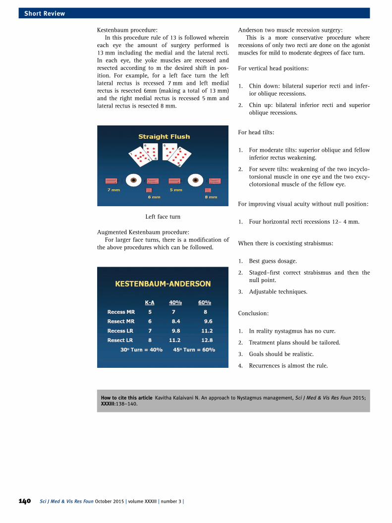
Kestenbaum procedure:In this procedure rule of 13 is followed wherein
each eye the amount of surgery performed is13 mm including the medial and the lateral recti.In each eye, the yoke muscles are recessed andresected according to m the desired shift in pos-ition. For example, for a left face turn the leftlateral rectus is recessed 7 mm and left medialrectus is resected 6mm (making a total of 13 mm)and the right medial rectus is recessed 5 mm andlateral rectus is resected 8 mm.
Left face turn
Augmented Kestenbaum procedure:For larger face turns, there is a modification of
the above procedures which can be followed.
Anderson two muscle recession surgery:This is a more conservative procedure where
recessions of only two recti are done on the agonistmuscles for mild to moderate degrees of face turn.
For vertical head positions:
1. Chin down: bilateral superior recti and infer-ior oblique recessions.
2. Chin up: bilateral inferior recti and superioroblique recessions.
For head tilts:
1. For moderate tilts: superior oblique and fellowinferior rectus weakening.
2. For severe tilts: weakening of the two incyclo-torsional muscle in one eye and the two excy-clotorsional muscle of the fellow eye.
For improving visual acuity without null position:
1. Four horizontal recti recessions 12– 4 mm.
When there is coexisting strabismus:
1. Best guess dosage.
2. Staged–first correct strabismus and then thenull point.
3. Adjustable techniques.
Conclusion:
1. In reality nystagmus has no cure.
2. Treatment plans should be tailored.
3. Goals should be realistic.
4. Recurrences is almost the rule.
How to cite this article Kavitha Kalaivani N. An approach to Nystagmus management, Sci J Med & Vis Res Foun 2015;XXXIII:138–140.
140 Sci J Med & Vis Res Foun October 2015 | volume XXXIII | number 3 |
Short Review

Pediatric low vision care services: a new era
Sarika Gopalakrishnan and Sumita Agarkar
IntroductionThe major causes of blindness in children varywidely from region to region, being largely deter-mined by socioeconomic development, and theavailability of primary health care and low visioncare services.1,3 The prevalence of blindness inchildren ranges from ∼0.3/1000 children in afflu-ent regions to 1.5/1000 in the poorest communi-ties. Reliable population-based data on the causesof blindness in children are difficult to obtain indeveloping countries.2 There is an increasingawareness about the needs of students with lowvision, particularly in developing countries whereprograms of integrated education are being devel-oped. However, the appropriate low vision servicesare usually mandatory in order to improve theirresidual vision.
Definition of low visionA child with low vision is one who has impairmentof visual functioning even after treatment and/orstandard refractive correction, and has a visualacuity of <6/18 to light perception, or a visual fieldof <10° from the point of fixation in the better eye,
but who uses, or is potentially able to use, visionfor the planning and/or execution of a task forwhich vision is essential (WHO, 1992).
Impact of low visionFunctionally, low vision is described as irreversiblevisual loss and a reduced ability to perform manydaily activities, such as recognizing people in thestreet, reading blackboards, writing at the samespeed as peers, and playing with friends.
Causes of low visionThe major causes of visual impairment amongchildren includes Stargardt’s disease, Myopicdegeneration, Oculocutaneous Albinism, RetinitisPigmentosa, Maculopathy, Optic Atrophy, Cornealopacities, Rod cone dystrophy and other heredo-macular degenerations.3
In the year 2010The mean number of children seen in the LowVision Care clinic per year was found to be 1500.The referral criteria included children with lowvision and constricted field of vision.
Approach Before 2010 After 2010
History General history, family historyand birth history
Classroom environment history, academicperformance
Vision assessment Regular method of visionassessment with usual logMARchart
Functional vision assessment for pre-verbalchildren with Cardiff acuity card, rudimentaryvision kit and LEA symbols
Management Usually spectacles or opticalmagnifiers would be the choice
Additionally a wide range of non-optical devicesand electronic assistive devices are available
Advice Generalized case summarywould be given
Specific letter to school management is beinggiven based on the needs (e.g. seatingarrangements, compensatory time duringexaminations, use of low vision devices, etc.)
Referral Most of the times children willbe referred to blind school
Children will be referred to integrated orinclusive mode of schooling
New initiatives
1. Awareness on referral criteria among eyecare professional including Ophthalmologistswas created.
2. The range of optical devices and non-opticaldevices were extended significantly, so thatappropriate magnification was chosen fortrial and final prescription.
3. Latest varieties of electronic assistive deviceswere introduced from different parts of theworld.
4. Tools and devices used for daily living activ-ities were included in the stock.
5. All the devices were purchased from all overthe world and for lower cost.
6. Primary low vision care service was intro-duced in all the floors and branches of thetertiary eye care center, so that more numberof patients got benefited.
7. Organizational membership was received fromBookshare for accessing DAISY books, whichcan be useful for patients with print disabilities.
Sci J Med & Vis Res Foun October 2015 | volume XXXIII | number 3 | 141
Update

8. Psychological counseling on self-motivationand need for special schooling are being pro-vided to the parents whose children are visuallyimpaired.
9. Braille training and Computer JAWS soft-ware demonstration are provided for childrenwith low vision.
10. Smart Cane demonstration for orientationand mobility training.
In the year 2015The mean number of children seen per year hasincreased to 3200 which is twice the amount seen5 years back. The referral criteria have beenextended such that pre-schoolers are also referredto the low vision care clinic in order to decidetheir mode of education and for appropriateguidance.
Transformation in the trend
Sl.No
Visual task Before 2010 2010–2014 From 2015
1. Viewing board Approachmagnification
Conventional telescope ONYX electronicassistive device
2. Reading tasks Bifocal spectacles Dome magnifier Mouse model CCTV/portable CCTV
3. Writing tasks Cut away standmagnifier
Spectacle magnifier Quick look zoom CCTV
4. Watching TV Approachmagnification
SEE TV binoculartelescope
Snow 7HD videomagnifier
5. Computer External screenmagnifier
In-built modifications/JAWS/MAGIC
Newer software/latesttechnologies
6. Mobile Tactile cues forkeypad
TALKS In-built applications/android accessibilitysettings
7. Non-optical devices Four linednotebooks
Typoscope/letter writer Thick lined notebooks
8. Writing tool Scribe Writing in larger size Bold pencils/bold pens
9. Activities of dailyliving
Time calculation Help from others Large dialhigh-contrast clock
Talking clock
Color identification Help from others Color labeling ofpencils/pens
Color recognizer
Currency identification Help from others Notex Look tel money readerAPP
10. Recreation Nil Increased taskillumination
Jumbo playing cards
Mode of schooling
1. Mainstream: School with normal childrenwithout any disability.
2. Integrated school: A school where both nor-mally sighted and visually impaired studentswill be studying in the same class.(a) Resource model: The school is provided
with the resource set up, where a teacherwill be available all the day. During thefree hours, the students with visualimpairment go to the resource room forguidance.
(b) Itinerant model: The Resource teacher(Special Educator) goes to different
schools and visually impaired children’splace to teach them.
3. Inclusive school: Mild to moderate form of alltypes of differently abled children will beincluded along with the normal children.
4. Special school: A school for visually impaired/deaf-mutism/mentally retarded/learning dis-ability/any specific disability.
Rights and considerationScholarship
1. The Union Ministry of Welfare since 1955 hasbeen operating through the state Governments
142 Sci J Med & Vis Res Foun October 2015 | volume XXXIII | number 3 |
Update

and Union territories a scheme of scholarshipsawarded to challenged person for pursuingeducation in special schools being run bynon-government organizations.
2. The Rate of scholarships is Rs.1000/- perannum for cases hailing from the lower socio-economic status and is renewable from year toyear.
3. In case of severely challenged persons whorequire special arrangements for transporta-tion, an additional monthly allowance ofRs.50/- is sanctioned.
The following allowances and facilities are providedunder this scheme
1. Books and Stationary allowance of Rs.400/-per annum.
2. Uniform allowance of Rs. 50/- per annum.
3. Transport allowance of Rs.50/- per month (ifa challenged child admitted under the schemeresides in a hostel of the school within theschool premises, no transportation chargeswould be admissible).
4. Reader allowance of Rs.50/- per month incase of blind children after class V.
5. Escort allowance for severely handicappedchildren with lower extremity disabilities atthe rate of Rs.75/- per month.
6. Annual cost of equipment subject to amaximum of Rs.2000/- per student for aperiod of 5 years.
7. The tuition payable and actually paid by theGovernment servant is reimbursable subject toRs.50/- per month per child in the case ofmultiple disabled children.
Railways
1. 75% concession in the basic fare in the firstand second class is allowed to persons withmultiple disabilities accompanied by anescort.
2. 50% concession in the first and second classmonthly/quarterly season fares both for theindividual with disability and his/her escortover suburban and non-suburban section ofIndian railways is allowed.
Roadways
1. Most State Governments having state ownedand operated transport undertakings or cor-porations allow subsidized/free bus travel in
the city and rural routes, and an escort ischarged fifty percent of the fare.
ConclusionMajority of the students with low vision need lowvision services in order to perform better.Accurate refraction is important in the students.Early diagnosis and intervention helps in prevent-ing the vision loss which will lead the students tocontinue their academic activities without anyinterference. Many children need additionalsupport from the Government in order to continuetheir education. The need for awareness on man-agement and referral of children with Low visionhas to be improved in this Millennium due toincreasing number of children who discontinuetheir schooling due to Low vision. Awarenessamong eye care professionals and parents is man-datory for bringing up these children with Lowvision.
References1. Gilbert Clare AllenFoster. Childhood blindness in the context of
VISION 2020: the right to sight. Bull World Health Organ 2001;79.3: 227–232.
2. Sharma MK, et al. A profile of low vision among the blindschool students in Lumbini Zone of Nepal. Nepalese JOphthalmol 2010; 2.2: 127–131.
3. Shah Mufarriq, et al. Causes of visual impairment in childrenwith low vision. J Coll Phys Surg Pak 2011; 21.2: 88–92.
Case 1:General history: A 14-year-old female diagnosedwith High Myopia and associated retinal findingof dry macula (Myopic Retinal Degeneration) wasreferred to the Low Vision Care Clinic as she haddiscontinued school 3 months back. She wasaccompanied by her parents. There was no familyhistory of parental consanguinity. Her youngersister had myopia (around -3.00D and no visualimpairment). There was no other relevant history.
Previous low vision care: nil
Present complaints:
Distance visual tasks: The patient had difficultyseeing the blackboard and recognizing the facesof people who were more than 3 m away. Shewatched television from a distance of 1 m.
Near visual tasks: The patient was able tomanage at a close working distance of around15 cm.
ADL/mobility: These tasks were independent.
Light sensitivity: There were no complaintsregarding light sensitivity.
Additional history: Classroom environment
School: Private school (English medium), StateBoard of Education syllabus.
Sci J Med & Vis Res Foun October 2015 | volume XXXIII | number 3 | 143
Update

Medium of teaching: English.
Class strength: 30.
Academic performance of patient: Average (50–60%).
Reason for discontinuing school: Unable to seeblackboard from front bench at a distance of 3 m.
Parents say teacher wrote letters of size aboutone finger length (∼2.5 inches or 6–7 cms).
Color of board: Black.
Letters: White chalk used, colored chalk: rare.
Class lighting: Sufficient (no glare and no com-plaints of dim/poor lighting, including cloudydays).
Internal lighting present in class: three fluores-cent lights.
External lighting: three large windows.
Seating position: Third row, center.(No flexibility in seating, not allowed to copy
or reference friends notebook).
Visual requirements: Seeing blackboard.
Examination:
Previous glass prescription:OD: −13.50 DS/− 2.00C × 50,OS: −12.00 DS/ − 1.25 DC × 45.
Distance visual acuity with glasses:OD: 3/30+2, 6/60+2,OS: 3/19, 6/38,B/O: 3/19, 6/38.
Near visual acuity with glasses: OD/OS/B/O: N8@ 15 cm.
Reading speed: 40 wpm.
Unaided near visual acuity: B/O: N5 @ 10 cm
Reading speed: 60 wpm.
Refraction:
Retinoscopy:OD: −11.50DS/ − 1.00 DC × 60,OS: −11.25 DS/ − 1.25 DC × 120.
Dynamic retinoscopy: (MEM)
Accommodative Lag: +1.00 DS to +1.25DS
Subjective refractionOD: −11.50DS/ − 1.00 DC × 60 [3/9.5, 6/19],OS: −11.25 DS/ − 1.25 DC × 120 [3/9.5, 6/19].
ADD: OU: Does not prefer ADD
Unaided near visual acuity: N5 with ease @10 cms
Low contrast visual acuity: 3/19 [6/38] @Borderline impairment.
D15: Normal,
Amsler: No scotoma detected,
Confrontation: Normal peripheral fields.
LVD Trial for distance:
Required visual acuity: 6/6
Magnification required: 19/6 = 3 ×
Trial 1: 3× Monocular Handheld Telescope: 6/6with ease.
Telescope training was given—able to localize,hold fixation, scan, track and copy from text onsample blackboard.
Copies text at speed of 30–40 wpm.
LVD trial for near: Reads up to N8 withcorrection.
Reads up to N4 unaided.
Trial 1: Add +1.00DS to +5.00DS tried in 1.00Dsteps. Patient does not report significant differ-ence/improvement
Trial 2: With 4× Dome Magnifier: N4; Reads withease @ 40 cms Reading speed: 80 wpm.
Final Rx:
1. New R× for distance.
2. 3× Monocular handheld telescope for use withblackboard.
3. To use dome magnifier for prolonged readingtasks, to reduce eyestrain, reference atlas,maps, dictionaries or fine print.
4. To use reading stand for prolonged readingtasks to aid posture and maintain workingdistance.
Other advice: The condition and prognosis wereclearly explained to the parents. The parents werereassured about closer working distance and theneed to use a magnifier for comfortable reading atclose working distance was explained. The childwas advised to continue her schooling with thehelp of low vision care. They were also counseledabout available options for computer modificationand magnification.
Case 2:General history: An 8-year-old-Female diagnosedwith Congenital Stationary Night Blindness wasaccompanied by her parents to the low vision careclinic. Her difficulty with dimly lit environmentwas noticed by the parents at around 1 year ofage. She was staying with her parents and twoelder brothers. She was currently studying in StdII. Her parents did not have a consanguineousmarriage but her mother’s age was around 34when the child was born. Her father who was
144 Sci J Med & Vis Res Foun October 2015 | volume XXXIII | number 3 |
Update

working in an automobile assembly company wasthe source of family income.
Previous history: She had no history of using anylow vision devices.
External observation showed she had good fix-ation but difficulty in searching for a pen amongvarious items placed on the table.
Present complaints:
Distance visual tasks: Difficulty with seeing theblackboard in the classroom (3 m), seeing objectsat distance while sightseeing. She was able tomanage all near acuity tasks at a closer workingdistance.
Mobility and ADL: She was able to move aroundindependently but had difficulty in dim light andtook long time to adjust to dimly lit surroundings.She had independent Activities of Daily Livingwithin the limitations of her age.
Additional history: Classroom environment:
1. Seated at 3 m from blackboard.
2. Teacher uses white chalk. Colored chalk usedoccasionally.
3. Allowed to come close to the board.
4. No note-taking until class IV in the school.
5. Artificial room lighting—five fluorescent lights.
6. Class strength: 40 students.
7. Medium of instruction: English.
8. Type of school: Private.
9. Syllabus: State board.
Patient requirement: Seeing blackboard.
Examination:
Visual acuity:Distance Visual Acuity with Bailey Lovie
LogMAR chart: OD/OS/B/o: 3/12 [6/24]Near visual acuity: N10 @25 cm with the MN
Read chart at 10 wpmgood reading skills.There was no difference between monocular
and binocular visual acuities. There was no sig-nificant refractive error on objective refraction.
Dynamic retinoscopy: Accommodative lag:+1.00DS.
Cycloplegic refraction: No refractive error requir-ing correction detected.
Low contrast visual acuity with Bailey LovieLow Contrast LogMAR chart was 3/15[6/30](Within normal limits).
Color vision: Able to match a few colors on theD15 panel but could not comprehend the entire test.
Confrontation: Mild peripheral field deficitbeyond 50° visual field.
Trial of low vision devices(1) 4× monocular handheld telescope: 6/6 (no
specific preference for right or left eye)NearADD: OD/OS: +2.00DS: N4 @ 20 cm.The child seemed to be able to be able to read
better and reading speed improved to 25 wpm forfiner print (>N10 size).
Final R × :
1. New Bifocal R× with +2.00DS ADD.
2. The parents were advised to purchase overthe counter binoculars of 3× or moremagnification.
3. A letter was given to the school to provide aseparate seating arrangement closer to theblackboard at <2 m when required. (Currently,students were not required to copy any notesfrom the blackboard.)
Advice:The child was advised to be seated in a separate
chair (in front of the front row) closer to board at∼2 m. Letter was given to school managementrecommending closer seating arrangement,increased lighting level and compensatory timefor completing her examinations. The parentswere advised to increase the illumination level athome especially during night time. The childneeds to be instructed on caution regardingmobility tasks. Option of low vision devices wereexplained to parents which will be beneficial inthe future. The patient was advised to come for anannual low vision care visit with her school booksand class notes.
Sci J Med & Vis Res Foun October 2015 | volume XXXIII | number 3 | 145
Update

APPENDIX 1
CLASS ROOM ENVIRONMENT
Standard: Informant:
Mode of education: a) Special school b) Regular school c) Private tutor
School name/College name:
Class teacher/Principal name:
Address of the school/ college:
1. Class strength:
2. Seating position: Center/Corner
3. Approx distance from board:
4. Which bench/row:
5. Color of writing material on board:
6. Color of the board:
7. Number of windows in class:
8. Sitting beside window:
9. Amount of light in class: Dimly illuminated/Optimally illuminated/Glaring (too bright)
Academic performance:
Good peer-group interaction: Yes/No Good parent–teacher interaction: Yes / No
Is there any difficulty in copying from black board: Yes/No
If yes: managing by:
Copying from friends/Going close to the black board/Taking friends note-booksto home/Teacher’s dictation.
Others:
Using extra hours for writing exams: Yes/No
Use of special assistance: Yes/No
If yes: Scribe/Braille/Talking books/Audio cassettes
Others:
Since how long:
Any difficulty with special assistance:
Is the teacher co-operative and ready to give individual attention: Yes/No/NA:
Will the school management be ready to make changes in the setup: Yes/No/NA
Letter to the class teacher required: Yes/No/NA
146 Sci J Med & Vis Res Foun October 2015 | volume XXXIII | number 3 |
Update

APPENDIX 2
LETTER TO SCHOOL MANAGEMENT
LOW VISION CARE CLINIC Date:
TO WHOMSOEVER IT MAY CONCERN
This is to state that Master/Miss.______________ who is studying ___STD in your school (ReferenceMRD No.: __________ ) was evaluated in the Low Vision Care Clinic. He was noted to have _________Visual Impairment in both the eyes due to retinal problem.
This vision will not be sufficient to view the black board comfortably. He needs to be seated closer tothe black board (i.e. 1 m distance) or in the front row (without rotation). Most of his learning will bethrough listening to the classes, so dictating as much as possible will help him in a large way.
Kindly, allow him to copy notes from his friend’s notebook in case he is not able to copy from the blackboard directly. In addition, he can be given compensatory time concession of minimum 30 min for allexaminations (school level and for board exams).
Please extend your co-operation so that his ocular condition doesn’t interfere with his academicperformance.
Thank you for your kind co-operation.
Yours sincerely,(_________________)OptometristLow Vision Care ClinicSankara NethralayaChennaiTamil NaduPhone: 044-42271519
How to cite this article Gopalakrishnan S, Agarkar S. Pediatric low vision care services: a new era, Sci J Med & VisRes Foun 2015;XXXIII:141–147.
Sci J Med & Vis Res Foun October 2015 | volume XXXIII | number 3 | 147
Update


Cortical visual impairment
Preeti Patil Chhablani
Cortical visual impairment (CVI) is emerging asone of the important causes of blindness or visualimpairment in India. This increasing trend is likelyto continue as further advances in the field of neo-natology and fetal medicine increase the chances ofsurvival of a premature infant or an infant withmultiple congenital abnormalities. Previous studiesfrom developed nations have shown that infantsborn prematurely and those with low birth weightare at a risk of severe ophthalmic impairment.1,2
What is CVI?The current definition of cerebral or CVI includesall visual dysfunctions caused by damage to ormalfunctioning of the retrochiasmatic visual path-ways in the absence of damage to the anteriorvisual pathways, or any major ocular disease.3
We need to remember that CVI does not implytotal blindness and is a spectrum of vision lossranging from mild visual impairment to completevisual deprivation.
PathogenesisThe central nervous system in a newborn is sus-ceptible to various damaging influences such ashypoxia–anoxia, hypoglycemia or a combinationof both. Two distinct subgroups of cortical visualloss in terms of the areas of damage and the timeof injury have been described.4 Injury in full termneonates, predominantly involves the striate andperistriate cortex, whereas in preterm neonates,injury involves the subcortical white matter, includ-ing the optic radiations. This results in different pat-terns of damage as seen on neuroimaging and alsoresults in different clinical manifestations.
In a full term neonate, the vascular supply ofthe brain is derived from the major cerebral arter-ies and its watershed areas lie at the interfacesbetween the major cerebral arterial distributions.5–7
Hence, any hypoxic-ischemic injury produceswatershed infarctions in the parieto-occipital andparasagittal cortex, resulting in cortical visualloss. Whereas, in the developing brain, the cortexand underlying white matter receive their bloodsupply from branches of the blood vessels on thesurface of the cerebral hemispheres and the water-shed zone lies within the periventricular whitematter.8 Hence, preterm injury to the brain resultsin injury to the subcortical white matter, resultingin periventricular leukomalacia (PVL).8 Othercauses of injury in neonates include birth trauma,intraventricular hemorrhage and hypoglycemia.Neonatal hypoglycemia, in particular, in known tocause significant visual deficits and neuroimaging
in these cases shows restriction on diffusion pre-dominantly in the occipital areas.9
CausesA wide variety of insults to the central nervoussystem can result in CVI. These include: perinatalconditions such as birth trauma, hypoxia–anoxia,hypoglycemia and post-natal conditions such assepsis, meningitis, encephalitis, accidental andnon-accidental trauma, cerebral malformations suchas Chiari malformations, Dandy–Walker complex,hydranencephaly, porencephalic cysts, metabolic andneurodegenerative conditions such as MELAS (meta-bolic encephalopathy, lactic acidosis and strokelikeepisodes), Fabry’s disease, Leigh’s disease, X-linkedadrenoleukodystrophy and hydrocephalus.10
Clinical presentation and diagnosisAs mentioned previously, CVI can manifest as mildvisual loss with a slight delay in achievement ofvisual developmental milestones or as near total ortotal blindness. These babies are most frequentlybrought to the ophthalmologist with complaints of“not seeing the mother’s face”, poor visual responseto light, “shaking of eyes” or nystagmus.
These children often have multiple other disabil-ities and one must remember to make the child com-fortable before starting the examination. Responsesto bright light, colored objects and human faces mustbe assessed. Some of these babies may respond betterin a supine position and in dim light.
One must look for the presence of nystagmus,strabismus and ocular motility deficits and pupil-lary responses. Cycloplegic refraction must bedone along with a thorough fundus examination.
Nystagmus is rare or absent in children withCVI. However, roving eye movements or occa-sional bursts of nystagmus may be seen and thepresence of such eye movements signifies severevisual impairment.4 An intact geniculostriatepathway is a prerequisite for the development ofcongenital nystagmus and hence nystagmus isabsent in extensive posterior pathway disease.11
However, some children with combined anteriorand posterior visual pathway disease may presentwith CVI and nystagmus.12
Strabismus is common in children with CVI.Both esotropia and exotropia have been reported.Esotropia is more common in children with PVLand may be associated with latent nystagmus, dis-sociated vertical deviation and ‘A’ pattern.10 Otherstudies have shown that cortical visual loss isassociated with exotropia more commonly thanesotropia. Also, children with more extensive
Consultant, Paediatricophthalmology, Strabismus andNeuroophthalmology Service,L V Prasad Eye Institute,Kallam Anji Reddy Campus,Hyderabad, India
Correspondence:Preeti Patil Chhablani,Consultant, PaediatricOphthalmology, Strabismus andNeuroophthalmology Service,L V Prasad Eye Institute,Kallam Anji Reddy Campus,Hyderabad, India.Email: [email protected]
148 Sci J Med & Vis Res Foun October 2015 | volume XXXIII | number 3 |
Alumni Corner

neurological damage may have congenitalexotropia.10,12
Other ocular motor abnormalities that have beennoted in these children include tonic downgaze,horizontal conjugate gaze deviation and defectivesmooth pursuits and saccadic eye movements.10,13
Fundus examination is extremely important todetect the presence of associated optic atrophyand also to rule out other conditions with similarclinical presentation such as retinal dystrophies,congenital macular scars, sequelae of retinopathyof prematurity, etc.
In cases where the pathological processesaffecting the posterior visual pathways and thecerebral cortex have not affected the anteriorvisual pathway, the fundus is usually normal andthe disk appears healthy. However, studies involv-ing children with retrogeniculate vision loss, havedescribed a variety of optic nerve abnormalitiesincluding optic atrophy, optic nerve hypoplasiaand pseudoglaucomatous cupping.4,12
In additional to structural damage, one mustkeep in mind that these children also have signifi-cant functional deficits such as visual fielddefects, which include generalized field restriction,central scotomas, homonymous hemianopias andaltitudinal defects.10,12
One must keep in mind that vision is a complexfunction and has multiple components. These chil-dren may show other visual functional abnormalitieswhich may be difficult to quantify, such as difficul-ties in visual processing, difficulty in simultaneousperception and “crowding” phenomenon. Otherpeculiar visual traits include a tendency to stare atbright objects, such as fluorescent room lights or thesun. This is known as “light gazing” and is regardedas a sign of severe visual impairment.14
These children also, often function better in afamiliar environment, may show a better visualperformance for objects in motion as comparedwith static objects and are better at identificationof colors as compared to their perception of form.15
Some children also complain of photophobia.Diagnosis is confirmed by neuroimaging.
While imaging of the infant brain is possible byultrasound and computed tomography, magneticresonance imaging (MRI) is the preferred modality.MRI can help to pick up even the subtle changesof PVL such as abnormal dilatation and irregular-ity of the lateral ventricles, high intensity signalsin the periventricular white matter on T2 weightedimages and periventricular gliosis. Severe hypoxicinjury may lead to encephalomalacia with sur-rounding gliosis.10 Other abnormalities seeninclude thinning of the corpus callosum, alteredsignals from the thalamus and putamen and cere-bellar atrophy.10,12
It is not clearly known to what extent PVL andCVI and the associated neurological abnormalitiesaffect visual evoked potentials (VEP), but ingeneral, the absence of a flash VEP is associatedwith a poor prognosis.10
Electroretinogram may help by ruling outretinal dystrophies, which may sometimes presenta similar clinical picture.
ManagementEarly diagnosis and intervention is the key to man-agement in these cases. A team approach integrat-ing the services of a pediatric ophthalmologist,pediatrician, physiotherapist, speech therapist andvision therapist is desirable. Parental counseling isof utmost importance and one must remember thatwhile it is definitely important to provide theparents a realistic picture of their child’s condition,encouraging the parents to start rehabilitation andremain positive in their outlook may go a long waytoward bettering the child’s life.
References1. Cooke RW, Foulder-Hughes L, Newsham D, Clarke D. Ophthalmic
impairment at 7 years of age in children born very preterm. ArchDis Child Fetal Neonatal Ed 2004; 89: F249–F253.
2. Robaei D, Kifley A, Gole GA, Mitchell P. The impact of modestprematurity on visual function at age 6 years: findings from apopulation-based study. Arch Ophthalmol 2006; 124: 871–877.
3. Boot FH, Pel JJ, van der Steen J, Evenhuis HM. Cerebral visualimpairment: which perceptive visual dysfunctions can beexpected in children with brain damage? A systematic review.Res Dev Disabl 2010; 31: 1149–1159.
4. Brodsky MC, Fray KJ, Glasier CM. Perinatal cortical andsubcortical visual loss: mechanisms of injury and associatedophthalmologic signs. Ophthalmology 2002; 109: 85–94.
5. Flodmark O, Jan JE, Wong PK. Computed tomography of thebrains of children with cortical visual impairment. Dev MedChild Neurol 1990; 32: 611–620.
6. Barkovich A. Pediatric Neuroimaging. Philadelphia: LippincottWilliams & Wilkins, 2000.
7. Volpe J. Neurology of the Newborn, 3rd ed. Philadelphia: W.B.Saunders, 1995; 403–463.
8. Barkovich A. Pediatric Neuroimaging. Philadelphia: LippincottWilliams & Wilkins, 2000.
9. Tam EW, Widjaja E, Blaser SI, et al. Occipital lobe injury andcortical visual outcomes after neonatal hypoglycemia. Pediatrics2008; 122: 507–512.
10. Brodsky M. The Apparently Blind Infant. In PediatricNeuro-Ophthalmology, Edition. New York: Springer, 2010; 1–58.
11. Fielder AR, Evans NM. Is the geniculostriate system aprerequisite for nystagmus? Eye (Lond) 1988; 2 (Pt 6): 628–635.
12. Jacobson LK, Dutton GN. Periventricular leukomalacia: animportant cause of visual ocular motility dysfunction inchildren. Surv Ophthalmol 2000; 45: 1–13.
13. Jacobson L, Ygge J, Flodmark O. Nystagmus in periventricularleucomalacia. Br J Ophthalmol 1998; 82: 1026–1032.
14. Jan JE, Groenveld M, Sykanda AM. Light-gazing by visuallyimpaired children. Dev Med Child Neurol 1990; 32: 755–759.
15. Jan JE, Groenveld M, Sykanda AM, Hoyt CS. Behaviouralcharacteristics of children with permanent cortical visualimpairment. Dev Med Child Neurol 1987; 29: 571–576.
How to cite this article Chhablani PP. Cortical visual impairment, Sci J Med & Vis Res Foun 2015;XXXIII:148–149.
Sci J Med & Vis Res Foun October 2015 | volume XXXIII | number 3 | 149
Alumni Corner

List of fellows trained by Department of PediatricOphthalmology, Sankara Nethralaya
Year Initial Name
April 1999 HAM Dr Hemalini Mehta
April 2001 MK Dr Madhu Karna
April 2002 AMJ Dr Aarti Jhamwar
October 2002 KKV Dr Kavitha Kalaivani Natarajan
October 2003 LK Dr Lov Kochgaway (Orbis)
MSH Dr Manish Sharma (Orbis)
April 2004 ST Dr Satish Thomas (Orbis)
RSA Dr R Sudhindra (Orbis)
October 2004 SKL Dr Shalini Kaul (Orbis)
ASV Dr Anand S Vajawade (Orbis )
April 2005 HLV Dr Hemalatha Vidyashankar (Orbis)
RPH Dr Ramprakash (Orbis)
SSY Dr Sharath Shetty (Orbis)
October 2005 CB Dr Charuta Bhadre (Orbis)
CSZ Dr C Sadiq Zamir (Orbis)
AAT Dr Amol Anil Tamhane (Orbis)
October 2006 SRK Dr Sai Rani (Orbis)
KM Dr Kaushik Murali (Orbis)
VAS Dr Varshini Shankar (SN)
MRC Dr Mayee Rishikesh Charudatta (SN)
April 2007 NUV Dr Naik Uday Vithal (Orbis)
PSM Dr Prashant kumar S Murhe (Orbis)
October 2007 AMA Dr Amit Maitreya (Orbis)
BCK Dr Basavaraju C Kadiwal (SN)
April 2008 JGM Dr Jay Mahida (Orbis)
BPA Dr Bhujbal Pritamsingh Appasaheb (Orbis)
October 2008 ATK Dr Ajit Kumar Sinha (Orbis)
PNT Dr Parag N Tupe (Orbis)
October 2009 AGM Dr Amula Gangaprasad Muthaiah
PAP Dr Preeti Ajit Patil
April 2010 HGM Dr Harsha G Mangalgi
RBR Dr Resmi Bhaskar
October 2010 PBP Dr Prathiba Patil Bapusaheb (SNK)
SFK Dr Mohd Sarafraz Khan
RRA Dr Renu R Athanikar
April 2011 RSK Dr R Srikanth
VAR Dr Akila Ramkumar
RMA Dr Radhimalar Anand
SVS Dr Sanil Vijaybhai Shah (SNK)
October 2011 SDN Dr Sujay Siddharthan
AKR Dr Anushree Vijay Kaduskar
PMH Dr Preeti M Hurakadli (SNK)
April 2012 SGI Dr Shama Ali Goliyadi
NJC Dr Nidhi J Chopra
October 2012 RJD Dr Roshani Jaideep Desai
October 2012 STS Dr Sudipta Sahana
October 2012 SPR Dr Shalaka Ravindra Paralkar
October 2012 AE Dr Aarti Elhence
April 2013 MSR Dr Shruti Rajendra Mittal
April 2013 ARN Dr Anbarasi Sherren
October 2013 SNA Dr Soumya Nambiar
October 2013 JSP Dr Jayesh Suresh Patil
October 2013 KSH Dr Khyati Jain
April 2014 BRV Dr Bhatt Riddhi Vijay
April 2014 PPL Dr Prakash Parimal
April 2014 VVG Dr Varada Vinay Gokhle
150 Sci J Med & Vis Res Foun October 2015 | volume XXXIII | number 3 |

Last word
Lessons learnt in Paediatric ophthalmologyOpthalmic and otherwise!
Many summers back when I joined Sankara Nethralaya, as was the trend then I was trained in vitreoretinal procedures but somewhere down the line it was decided that I should do Paediatric ophthalmology. Thirty seven years later when I look back, it was a momentous decision that changed my life in many ways. I was fortunate to train under doyens of this fi eld like Dr Marshal Parks, Dr Arthur Jampolasky, Dr Alan Scott and Dr Prem Prakash. They were true visionaries and pioneers who changed the way world looked at Paediatric ophthalmology.
Being a Paediatric ophthalmologist has taught me lessons in patience, perseverance and hope. I have learned to communicate with non-verbal children, special children as well as parents and often grandparents too. I have learned to take in my stride when the room furniture gets rearranged by the patient while I talk to the patient. I am not surprised when an enveloped mail to me contains lenses from the trial set which parents have discovered after reaching home. I learnt not to assume anything when an elderly couple brought in a young child who turned out to be their sixteenth offspring. Similarly, I learnt to be non-judgmental when another of my overseas patient requested a squint correction so that he could marry a third time!
In a career spanning 37 years and still counting, there have been many changes in the way we practice ophthalmology, but one thing that has remained unchanged is the team work. Paediatric eye care, in particular, needs a team of optometrists, anaesthetists, paediatricians, nurses and counsellors. Together they make us look good as doctors. I am grateful for working with a dedicated team. Lest I forget, the team also includes parents and caregivers who need reassurance and empathy from us while being honest about the goals of treatment. It is their trust in us that allows us to work with children. It is a responsibility which should not sit lightly. This means constant vigilance, checking whether it is the correct eye, correct patient, correct surgery and then doing it all over again once more!
However, all said and done, if clock were to turn back, I am sure I will choose Paediatric ophthalmology all over again.
Vice President, Medical Research Foundation, Director, Paediatric ophthalmology, Sankara Nethralaya, Chennai
Correspondence to: Dr T S Surendran, Vice President, Medical Research Foundation, Sankara Nethralaya, ChennaiEmail: [email protected]
Dr T S Surendran
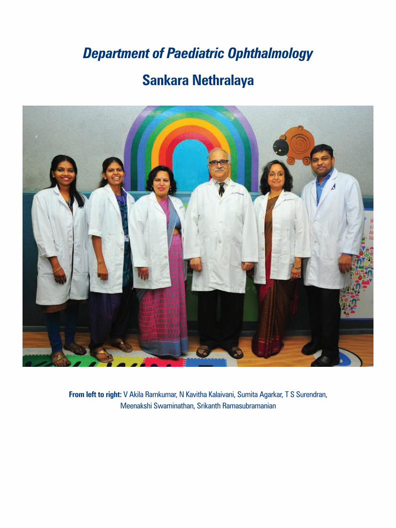
Department of Paediatric Ophthalmology
Sankara Nethralaya
From left to right: V Akila Ramkumar, N Kavitha Kalaivani, Sumita Agarkar, T S Surendran, Meenakshi Swaminathan, Srikanth Ramasubramanian