Embed Size (px)
Citation preview

Scientific Article
Treatment of fluorosed and white-spot humanenamel with calcium sucrose phosphate in vitroPamela Den Besten, DDS, MS Nina Giambro, DDS, MS
Abstract
A number of treatments have been devised to improvethe appearance of fluorosed enamel. However, many ofthese have been empirically based, and the success of thevarious treatment regimens have not been quantitated. Inthis study, the relative whiteness of normal, mildlyfluorosed, moderately fluorosed, and carious white-spotlesions on extracted teeth was quantitated by light reflec-tance using a Minolta Chroma Meter. The color was againdetermined following a number of treatment regimens toassess the potential use of various agents in treating theenamel lesions. Treatment of the enamel with a 35% hy-drogen peroxide gel resulted in a significantly increasedwhitening, which was not reduced by subsequent treat-ment (P < 0.05). Removal of the enamel surface with a den-tal bur, followed by treatment with 5.25% sodium hy-pochlorite and placement in an artificial saliva was suc-cessful for returning white-spot lesions to a normal enamelcolor. Treatment of enamel with 5.25% sodium hypochlo-rite followed by calcium sucrose phosphate paste and place-ment in artificial saliva was most successful in returningboth white-spot and fluorosed lesions to a normal color.SEM imaging of the calcium sucrose phosphate treatedenamel suggests that this treatment filled the porousenamel, resulting in a normal light reflectance from theenamel. (Pediatr Dent 17:340-45, 1995)
E namel fluorosis is a porosity in the enamel re-sulting from exposure of the developing toothto above-optimal levels of fluoride. The poros-
ity is subsurface to a well-mineralized surface layer inall but severely fluorosed enamel, where the surfacelayer is broken down, exposing the porous enamel di-rectly to the oral environment. The degree of porosityand the depth of the lesions varies depending on thefluoride exposure. The chalky appearance of fluorosedteeth reflects the degree of porosity, which can be asmuch as 25% pore volume and may extend to thedentinoenamel junction in severely fluorosed teeth.1
Several studies have suggested that mild fluoroticlesions repair with time. This suggests a decrease
in subsurface porosity, possibly by remineralizationthrough long-term exposure to saliva. 2 Indeed, someof the proposed empirical treatments to improve the es-thetics of fluorosed enamel may act to remove theouter enamel surface and allow more rapid sub-surface mineralization of the fluorotic lesion throughsaliva exposure.
A number of these treatments to improve the esthet-ics of discolored or fluorosed teeth have been describedin the clinical literature. These include rubbing the teethwith 18% hydrochloric acid both with and withoutheat,B. 4 and treating with hydrogen peroxide with orwithout heat.5-s Techniques using hydrochloric acidcombined with hydrogen peroxide or heat have beendescribed.9 Croll1° suggested that all these techniquescould be enhanced by microabrasion or surface layerremoval. Pretreating the enamel with sodium hy-pochlorite followed by dental adhesive resin has beensuggested for severely fluorosed teeth.11
Calcium sucrose phosphate paste (CaSP) as a treat-ment for fluorosed enamel first was reported in 1982by Powell and Craig22 The treatment protocol was tofirst pumice the teeth, then to apply a 2% sodium fluo-ride solution followed by a thick layer of 40% CaSP gel.The rationale for first applying a fluoride solution andthen applying CaSP was not provided. A substantialimprovement in esthetics was reported, based on sub-jective viewing of photographs taken before and aftertreatment. In 1986 Myers and Lyon13 used a differentprotocol. Teeth were pumiced, etched with 37% phos-phoric acid for 2 rain, a 2% sodium fluoride was appliedfor 4 min, and finally a thick layer of 40% CaSP gel wasapplied for at least 30 min. The patient was advised torinse at home with sodium monofluorphosphate solu-tion. A rationale for this protocol was not provided, butthe reported results were similar to those reported byPowell and Craig22
Though most of these reports are case reviews docu-mented by photographs, some of the treatments appearpromising for the esthetic improvement of enamel opaci-ties. In particular, the use of CaSP appears to be a poten-tially effective approach. In this study we have quanti-
340 American Academy of Pediatric Dentistry Pediatric Dentistry- 17:5, 1995
tated the change in appearance of fluorotic and white-spot lesions in extracted teeth before and after varioustreatments, using a colorimeter to measure the surfacecolor reflectance of the enamel. Colorimeters have beenused successfully to study the color of ceramics and thecolor changes of teeth before and after bleaching withhydrogen peroxide.14, is Therefore the use of a colorim-eter in this study allowed an objective determination ofthe color of fluorosed and white-spot enamel lesionsfollowing treatment, relative to normal enamel.
Materials and methods
Sample collectionA total of 177 human teeth were collected; 62 with
various degrees of fluorosis were from ColoradoSprings, Colorado (water supply containing 2-3 ppmF). Sixty-nine teeth with normal macroscopic appear-ance were collected in Boston, Massachusetts. Forty-sixteeth with various white-spot lesions, some with severedecalcification around orthodontic brackets, also werecollected from the Boston area. All teeth were lightlycleaned with flour of pumice and stored in a moist at-mosphere at 4°C until use. The fluorosed teeth wereclassified into three general categories of mildly, mod-erately or severely fluorosed according to Dean’s index26Teeth with severe fluorosis, resulting in a darkening ofthe enamel were excluded from further study of treat-ment regimens.
Characterization by measurementsof light reflectance
Initial photographs were taken of all teeth, and the.whiteness of the enamel lesions was quantitated bymeasuring color reflectance using a Minolta ChromaMeter CR24I (Minolta, Ramsey, NJ). To standardizemeasurements, enamel blocks containing the lesion ofinterest were cut using a diamond dental bur withoutdisturbing the outer enamel surface. A 4-mm-diameterwindow was made by placing a circular piece ofparafilm of the same diameter over the lesion, and cov-ering it and the surrounding enamel with Deltonlight cured pit and fissure sealant (Johnson andJohnson, New Brunswick, NJ). After polymerizationof the sealant, the parafilm was carefully removed,revealing the enamel lesion.
Light surface reflectance measurements were ex-pressed in L*a*b* color space measurements estab-lished by the Commission de L’Eclairage in 1978,17 andare related to human color perception in all three colordimensions. L* values represent color gradients fromwhite to black, a* values represent color gradients fromgreen to red, and b* values represent color gradientsfrom blue to yellow. Only L* value measurements oflight surface reflectance were used in this study withwhiter colors having a higher reading, and darker col-ors a lower reading.
To ensure a reproducible position of each enamellesion with regard to the Chroma Meter, a wax mold for
each block was made and stored for future use. Thereproducibility of the method was tested by making 10subsequent L* measurements on one lesion. The L*-valueof the enamel was measured, the block with its wax basewas removed from the stand, and then was replaced fora second measurement. This was repeated 10 times, witha standard error of measurement of 0.9 %, showing highreproducibility of the measurement process. Prelimi-nary studies showed that L* values did not change whenthe teeth were dry versus wet, so for convenience allmeasurements were done with the teeth dry.
Treatment protocols
The teeth were divided into three groups includingnormal, fluorotic (both mild and moderately fluorosedteeth), and white-spot lesions secondary to orthodon-tic treatment. The effects of various treatments on theseteeth have been grouped as: 1) use of sodium hypochlo-rite, 2) use of hydrogen peroxide, 3) use of calcium su-crose phosphate. Teeth were placed into treatmentgroups of 4-10 teeth. Due to constraints on the num-ber of available teeth, teeth with white-spot lesionswere not measured for a color change following expo-sure to artificial saliva alone (group la). Initial studieswere done with 10 teeth per treatment group.However, the desirability of further treatment proto-cols combined with a limited number of available teethresulted in fewer teeth per treatment group in the laterstudies, With a final variability in the number of teethper treatment group.
In treatment group 1, the effect of sodium hypochlo-rite on fluorosed and white-spot enamel was deter-mined. In group la, teeth were immersed in artificialsaliva for 24 hr. In group lb, 5.25% sodium hypochlo-rite was placed on the enamel window for 20 rain, andthe enamel was then rinsed with deionized water.Teeth in group lc were treated the same as in grouplb with additional immersion of the tooth in artificialsaliva for 24 hr. In group ld, the surface of the enamelwas removed by mechanical abrasion using a dentalgreen stone, followed by a 20-min exposure to 5.25%sodium hypochlorite, a rinse with deionized water, andplacement in artificial saliva for 24 hr. The ion concen-tration in the artificial saliva was similar to that foundin whole salivala, 19 and was composed of 0.8 mmol/Lcalcium, 3 mmol/L PO4, 150 mmol/L KC1, and 20mmol/L cacodylate buffer at pH 7.
In treatment group 2, the effect of hydrogen perox-ide on fluorosed and white-spot enamel was deter-mined. The exposed enamel lesions were: in group 2a,covered with a 35% hydrogen peroxide gel (Starbritebleaching system, Stardent Laboratories, Midvale, UT)for 20 min and rinsed with deionized water; in 2b,treated as above followed by immersion in artificialsaliva for 24 hr.
The effect of calcium sucrose phosphate paste(CaSP) on enamel lesions was determined in treatmentgroup 3. The CaSP paste (marketed as a toothpaste for
Ped&tric Dentistry- 17:5, 1995 American Academy of Pediatric Dentistry 341

dentin sensitivity, under the trade name Fluoran,(Creighton Pharmaceuticals, Sidney, Australia) wasused as a source of CaSP. This paste contained 10%CaSP/calcium orthophosphate complex. In group 3a,CaSP was applied to the teeth and rubbed into theenamel with a cotton pellet for I min, followed by a 24-hr exposure to artificial saliva. The enamel from group3b was exposed to 35% hydrogen peroxide for 20 min,rinsed with deionized water, and treated with CaSP asdescribed above, followed by a 24-hr exposure to arti-ficial saliva. In group 3c, the enamel was exposed to5.25% sodium hypochlorite for 20 min, rinsed withdeionized water, and treated with CaSP as describedabove, followed by a 24-hr exposure to artificial saliva.
During these procedures, only the circular experi-mental windows were exposed, with the remainingsurfaces of the blocks protected by dental sealant anddental wax. Blocks were suspended from a wax-cov-ered glass rod in a capped test tube, so that the win-dow on the enamel block was totally exposed to theartificial saliva. The significance of the differences be-tween the means of the control (normal) and experi-mental groups was evaluated using a two-tailedStudent’s t-test for unpaired samples. The paired testwas used to compare pre- and post-treatment L*-values. Finally, post-treatment L* values were com-pared with the control L*-value (normal group) withan unpaired two-tailed Student’s t-test to determinethe effectiveness of the various treatments. Values oft indicating P < 0.05 were regarded as statisticallysignificant differences.
Scanning electron microscopy
Three fluorosed and two normal teeth treated withCaSP paste were prepared for SEM. Grooves were scoredwith a diamond disc on either side of the window, thenfractured either at room temperature or under liquidnitrogen with a chisel. This procedure resulted in rela-tively flat fractured surfaces, which are necessary forcomparative elemental analysis. The fractured surfaceswere mounted parallel to the surface of 1-cm-diametercarbon stub with carbon paint, and were then coatedwith a thin layer of carbon in a JEOL 4 vacuum evapo-rator. Specimens were viewed in a JEOL 6400 SEM(JEOL, Peabody, MA). Samples were imaged in both sec-ondary (SEI) and back-scattered (BEI) electron modes.
ResultsColor measurement
The initial L*-values for the different groups wereall significantly different from that of normal enamel.The L*-value for mildly fluorosed teeth (N = 28) showeda whiter color, as indicated by a significantly higher L*value of 78 + 3.9, compared with normal teeth (N = 56),which had an L*-value of 69 + 2.9. Likewise, moderatelyfluorosed teeth (N = 28) were whiter with an L*-valueof 83 + 3.5, compared with normal and mildly fluorosedteeth. The severely fluorosed teeth (N = 6) were darker
with a lower L*-value of 58 + 6.4 compared with nor-mal, mildly, and moderately fluorosed teeth. Thewhiteness of the enamel for the white-spot lesions (N 36) was L* = 80 + 3.5, which was similar to the color ofmoderately fluorosed teeth.
The effects of the various treatment protocols on theL*-values are shown in Tables 1, 2, and 3. Use of artifi-cial saliva alone had no effect on the enamel color offluorosed teeth. When the enamel was treated withsodium hypochlorite in combination with immersionin artificial saliva or with mechanical removal of theenamel surface (Table 1), there was no effect on the colorof the fluorosed enamel. Treatment with hydrogenperoxide (Table 2) caused a significantly increasedwhitening (higher L*-values) of the enamelin all groups.The whitening effect of peroxide remained in teeth thatwere subsequently placed in artificial saliva and in teeththat were subsequently treated with CaSP (Table 3). Use
TABLE 1. TREATMENT Group 1 :
USE OF SODIUM HYPOCHLOrlTE
Mean L *-values (SD)
a) Artificial saliva (no hypochlorite)
Normal Fluorotic White Spot
N=5 N=6pre- 68.1 (2.8) 75.9 (1.8)"post- 69.4 (2.2) 74.8 (1.7)"
b) Sodium hypochlorite (alone)
Normal Fluorotic White Spot
N= 10 N= 10 N= 10pre- 70.0 (3.3) 81.4 (3.9)" 82.7 (3.0)"post- 69.5 (3.1) 79.9 (3.7)" 80.9 (2.8)"
c) Sodium hypochlorite followed by artificial saliva
Normal Fluorotic White Spot
N=7 N=4pre- 70.5 (2.5) 82.5 (0.9)"post- 69.0 (1.4) 81.5 (1.5)"
d) Surface abrasion, sodium hypochloritefollowed by artificial saliva
Normal Fluorotic White Spot
N=7 t¢=6 N=4pre- 68.9 (2.6) 84.3 (5.7)" 83.9 (2.1)"post- 70.2 (2.1) 82.2 (4.2)" 68.7 (0.8)~
¯ L-value is significantly different from the L-value fornormal enamel (P < 0.05).
~"Post-treatment value is significantly different from pre-treatment value (P< 0.05).
342 American Academy of Pediatric Dentistry Pediatric Dentistry - 17:5,1995
TABLE 2. TREATMENT GROUP 2:USE OF HYDROGEN PEROXIDE
Mean L*-values (SD)
Normal Fluorotic White Spot
a) Hydrogen peroxide (alone)
pre-post-
N = 10
68.6 (2.3)74.9 (2.6)*f
N =10
81.12(3.8)*88.2 (3.1)*1-
N = 4
81.3(0.6)*86.9 (1.0)'*
b) Hydrogen peroxide followed by artificial saliva
N = 4 N = 6 N=6
pre- 69.1 (2.6) 78.8 (2.5)' 77.1 (2.7)*post- 76.8(2.5)*t 83.3(1.7)'+ 80.9(2.2)*
• L*-value is significantly different from the L*-value fornormal enamel (P< 0.05).
t Post-treatment value is significantly different from pre-treatment value (P< 0.05).
TABLE 3. TREATMENT GROUP 3:USE OF CALCIUM SUCROSE PHOSPHATE
Mean L*-values (SD)
Normal Fluorotic White Spot
a) Calcium sucrose phosphate followed by artificial saliva
pre-post-
N = 6
70.1 (3.0)69.5 (1.6)
N = 6
79.1 (6.6)'72.8 (2.7)f
N = 4
83.0 (2.4)*75.6(1.8)+
b) Hydrogen peroxide, calcium sucrose phosphatefollowed by artificial saliva
pre-post-
N = 1 0
68.6 (2.3)72.4(1.3)*+
N =10
81.16(3.82)*84.27 (3.23)*
N = 4
81.26(0.65)*86.16 (2.65)*+
c) Sodium hypochlorite, calcium sucrose phosphatefollowed by artificial saliva
pre-post-
N = 1 0
70.1 (3.3)69.7 (2.9)
N =10
81.4(4.0)*70.6 (3.40)t
N =10
82.7(3.1)*70.7 (4.9)+
• Value is significantly different from the L*-value for normalenamel (P<0.05).
t Post-treatment value is significantly different from pre-treatment value (P< 0.05).
of CaSP paste either alone or with sodium hypochloriteon the enamel surface produced a significant decreasein whiteness (lower L*-value) of fluorotic enamel toresult in a color similar to normal enamel (Table 3).
Unlike fluorosed enamel, the color of white-spot le-sions returned to that of normal enamel following me-chanical surface removal, sodium hypochlorite, and
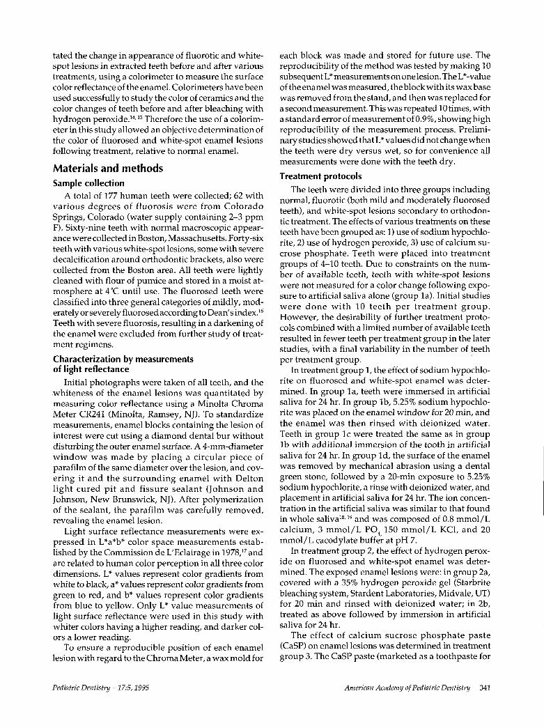
Fig 1. Back-scattered electron image of moderately fluor-osed human enamel after treatment with sodium hypo-chlorite, calcium sucrose phosphate paste and artificialsaliva. A globular material consisting of large granules(arrow) is compacted into the enamel rods. Bar = 10 Lim.
artificial saliva. A treatment group using mechanicalsurface removal alone was not included, so the relativeimportance of the sodium hypochlorite in this treat-ment regimen can not be determined. The use of CaSPon white-spot lesions prior to placement in the artifi-cial saliva resulted in some color change with a signifi-cantly reduced L*-value. However, pretreatment withsodium hypochlorite prior to use of CaSP and immer-sion in artificial saliva was more effective in returningthe color of both fluorosed and white-spot lesions tothat of normal enamel.
SEM observationsBoth scanning and back-scatter electron imaging of
normal enamel treated with CaSP paste, showed aregular appearance of enamel rods. However, treat-ment of moderately fluorosed teeth with CaSP resultedin the appearance of an irregular-shaped globular ma-terial compacted between regular enamel rods (Fig 1).In scanning and in back-scattered mode it was evidentthat this material was located between the enamel rodsbelow the enamel surface, and was not merely an arti-fact from surface contamination.
DiscussionThe appearance of white-spot lesions and fluorotic
enamel results from a subsurface porosity in the enamelbelow a well mineralized surface layer.21121 This subsur-face porosity, which increases with increasing severityof fluorosis,22 results in a whiter appearance to theenamel lesion. In this study, fluorosed and normalenamel were characterized in vitro by measuring thesurface color reflectance of the enamel. The measure-ment of surface color reflectance showed that theenamel whiteness, as measured by the L*-value, couldbe directly correlated with the visual assessment ofteeth as being fluorosed or having white spots. The
Pediatric Dentistry -17:5,2995 American Academy of Pediatric Dentistry 343
sensitivity of the color measurement technique isshown by the significant increase in L*-values of mod-erately fluorosed enamel compared with mildlyfluorosed enamel. With increasing fluorosis enamelbecomes more porous. Therefore, the increased white-ness (higher L*-values) of the moderately fluorosedteeth is likely related to the increased enamel porosityin these teeth. Quantitative evaluation of the color ofboth fluorosed and white-spot enamel using theMinolta Chroma Meter proved to be a sensitive methodshowing significant differences in the color of fluorosedand white-spot lesions, compared with unaffectedenamel. This quantitative measurement of the white-ness of a lesion allowed an objective evaluation of theeffect of various treatment regimens in altering thecolor of fluorosed and white-spot enamel.
The rationale for treating fluorosed enamel withoxidizing agents such as hydrogen peroxide and so-dium hypochlorite was that removing surface organicmaterial could result in a more ready diffusion of cal~cium and phosphate into the enamel. This increaseddiffusion of mineral into the lesion may increaseremineralization within the subsurface lesion. Enamelwas treated with both artificial saliva anddeproteinizing agents (hypochlorite and peroxide) bothtogether and separately to .determine the Unique con-tribution of each to changes in the enamel.
Treating the enamel with hydrogen peroxide wasfound to cause significant whitening of all teeth. Thiswhitening effect was unchanged by any subsequenttreatment regimens. Although a whitening effect maymask the fluorotic lesion by causing the less fluoroticenamel to become whiter, it creates a color that is lighterthan the normal range of human tooth enamel. It there-fore does not appear to be indicated for treatment offluorotic or white-spot lesions.
Unlike hydrogen peroxide treatment, treating theenamel surface with a 5.25% sodium hypochlorite re-sulted in no changes in the color of either fluorosed orwhite-spot lesions. Previous studies have shown hy-pochlorite to be an effective deproteinizing agent andto increase penetration of mineral into carious enamellesionsd3. 24 In these studies, use of sodium hypochlo-
rite to remove the organic surface layer was effectivein treating enamel lesions in combination with CaSP.
Mechanical removal of the tooth surface appearedto be effective for changing the color of white-spot le-sions into the range of normal enamel. This may be dueto the shallow subsurface nature of this lesion, Whenthe highly mineralized surface area of these lesions ismechanically removed, the underlying shallow lesionmay be remineralized in the artificial saliva. It is likelythat removing the surface enamel through use of a den-tal bur or by "microabrasion" also would be effectivein very mildly fluorosed teeth, where the subsurfaceporosity is minimal and is limited to the outer 50-100~tm of enamel surface. However, in moderatelyfluorosed teeth, where the porosity is greater, simplyremoving the surface without further "filling in" of the
344 American Academy of Pecliatric Dentistry
pores is not likely to be as effective. This suggests thatcarefully assessing the severity of the lesion is impor-tant prior to treatment.
Using CaSP alone decreased L*-values toward val-ues comparable to normal enamel for both fluorosedand white-spot enamel without affecting normal toothcolor. However, pretreatment with sodium hypochlo-rite, followed by CaSP and exposure to an artificial sa-liva was the most successful treatment protocol, result-ing in L*-values similar to normal enamel for bothfluorosed and white-spot lesions,
CaSP is a fine white powder with a bland, neutraltaste. It is a mixture of calcium sucrose mono- anddiphosphates, disucrose monophosphate, and inor-ganic calcium orthophosphate, containing approxi-mately 11% calcium, 9.5% organic phosphorous, and2.5% inorganic phosphorous. Prior to the reported useof CaSP in treatment of fluorosed enamel, it had beenused as a food additive to reduce the incidence of den-tal caries in children,2~a7 and for desensitizing dentin.-~s
High concentrations of calcium and orthophosphateions in CaSP were cited as b~ing responsible for therapid remineralization of softened enamel and for de-sensitizing dentin. However, the mechanisms of actionof CaSP appear to be poorly defined.
When moderately fluorosed teeth that had beentreated with 10% CaSP paste were examined by scan-ning electron microscopy, a globular material with agranular appearance was found. Back-scattered scan-ning electron microscopy also showed electron absor- "bent areas of material that had been deposited withinthe rods. These results suggest that treatment with theCaSP paste resulted in filling the subsurface porousspaces rather than simply a remineralization effect aspreviously suggested. Filling the porous enamel wouldalter the light reflectance and hence the color of theporous fluorosed or white-spot enamel, returning thecolor to normal. This suggests that with more severewhite opaque lesions, such as found in mildly andmoderately fluorosed teeth as classified by Dean, anactual filling of the pores may necessary for an accept-able clinical result.
The quantitative results obtained in this in vitrostudy, along with previous reports, suggest that CaSPcan be an effective treatment for enamel opacities. Fur-ther studies to compare different concentrations as wellas the long-term effect of this material on tooth enamelare warranted. Although CaSP paste is not currentlycommercially available, it is potentially useful in fur-ther developing treatments to be used for this impor-tant aspect of esthetic dentistry.
Conclusions1. Changes in the appearance of enamel can be
quantitated by measuring light reflectance.
2. Effective treatment of enamel lesions such asmoderately fluorosed enamel differ from treat-ment of less porous lesions such as in very mildlyfluorosed enamel or white-spot lesions.
Pediatric Dentistry - 17:5,1995

3. Treatment of enamel with 5.25% sodium hy-pochlorite followed by a calcium sucrose phos-
phate paste was most effective in changing theappearance of fluorosed and white-spot lesions
into a color range for normal enamel.
Dr. Den Besten is an associate professor and chair, pediatric den-tistry, University of California, San Francisco. Dr. Giambro is inorthodontic private practice in Utrecht, The Netherlands.
The authors acknowledge the expertise of Dr. Kenneth Prostackin completing the SEM studies and thank Dr. James Kruse of Colo-rado Springs, Colorado, who provided the fluorosed teeth.
1. Fejerskov O, Silverstone LM, Melsen B and Moller IJ: His-tological features of fluorosed human dental enamel. Car-ies Res 9:190-210, 1975.
2. Christensen J, Larsen M, Fejerskov O: Effect of a mineraliz-ing solution on sections of fluorosed human dental enamelin vitro. Caries Res 13:47-56, 1979.
3. McCloskey RJ: A technique for removal of fluorosis stains.J Am Dent Assoc 109:63-64, 1984.
4. Croll TP, Cavanaugh RR: Enamel color modifications bycontrolled hydrochloric acid-pumice abrasion. I. Techniquesand examples. Quintessence Int 17:81-87, 1986.
5. Ames JW: Removing stains from mottled enamel. Am DentAssoc J 24:1674-77, 1937.
6. Younger HB: Bleaching fluoride stain from mottled enamel.Texas D J 57:380-82, 1939.
7. Chandra S, Chawla TN: Clinical evaluation of heat methodfor bleaching of discolored mottled teeth. J Indian DentAssoc 46:313-18, 1974.
8. Chandra S, Chawla TN: Clinical evaluation of the sandpa-per disc method for removing fluorosis stains from teeth. JAm Dent Assoc 90:1273-76, 1975.
9. Murrin JJ, Barkmeier WM: Chemical treatment of vital teethwith intrinsic stain. Ohio Dent J 56:6-10, 1982.
10. Croll TP: Enamel microabrasion: the technique. Quintes-sence Int 20:395-400, 1989.
11. Belkhir MS, Douki N: A new concept for removal of dentalfluorosis stains. J Endodont 17:288-92, 1991.
12. Powell K, Craig GG: A simple technique for the aestheticimprovement of fluorotic-like lesions. ASDC J Dent Child49:112-17, 1982.
13. Myers D, Lyon TC Jr: Treatment of fluorosis-like lesions withcalcium sucrose phosphate gel. Pediatr Dent 8:213-15,1986.
14. Rosensteil SF, Porter SS, Johnston WM: Colour measure-ments of all ceramic crown systems. J Oral Rehabil 16:491-501, 1989.
15. Rosensteil SF, Gegauff AG, Johnston WM: Duration of toothcolor change after bleaching. J Amer Dent Assoc 123:54-59,1991.
16 Dean HT: Classification of mottled enamel diagnosis. AmerDent Assoc J 21:1421-26, 1934.
17. Commission internationale de L’Eclairge, Recommenda-tions on uniform colour spaces, colour difference equationsand psychometric colour terms. Paris: Bureau Central de laDIE suppl. 2 to pub 15, 1978.
18. Featherstone JDB, Cutress TW, Rodgers BE, Dennison PJ:Remineralization of artificial caries-like lesions in vivo bya self-administered mouthrinse or paste. Caries Res 16:235-42, 1982.
19. Featherstone JDB, Zero DT: Laboratory and human studiesto elucidate the mechanism of action of fluoride-containingdentifrices. In: Clinical and Biological Aspects of Dentifrices,Embery G, Rolla G, Eds. Oxford: Oxford University Press,1991, pp 1-14.
20. Fejerskov O, Yaeger JA, Thylstrup A: Microradiography ofacute and chronic administration of fluoride on human andrat dentin and enamel. Arch Oral Biol 24:123-30, 1979.
21. Newbrun E, Brudevold F: Studies on the physical proper-ties of fluorosed enamel. I. Micrographic analysis. Arch OralBiol 2:15-20, 1960.
22. Thylstrup A, Fejerskov O: Clinical appearance of dentalfluorosis in permanent teeth in relation to histologicchanges. Community Dent Oral Epidemiol 6:315-28, 1978.
23. Robinson C, Kirkham J: The effect of fluoride on the devel-oping mineralized tissues. J Dent Res 69:685-91, 1990.
24. Christensen L, Larsen M, Fejerskov O: Effect of a mineral-izing solution on sections of fluorosed human dental enamelin vitro. Caries Res 13:47-56, 1979.
25. Harris R, Schamschula RG, Gregory G, Roots M, BeveridgeJ: Observations on the cariostatic effect of calcium sucrosephosphate in a group of children aged 5-17. Preliminaryreport. Aust Dent J 12:105-113, 1967.
26. Harris R, Schamschula RG, Beveridge J, Gregory G: Thecariostatic effect of calcium sucrose phosphate in a groupof children aged 5-17 years. Aust Dent J 13:32-39, 1968.
27. Clarke NG, Fanning EA: Plaque pH and calcium sucrosephosphate: a telemetric study. Aust Dent J 16:13-16, 1971.
28. Lilienthal B, Napper DH, Smythe BM: The hardening and soft-ening of human tooth enamel. Aust Dent J 13:219-30, 1968.
Pediatric Dentistry- 17:5, 1995 American Academy of Pediatric Dentistry 345