Embed Size (px)
Citation preview

1
Andrew Lisowski, M.S., HTL (A.S.C.P.)
Science of H & E

“The desired end result of a tissue stained with hematoxylin and eosin is based upon what seems to be almost infinite factors. Pathologists have individual preferences for section thickness, intensities, and shades. The choice of which reagents to use must take into consideration: cost, method of staining, option of purchasing commercially-prepared or technician-prepared reagents, safety, administration policies, convenience, availability, quality, technical limitations, as well as personal preference.”
Hematoxylin and Eosin Staining
2
Guidelines for Hematoxylin and Eosin StainingNational Society for Histotechnology

Why Do We Stain?
3
In order to deliver a medical diagnosis, tissues must be examined under a microscope.Once a tissue specimen has been processed by a histology lab and transferred onto a glass slide, it needs to be appropriately stained for microscopic evaluation. This is because unstained tissue lacks contrast: when viewed under the microscope, everything appears in uniform dull grey color.
Unstained tissue H&E stained tissue

What Does "Staining" Do?
4
Contrasts different cells
Highlights particular features of interest
Illustrates different cell structures
Detects infiltrations or deposits in the tissue
Detect pathogens
There are different staining techniques to reveal different structures of the cell
H&E stain showing extensive iron deposits
Placenta’s large blood vessels
Superbly contrasted GI cells

As its name suggests, H&E stain makes use of a combination of two dyes – hematoxylin and eosin.
It is often termed as “routine staining” as it is the most common way of coloring otherwise transparent tissue specimen. H&E is fast and relatively inexpensive method of assessing tissue morphology. First used almost 150 years ago, it is still used today with little changes.
What is H&E Staining?
5

Staining does not produce color randomly; instead, the dyes exploit differences in the chemistry of the tissue to differentially dye various components.Ionic bonding is the most important type of bonding that occurs in histologic staining techniques. It involves electrostatic attraction between ions of opposite charge, one of which is in the tissue, and the second of which is in the dye. Hematoxylin is positively charged and can react
with negatively charged cell components, such as nucleic acids in the nucleus. These stain blue as a result.
Eosin is negatively charged and can react with positively charged components in the tissue, such as amino groups in proteins in the cytoplasm. These stain shades of red to pink as a result.
How is it Done?
6
Example of H&E stain

Long history of use, staining method published by Böhmer & Fischer in 1875 H&E is the primary diagnostic technique for the evaluation of morphology and the changes
associated with the disease process H&E remains the most frequently used tissue stain worldwide with an estimated 2.5 to 3
million slides stained per day It is a useful all-purpose stain that is quick and easy to use, which may explain why it has
stood the test of time Customer expectations or preferences are extremely subjective
H&E Stain
7

H&E Staining Procedure Step-by-Step
8
Procedure involves several steps and regents divided into groups based on their function.
Deparaffinization or Dewaxing steps:
Typically done by solvents like xylene or xylene substitutes to remove paraffin from sections adhered to the glass slide.
Staining pathway
Wa
ter
rin
se
X A
Dif
fere
nti
ato
r
Blu
ing
X A A A A X XA
Eosi
n
He
ma
tox
ylin
X-solventA-alcohol
Wa
ter
rin
se
Wa
ter
rin
se
Wa
ter
rin
se
Wa
ter
rin
se
X A
Dif
fere
nti
ato
r
Blu
ing
X A A A A X XA
Eosi
n
He
ma
tox
ylin
X-solventA-alcohol
Wa
ter
rin
se
Wa
ter
rin
se
Wa
ter
rin
se
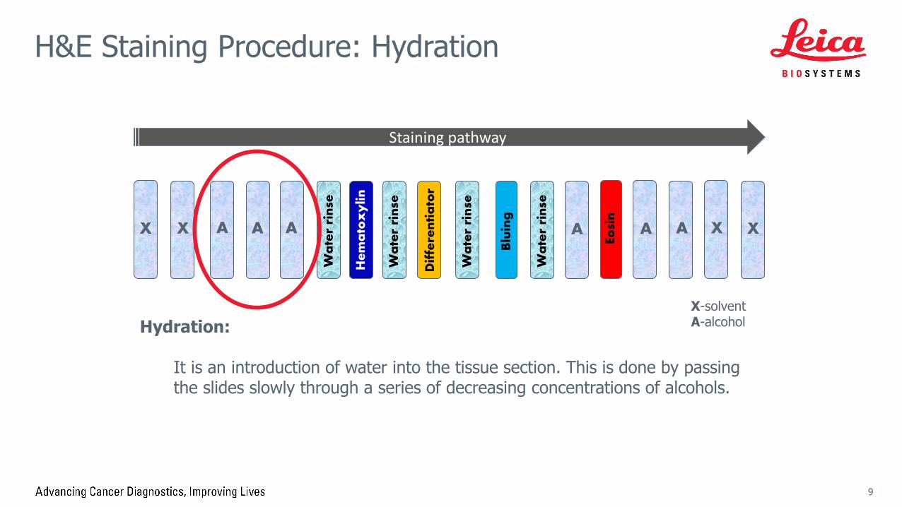
H&E Staining Procedure: Hydration
9
Hydration:
It is an introduction of water into the tissue section. This is done by passing the slides slowly through a series of decreasing concentrations of alcohols.
Staining pathway

Wa
ter
rin
se
X A
Dif
fere
nti
ato
r
Blu
ing
X A A A A X XA
Eosi
n
He
ma
tox
ylin
X-solventA-alcohol
Wa
ter
rin
se
Wa
ter
rin
se
Wa
ter
rin
se
H&E Staining Procedure: Staining
10
Primary Staining:
Hematoxylin is used after deparaffinization and hydration. It stains the nucleus of the cell, specifically, the chromatin within the nucleus and the nuclear membrane. The nucleoplasm of the nucleus should remain unstained; this allows the staining pattern of the chromatin to be seen easily.
Staining pathway
Wa
ter
rin
se
X A
Dif
fere
nti
ato
r
Blu
ing
X A A A A X XA
Eosi
n
He
ma
tox
ylin
X-solventA-alcohol
Wa
ter
rin
se
Wa
ter
rin
se
Wa
ter
rin
se
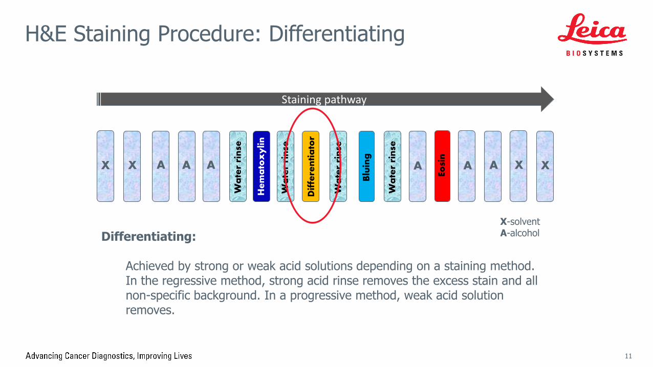
H&E Staining Procedure: Differentiating
11
Differentiating:
Achieved by strong or weak acid solutions depending on a staining method. In the regressive method, strong acid rinse removes the excess stain and all non-specific background. In a progressive method, weak acid solution removes.
Staining pathway

Wa
ter
rin
se
X A
Dif
fere
nti
ato
r
Blu
ing
X A A A A X XA
Eosi
n
He
ma
tox
ylin
X-solventA-alcohol
Wa
ter
rin
se
Wa
ter
rin
se
Wa
ter
rin
se
H&E Staining Procedure: Bluing
12
Bluing Step:
Bluing reagents vary from ammonia solutions, tap water, Scott’s solution, Blue Buffer 8, lithium and magnesium carbonate solutions. Bluing changes the reddish-purple hematoxylin to a blue or purple-blue color. It is a pH dependent reaction and occurs in an alkaline solution.
Staining pathway

Wa
ter
rin
se
X A
Dif
fere
nti
ato
r
Blu
ing
X A A A A X XA
Eosi
n
He
ma
tox
ylin
X-solventA-alcohol
Wa
ter
rin
se
Wa
ter
rin
se
Wa
ter
rin
se
H&E Staining Procedure: Neutralizing
13
Neutralizing Step:
When using an alcoholic eosin this step must contain 95% (or similar %) alcohol; when choosing an aqueous eosin, water is used. This is to saturate sections with the same diluent that makes up an eosin used.
Staining pathway

Wa
ter
rin
se
X A
Dif
fere
nti
ato
r
Blu
ing
X A A A A X XA
Eosi
n
He
ma
tox
ylin
X-solventA-alcohol
Wa
ter
rin
se
Wa
ter
rin
se
Wa
ter
rin
se
H&E Staining Procedure: Counterstaining
14
Secondary Staining or Counterstaining:
Eosin stains nearly everything that hematoxylin will not stain. When applied correctly, eosin produces three different hues which can be used to differentiate various tissue elements; red blood cells stain dark reddish orange, collagen stains a lighter pastel pink, and smooth muscle stains bright pink.
Staining pathway

Wa
ter
rin
se
X A
Dif
fere
nti
ato
r
Blu
ing
X A A A A X XA
Eosi
n
He
ma
tox
ylin
X-solventA-alcohol
Wa
ter
rin
se
Wa
ter
rin
se
Wa
ter
rin
se
H&E Staining Procedure: Dehydration
15
Dehydration:
Removal of water from the tissue section. Increasing concentrations of alcohol after eosin staining are to remove water from the tissue section. Concentration of alcohols following the stain is important. Since eosin is very soluble in water, it is easily removed from already stained section by alcohol that is less than 100%.
Staining pathway

Wa
ter
rin
se
X A
Dif
fere
nti
ato
r
Blu
ing
X A A A A X XA
Eosi
n
He
ma
tox
ylin
X-solventA-alcohol
Wa
ter
rin
se
Wa
ter
rin
se
Wa
ter
rin
se
H&E Staining Procedure: Clearing
16
Clearing:
Displacement of alcohol from the tissue sections with the clearant (usually xylene or xylene substitutes) to assure miscibility when coverslipping with xylene, toluene or other petroleum-based mounting media.
Staining pathway
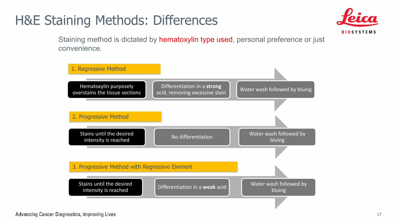
H&E Staining Methods: Differences
17
Stains until the desired intensity is reached No differentiation Water wash followed by
bluing
Stains until the desired intensity is reached Differentiation in a weak acid Water wash followed by
bluing
1. Regressive Method
2. Progressive Method
3. Progressive Method with Regressive Element
Staining method is dictated by hematoxylin type used, personal preference or just convenience.
Hematoxylin purposely overstains the tissue sections
Differentiation in a strong acid, removing excessive stain Water wash followed by bluing

Staining Overview: Hematoxylin
18
Hematoxylin comes from a logwood tree that grows in Central and South America. Hematoxylin alone is not technically a dye, and will not directly stain tissues. First, it needs to be oxidized to hematein. It is done by adding chemical oxidizing agents and also
done naturally in a process called ripening when hematoxylin is exposed to air. Hematoxylin ripens throughout its life.
Secondly, it needs to be complexed with a “mordant” (from French “mordre” – to bite, grip), typically aluminum ion, that helps it link to the tissue.
Hematoxylin in complex with aluminum is positively charged and can react with negatively charged cell components, such as nucleic acids in the nucleus. These stain blue as a result.
MordantOxidation

Hematoxylin binds to its target, i.e. nucleic acid, but also binds to large proteins creating unwanted background. While to some pathologists this is negligible, others prefer no background caused by hematoxylin.
Hematoxylin
19
Target: DNA Background: mucins, large proteins
Tissue section
Nucleic acid (DNA)
Glass slide

Unwanted hematoxylin background or non-specific staining can be removed in a differentiating step by acidic solution.
Differentiation by Acid Solutions
20
Differentiation breaks the bond between mordant and the tissue
Tissue sectionNucleic acid (DNA)
Glass slide

Over-differentiation may cause tissue loss or tissue lifting. Choice of differentiator (strong, medium or weak acid) and immersion time are
crucial for keeping tissue sample on the surface of the glass safely.
Differentiation by Acid Solutions
21
Glass slide
Degree of tissue loss or tissue lifting might depend on slide type used (coated vs. non-coated)
Broken bonds

Properly done differentiation removes non-specific hematoxylin staining.
Removing the Background
22
Background staining: goblet cells Background-free goblet cells
Example: Goblet cells in GI track contain mucins that are non-specifically stained with hematoxylin causing the background that can be removed with differentiator
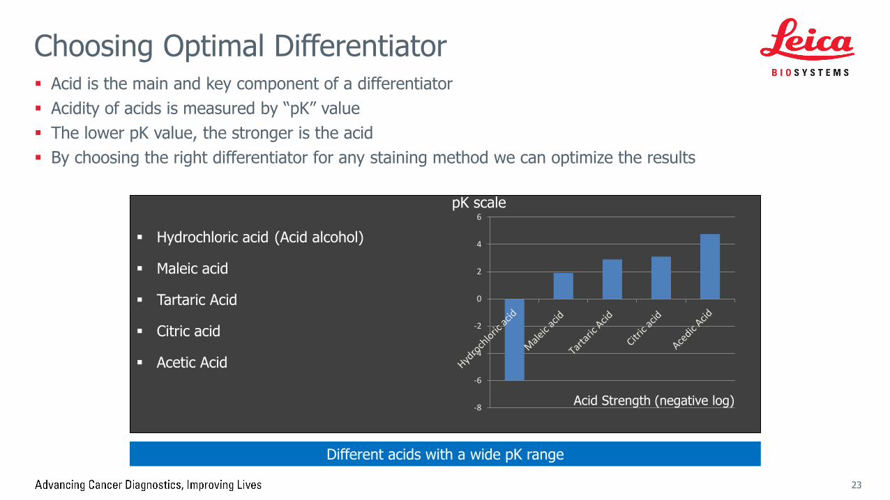
Acid is the main and key component of a differentiator Acidity of acids is measured by “pK” value The lower pK value, the stronger is the acid By choosing the right differentiator for any staining method we can optimize the results
Choosing Optimal Differentiator
23
Hydrochloric acid (Acid alcohol)
Maleic acid
Tartaric Acid
Citric acid
Acetic Acid
-8
-6
-4
-2
0
2
4
6pK scale
Acid Strength (negative log)
Different acids with a wide pK range

Hematoxylin Bluing
24
o Tap water
o Ammonia water
o Scott’s tap water
o Buffered solutions
pH may be unpredictable
pH may be too high (section loss may occur)
more effective at maintaining the optimal pH (less risk of tissue loss)
Bluing is necessary to convert hematoxylin reddish purple nuclear coloration to a crisp blue/purple. Changing the color to blue gives a much better contrast with the red (eosin) counterstain.
There are numerous formulations of bluing reagents available. Choose the right product with stable pH and gentle mode of action.

Eosin Y is a negatively charged synthetic dye that binds to positively charged molecules in tissue sample.
It is soluble in either water or alcohol so aqueous or alcoholic eosin version are common as counterstains.
Good quality eosin generates three shades of red but there are also variants that produce fourth color.
Eosin Staining
25
Eosin moleculePowdered Eosin Y

H&E stained kidney
Good counterstain will not only contrast sharply with the blue nuclei, but it will allow the non-nuclear tissue components to be clearly differentiated from each other by three different shades of eosin.
Eosin Staining: Result
26
2. Connective tissue paler shade of pink
3. Red blood cells intensly red
1. Cytoplasm/muscle pink-orange
Nuclei blue
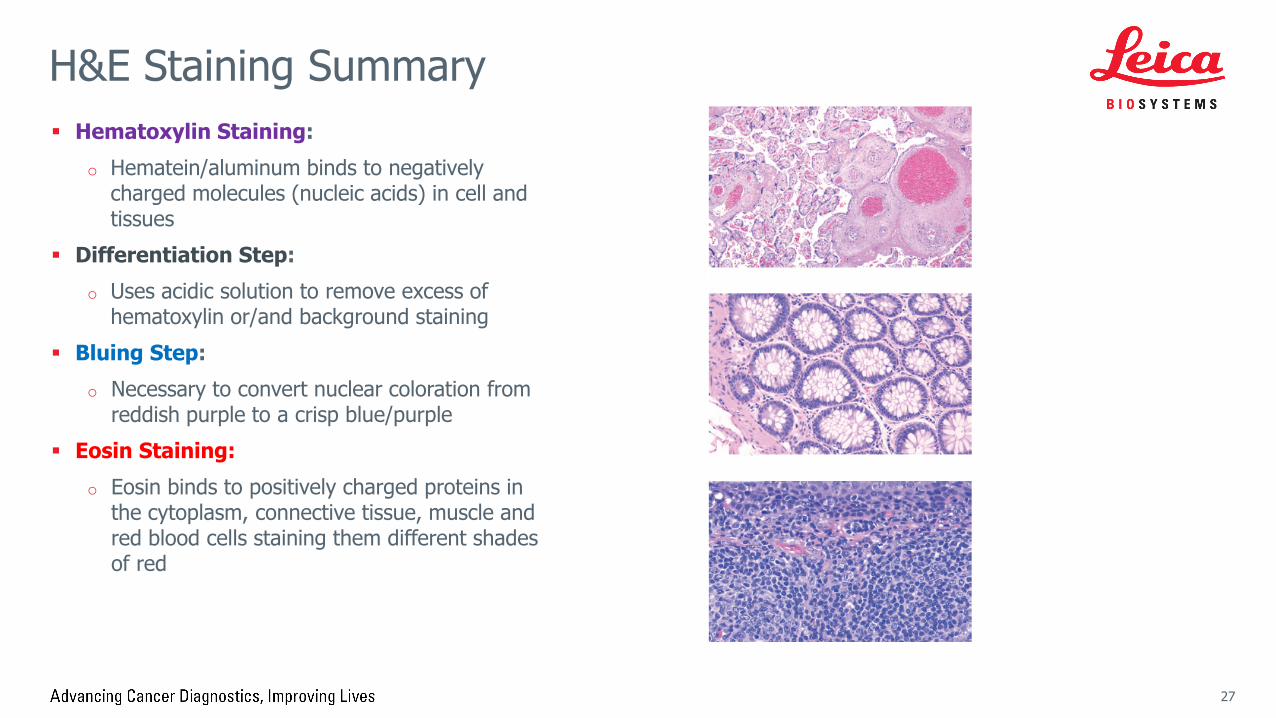
Hematoxylin Staining:
o Hematein/aluminum binds to negatively charged molecules (nucleic acids) in cell and tissues
Differentiation Step:
o Uses acidic solution to remove excess of hematoxylin or/and background staining
Bluing Step:
o Necessary to convert nuclear coloration from reddish purple to a crisp blue/purple
Eosin Staining:
o Eosin binds to positively charged proteins in the cytoplasm, connective tissue, muscle and red blood cells staining them different shades of red
H&E Staining Summary
27

Copyright © 2019 Leica Biosystems, a division of Leica Microsystems Inc. All rights reserved. LEICA and the Leica Logo are registered trademarks of Leica Microsystems IR GmbH.
190137 Rev A ∙ 02/2019
This reference document is presented as a service to health care professionals by Leica Biosystems and has been compiled from available literature. Although every effort has been made to report faithfully the information, Leica Biosystems cannot be held responsible for the correctness. This document is not intended to be, and should not be construed as medical advice. For any use, the product information guides, inserts and operation manuals of the various drugs and devices should be consulted. Leica Biosystems and the editors disclaim any liability arising directly or indirectly from the use of drugs, devices, techniques or procedures described in this reference document.