Embed Size (px)
Citation preview

Mahatma Jyotiba Fule College of VeterinaryScience and Animal Husbandry, Chomu (Raj.)
PRESENTED BY:Dr. Vikram Punia
2020-2021Assistant professor
DEPARTMENT OF VETERINARY PARASITOLOGY
COLLEGE OF VETERINARY SCIENCE & ANIMAL HUSBANDRY, CHOMU (RAJ.)
HYMENOLEPIDIDAE , TAPEWORMS OF DOG

•
•
•
•
•
•
•
•
•
•
Family :Hymenolepididae Cestodes are usually narrow and thread- like in appearance.
Tape worm are small to medium in size with retractable bearing
a single circle of hooks.
Rostellum bears 8-10 hooks.
Suckers are unarmed .
Single set reproductive organs in each proglottid.
Ovary is median in position, lobed and transversely elongate.
Testes are up to 4 in number and arranged in a row or a triangle.
Vitelline glands are potato- shaped.
Uterus is sac- like.
Cirrus is spinosed.

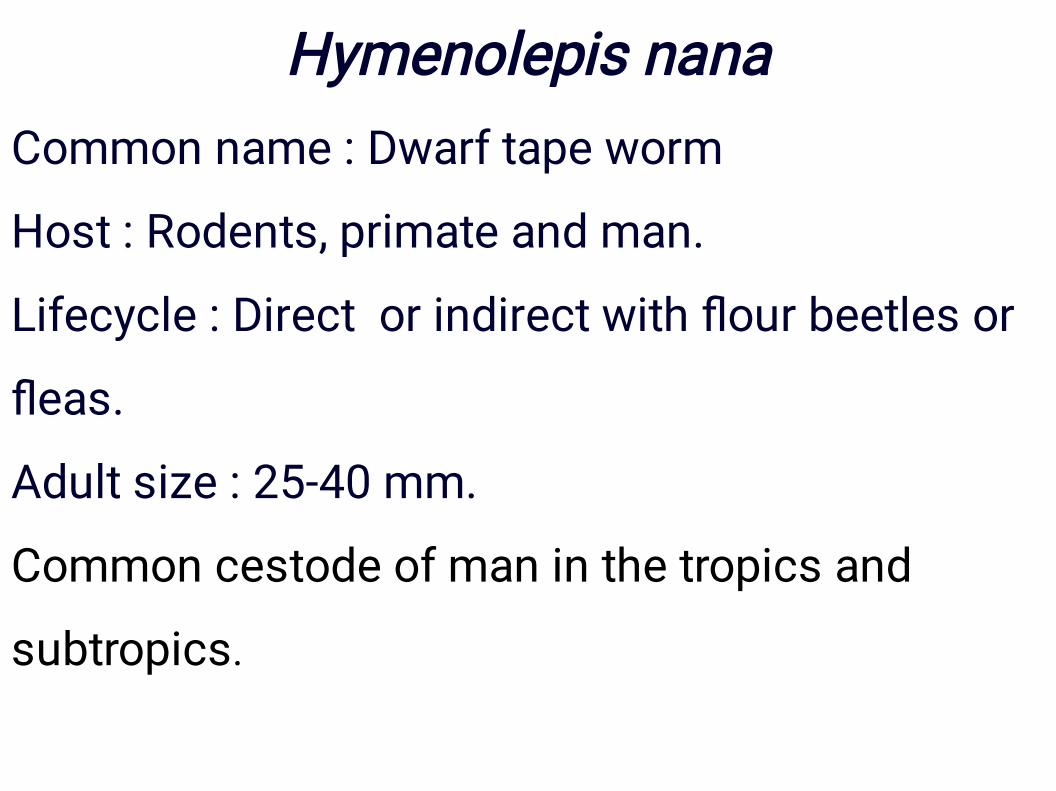
Hymenolepis nanaCommon name : Dwarf tape worm
Host : Rodents, primate and man.
Lifecycle : Direct or indirect with flour beetles or
fleas.
Adult size : 25-40 mm.
Common cestode of man in the tropics and
subtropics.

•
•
Eggs
Eggs are oval or sub-spehrical and smaller, ranging
40-60 um x 30-50 um. On the inner membrane are two
poles, from which 4 to 8 polar filaments spread out
between the two membranes.
The oncosphere has six hooks. Notice in picture the
extra detail of the two polar thickenings on the
membrane of the oncosphere with filaments extending
into the space around the hexacanth embryo.

Egg of Hymenolepis nana

Lifecycle
Lifecycle is direct and cysticercoids develop within the villi of
small intestine.
Self – infection through ingestion of eggs containing the
developing cysticercoids discharged with own faeces. Auto-
infection where the cysticercoids while in intestine hatch out from
the eggs and attach to mucosa to grow into adult worm.
Pre-patent period 16 days.


Pathogenesis
occur only with heavy infections and are most apparent in
children.
Gastrointestinal discomfort
Diarrhea
Weakness
Poor appetite/anorexia
Headaches
Complications: discomfort and dehydration from prolonged
diarrhea

Diagnostic Tests:
Examination of the stool for eggs and parasites confirms the
diagnosis. Concentration techniques and repeated examinations
will increase the likelihood of detecting light infections. See
Morphology.
Treatment
Praziquatel as a single dose is currently the treatment of choice.
Expect a full recovery following treatment. Both Niclosamide and
praziquantel have been very effective and had minimal side
effects. Niclosamide causes death to the tapeworm by interfering
with oxidative phosphorylation, use 40 mg/kg for one dose only in
children. Praziquantel causes paralysis and death use 25 mg/kg
for one dose only

•
•
•
•
•
•
The tapeworms of dog are as follows:
Dipylidium caninum
Taenia hydatigena
Taenia multiceps
Echinococcus granulosus
Mesocestoides lineatus
Diphyllobothrium latum

Dipylidium caninum
Common name :Double pored dog tapeworm.
It also occurs in man and cat.
Location : Small intestine
I/H : Dog flea: (Ctenocephalides canis), Dog lice:
Trichodectes canis and Heterodoxus spiniger)
Metacestode stage : Cysticercoid

•
•
•
•
•
Morphology
Retractable rostellum armed with three or four rows
of rose thorn shaped hooks.
Each segment contains two sets of genital organ.
Vitelline glands and ovary form a mass on either side
resembling a bunch of grapes.
In the gravid segment uterus are replaced by egg
capsule or egg packets.
Egg packets contain 30 eggs per packet. Gravid
segments are elongate and oval in shape
resembling cucumber seed shape.
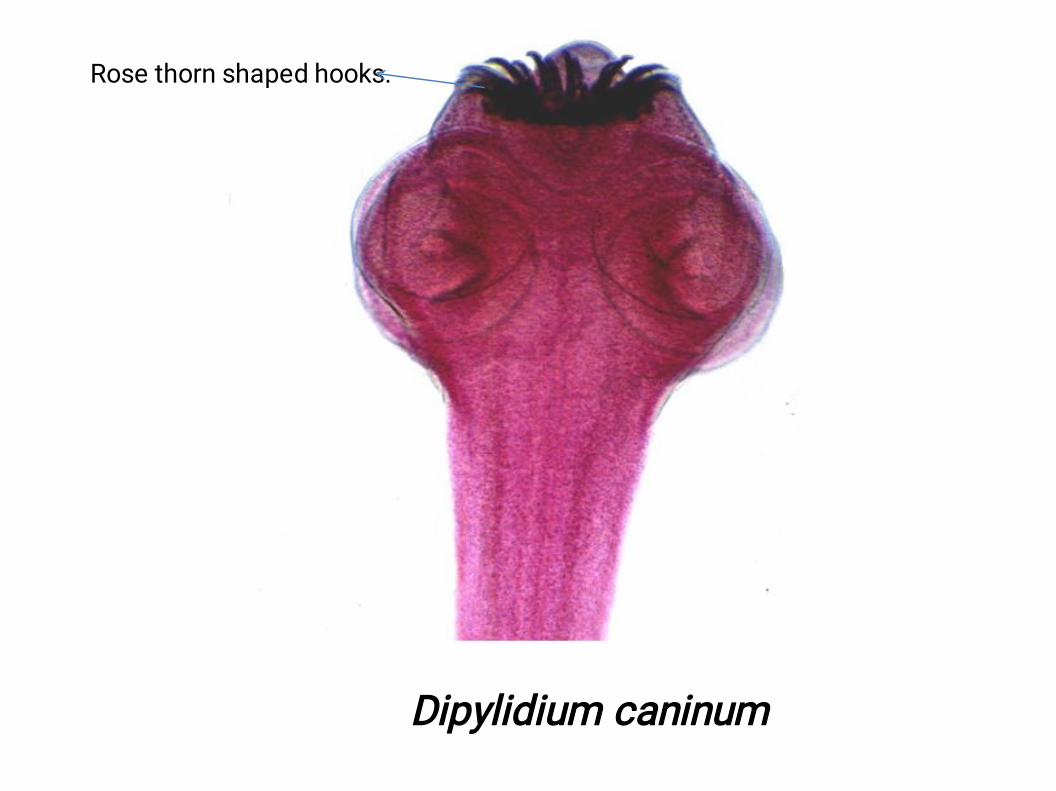
Rose thorn shaped hooks.
Dipylidium caninum


D. caninum egg packet, containing 8 visible eggs
D. caninum eggs are round to oval (average size 35 to 40 µm;
range 31 to 50 µm by 27 to 48 µm) and contain an oncosphere
that has 6 hooklets. Proglottids of D. caninum contain
characteristic egg packets that are round to ovoid and contain 5
to 15 or more eggs each.
•
•
•
•
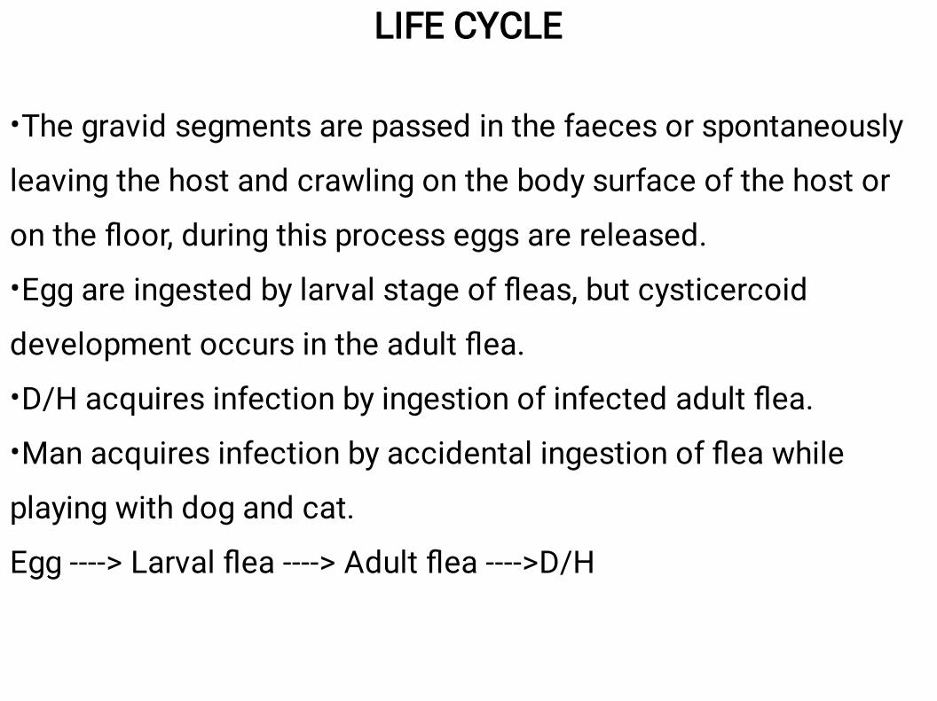
LIFE CYCLE
The gravid segments are passed in the faeces or spontaneously
leaving the host and crawling on the body surface of the host or
on the floor, during this process eggs are released.
Egg are ingested by larval stage of fleas, but cysticercoid
development occurs in the adult flea.
D/H acquires infection by ingestion of infected adult flea.
Man acquires infection by accidental ingestion of flea while
playing with dog and cat.
Egg ----> Larval flea ----> Adult flea ---->D/H


•
•
PATHOGENESIS
It depends upon the age of host. Adult worms are not
pathogenic to dog but heavy infection causes abdominal
pain, un-thriftiness, diarrhea or constipation and rarely
intestinal obstruction may occur.
When gravid segment leave the intestines they cause
severe irritation around the perianal area and due to
constant irritation the dog will dragging of the anus over
the ground. This condition is known as “anal pruritus”.
•
•
•
•
•
•
•
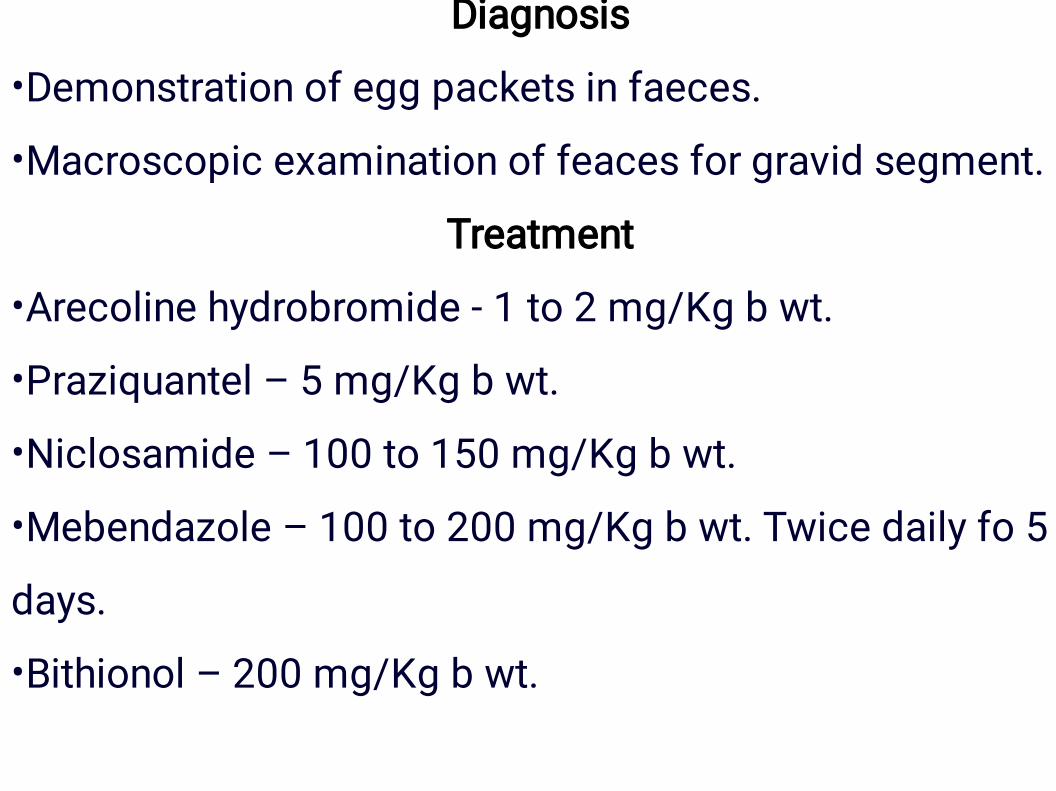
Diagnosis
Demonstration of egg packets in faeces.
Macroscopic examination of feaces for gravid segment.
Treatment
Arecoline hydrobromide - 1 to 2 mg/Kg b wt.
Praziquantel – 5 mg/Kg b wt.
Niclosamide – 100 to 150 mg/Kg b wt.
Mebendazole – 100 to 200 mg/Kg b wt. Twice daily fo 5
days.
Bithionol – 200 mg/Kg b wt.