Embed Size (px)
Citation preview

School of Health Sciences
Delon and Elizabeth Hampton Hall of Civil Engineering, Room 1173 | 550 Stadium Mall Drive | West Lafayette, IN 47907-2051
(765) 494-1419 | Fax: (765) 496-1377 | www.healthsciences.purdue.edu
August 15, 2019
Re: Research Projects
To our MP students,
There are many research projects, at the MS and PhD level, available to you.
Dr. Keith Stantz (Purdue)
Project 1: Advanced Dynamic Contrast Enhanced Imaging
Introduction: Therapeutic resistance is strongly correlated with the microenvironment of the tumor, or TME.
One such TME niche is the formation of necrotic regions/foci within the tumor, with is pathologically correlated
to poor patient outcome. This is because these regions influence important oncological factors such as
metabolism, cancer stem/progenitor-like phenotype, and immune function in order to escape radiation therapy.
Goal: To developed advanced imaging (DCE) methods to image these local TME changes within the tumor.
Innovation: To date, dynamic contrast enhanced imaging utilize biophysical models that assume contrast agents
are spatially localized in order to monitor tumor physiology. Such an assumption severely limits our ability to
detect and quantify TME regions in primary tumors and metastases. Instead, 3D physics models of contrast
transport will be developed to account for the non local effects, significantly enhancing our diagnostic capability
to detect and target these regions with x-ray, proton, and neutron beam radiation.
Approach: The student will develop the mathematic (and computational) models of contrast transport based on
physics based models of convection and diffusion. These models will be tested on CT/MRI data. This working
group includes an undergraduate also working in this area.
Project 2: Radiation induce immunology
Introduction: A recent discovery by researchers at the Cancer Center in Cornell demonstrated that a large dose
fraction can stimulate an adaptive immune response, thus allowing a patient’s immune system to recognize their
own cancer/distant metastasis. This technique has recently been tested in cancer patients. Large doses have been
applied to a patient’s primary tumor, which stimulated their immune system to recognize and attack distant
metastases. However, this affect remains difficult to reproduce in most cancers with varying levels of success.
Objective: To test a new hypothesis on how to overcome a key limitation in this technique.
Innovation: We propose that charged particle beams (protons, carbon ions) can be used to elicit the above
immune response compared to x-rays.
Approach: The student will perform in vitro radiation biology studies in cancer cells exposed to radiation under
different microenvironments. The student will be trained to culture cancer cells, expose these cancer cells to
radiation under different tumor environments, and perform assays to measure the immunogenic response. This
student will work with both a PhD student and an undergraduate student investigating this area of radiation
therapy.
School of Health Sciences
Delon and Elizabeth Hampton Hall of Civil Engineering, Room 1173 | 550 Stadium Mall Drive | West Lafayette, IN 47907-2051
(765) 494-1419 | Fax: (765) 496-1377 | www.healthsciences.purdue.edu
Project 3: Developing a new scanner modality to image in 3D charge particle dosimetry
Introduction: Charged particle beams, specifically proton beam therapy, is quickly advancing, with new
facilities being constructed every year. The challenge with proton beam therapy is the unique dose deposition
(Bragg peak and distal edge), which if applied incorrectly, can significantly impact patient treatment and
outcome. Through the use of thermoacoustics (ultrasound-based imaging), the dose deposition can be obtained
in real-time to verify delivery in patients and in advanced radiation therapy (see previous two projects).
Innovation: This is a new imaging modality patented by Dr. Stantz and his collaborators.
Approach: There is a fair amount of simulation research both in radiation dose from protons, alphas, carbon
ions, and neutrons, and 3D imaging of the scanner design, to determine the feasibility and implementation of this
technology. There is an opportunity to explore how thermoacoustic imaging can be used in FLASH therapy. The
student interested in this project will work with both PhD and undergraduate students.
Dr. Nie (Purdue)
Projects:
• Explore the opportunities for using synchrotron X-ray technologies on health issues related to
metals and trace elements
• Development of non-invasive neutron activation analysis (NAA) technology to quantify metals in human
tissue in vivo
• Development of non-invasive x-ray fluorescence (XRF) technology to quantify metals in human tissue in
vivo
• Development of associated partial neutron imaging (APNI) technology for elemental analysis in medical
diagnostic
• Development of neutron boron capture therapy technology for disease treatment
• Development of synchrotron XRF technology for elemental mapping in biological samples
Dr. Colleen DesRosiers (IUSM)
DesRosiers, Colleen M [email protected]
Project 1: Evaluating the dosimetric differences between a CT planning scan image generated with 12 bit
versus 16 bit processor
Background: Radiation dose distributions are dependent on electron density of the medium irradiated. In a
treatment planning system (TPS) the electron density is calculated based on HU to electron density conversion
table. The HU scale depends on the processing; specifically, a 16 bit reconstructed image will have a broader
HU range than a 12 bit, which may impact the resulting dose distribution.
Experiment:
1. A clinical CT scan will be performed on an electron density phantom. The image will be reconstructed with
12 bits and 16 bits.
2. Both scans will be imported into the TPS and dose distributions from an identical treatment plan will be
generated using the HU/electron density conversion table for 12 bits.
3. DVHs and point doses will be compared between the two image sets.
School of Health Sciences
Delon and Elizabeth Hampton Hall of Civil Engineering, Room 1173 | 550 Stadium Mall Drive | West Lafayette, IN 47907-2051
(765) 494-1419 | Fax: (765) 496-1377 | www.healthsciences.purdue.edu
4. A HU/electron density conversion table will be created for 16 bits and dose calculations will be generated
and compared with calculations performed using the 12 bit scale.
Project 2: To standardize the block transmission factors for lung blocks in the TBI setting, based on
patient/block geometry.
Background: Attenuation coefficients are based on transmission measurements made in good geometry (narrow
fields, large distance). For total body irradiation (TBI), 50% transmission blocks are used to shield the lungs to
be less than 900 cGy to reduce the risk of radiation pneumonitis. However, determining the appropriate
thickness of the blocks to produce the desired dose will depend on the size of the blocks, which can vary from
about 20 cm2 to 100 cm2, depending upon the age/size of the patient.
Experiment:
1. Three sizes of blocks will be cut to at least three different thickness (total number of blocks = 9) and
transmission measurements will be made in the TBI setting.
2. Dose calculations will be performed based on typical patient geometry (lung/patient sizes) for infants,
children/small adults, and adults
3. A spreadsheet will be designed to calculate the optimum thickness of the blocks to deliver the desired dose
to the lungs, based on the lung size and patient geometry.
Dr. Dydak (Purdue)
Project 1) Manganese Toxicity in Welders The inhalation of the metal manganese (Mn) in welding fumes puts thousands of welders at risk of Mn toxicity, which results in mood changes, cognitive deficits and eventually motor symptoms similar to Parkinson’s disease. Our group is following a local cohort of welders since 2013 to study the dynamics of deposition of Mn in the brain via MRI, resulting changes in neurochemistry via MRS, as well as correlations with cognitive and motor function, mood, exposure metrics and more. Projects that relate to this overall study are listed below. All may be combined with getting trained as secondary/primary operator of the MRI scanner and help with data acquisition in our ongoing welder study: 1.a Analysis of brain Mn and Iron levels Toenail Mn levels have been shown to be an easy-to-access marker of personal exposure over the past
year. However, brain Mn levels are more dynamic and have a half-life of about 3-6 months. This study tries to find whether a correlation between toenail Mn and brain Mn can be found for a certain timing of data collection, to provide a marker for individual risk for Mn toxicity in welders. Tasks include to learn to run and analyze quantitative MRI scans (T1 maps, T2* maps, QSM) to calculate brain Mn and brain iron levels. This involves the use of Matlab pipelines, FreeSurfer and other imaging analysis software, and to become trained as secondary (and possibly primary) MRI operator. Requirement: Some knowledge of Matlab or other programming language
1.b Generation of individual Mn deposition maps

School of Health Sciences
Delon and Elizabeth Hampton Hall of Civil Engineering, Room 1173 | 550 Stadium Mall Drive | West Lafayette, IN 47907-2051
(765) 494-1419 | Fax: (765) 496-1377 | www.healthsciences.purdue.edu
We recently developed an analysis pipeline to general whole-brain Mn deposition maps, which depict all brain areas which show statistically significant increases in R1 in a group comparison between welders and controls. The next step is to try generating individual brain Mn maps, by comparing individual R1 values
with “normal” values on a pixel-by-pixel basis. Task: Using our existing R1 maps, representing brain Mn levels, from >30 welders and 20 controls, first the “normal” maps need to be generated, and different techniques to generate individual maps will be explored (p-values, difference images, etc). The student will first need to learn the existing whole-brain analysis pipeline, involving Matlab and FreeSurfer, and then adjust the pipeline accordingly. Requirement: Some knowledge of Matlab or other programming language
1.c Analysis of oxidative stress markers in welders Oxidative stress has been implicated in the mechanism of Mn toxicity by animal and cell studies. Yet,
markers of oxidative stress have never been explored in humans exposed to Mn. Glutathione (GSH) is a validated marker of oxidative stress and produces a very small signal in 1H MR Spectroscopy. While usually editing techniques need to be used to obtain a reliable signal from GSH, recent publications indicate that regular short TE PRESS spectra may give the same sensitivity to measure GSH. Our short TE PRESS data from >30 welders and >20 controls has not ever been analyzed for GSH. Task: This project includes to run statistical analysis on existing GSH quantification levels, but may also require to rerun the quantification of the existing data with other parameters using LCModel, a spectroscopy quantification tool. Requirements: Some knowledge of statistical analysis (group differences, regression), excel and interest in learning about MRS
1.d Optimization of MRS for simultaneous GABA and GSH editing GABA, the main inhibitory neurotransmitter, has been shown to be elevated in the thalamus of Mn
exposed subjects by our group. Equally, oxidative stress is implicated in the mechanism of Mn toxicity, but changes in glutathione (GSH), a marker of oxidative stress, have not yet been measured in humans exposed to Mn. In the same “toenail study” as above we acquire both GABA and GSH with the same MR Spectroscopy (MRS) sequence called HERMES. Yet, the results of simultaneous acquisition of GABA and GSH via HERMES have not yet yielded same quality results as individual acquisitions using MEGA-PRESS, and requires a systematic comparison and optimization of parameters between HERMES and MEGA-PRESS. The student taking on this study will later also be involved in MRS data analysis of the GABA and GSH data, and its correlation to brain Mn, toenail levels and exposure. Tasks include to learn to run and analyze edited MRS scans, using Matlab pipelines, and to become trained as secondary (and possibly primary) MRI operator. Requirement: Some knowledge of Matlab
Project 2: Laterality of GABA concentrations in Autism and normal developing children Imbalances of the neurotransmitter system, esp. of the main inhibitory neurotransmitter GABA, has been implicated in Autism Spectrum Disorder (ASD). Several studies have found altered GABA levels in different brain regions, and some of the results are conflicting. Several studies also saw left-right differences in GABA levels between the two brain hemispheres in autistic children, but could not rule out technical confounders (placing of the VOI, etc). Equally, it has been suggested that the developing brain in general may show a laterality in GABA levels. Yet, due to time constraints, most studies only investigate a brain region on one side of the brain. Task: Using our 3D motion-insensitive GABA-MRSI technique allows to map both sides of the brain simultaneously. Using this data, our brain-region-specific analysis approach enables us to calculate GABA levels for specific brain regions using masks and

School of Health Sciences
Delon and Elizabeth Hampton Hall of Civil Engineering, Room 1173 | 550 Stadium Mall Drive | West Lafayette, IN 47907-2051
(765) 494-1419 | Fax: (765) 496-1377 | www.healthsciences.purdue.edu
thus be able to compare left and right GABA levels for several brain regions. This study is new and will include data acquisition in several healthy and autistic children, and learning to use our brain-region-specific analysis pipeline, which involves Matlab, LCModel and other image analysis tools. Requirements: Some knowledge of Matlab; commitment to the research project for more than one semester
Project 3: Oxidative Stress and GABA in Post-Traumatic Stress Disorder (PTSD) Smoking is of higher prevalence in subjects suffering from PTSD than in the normal population. Oxidative stress and imbalances of the neurotransmitter system have been implicated both with PTSD and addictive behavior in the past. This funded study (PI: Dr. Meden Isaac-Laam from Purdue Northerwestern) will be investigating several brain regions for changes in GSH, a marker for oxidative stress, and GABA, the main inhibitory neurotransmitter in adult subjects suffering from PTSD, and study differences in smokers vs non-smokers using an optimized HERMES sequence (overlap with project 1.c!). Tasks will include to help with the optimization of the HERMES sequence and the study protocol, help with data acquisition (i.e. becoming secondary and primary operator of the MRI scanner) and spectral processing. Requirements: Some physics background to understand the basics of MRS, some Matlab knowledge, and commitment for more than one semester.
Dr. Harris (Purdue) [email protected]
Projects: The following MS level projects in MHP would be done in conjunction with Dr. Michael Martin, RSO at IUPUI • Analysis of decontamination and adhesion characteristics of different radiopharmaceuticals on different
surfaces (comprehensive analysis of several and newer radiopharmaceuticals, including Lutathera (177Lu-dotattae), Xofigo (223RaCl), various diagnostic Tc-99m, NetSpot (68Ga-dotatate), and study drugs like 177Lu-PSMA and 68Ga-PSMA)
• Analysis of dosimetry for varios positions and geometries in fluoroscopy and their impact on stadd radiation dose
• Development of program for patient release criteria based on updated NRC guidance (https://www.govinfo.gov/content/pkg/FR-2019-08-09/pdf/2019-17060.pdf?utm_source=federalregister.gov&utm_medium=email&utm_campaign=subscription+mailing+list) , and using multi-time point patient specific uptake data.
• Development of radiation safety guidelines, practices, and implementation for GammaPod therapy device (breast stereotactic radiosurgery) – tentative
• Analysis of long-lived contaminants in investigational radiopharmaceuticals (INDs) and implications to patient dosimetry • Development of locally produced radionuclides for basic development of radiotherapeutic drugs at the
in-vitro and in-vivo level (e.g. small scale activation of NCA Lu-177, possibly others)
Radiation Oncology Clinical/Research Opportunities
Yi Le, PhD, DABR
Associate Professor
Department of Radiation Oncology
Indiana University School of Medicine
Email: [email protected]: 317-962-3388

Introduction
Indiana University/Purdue University Joint Medical Physics Program
• Associate Director (IUSM) – Yi Le
• Helping students interested in radiation therapy physics• Coordinating, collaborating activities between Purdue and IU Radiation
Oncology
Paths:
• Radiation Oncology Physics (core course HSCI 572)
• Radiotherapy Clinical competence I/II (selective HSCI 690)
• Clinical/Research projects• Open both to MS and PhD students, part time and no financial support

Predict Survival Outcome for Lung Cancer Patients Treated with Radiotherapy using PET Imaging
Yong Yue, PhD, DABR
Associate Professor
Department of Radiation Oncology
Indiana University School of Medicine
Email: [email protected]: 317-962-3549
Background
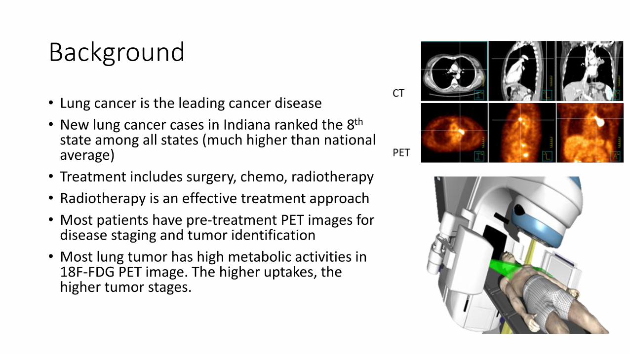
• Lung cancer is the leading cancer disease
• New lung cancer cases in Indiana ranked the 8th
state among all states (much higher than national average)
• Treatment includes surgery, chemo, radiotherapy
• Radiotherapy is an effective treatment approach
• Most patients have pre-treatment PET images for disease staging and tumor identification
• Most lung tumor has high metabolic activities in 18F-FDG PET image. The higher uptakes, the higher tumor stages.

Aims and Approach
Study goals:
• Identify the correlation between FDG activity and patient survival outcome
• Develop treatment strategy to improve treatment outcome
Methods:
• Data collections: image and clinical data
• Image registration: deformable
• Eclipse external beam treatment planning
• Image segmentation
• Data analysis: correlation, regression, and deep learning
Requirements: computer programming, algorithm development

In-treatment Room MR-Ultrasound Image-guided Radiotherapy for Prostate Cancer Patients
Yong Yue, PhD and Yi Le, PhD
Department of Radiation Oncology
Indiana University School of Medicine
Email: [email protected]: 317-962-3549
Email: [email protected]: 317-962-3388
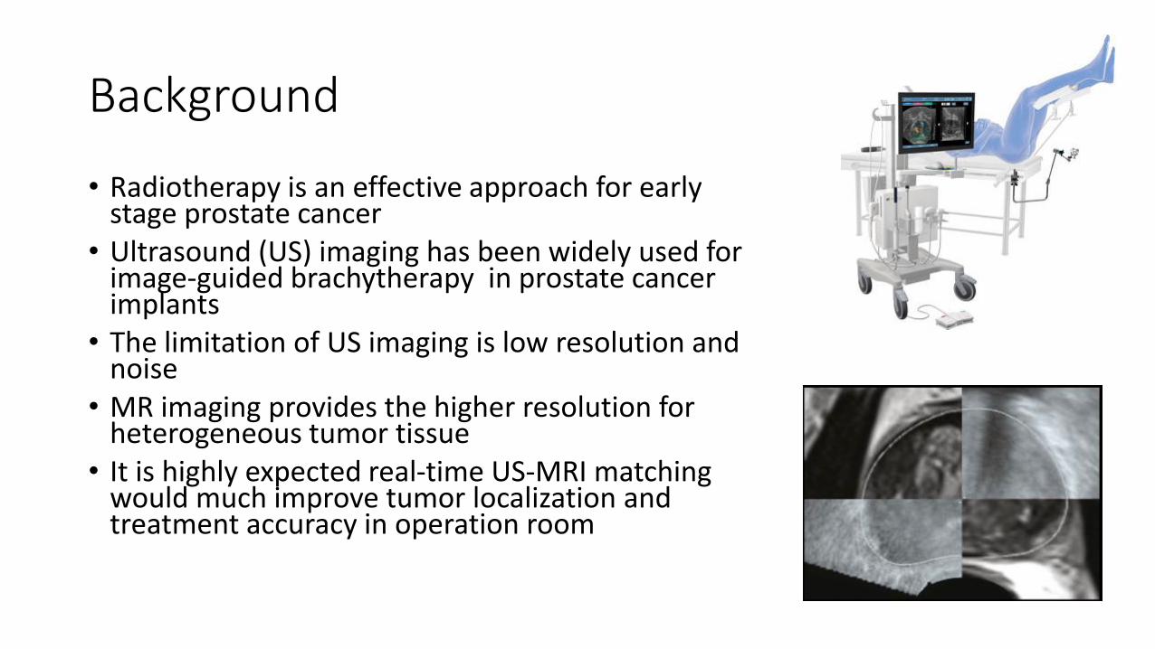
Background
• Radiotherapy is an effective approach for early stage prostate cancer
• Ultrasound (US) imaging has been widely used for image-guided brachytherapy in prostate cancer implants
• The limitation of US imaging is low resolution and noise
• MR imaging provides the higher resolution for heterogeneous tumor tissue
• It is highly expected real-time US-MRI matching would much improve tumor localization and treatment accuracy in operation room

Aims and Approach
Study goals: • Real-time ultrasound and MRI image registration• In operation room implementation and work flow setup for prostate
brachytherapyApproaches:• MRI and US image data collection• 3D Slicer software • Novel image registration approach development• Brachytherapy treatment planning
Requirements: computer programming, algorithm development

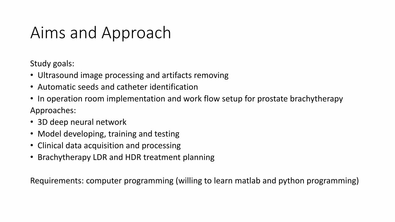
Improve image quality and auto-catheters/seeds segmentation in ultrasound based prostate
brachytherapy
Yi Le, PhD and Yong Yue, PhD
Department of Radiation Oncology
Indiana University School of Medicine
Email: [email protected]: 317-962-3549
Email: [email protected]: 317-962-3388

Background
• Ultrasound (US) imaging has been widely used for image-guided brachytherapy in prostate cancer implants for both LDR seed implant and HDR afterloader treatment
• The limitation of US imaging is low resolution, noise and artifacts from seeds and catheters
• Better imaging processing to remove artifacts and automatic seeds and catheters identification will greatly improve implant quality and clinical workflow
Aims and Approach
Study goals:
• Ultrasound image processing and artifacts removing
• Automatic seeds and catheter identification
• In operation room implementation and work flow setup for prostate brachytherapy
Approaches:
• 3D deep neural network
• Model developing, training and testing
• Clinical data acquisition and processing
• Brachytherapy LDR and HDR treatment planning
Requirements: computer programming (willing to learn matlab and python programming)

Small field dosimetry using EPID and portal dosimetry
Yi Le, PhD, DABR
Associate Professor
Department of Radiation Oncology
Indiana University School of Medicine
Email: [email protected]: 317-962-3388

Hands on project
• MV portal imaging originally used for localization
• Recently used as dosimetric verification method for IMRT/VMAT
• Use portal imager for small field dosmetricmeasurement
• Feasibility of output and profile measurement comparing to small ion chamber, diode and film measurement
• Additional dosimetry modeling for small field SRS/SBRT treatment QA

Research ProjectsColin Huang, PhD, Assistant Professor/Medical Physicist
Radiation Oncology, IU School of Medicine
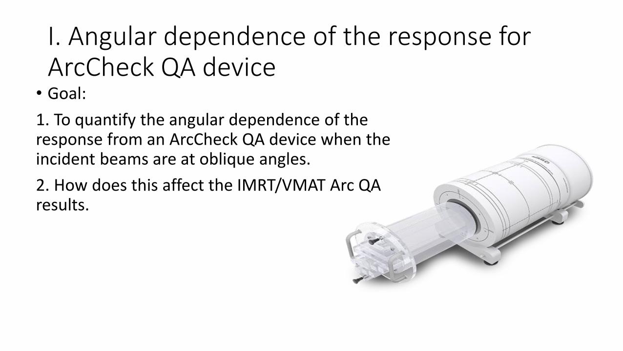
• Goal:
1. To quantify the angular dependence of the response from an ArcCheck QA device when the incident beams are at oblique angles.
2. How does this affect the IMRT/VMAT Arc QA results.
I. Angular dependence of the response for ArcCheck QA device

• Learn to operate the linear accelerator, including setting collimator, gantry, and couch angles; setting field size beam energy, MUs and beam on. (under supervision)
• Become an expert of using the ArchCheck QA software and device to do measurements and perform analysis.
What can you get from this project

• Learn to create IMRT/VMAT Arc QA plans from original plan
• Learn to deliver a IMRT/VMAT Arc QA plan and use ArcCheck to analyze the QA results.
• Write a conference abstract
What can you get from this project

Flash Radiotherapy and PHASOR
Peter Maxim, PhD, DABR
Associate Professor and Director of Medical Physics
Department of Radiation Oncology
Indiana University School of Medicine
Email: [email protected]: 317-944-1185
• Potential Ph.D. thesis
• Potential Ph.D. thesis