Embed Size (px)
Citation preview

RESEARCH ARTICLE 1 2
SCD1 and SCD2 Form a Complex that Functions with the Exocyst and 3
RabE1 in Exocytosis and Cytokinesis 4
5 Jonathan Russell Mayersa, Tianwei Hub, Chao Wangb, Jessica J. Cárdenasa Yuqi Tana, 6 Jianwei Panb,c, Sebastian Y. Bednareka* 7 8 a Department of Biochemistry University of Wisconsin-Madison, Madison, Wisconsin 53706 9 b Ministry of Education Key Laboratory of Cell Activities and Stress Adaptations, School of Life 10 Sciences, Lanzhou University, Lanzhou 730000, China 11 c College of Chemistry and Life Sciences, Zhejiang Normal University, Jinhua 321004, China 12 *Corresponding author: [email protected] 13 14 Short Title: The SCD Complex Functions in Exocytosis 15 16 One-sentence summary: Plants have evolved to utilize the SCD complex, which is 17 architecturally distinct from—but performs similar functions as—the structurally-related exocytic 18 yeast and mammalian Sec2/Rabin8 Rab GEFs. 19 20 The author responsible for distribution of materials integral to the findings presented in this 21 article in accordance with the policy described in the Instructions for Authors (www.plantcell.org) 22 is: Sebastian Y. Bednarek ([email protected]). 23 24 ABSTRACT 25 Although exocytosis is critical for the proper trafficking of materials to the plasma membrane, 26 relatively little is known about the mechanistic details of post-Golgi trafficking in plants. Here we 27 demonstrate that the DENN (Differentially Expressed in Normal and Neoplastic cells) domain 28 protein SCD1 (STOMATAL CYTOKINESIS DEFECTIVE 1) and SCD2 form a previously 29 unknown protein complex, the SCD complex, that functionally interacts with subunits of the 30 exocyst complex and a member of the RabE1 family of GTPases in Arabidopsis thaliana. 31 Consistent with a role in post-Golgi trafficking, scd1 and scd2 mutants display defects in 32 exocytosis and recycling of PIN2-GFP. Perturbation of exocytosis using the small molecule 33 Endosidin2 results in growth inhibition and PIN2-GFP trafficking defects in scd1 and scd2 34 mutants. In addition to the exocyst, the SCD complex binds in a nucleotide state-specific 35 manner with Sec4p/Rab8-related RabE1 GTPases and overexpression of wild-type RabE1 36 rescues scd1 temperature-sensitive mutants. Furthermore, SCD1 colocalizes with the exocyst 37 subunit, SEC15B, and RabE1 at the cell plate and in distinct punctae at or near the plasma 38 membrane. Our findings reveal a mechanism for plant exocytosis, through the identification and 39 characterization of a protein interaction network that includes the SCD complex, RabE1, and the 40 exocyst. 41 42 43
44
Plant Cell Advance Publication. Published on September 29, 2017, doi:10.1105/tpc.17.00409
©2017 American Society of Plant Biologists. All Rights Reserved

INTRODUCTION 45
Vesicular trafficking to and from the plasma membrane is paramount to plant 46
growth and development, as it facilitates multiple important processes including cell wall 47
biosynthesis, nutrient uptake, hormone signaling, and pathogen defense (Takano et al., 48
2005; Tanaka et al., 2006; Robatzek, 2007). Similarly, in dividing cells exocytic and 49
endocytic trafficking pathways are essential for the formation of the cytokinetic organelle 50
known as the cell plate. 51
Proper regulation and function of biosynthetic secretory and endocytic 52
membrane trafficking pathways depend on stage-specific Rab GTPases. In their GTP-53
bound form, Rabs recruit divergent effectors to coordinate the formation, transport, 54
tethering and fusion of transport vesicles and organelles. To function properly, Rabs 55
must continually cycle between active and inactive forms through the exchange of GTP 56
and GDP via interactions with Rab GEFs (guanine nucleotide exchange factors) and 57
Rab GAPs (GTPase activating proteins)(Stenmark, 2009). Thus, the association of Rab 58
GTPases with downstream effector proteins is inherently dependent upon their 59
interactions with GEFs and GAPs. Surprisingly, of the 57 Arabidopsis thaliana Rabs, 60
only a few of their GEFs/GAPs and downstream effector proteins are known (Preuss et 61
al., 2006; Goh et al., 2007; Camacho et al., 2009; Thellmann et al., 2010; Qi and Zheng, 62
2011; Fukuda et al., 2013; Singh et al., 2014). Therefore, despite recent advances in 63
our knowledge of Rab function in plants, a significant gap remains in our understanding 64
of the molecular machinery involved in their regulation. 65
In yeast, communication between the GEF Sec2p and the Rab GTPases Ypt32p 66
and Sec4p establishes a functional connection between cargo-containing Golgi-derived 67
exocytic vesicles and the molecular machinery necessary for their targeting and fusion 68
with the plasma membrane (Ortiz et al., 2002). Specifically, Ypt32p recruits Sec2p to 69
the trans-Golgi network which in turn binds and activates the Sec4p, which facilitates 70
interactions with the exocyst complex, an evolutionarily conserved eight-subunit 71
complex consisting of Sec3, Sec5, Sec6, Sec8, Sec10, Sec15, Exo70, and Exo84, to 72
promote docking with the plasma membrane prior to fusion (Hammer and Sellers, 2012; 73
Wu and Guo, 2015; Vukasinovic and Zarsky, 2016). A similar mechanism occurs in 74
mammalian cells, in which a Rab cascade comprising Rab11, Rab8 and the Rab8 GEF 75

3
Rabin8 promotes exocyst-dependent vesicle targeting to the plasma membrane 76
(Knodler et al., 2010; Westlake et al., 2011; Mizuno-Yamasaki et al., 2012). 77
Similar to its function in yeast and mammalian cells (Heider and Munson, 2012; 78
Wu and Guo, 2015), the exocyst plays an important role in the trafficking of materials to 79
the plasma membrane, and also functions in multiple steps during cytokinesis and cell 80
plate formation in plants (McMichael and Bednarek, 2013; Rybak et al., 2014). 81
Arabidopsis exocyst subunit mutants exhibit developmental phenotypes including 82
dwarfism, improper guard cell cytokinesis, and cell plate maturation defects (Fendrych 83
et al., 2010; Drdova et al., 2013; Rybak et al., 2014; Wu and Guo, 2015). However, 84
although homologs for Ypt32/Rab11, Sec4p/Rab8 and exocyst subunits are known to 85
function in exocytosis and cytokinesis in plants, the molecular details of their 86
interactions are not known. 87
Arabidopsis scd1 (stomatal cytokinesis defective 1) and scd2 mutants exhibit 88
strikingly similar phenotypes to exocyst mutants: plants are dwarfed and have defects in 89
cell division and expansion that result in guard cell cytokinesis and root hair 90
morphogenesis defects (Falbel et al., 2003; Korasick et al., 2010; McMichael et al., 91
2013). In addition, SCD1 and SCD2 genetically interact and are associated with 92
clathrin-coated vesicles (CCVs), suggesting a role for these proteins in membrane 93
trafficking during cytokinesis and cell expansion including endocytosis (McMichael and 94
Bednarek, 2013; McMichael et al., 2013). The SCD1 protein is defined by an N-terminal 95
DENN (Differentially Expressed in Normal and Neoplastic cells) domain that in 96
metazoans has been demonstrated to activate Rab GTPases (Marat and McPherson, 97
2010; Yoshimura et al., 2010; Marat et al., 2012); however, a connection between 98
DENN-domain proteins and Rab GTPases has not been established in plants. 99
Members of the plant RabE1 GTPase (RabE1a-e) family are most closely 100
related to mammalian Rab8 and S. cerevisiae Sec4p (Rutherford and Moore, 101
2002). Live-cell imaging has demonstrated that RabE1d and RabE1c localize to the 102
Golgi stacks, plasma membrane, and the cell plate in dividing cells (Zheng et al., 2005; 103
Chow et al., 2008; Speth et al., 2009). Moreover, functional studies have indicated that 104
RabE1s act in post-Golgi trafficking to the plasma membrane and cell plate (Speth et 105
al., 2009; Ahn et al., 2013). Similar to the scd1 and scd2 mutant phenotypes, silencing 106

4
of RabE1 expression in Nicotiana benthamiana results in defective guard mother cell 107
cytokinesis, and overexpression of dominant-negative mutant RabE1 in plants 108
manifests in shoot and root growth defects (Speth et al., 2009; Ahn et al., 2013). 109
To date, only a few factors that function to regulate the exocyst complex have 110
been identified in plants (Lavy et al., 2007; Hazak et al., 2010). Here we show, through 111
the use of proteomics, in vitro binding studies, cargo trafficking assays, and 112
colocalization analysis, that SCD1 and SCD2 are subunits of a protein complex, which 113
we refer to as the SCD complex, that functions together with RabE1s and the exocyst in 114
a protein interaction network to mediate post-Golgi trafficking to the plasma 115
membrane and cell plate. 116
117
118

5
RESULTS 119
SCD1 and SCD2 are Subunits of an Oligomeric Protein Complex 120
Previously we demonstrated that SCD1 and SCD2 function in membrane 121
transport required for cytokinesis and cell expansion (McMichael et al., 2013). To further 122
define the protein interaction network of SCD1 and SCD2, we generated Arabidopsis 123
cell lines (PSB-d) that express N-terminal G-protein/Streptavidin-binding peptide (GS)-124
tagged SCD1 (GS-SCD1), SCD2 (GS-SCD2), or GFP (GS-GFP) fusion proteins. GS-125
SCD1, GS-SCD2, and GS-GFP were purified from Arabidopsis cell extracts using 126
tandem affinity purification (TAP) chromatography as described (Van Leene et al., 127
2011). TAP elutions were analyzed by SDS-PAGE (Figure 1A, Supplemental Figure 1) 128
and liquid chromatography tandem mass spectrometry (LC/MS/MS) (Figure 1B, 129
Supplemental Data Set 1). The major polypeptides detected in the TAP GS-SCD1 and 130
GS-SCD2 elutions migrated in SDS-PAGE at the expected molecular mass of GS-131
SCD1 (~140 kD) and GS-SCD2 (~70 kD), respectively, with the G-protein removed 132
during purification as described (Van Leene et al., 2011) (Figure 1A). In addition, 133
polypeptides of the expected size for endogenous SCD1 (~132 kD) and SCD2 (~64 kD) 134
were observed in the GS-SCD1 and GS-SCD2 TAP fractions, respectively. LC/MS/MS 135
analysis confirmed that endogenous untagged SCD2 co-purified with GS-SCD1 and 136
vice versa (Figure 1B, Supplemental Data Set 1) indicating that SCD1 and SCD2 137
physically interact. The association of endogenous untagged SCD1 and SCD2 was 138
further analyzed using independent methods. Immunoprecipitation of untransformed 139
Arabidopsis cell lysate using anti-SCD2 antibodies demonstrated co-140
immunoprecipitation of SCD1 with SCD2 under standard (100mM KCl) and higher salt 141
(300mM KCl) conditions, indicating that the association of SCD1 and SCD2 is salt 142
stable (Figure 1D). Furthermore, SCD1 and SCD2 were found to co-fractionate by 143
velocity gradient sedimentation analysis, which separates proteins based on their native 144
molecular mass and hydrodynamic properties (Harding, 1999). Specifically, Arabidopsis 145
cell extracts were fractionated by glycerol gradient sedimentation and analyzed by 146
immunoblotting with antibodies against both SCD1 and SCD2 (Figure 1C). Quantitative 147
analysis of the immunoblots revealed that endogenous SCD1 and SCD2 fractionated in 148
separate slower sedimenting peaks corresponding to their expected monomeric forms 149

6
(molecular masses 132 and 64 kD, respectively), as well as in an overlapping ~17S 150

7
peak with an estimated molecular mass of ~430 kD, which is larger than expected for a 151
complex containing only single copies of SCD1 and SCD2, suggesting that SCD1 and 152
SCD2 are subunits of a multimeric protein complex. In addition, LC/MS/MS analysis of 153
proteins that co-purified with GS-SCD1 and GS-SCD2 identified two SCD2-like 154
polypeptides encoded by At5g23700 and At5g13260, which we have named SCD2c 155
and SCD2b respectively. SCD2c and SCD2b share 75% and 56% amino acid identity 156
with SCD2, respectively (Figure 1B, Supplemental Data Set 1). Similar to SCD2, these 157
two SCD2-like proteins are predicted to contain two centrally located coiled-coil 158
domains as well as a ProDom-defined (Corpet et al., 1998), plant-specific domain of 159
unknown function, PD147848, at their respective carboxyl termini. LC/MS/MS analysis 160
of proteins that co-purified with GS-GFP did not show similar enrichment profiles of 161
interacting proteins (Supplemental Data Set 1, Supplemental Table 1). A small amount 162
of RabE1c co-purified with GS-GFP, but its abundance in the GS-SCD1 and GS-SCD2 163
samples was 9- and 29-fold higher respectively, based on Exponentially Modified 164
Protein Abundance Index (emPAI) as determined by Mascot (Supplemental Table 1). 165
166
scd Mutants Display Post-Golgi Trafficking Defects 167
Arabidopsis scd1 and scd2 mutant plants exhibit cytokinesis and cell expansion 168
defects, including impaired internalization of the endocytic tracer dye, FM4-64, 169
indicative of potential roles in cell plate and plasma membrane trafficking (Falbel et al., 170
2003; McMichael et al., 2013). LC/MS/MS analysis of GS-TAP experiments have 171
identified exocyst subunits (SEC3A, SEC5, SEC6, SEC10, SEC15B, EXO70A1, and 172
EXO84B) as potential interactors with SCD1 and SCD2, suggesting that the SCD 173
complex may function in exocytic vesicle trafficking to the cell plate and plasma 174
membrane (Figure 1B, Supplemental Data Set 1). To test this, we assayed the 175
trafficking of the plant plasma membrane protein PIN-FORMED2 fused to green 176
fluorescent protein (PIN2-GFP) (Xu and Scheres, 2005) in scd1 and scd2 mutants. 177
PIN2 is a member of the PIN family of transport proteins that functions to transport 178
auxin, a crucial developmental hormone, across the plasma membrane (Krecek et al., 179
2009). PIN2-GFP is trafficked from the trans-Golgi network/early endosome to the 180
plasma membrane where it can undergo rounds of constitutive endocytosis and 181

8
recycling from endosomes to the plasma membrane or be transported to the vacuole for 182

9
degradation (Lofke et al., 2013; Wang et al., 2013). To monitor GFP-PIN2 trafficking, 183
wild-type (Col-0) and loss-of-function scd1-2 and scd2-1 mutant plants expressing 184
PIN2-GFP, under control of its native promoter (ProPIN2:PIN2-GFP), were treated with 185
the reversible vesicle trafficking inhibitor Brefeldin A (BFA), which causes formation of 186
intracellular endosomal and trans-Golgi compartment aggregates (BFA bodies) (Rosa et 187
al., 1992; Ritzenthaler et al., 2002; Wang et al., 2013). Following 50 µM BFA treatment 188
for 60 min, de novo synthesized and internalized PIN2-GFP accumulated in 1–2 BFA 189
bodies/root cell in scd1-2, scd2-1, and wild-type plants as monitored by confocal 190
microscopy (Figure 2). Time course assays demonstrated that the levels of intracellular 191
PIN2-GFP decreased upon BFA removal in wild-type, scd1-2, and scd2-1 root cells, 192
indicative of the restoration of protein trafficking to the plasma membrane (Dhonukshe 193
et al., 2007). Relative to wild-type however, scd1-2 and scd2-1 mutants displayed a 194
significant delay in the rate of loss of PIN2-GFP-positive BFA bodies per cell and higher 195
PIN2-GFP fluorescent signal intensity (Figure 2A-2O). 196
To distinguish between the trafficking of de novo synthesized and internalized 197
PIN2-GFP, BFA washout experiments were performed in the presence of cycloheximide 198
(CHX), an inhibitor of eukaryotic translation (Schneider-Poetsch et al., 2010). In the 199
presence of CHX and BFA, the number of PIN2-GFP labeled BFA bodies/cell was found 200
to be reduced in both scd mutants compared to wild type (Figure 2P), indicative of an 201
endocytosis defect and consistent with previous data showing that scd1 and scd2 202
mutants exhibit defects in the uptake of the endocytic tracer dye, FM4-64 (McMichael et 203
al., 2013). In addition to a reduction in endocytosis, recycling of PIN2-GFP from BFA 204
bodies was delayed in scd mutants. As shown in Figure 2P-2Q, after normalization for 205
the initial number of BFA bodies per root cell in the presence of CHX, we observed a 206
decrease in the rate of PIN2-GFP-labeled BFA body disappearance in scd1-2 and scd2-207
1 mutants following removal of BFA. Similarly, scd mutants had increased PIN2-GFP 208
intensity per BFA body area per cell after washout (Figure 2R). Taken together, our 209
analysis of PIN2-GFP trafficking in the scd mutants indicates that the SCD complex 210
functions in exocytosis and endosomal recycling of plasma membrane proteins. 211
212
The SCD Complex Communicates with the Exocyst Complex 213

10
Given that the SCD complex functions in post-Golgi trafficking, and the 214
identification of exocyst subunits as putative SCD complex interactors (Figure 1B, 215
Supplemental Data Set 1), we further explored the relationship between the SCD 216
complex and the exocyst. To determine if SCD complex-mediated post-Golgi trafficking 217
to the plasma membrane is dependent on exocyst function, we performed 218
pharmacological studies with the small molecule Endosidin2 (ES2), which selectively 219
inhibits exocytosis by binding to exocyst subunit Exo70 (Zhang et al., 2016). As 220
previously shown, ES2 treatment of wild-type seedlings expressing PIN2-GFP blocks 221
exocytosis and endosomal recycling, resulting in the intracellular accumulation of PIN2-222
GFP in punctate structures (Figure 3A) that are distinct from BFA bodies (Zhang et al., 223
2016). Interestingly, ES2 was found to enhance the intracellular accumulation of PIN2-224
GFP in both scd1 and scd2 mutants compared to wild-type plants. Specifically, scd1-2 225
and scd2-1 mutant root cells treated with 40 µM ES2 showed 2.3-fold and 2.6-fold 226
elevation, respectively, in the average number of PIN2-GFP labeled-punctae/cell 227
(Figure 3A-3D) relative to ES2-treated wild-type (Col-0) root cells. These intracellular 228
PIN2-GFP punctae were not detectable in untreated Col-0 and scd mutants (Figure 2S-229
U). To confirm the presence of internalized PIN2-GFP in ES2 bodies, experiments were 230
performed with CHX. Treatment with both ES2 and CHX together enhanced intracellular 231
accumulation of PIN2-GFP, likely due to inhibition of recycling PIN2-GFP back to the 232
plasma membrane by ES2 (Supplemental Figure 2). 233
Homozygous temperature-sensitive scd1-1 grown above the permissive 234
temperature of 16°C and loss-of-function scd1-2 and scd2-1 plants are dwarfed and 235
display numerous growth defects including stunted roots (Falbel et al., 2003; McMichael 236
et al., 2013). To assess the effects of ES2 on root growth, wild-type (Col-0) and scd 237
mutant seedlings were germinated and grown for 13 days in the presence of DMSO or 238
30 µM ES2 and root length was measured. Relative to DMSO-treated control seedlings, 239
ES2-treated wild-type (Col-0) seedlings showed a ~10% reduction in root length. By 240
contrast, ES2 inhibited scd1-1, scd1-2 and scd2-1 seedling root growth by ~50%, ~80%, 241
and ~80%, respectively, relative to DMSO-treated scd mutant seedlings (Figure 3E, 3F). 242
The observed enhancement of growth defects observed in ES2-treated scd1 and scd2 243
mutants provides further support for a functional connection between the SCD and 244
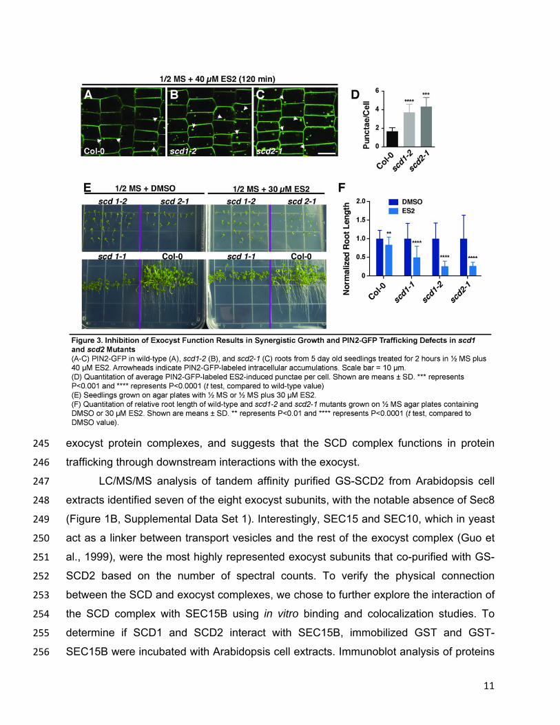
11
exocyst protein complexes, and suggests that the SCD complex functions in protein 245
trafficking through downstream interactions with the exocyst. 246
LC/MS/MS analysis of tandem affinity purified GS-SCD2 from Arabidopsis cell 247
extracts identified seven of the eight exocyst subunits, with the notable absence of Sec8 248
(Figure 1B, Supplemental Data Set 1). Interestingly, SEC15 and SEC10, which in yeast 249
act as a linker between transport vesicles and the rest of the exocyst complex (Guo et 250
al., 1999), were the most highly represented exocyst subunits that co-purified with GS-251
SCD2 based on the number of spectral counts. To verify the physical connection 252
between the SCD and exocyst complexes, we chose to further explore the interaction of 253
the SCD complex with SEC15B using in vitro binding and colocalization studies. To 254
determine if SCD1 and SCD2 interact with SEC15B, immobilized GST and GST-255
SEC15B were incubated with Arabidopsis cell extracts. Immunoblot analysis of proteins 256

12
bound to GST and GST-SEC15B demonstrated that SCD1 and SCD2 interacted with 257
GST-SEC15B and not with the GST negative control (Figure 4A). 258
Next, we utilized CLSM to assess whether GFP-SCD1 and red fluorescent 259
protein (RFP)-SEC15B colocalize. As previously shown (Fendrych et al., 2010; Rybak 260
et al., 2014), RFP-SEC15B predominantly localized to the cell plate and to punctate 261

13
structures at or adjacent to the plasma membrane in dividing and non-dividing cells, 262
respectively (Figure 4C, 4F). Similarly, SCD1 was associated with punctae closely 263
associated with the plasma membrane (McMichael et al., 2013) (Figure 4E) as well as 264
the cell plate in dividing cells (Figure 4B), which had previously not been reported. 265
Fluorescence intensity line scans across the cell plate (Figure 4D) and punctate 266
structures (Figure 4G) revealed overlapping intensity profiles of GFP-SCD1 and RFP-267
SEC15B consistent with colocalization (Figure 4H-4J). Furthermore, colocalization was 268
quantitatively verified for image pairs using the Costes randomization test (Costes et al., 269
2004). Images were analyzed with 100 Costes iterations returning a Costes P-Value of 270
1.00 for both punctae and cell plate localization with Pearson’s R values of 0.65 and 271
0.61 respectively. 272
Next, we utilized CLSM to assess whether SCD function is required for the 273
localization of the exocyst. As shown previously (Fendrych et al., 2010), GFP-SEC15B 274
was associated with the plasma membrane in non-dividing cells. However, the 275
subcellular distribution of GFP-SEC15B was altered in scd1-2 mutant root cells 276
compared to wild type (Figure 4K, 4L), with the most dramatic differences seen near the 277
plasma membrane. Line scan intensity measurements also demonstrated an increase in 278
the intracellular levels of GFP-SEC15B in scd1-2 mutants relative to wild type (Figure 279
4M, 4N). These data, together with the ES2 inhibitor and in vitro binding studies, 280
indicate that the SCD complex and exocyst function together to mediate post-Golgi 281
vesicle trafficking to the plasma membrane. 282
283
The SCD Complex Selectively Interacts with RabE1 GTPases 284
Given that SCD1 contains a tripartite DENN domain that has been demonstrated 285
in other systems to function as a GEF for Rab GTPases (Marat and McPherson, 2010; 286
Yoshimura et al., 2010; Marat et al., 2012), the SCD complex may function through 287
interactions with Rab GTPases to regulate exocytic vesicle trafficking. Consistent with 288
this, LC/MS/MS analysis of proteins associated with SCD1 and SCD2 identified all 289
members of the RabE1 family; specifically, RabE1a-e were identified by TAP and Co-IP 290
of GS-tagged and endogenous SCD proteins (Figure 1B, Supplemental Data Set 1). 291
Previously published reports have implicated RabE1 GTPases in post-Golgi trafficking 292

14
to the plasma membrane and cell plate (Zheng et al., 2005; Speth et al., 2009; Ahn et 293
al., 2013), suggesting that the SCD complex’s role in membrane trafficking may be 294
through its interaction with RabE1. We utilized colocalization analysis and in vitro 295
binding studies to validate the interaction of the SCD complex with RabE1 GTPases. 296
CLSM imaging of root cells from Arabidopsis lines that express GFP-SCD1 and 297
mO-RabE1c showed colocalization between GFP-SCD1 and mO-RabE1c at distinct 298
intracellular punctae, some of which appear at or near the plasma membrane, as well 299
as at the cell plate (Figure 5A-5H). Fluorescence intensity line scans across the cell 300
plate (Figure 5C, 5G) and punctate structures (Figure 5F, 5H) revealed overlapping 301
intensity profiles of GFP-SCD1 and mO-RabE1c consistent with colocalization. 302
Additionally, colocalization was quantitatively verified for image pairs as described 303
above, with 100 Costes iterations returning a Costes P-value of 1.00 for both punctae 304
and cell plate localization with Pearson’s R values of 0.50 and 0.61 respectively. 305
To confirm the specificity of RabE1 GTPase interaction with the SCD complex, in 306
vitro binding studies were performed with GST-tagged RabE1 and members of other 307
Arabidopsis Rab GTPase families that function in various stages of the plant 308
biosynthetic secretory and endocytic pathways. Following incubation of immobilized 309
candidate GST-Rab fusion proteins with Arabidopsis cell extracts, bound proteins were 310
eluted and analyzed by immunoblotting. As shown in Figure 5I, SCD1 selectively bound 311
to RabE1c. Binding of SCD1 to GST alone, or to GST-tagged members of other Rab 312
GTPases families including RabA5, A4, A2, C1, D2, G3, and H1, was not detected 313
(Figure 5I, 5J). SCD complex association with any other Rab families was also not 314
detected by LC/MS/MS (Supplemental Data Set 1). Furthermore, both SCD1 and SCD2 315
bound to all RabE1 family members in vitro, (Figure 5J) but not to the GST control. 316
To further define the interaction between RabE1 and the SCD complex, we 317
tested the binding of SCD proteins from Arabidopsis cell extract to nucleotide state-318
specific RabE1c mutants in vitro. SCD1 and SCD2 bound to either wild-type RabE1c or 319
to the S29N mutant RabE1c, RabE1cS29N, which is analogous to dominant inhibitory H-320
rasS17N mutant that displays reduced nucleotide affinity (Farnsworth and Feig, 1991; 321
Nassar et al., 2010). By contrast, no binding was observed to the RabE1cQ74L mutant 322
(Figure 5K), which is predicted to have a reduced intrinsic hydrolysis rate of GTP as 323

15
demonstrated for other Rab proteins (Walworth et al., 1998). These data demonstrate 324
that the SCD1 and SCD2 bind RabE1c in a nucleotide-dependent manner. 325
To test whether the SCD complex functions in the activation of RabE, we 326
determined if overexpression of wild-type RabE1 would rescue the conditional growth 327
and developmental defects of partial loss-of-function scd1-1 mutants. Precedence for 328
this has been established as Rab Sec4p was shown to rescue the temperature-329
sensitive growth defects of mutant alleles of its GEF Sec2p, in S. cerevisiae (Nair et al., 330

16
1990; Walch-Solimena et al., 1997). To determine if RabE1 overexpression rescues the 331

17
scd1 mutant, we generated multiple independent transgenic scd1-1 mutant lines that 332
express wild-type N-terminal mOrange (mO)-tagged RabE1c (mO-RabE1c) under the 333
control of the constitutive 35S-CaMV promoter. Whereas partial loss-of-function scd1-1 334
mutant plants exhibit temperature-sensitive growth and stomatal cytokinesis defects at 335
temperatures at or above 22°C (Falbel et al., 2003; McMichael et al., 2013), scd1-1 336
mO-RabE1c grown at 22°C were found to be phenotypically similar to wild-type (Col-0) 337
plants at various stages of development including leaf expansion, inflorescence growth, 338
and guard cell formation (Figure 6A-6C). By contrast, overexpression of mO-RabD2b 339
(scd1-1 mO-RabD2b), which is involved in ER to Golgi trafficking (Zheng et al., 2005) 340
failed to suppress the growth defects of scd1-1 plants (Supplemental Figure 3A-3B). 341
To determine if RabE1 suppression of the scd1-1 phenotype was dependent on 342
nucleotide state we examined the effect of overexpression of GTPase deficient 343
“constitutively active” RabE1cQ74L and “inactive” RabE1cS29N mutants. Previous studies 344
have reported that expression of GFP-RabE1dQ74L in wild-type plants did not 345
significantly affect plant growth and development (Speth et al., 2009; Ahn et al., 2013). 346
Similar to scd1-1 plants that expressed wild-type mO-RabE1c, the scd1-1 mO-347
RabE1cQ74L lines showed significant rescue of the scd1-1 mutant growth and 348
development defects (Figure 6A-6C). However, whereas bolt height and stomatal 349
development were restored in the scd1 mO-RabE1cQ74L lines, the level of suppression 350
of the scd1-1 phenotype was significantly reduced relative to scd1-1 plants expressing 351
wild-type mO-RabE1c (Figure 6A-6C). In contrast to scd1-1 mO-RabE1c and scd1-1 352
mO-RabE1cQ74L lines, scd1-1 mO-RabE1cS29N plants were phenotypically 353
indistinguishable from the parental scd1-1 lines (Figure 6A-6C). Overexpression of mO-354
RabE1cS29N did not suppress or enhance growth and guard cell cytokinesis defects 355
associated with the scd1-1 mutation, providing further evidence that the scd1-1 356
phenotype is likely due to defects in its nucleotide state-specific interaction with RabE1. 357
Expression of mO-RabD2b, mO-RabE1c, mO-RabE1cQ74L, and mO-RabE1cS29N was 358
confirmed by RT-PCR and immunoblot analysis (Supplemental Figure 3C, 3D). Taken 359
together, the biochemical interaction and genetic suppression studies suggest that the 360
SCD complex is required in some manner for RabE1 activation. 361
362

18
363

19
DISCUSSION 364
Here we demonstrate that the SCD1 and SCD2 proteins are members of the 365
SCD complex that functions in concert with the exocyst and RabE1s in post-Golgi 366
trafficking to the plasma membrane and cell plate (Figure 7). This conclusion is 367
supported by the results presented in this work as well as the previous phenotypic 368
characterization of scd, rabE1, and exocyst mutants (Speth et al., 2009; Fendrych et al., 369
2010; Ahn et al., 2013; Drdova et al., 2013; Rybak et al., 2014; Wu and Guo, 2015). 370
Exocyst mutants, including exo84b, sec6, and scd mutants show similar developmental 371
phenotypes including dwarfism, reduced leaf pavement cell expansion, and aberrant 372
guard mother cell cytokinesis (Falbel et al., 2003; Fendrych et al., 2010; McMichael et 373
al., 2013; Wu et al., 2013). At the cellular level, scd1 and scd2 share similar defects as 374
exocyst subunit loss-of-function mutants and ES2-treated cells (Zhang et al., 2016) in 375
exocytosis and endosomal recycling of the plasma membrane protein, PIN2-GFP, in the 376
presence of BFA (Drdova et al., 2013) (Figure 2), and show an accumulation of 377
secretory vesicles, indicating that the SCD and exocyst complexes function in post-378
Golgi vesicle targeting and fusion (Falbel et al., 2003; Fendrych et al., 2010). 379
Importantly, the SCD and exocyst complexes were found to colocalize at the cell plate 380
and in punctae adjacent to the plasma membrane (Figure 4B-4J) and to biochemically 381
interact by proteomic analysis of the SCD complex (Figure 1; Supplemental Data Set 1) 382
and by in vitro binding studies (Figure 4A). The interaction of the SCD and exocyst 383
complexes is further supported by the finding that ES2 inhibition of exocyst function 384
enhances growth and trafficking defects of scd1 and scd2 mutants (Figure 3). Given the 385
large number of EXO70 subunits, 23 in the Arabidopsis genome (Cvrckova et al., 2012), 386
ES2 experiments cannot distinguish whether the SCD complex functions in concert 387
solely with EXO70A1 and/or other EXO70-containing exocyst complexes in the 388
trafficking of proteins to the plasma membrane. 389
In addition to its association with the exocyst, the SCD complex was found to 390
interact with RabE1 GTPases, which have previously been shown to localize to the 391
Golgi, plasma membrane, and the cell plate (Zheng et al., 2005; Chow et al., 2008; 392
Speth et al., 2009). Similar to scd1 and scd2 mutants, silencing of NbRabE1 expression 393
or expression of dominant-negative NbRabE1S29N results in defects in protein trafficking 394

20
from the Golgi to the plasma membrane and inhibition of N. benthamiana plant growth 395
accompanied by defects in guard cell cytokinesis, polarized root hair expansion, and 396
pathogen defense responses (Falbel et al., 2003; Zheng et al., 2005; Speth et al., 2009; 397

21
Korasick et al., 2010; Ahn et al., 2013; McMichael et al., 2013). In this study, we show 398
that RabE1s bind to the SCD complex in a nucleotide-state specific manner and 399
colocalize at the cell plate and in punctae at or near the plasma membrane (Figure 1, 400
Supplemental Data Set 1, and Figure 5). 401
402
The SCD Complex Biochemically and Genetically Interacts with RabE1 403
The SCD1 subunit of the SCD complex contains an N-terminal tripartite DENN 404
domain, which in other systems has been demonstrated to possess GEF activity for 405
specific Rab GTPases (Allaire et al., 2010; Yoshimura et al., 2010), defining the DENN 406
domain as a protein module characteristic of vesicle-trafficking regulators. In particular, 407
DENN-domain-containing proteins connecdenn 1–3 are CCV-associated GEFs for 408
Rab35, which functions in CCV trafficking, endosomal recycling, actin regulation, and 409
cytokinesis in animals (Allaire et al., 2006; Kouranti et al., 2006; Patino-Lopez et al., 410
2008; Allaire et al., 2010; Marat et al., 2012). Our data showing that SCD complex binds 411
to “inactive” RabE1cS29N and not the “constitutively active” RabE1cQ74L (Figure 5K) is 412
intriguing and suggests that the SCD complex functions to activate members of the 413
RabE1 family. This hypothesis is further supported by the genetic evidence that 414
overexpression of wild-type RabE1c and constitutively active RabE1cQ74L, but not the 415
inactive RabE1cS29N mutant protein, suppressed the growth and developmental defects 416
in scd1-1 mutants (Figure 6). In a similar manner, overexpression of Sec4p in S. 417
cerevisiae rescues the temperature-sensitive growth defects of mutant alleles of its 418
GEF, Sec2p (Nair et al., 1990; Walch-Solimena et al., 1997), and expression of 419
“constitutively active” mutant RAB-A1cQ72L partially rescues the growth and 420
development of attrs130 mutant plants (Qi et al., 2011), which have defects in the plant 421
homolog of the yeast and mammalian Ypt32/Rab11 TRAPPII complex. In all these 422
cases, it is likely that when overexpressed the levels of GTP-bound or constitutive-423
active Rab GTPase are sufficient to overcome the need for GEF-catalyzed activation to 424
support vesicle trafficking. 425
In S. cerevisiae and mammalian cells, Sec4p and Rab8 are activated through the 426
structurally related GEFs Sec2p and Rabin8, respectively (Guo et al., 2013). Vesicle-427
associated GTP-bound Sec4p/Rab8 GTPases and their cognate GEFs interact with the 428

22
exocyst Sec15 subunit to promote docking and fusion of exocytic vesicles at the plasma 429
membrane (Huber et al., 1993; Guo et al., 1999; Feng et al., 2012). In contrast to 430
Sec4p- and Rab8-dependent trafficking, our understanding of the effectors involved in 431
RabE1 activation and post-Golgi vesicle docking/fusion is limited and unlike the 432
Sec4p/Rab8 GEFs, Sec2p and Rabin 8, and other DENN GEFs (Dong et al., 2007; 433
Allaire et al., 2010; Yoshimura et al., 2010; Marat et al., 2012; Vetter et al., 2015), the 434
architecture of the multisubunit SCD complex, with the exception of the SCD1 DENN 435
domain, is fundamentally distinct. Of interest are future experiments to determine if the 436
SCD complex functions as a GEF for RabE1. A complication in testing its GEF activity is 437
the multisubunit nature of the SCD complex as we are only able to purify relatively low 438
quantities of the complex from tissue-cultured cells, and individual subunits and 439
domains including the SCD1 DENN domain are not stable when individually purified, 440
thereby necessitating reconstitution of the intact complex. In addition, it will be important 441
to determine the molecular function(s) of the plant-specific protein SCD2. Furthermore, 442
the presence of two SCD2-like proteins (AT5G13260 and AT5G23700) in GS-SCD1, 443
GS-SCD2, and anti-SCD2 Co-IPs provides interesting insight for other subunits in the 444
SCD complex. While more work is required to fully delineate the subunit identity and 445
stoichiometry of the complex, it is tempting to speculate, given the co-fractionation of 446
these two proteins with SCD1 and SCD2, that they may be additional subunits of the 447
SCD complex. 448
Although we favor a model in which the SCD complex functions in the activation 449
of RabE1, we cannot rule out potential alternative roles for the SCD complex in RabE1-450
dependent membrane trafficking. In addition to GEFs and GAPs, Rab GTPases are 451
regulated by other factors including guanine nucleotide release factors, GDP 452
dissociation inhibitors (GDI), and GDI displacement factors (GDF), which serve to 453
deliver Rabs to membranes (Stenmark, 2009). The selective interaction of SCD 454
complex with bacterially expressed (i.e. non-prenylated) RabE1, (Figure 5) however, 455
appears to be distinct from that of GDIs which bind with high affinity exclusively to the 456
prenylated GDP-form of Rabs (Araki et al., 1990; Pylypenko et al., 2006) and display 457
limited specificity in Rab binding (Grosshans et al., 2006). Similarly, the GDF, Yip3 458
(Sivars et al., 2003), and the mammalian guanine-nucleotide release factor MSS4, 459

23
which has been proposed to function as a chaperone for nucleotide-free Rabs (Itzen et 460
al., 2006), bind to a number of distinct Rabs, unlike the SCD complex, which binds 461
selectively to RabE1s but not to other early or late secretory pathway Rabs (Figure 5I; 462
Supplemental Data Set 1). 463
464
The SCD Complex Functions in Exocytosis, Recycling and Endocytosis 465
The highly dynamic process of cell plate formation involves vesicle-mediated 466
delivery and retrieval of membrane from the division plane. Likewise, in non-dividing 467
cells the coordination of exocytosis and endocytosis is critical for polarized cell 468
expansion and the polarized distribution of plasma-membrane proteins. Interestingly, 469
scd1 and scd2 mutants display defects not only in exocytosis but also in the uptake of 470
the endocytic tracer dye FM4-64 (McMichael et al., 2013), and PIN2-GFP endocytosis 471
(Figure 2P, 2R). While the SCD Complex interaction network is composed of proteins 472
involved in exocytosis, scd mutants exhibit defects in both endocytosis and exocytosis 473
(Figure 7). Whether the SCD proteins function directly or indirectly in endocytosis 474
remains to be determined. Consistent with the latter, defects in retrograde trafficking 475
have been demonstrated to affect anterograde trafficking; for example, in human cells, 476
defects in GARP and VAMP4-dependent endosome to trans-Golgi network trafficking 477
result in an inhibition of recycling of factors necessary for the trafficking of proteins to 478
the plasma membrane (Hirata et al., 2015). By contrast, however, treatment of plant 479
cells with the small molecule Endosidin 16 (ES16), which inhibits RabA-dependent 480
exocytosis, does not affect endocytosis (Li et al., 2017). Nevertheless, it cannot be 481
excluded that the SCD/RabE1 and ES16-sensitive/RabA trafficking pathways are 482
distinct and that the SCD-dependent exocytic pathway is responsible for transporting 483
factors required for endocytosis and/or that the SCD complex functions directly in 484
endocytosis (Figure 7). Indeed, in S. cerevisiae, Sec4p and Sec2p have also been 485
shown to function in exocytosis and endocytosis (Riezman, 1985; Johansen et al., 486
2016) and work in diverse systems, including yeast, metazoans, and Trypanosoma 487
brucei has demonstrated a role for the exocyst in endocytosis (Riezman, 1985; Sommer 488
et al., 2005; Jose et al., 2015; Boehm et al., 2017). 489

24
Although scd1 and scd2 mutants exhibit strong growth and developmental 490
defects, PIN2-GFP exocytosis and/or endosomal recycling were found to be only 491
partially impaired in mutant root cells (Figure 2). This may reflect the existence of 492
RabE1 independent post-Golgi trafficking pathways including RabA-dependent 493
pathways that could compensate for the loss, in scd mutants, of RabE1-dependent 494
trafficking to the cell plate and/or plasma membrane. An alternative, but not mutually 495
exclusive, possibility is that the severe developmental defects observed in scd mutants 496
could be due to the lowered efficiency, as opposed to total inhibition, of the trafficking of 497
critical cell-type specific factors necessary at distinct times in plant development. 498
Our work demonstrates that SCD1 and SCD2 form a complex that functions in 499
the post-Golgi trafficking to the plasma membrane and cell plate, a process that is 500
mediated through interactions with RabE1 and the exocyst complex. 501
502
503

25
METHODS 504
505
Plant Materials 506
Columbia-0 lines used in this study were obtained from ABRC. scd1-1, scd1-2, and 507
scd2-1 lines are described in (Falbel et al., 2003; McMichael et al., 2013). PIN2-GFP 508
lines are described in (Xu and Scheres, 2005). 509
510
Transgenic Plants/scd1-1 Mutant Rescue 511
Transgenic plants created in this study were generated using the floral dip method, 512
using Agrobacterium tumefaciens strain EHA105 (Clough and Bent, 1998). Transgenic 513
scd1-1 mutant plants were transformed with the vector 35Spro:mO-RabE1c (or 514
35Spro:mO-RabD2b) (pSITE-II-5c) and the indicated point mutant constructs (Q74L or 515
S29N). Following selection with kanamycin on ½ MS +1% agar (w/v), T3 plants were 516
transferred to soil and grown at 22°C under 16-hour light (T12 fluorescent bulb ~160 517
µmol m-2 s-1) and 8-hour dark cycle and phenotypically analyzed for defects in bolt 518
height 60 days after germination. For the guard cell cytokinesis screen, stomata from 519
seedling leaves were analyzed in 14–20-day-old plants grown under continuous light 520
(T8 fluorescent bulb ~120 µmol m-2 s-1 grown on ½ MS + 0.6% agar (w/v). 521
Measurements included at least three independent transformants for each 522
overexpression line. Overexpression of mO-RabE1c and mutants was confirmed by 523
immunoblot analysis using anti-RabE1 antibodies (Speth et al., 2009), with anti-CDC48 524
served as a loading control (Rancour et al., 2002). 525
For GFP-SCD1 colocalization with RabE1c or SEC15B, scd1-1 ProSCD1:GFP-526
SCD1 (McMichael et al., 2013) lines were transformed as described above with either 527
35Spro:mO-RabE1c or UBQ10pro:RFP-SEC15B. Initial transformants were selected on 528
selective media (Kanamycin or Basta on ½ MS +0.6% agar) and propagated on soil. 529
Imaging of seedling roots was performed as described below. 530
531
Cloning/RT PCR 532
All PCR-generated cloning fragments used in this study were produced using Phusion 533
DNA polymerase (Thermo Fisher). All oligonucleotide primers were synthesized by 534

26
Integrated DNA Technologies. Detailed cloning information and primer sequences can 535
be found in Supplemental Tables 2 and 3. All clones were sequence verified by Sanger 536
Sequencing (UW-Madison Biotech Center). 537
Expression of mOrange-tagged Rabs in scd1-1 lines was verified by RT-PCR 538
with primers JM15 and JM16 specific for the mOrange tag, and isolation of mRNA was 539
verified in each sample using primers against UBQ10 (JM17 and JM18). Briefly, RNA 540
from seedlings was isolated using the RNeasy plant mini kit (Qiagen). Following 541
isolation 1 μg of RNA was used in a reverse transcriptase reaction with M-MuLV reverse 542
transcriptase (New England Biolabs) according to the manufacturer’s instructions. 543
Following RT-PCR, first-strand cDNA was diluted 25-fold in a PCR reaction using 544
primers JM15 and JM16 (mOrange) or JM17 and JM18 (UBQ10). 545
546
GST-Binding Experiments 547
GST-fusion proteins were purified using standard techniques. Briefly, frozen E. coli 548
pellets (containing IPTG induced over-night expression of GST-fusion proteins) were 549
thawed in GST buffer (PBS pH 7.4, 500mM NaCl, 0.1% Tween-20 and 1mM BME) in 550
the presence of 1mM lysozyme plus PMSF (1mM) and Benzamadine (1mM) then 551
subjected to sonication for 2 min of total on time (20sec on, 40sec off). Pellets were 552
clarified by centrifugation at 23,700 g before incubation with glutathione resin for 1 hour 553
at 4°C and batch washed 3x with GST buffer. For GST-pulldown experiments, 554
immobilized GST fusion proteins (50 μL) were incubated with 1.5 mL of T87 tissue 555
culture extract [3-day-old T87 cells were lysed in lysis buffer (50 mM Hepes pH 7.6, 100 556
mM KCl, 0.5 mM EDTA, 0.5 mM MgCl, 0.05% NP-40, and 10% glycerol + protease 557
and/or phosphatase inhibitors) as a 50:50 slurry of pelleted cells to lysis buffer with 558
depressurized nitrogen (1500 psi for 20 min on ice in a cell disruption vessel (Parr 559
Instruments)), and rotated for 15 min at 4°C after addition of 0.5% TX-100 followed by 560
clarification with ultracentrifugation at 134,878 g for 20 min in a TLA100.3 rotor] for 1 561
hour at 4°C then washed with lysis buffer (3x) before elution with 10mM glutathione in 562
elution buffer (50 mM Tris pH 8.1, 150 mM NaCl). Eluants were analyzed via SDS-563
PAGE and immunoblotting as described in figure legends. Affinity purified antibodies 564

27
against SCD1 and SCD2 were generated as described (Falbel et al., 2003; McMichael 565
et al., 2013). 566
567
Co-Immunoprecipitation/TAP Purification 568
Co-IPs were performed from cell extract prepared as described above (for each panel, 569
the same extract is used for each condition). For Co-IP, anti-SCD2 antibodies were 570
covalently linked to Protein A beads (Bio-Rad) using dimethylpimelimidate (Sigma). To 571
covalently link beads, Protein A beads were washed in PBS with 0.1% Tween-20 (PBS-572
t) three times. Beads were then resuspended in PBS-t plus 55 μg of SCD2 antibody 573
(McMichael et al., 2013) and incubated for 1 hour at room temperature. Following 574
incubation, beads were washed 3 times with PBS-t followed by 3 washes with 0.2 M 575
sodium borate pH 9.0. Dimethylpimelimidate (DMP, 22 mM) was added and the beads 576
were incubated for 30 min at room temperature. Beads were then washed 3 times with 577
0.2 M ethanolamine pH 8.5, 200 mM NaCl. For IP, antibody-linked beads (50 μL) were 578
incubated with ~ 1.5 mL of plant cell extract (prepared as described above) and washed 579
with lysis buffer (described above) 3 times, bound proteins were eluted with 100 mM 580
glycine and TCA precipitated. TCA pellets were analyzed using mass spectroscopy 581
(described below) or immunoblot techniques. 582
GS-TAP fusions to SCD1 and SCD2 (or GS-TAP GFP) were generated using 583
gateway cloning techniques into pKNGSTAP (Van Leene et al., 2011). Transgenic 584
PSBd cell lines were generated and TAP of GS-SCD1 and GS-SCD2 was performed as 585
described (Van Leene et al., 2011), with some modifications (see below). Briefly, 6-day-586
old transgenic PSBd cell lines expressing GS-SCD1, GS-SCD2, or GS-GFP were lysed 587
using depressurized nitrogen in lysis buffer (as described in GST-binding experiment 588
above). Following lysis, extracts were incubated with 0.5% TX-100 and clarified (as 589
described in GST-binding experiment above). After clarification, extracts were incubated 590
with human IgG Sepharose 6 (GE healthcare) for 1 hour at 4°C. Following incubation, 591
beads were batch washed with wash buffer (10 mM Tris pH 8.0, 150 mM NaCl, 0.1% 592
(v/v) NP-40, and 5% (v/v) ethylene glycol) 3 times, and incubated with ~200 units of 593
TEV protease for 60 min at 16°C. The supernatant was collected and incubated with 594
pre-equilibrated Streptavidin Sepharose Resin (GE Healthcare) for 1 hour at 4°C. 595

28
Following incubation, beads were batch washed with wash buffer 3x and eluted with 596
desthiobiotin in wash buffer. Eluants were TCA precipitated and analyzed by 597
immunoblot or mass spectroscopy analysis. 598
599
Mass Spectroscopy 600
Enzymatic “In Liquid” Digestion 601
Purified protein samples were TCA/acetone precipitated [10% TCA, 28% acetone final] 602
then pellets re-solubilized and denatured in 7.5 μl of 8M Urea / 50mM NH4HCO3 (pH8.5) 603
/ 1mM TrisHCl for 5 minutes. Samples were subsequently diluted to 30 μl for a reduction 604
step with 1.25 μl of 25 mM DTT, 2.5 μl MeOH, 18.75 μl 25mM NH4HCO3 (pH8.5). 605
Samples were incubated at 50̊C for 15 minutes and cooled on ice to room temperature 606
and then 1.5μl of 55 mM IAA was added for alkylation. Samples were incubated in 607
darkness at room temperature for 15 minutes. Reactions were quenched by adding 4 μl 608
of 25 mM DTT. Subsequently 2 μl of Trypsin/LysC solution (100ng/μl Trypsin/LysC Mix 609
from PROMEGA Corp. in 25mM NH4HCO3) and 12.5 μl of 25 mM NH4HCO3 (pH8.5) 610
were added to generate a 50 µl final volume. Digestion was conducted for 2 hours at 611
42°C then an additional 1 µl of trypsin/LysC solution was added. Digestion proceeded 612
overnight at 37°C. Reactions were terminated by acidification with 2.5% TFA 613
(Trifluoroacetic Acid) added to a 0.3% final concentration. 614
NanoLC-MS/MS 615
Digests were cleaned up using OMIX C18 SPE cartridges (Agilent, Palo Alto, CA) per 616
manufacturer protocol and eluted in 20 µl of 60/40/0.1% ACN/H2O/TFA, dried to 617
completion in the speed-vac and finally reconstituted in 20 µl of 0.1% formic acid. 618
Peptides were analyzed by nanoLC-MS/MS using the Agilent 1100 nanoflow system 619
(Agilent) connected to a new generation hybrid linear ion trap-orbitrap mass 620
spectrometer (LTQ-Orbitrap Elite™, Thermo Fisher Scientific) equipped with an EASY-621
Spray™ electrospray source. Chromatography of peptides prior to mass spectral 622
analysis was accomplished using capillary emitter column (PepMap® C18, 3 µM, 100 Å, 623
150x0.075 mm, Thermo Fisher Scientific) onto which 3 µl of extracted peptides was 624
automatically loaded. The NanoHPLC system delivered solvents A: 0.1% (v/v) formic 625
acid, and B: 99.9% (v/v) acetonitrile, 0.1% (v/v) formic acid, at 0.50 µL/min to load the 626

29
peptides (over a 30 minute period) and 0.2 µl/min to elute peptides directly into the 627
nano-electrospray with gradual gradient from 3% (v/v) B to 30% (v/v) B over 77 minutes 628
and concluded with 5 minute fast gradient from 30% (v/v) B to 50% (v/v) B, at which 629
time a 5 minute flash-out from 50-95% (v/v) B took place. As peptides eluted from the 630
HPLC-column/electrospray source survey MS scans were acquired in the Orbitrap with 631
a resolution of 120,000 followed by MS2 fragmentation of the 20 most intense peptides 632
detected in the MS1 scan from 300 to 2000 m/z; redundancy was limited by dynamic 633
exclusion. 634
Data analysis 635
Raw MS/MS data were converted to mgf file format using MSConvert (ProteoWizard: 636
Open Source Software for Rapid Proteomics Tools Development) for downstream 637
analysis. Resulting mgf files were used to search against Arabidopsis thaliana TAIR10 638
amino acid sequence database with a decoy reverse entries and a list of common 639
contaminants (70,857 total entries) using in-house Mascot search engine 2.2.07 (Matrix 640
Science) with variable Methionine oxidation with Asparagine and Glutamine 641
deamidation. Peptide mass tolerance was set at 15 ppm and fragment mass at 0.6 Da. 642
Protein annotations, significance of identification and spectral-based quantification was 643
done with help of Scaffold software (version 4.3.2, Proteome Software Inc., Portland, 644
OR). Peptide identifications were accepted if they exceeded specific database search 645
engine thresholds. Mascot identifications required that at least ion scores must be 646
greater than both the associated identity scores and 20. Protein identifications were 647
accepted if they contained at least 2 identified peptides. Protein probabilities were 648
assigned by the Protein Prophet algorithm (Nesvizhskii et al., 2003). Proteins that 649
contained similar peptides and could not be differentiated based on MS/MS analysis 650
alone were grouped to satisfy the principles of parsimony. 651
652
Glycerol Gradient Velocity Sedimentation 653
T87 tissue culture extract generated (as described in GST-pulldown above) and then 654
separated on a 10–40% (v/v) glycerol gradient by centrifugation at 35,000 rpm for 16 655
hours at 4°C in an SW 50.1 rotor. Following centrifugation, fractions were collected and 656
analyzed by immunoblot analysis following by SDS-PAGE as described in figure 657

30
legends. Densitometry was measured using Photoshop, graphs were generated in 658
GraphPad Prism. For protein standards, the following proteins were used: ovalbumin 659
(Sigma), bovine serum albumin (Sigma), yeast alcohol dehydrogenase (Sigma), sweet 660
potato beta-amylase (Sigma), catalase (Sigma), apoferritin (MP Biomedicals), 661
thyroglobulin(Sigma). Following centrifugation, SDS-PAGE gels were Coomassie 662
stained and quantified as described above and used to generate standard curves from 663
which experimental S-values were determined to make native molecular weight 664
determinations. 665
666
Microscopy 667
All colocalization experiments, ES2-treated PIN2-GFP experiments, and GFP-SEC15B 668
experiments were conducted on a CLSM (Nikon A1R-Si+). For colocalization studies, 669
roots of 5-day-old seedlings were grown on ½ MS plus 1% agar (continuous light T8 670
fluorescent bulb ~120 µmol m-2 s-1) (w/v) and imaged. For ES2 treatments, 5-day-old 671
seedlings grown on ½ MS 1% agar (w/v) plates were placed in ½ MS plus 40 µM ES2 672
for 2 hours at room temperature, roots were imaged by CLSM. A single focal plane was 673
used to determine the number of punctae per cell. For CHX plus ES2 experiments, 674
seedlings were grown as described above and left untreated, or pretreated with 50 µM 675
CHX for 30 min before additional treatment with 50 µM CHX with and without 40 µM 676
ES2 for 120 min. To verify our colocalization analysis, images were processed using 677
ImageJ Coloc 2 plug-in. Briefly, background subtraction from candidate images was 678
performed using rolling ball subtraction with a 200-pixel ball size. ROIs were selected 679
and run through Coloc 2 plug-in with 100 Costes randomizations using a PSF of 3. All 680
graphs were generated in GraphPad Prism. All line scans were performed using NIS-681
Elements software with a 2-pixel width, intensity over distance of the line scan was 682
plotted using GraphPad Prism. 683
For PIN2-GFP plus BFA trafficking experiments, seedlings were pretreated with 684
BFA for 60 min in ½ MS liquid media, and followed by washout with ½ MS liquid 685
medium for different lengths of time (0, 20, 40, and 60 min). For PIN2-GFP BFA+CHX 686
trafficking experiments, seedlings were pretreated with CHX for 30 min in ½ MS liquid 687
media, followed by washout with CHX and BFA for 60 min in ½ MS liquid media, and/or 688

31
finally by washout with ½ MS liquid medium for different lengths of time (0, 20, 40, and 689
60 min). Following treatment, seedlings were imaged by CLSM (Leica TCS SP5 AOBS). 690
691
Plant Growth with ES2 692
Wild-type (Col-0), scd1-1, scd1-2 and scd 2-1 mutant plants were grown vertically on ½ 693
MS 1% (w/v) agar with either DMSO or 30 µM ES2, under constant light (T8 fluorescent 694
bulb ~120 µmol m-2 s-1) at 22°C. Seedling images were taken using a digital camera 695
(Nikon CoolPix S8100) and the root length of 13-day-old seedlings was measured using 696
available tools in Photoshop. Graphs were generated using GraphPad Prism. 697
698
Quantification and Statistical Analysis 699
All statistically significance calculations were carried out using the Student’s t test using 700
GraphPad Prism software, and are shown in Supplemental Data Set 2. For PIN2-GFP 701
trafficking in the presence of BFA and/or CHX, sample sizes, P values are indicated in 702
the figure legends. For ES2-treated seedling growth experiments, P values are 703
indicated in the figure legend, sample sizes for DMSO treated are: Col-0 (26 roots), 704
scd1-1 (26 roots), scd1-2 (28 roots), scd 2-1 (10 roots). Sample sizes for ES2 treated 705
are: Col-0 (26 roots), scd1-1 (34 roots), scd1-2 (26 roots) and scd2-1 (17 roots). For 706
PIN2-GFP ES2 treatment experiments, the P values are indicated in the figure legends, 707
sample sizes are Col-0 (8 roots, 367 cells), scd1-2 (5 roots, 210 cells), and scd2-1 (4 708
roots, 221 cells). Significance calculations of colocalization experiments are described 709
in the “Microscopy” section. 710
711
Accession Numbers 712
The accession numbers for DNA sequences used in this study are as follows: 713
SCD1: AT1G4940, SCD2: AT3G48860, RabE1a: AT3G53610, RabE1b: AT5G59840, 714
RabE1c: AT3G46060, RabE1d: AT5G03520, RabE1e: AT3G09900, RabA5b: 715
AT3G07410, RabH1c: AT4G98890, RabC1: AT1G43890, RabA5e: AT1G05810, 716
RabA4a: AT5G65270, RabD2b: AT5G47200, SEC15B: AT4G02350. 717
718
Supplemental Data 719

32
Supplemental Figure 1. Tandem Affinity Purified GS-GFP. 720
Supplemental Figure 2. PIN2-GFP Trafficking in the Presence of CHX and ES2. 721
Supplemental Figure 3. RabD2b Overexpression Does Not Rescue scd1-1. 722
Supplemental Table 1. Mascot Protein Scores and Exponentially Modified Protein 723
Abundance Index (emPAI) for GS-TAP purifications. 724
Supplemental Table 2. Constructs Used in This Study 725
Supplemental Table 3. Primers Used in This Study 726
Supplemental Data Set 1. Detailed LC/MS/MS Data Sheet of Proteins Identified with 727
GS-TAP and Co-IP Experiments Performed in This Study. 728
Supplemental Data Set 2. T-test Tables 729
730
AUTHOR CONTRIBUTIONS 731
J.R.M., J.J.C., J.P., and S.Y.B. designed research; J.R.M., T. H., C. W., J.J.C., and732
Y.T., performed experiments; J.R.M., T. H., C. W., Y.T., J.P., and S.Y.B. analyzed data;733
J.R.M., and S.Y.B. wrote the paper.734
735
ACKNOWLEDGMENTS 736
The authors would like to thank members of the Bednarek lab and Richard Amasino for 737
helpful discussions and critically reading the manuscript. We also would like to thank 738
Elle Kielar Grevstad (UW-Madison Biochemistry) for assistance with confocal 739
microscopy and data analysis. We also thank Grzegorz Sabat for performing Mass 740
Spectroscopy (UW-Madison Biotechnology Center) and for help with data analysis. We 741
also like to thank Glenn Hicks for providing ES2 and Christopher Grefen for 742
pUBQN:XFP-Dest vectors. We also thank Sheng Yang He for the kind gift of RabE1 743
antibody. J.M. is supported by a fellowship from the National Institute of Health 744
(F32 GM112446-02). This work was supported by funding to S.Y.B from the UW 745
Madison College of Agriculture and Life Sciences and the National Science Foundation 746
(award No. 1121998 and 1614915), and to J.P. from the National Natural Science 747
Foundation of China (No. 31670283, 31370313, and 91317304). 748
749
750

Parsed CitationsAhn, C.S., Han, J., and Pai, H. (2013). Characterization of in vivo functions of Nicotiana benthamiana RabE1. Planta 237, 161-172.
Pubmed: Author and TitleCrossRef: Author and TitleGoogle Scholar: Author Only Title Only Author and Title
Allaire, P.D., Marat, A.L., Dall'Armi, C., Di Paolo, G., McPherson, P.S., and Ritter, B. (2010). The Connecdenn DENN domain: a GEF forRab35 mediating cargo-specific exit from early endosomes. Mol Cell 37, 370-382.
Pubmed: Author and TitleCrossRef: Author and TitleGoogle Scholar: Author Only Title Only Author and Title
Allaire, P.D., Ritter, B., Thomas, S., Burman, J.L., Denisov, A.Y., Legendre-Guillemin, V., Harper, S.Q., Davidson, B.L., Gehring, K., andMcPherson, P.S. (2006). Connecdenn, a novel DENN domain-containing protein of neuronal clathrin-coated vesicles functioning insynaptic vesicle endocytosis. J Neurosci 26, 13202-13212.
Pubmed: Author and TitleCrossRef: Author and TitleGoogle Scholar: Author Only Title Only Author and Title
Araki, S., Kikuchi, A., Hata, Y., Isomura, M., and Takai, Y. (1990). Regulation of reversible binding of smg p25A, a ras p21-like GTP-binding protein, to synaptic plasma membranes and vesicles by its specific regulatory protein, GDP dissociation inhibitor. J Biol Chem265, 13007-13015.
Pubmed: Author and TitleCrossRef: Author and TitleGoogle Scholar: Author Only Title Only Author and Title
Boehm, C.M., Obado, S., Gadelha, C., Kaupisch, A., Manna, P.T., Gould, G.W., Munson, M., Chait, B.T., Rout, M.P., and Field, M.C.(2017). The Trypanosome Exocyst: A Conserved Structure Revealing a New Role in Endocytosis. PLoS Pathog 13, e1006063.
Pubmed: Author and TitleCrossRef: Author and TitleGoogle Scholar: Author Only Title Only Author and Title
Camacho, L., Smertenko, A.P., Perez-Gomez, J., Hussey, P.J., and Moore, I. (2009). Arabidopsis Rab-E GTPases exhibit a novelinteraction with a plasma-membrane phosphatidylinositol-4-phosphate 5-kinase. J Cell Sci 122, 4383-4392.
Pubmed: Author and TitleCrossRef: Author and TitleGoogle Scholar: Author Only Title Only Author and Title
Chow, C.M., Neto, H., Foucart, C., and Moore, I. (2008). Rab-A2 and Rab-A3 GTPases define a trans-golgi endosomal membrane domainin Arabidopsis that contibutes substantially to the cell plate. Plant Cell 20, 101-123.
Pubmed: Author and TitleCrossRef: Author and TitleGoogle Scholar: Author Only Title Only Author and Title
Clough, S.J., and Bent, A.F. (1998). Floral dip: a simplified method for Agrobacterium-mediated transformation of Arabidopsis thaliana.Plant J 16, 735-743.
Pubmed: Author and TitleCrossRef: Author and TitleGoogle Scholar: Author Only Title Only Author and Title
Corpet, F., Gouzy, J., and Kahn, D. (1998). The ProDom database of protein domain families. Nucleic Acids Research 26, 323-326.Pubmed: Author and TitleCrossRef: Author and TitleGoogle Scholar: Author Only Title Only Author and Title
Costes, S.V., Daelemans, D., Cho, E.H., Dobbin, Z., Pavlakis, G., and Lockett, S. (2004). Automatic and quantitative measurement ofprotein-protein colocalization in live cells. Biophys 86, 3993-4003.
Pubmed: Author and TitleCrossRef: Author and TitleGoogle Scholar: Author Only Title Only Author and Title
Cvrckova, F., Grunt, M., Bezvoda, R., Hala, M., Kulich, I., Rawat, A., and Zarsky, V. (2012). Evolution of the land plant exocyst complexes.Front Plant Sci 3, 159.
Pubmed: Author and TitleCrossRef: Author and TitleGoogle Scholar: Author Only Title Only Author and Title
Dhonukshe, P., Aniento, F., Hwang, I., Robinson, D.G., Mravec, J., Stierhof, Y.D., and Friml, J. (2007). Clathrin-mediated constitutiveendocytosis of PIN auxin efflux carriers in Arabidopsis. Curr Biol 17, 520-527.
Pubmed: Author and TitleCrossRef: Author and TitleGoogle Scholar: Author Only Title Only Author and Title

Dong, G., Medkova, M., Novick, P., and Reinisch, K.M. (2007). A catalytic coiled coil: structural insights into the activation of the RabGTPase Sec4p by Sec2p. Mol Cell 25, 455-462.
Pubmed: Author and TitleCrossRef: Author and TitleGoogle Scholar: Author Only Title Only Author and Title
Drdova, E.J., Synek, L., Pecenkova, T., Hala, M., Kulich, I., Fowler, J.E., Murphy, A.S., and Zarsky, V. (2013). The exocyst complexcontributes to PIN auxin efflux carrier recycling and polar transport in Arabidopsis. Plant J 73, 709-719.
Pubmed: Author and TitleCrossRef: Author and TitleGoogle Scholar: Author Only Title Only Author and Title
Falbel, T.G., Koch, L.M., Nadeau, J.A., Sack, F.D., and Bednarek, S.Y. (2003). SCD1 is required for stomatal cytokinesis and polarizedcell expansion in Arabidopsis. Development 130, 4011-4024.
Pubmed: Author and TitleCrossRef: Author and TitleGoogle Scholar: Author Only Title Only Author and Title
Farnsworth, C.L., and Feig, L.A. (1991). Dominant inhibitory mutations in the mg(2+)-binding site of rash prevent its activation by gtp.Mol Cell Biol 10, 4822-4829.
Pubmed: Author and TitleCrossRef: Author and TitleGoogle Scholar: Author Only Title Only Author and Title
Fendrych, M., Synek, L., Pecenkova, T., Toupalova, H., Cole, R., Drdova, E., Nebesarova, J., Sedinova, M., Hala, M., Fowler, J.E., et al.(2010). The arabidopsis exocyst complex is involved in cytokinesis and cell plate maturation. The Plant Cell 22, 3053-3065.
Pubmed: Author and TitleCrossRef: Author and TitleGoogle Scholar: Author Only Title Only Author and Title
Feng, S., Knodler, A., Ren, J., Zhang, J., Zhang, X., Hong, Y., Huang, S., Peranen, J., and Guo, W. (2012). A Rab8 guanine nucleotideexchange factor-effector interaction network regulates primary ciliogenesis. J Biol Chem 287, 15602-15609.
Pubmed: Author and TitleCrossRef: Author and TitleGoogle Scholar: Author Only Title Only Author and Title
Fukuda, M., Wen, L., Satoh-Cruz, M., Kawagoe, Y., Nagamura, Y., Okita, T.W., Washida, H., Sugino, A., Ishino, S., Ishino, Y., et al. (2013).A guanine nucleotide exchange factor for Rab5 proteins is essential for intracellular transport of the proglutelin from the Golgiapparatus to the protein storage vacuole in rice endosperm. Plant Physiol 162, 663-674.
Pubmed: Author and TitleCrossRef: Author and TitleGoogle Scholar: Author Only Title Only Author and Title
Goh, T., Uchida, W., Arakawa, S., Ito, E., Daiobu, T., Ebine, K., Takeuchi, M., Sato, K., Ueda, T., and Nakano, O. (2007). VPS9a thecommon activatior for two distinct types of Rab5 GTPases, is essential for the development of Arabidopsis thaliana. Plant Cell 19, 3504-3515.
Pubmed: Author and TitleCrossRef: Author and TitleGoogle Scholar: Author Only Title Only Author and Title
Grosshans, B.L., Ortiz, D., and Novick, P. (2006). Rabs and their effectors: achieving specificity in membrane traffic. Proc Natl Acad SciU S A 103, 11821-11827.
Pubmed: Author and TitleCrossRef: Author and TitleGoogle Scholar: Author Only Title Only Author and Title
Guo, W., Roth, D., Walch-Solimena, C., and Novick, P. (1999). The exocyst is an effector for Sec4p, targeting secretory vesicles to sitesof exocytosis. EMBO J 18, 1071-1080.
Pubmed: Author and TitleCrossRef: Author and TitleGoogle Scholar: Author Only Title Only Author and Title
Guo, Z., Hou, X., Goody, R.S., and Itzen, A. (2013). Intermediates in the guanine nucleotide exchange reaction of Rab8 protein catalyzedby guanine nucleotide exchange factors Rabin8 and GRAB. J Biol Chem 288, 32466-32474.
Pubmed: Author and TitleCrossRef: Author and TitleGoogle Scholar: Author Only Title Only Author and Title
Hammer, J.A., and Sellers, J.R. (2012). Walking to work: roles of class V myosins as cargo transporters. Nat Rev Mol Cell Biol 13, 13-26.Pubmed: Author and TitleCrossRef: Author and TitleGoogle Scholar: Author Only Title Only Author and Title
Harding, S. (1999). Protein Hydrodynamics. In Protein: Comprehensive Treatise, G. Allen, ed., pp. 271-300.Pubmed: Author and Title
CrossRef: Author and TitleGoogle Scholar: Author Only Title Only Author and Title
Hazak, O., Bloch, D., Poraty, L., Sternberg, H., Zhang, J., Friml, J., and Yalovsky, S. (2010). A rho scaffold integrates the secretorysystem with feedback mechanisms in regulation of auxin distribution. PLoS Biol 8, e1000282.
Pubmed: Author and TitleCrossRef: Author and TitleGoogle Scholar: Author Only Title Only Author and Title
Heider, M., and Munson, M. (2012). Exorcising the exocyst complex. Traffic 13, 898-907.Pubmed: Author and TitleCrossRef: Author and TitleGoogle Scholar: Author Only Title Only Author and Title
Hirata, T., Fujita, M., Nakamura, S., Gotoh, K., Motooka, D., Murakami, Y., Maeda, Y., and Kinoshita, T. (2015). Post-Golgi anterogradetransport requires GARP-dependent endosome-to-TGN retrograde transport. Mol Biol Cell 26, 3071-3084.
Pubmed: Author and TitleCrossRef: Author and TitleGoogle Scholar: Author Only Title Only Author and Title
Huber, L.A., Pimplikar, S., Parton, R.G., Virta, H., Zerial, M., and Simons, K. (1993). Rab8, a small GTPase involved in vesicle trafficbetween the TGN and the basolateral plasma membrane. Journal Cell Biology 123, 35-45.
Pubmed: Author and TitleCrossRef: Author and TitleGoogle Scholar: Author Only Title Only Author and Title
Itzen, A., Pylypenko, O., Goody, R.S., Alexandrov, K., and Rak, A. (2006). Nucleotide exchange via local protein unfolding--structure ofRab8 in complex with MSS4. EMBO J 25, 1445-1455.
Pubmed: Author and TitleCrossRef: Author and TitleGoogle Scholar: Author Only Title Only Author and Title
Johansen, J., Alfaro, G., and Beh, C.T. (2016). Polarized Exocytosis Induces Compensatory Endocytosis by Sec4p-Regulated CorticalActin Polymerization. PLoS Biol 14, e1002534.
Pubmed: Author and TitleCrossRef: Author and TitleGoogle Scholar: Author Only Title Only Author and Title
Jose, M., Tollis, S., Nair, D., Mitteau, R., Velours, C., Massoni-Laporte, A., Royou, A., Sibarita, J.B., and McCusker, D. (2015). Aquantitative imaging-based screen reveals the exocyst as a network hub connecting endocytosis and exocytosis. Mol Biol Cell 26,2519-2534.
Pubmed: Author and TitleCrossRef: Author and TitleGoogle Scholar: Author Only Title Only Author and Title
Knodler, A., Feng, S., Zhang, J., Zhang, X., Das, A., Peranen, J., and Guo, W. (2010). Coordination of Rab8 and Rab11 in primaryciliogenesis. Proc Natl Acad Sci U S A 107, 6346-6351.
Pubmed: Author and TitleCrossRef: Author and TitleGoogle Scholar: Author Only Title Only Author and Title
Korasick, D.A., McMichael, C.M., Walker, K.A., Anderson, J.C., Bednarek, S.Y., and Heese, A. (2010). Novel functions of StomatalCytokinesis-Defective 1 (SCD1) in innate immune responses against bacteria. J Biol Chem 285, 23342-23350.
Pubmed: Author and TitleCrossRef: Author and TitleGoogle Scholar: Author Only Title Only Author and Title
Kouranti, I., Sachse, M., Arouche, N., Goud, B., and Echard, A. (2006). Rab35 regulates an endocytic recycling pathway essential for theterminal steps of cytokinesis. Curr Biol 16, 1719-1725.
Pubmed: Author and TitleCrossRef: Author and TitleGoogle Scholar: Author Only Title Only Author and Title
Krecek, P., Skupa, P., Libus, J., Naramoto, S., Tejos, R., Friml, J., and Zazimalova, E. (2009). The PIN-FORMED (PIN) protein family oftrasporters. Genome Biol 10, 249.
Pubmed: Author and TitleCrossRef: Author and TitleGoogle Scholar: Author Only Title Only Author and Title
Lavy, M., Bloch, D., Hazak, O., Gutman, I., Poraty, L., Sorek, N., Sternberg, H., and Yalovsky, S. (2007). A Novel ROP/RAC effector linkscell polarity, root-meristem maintenance, and vesicle trafficking. Curr Biol 17, 947-952.
Pubmed: Author and TitleCrossRef: Author and TitleGoogle Scholar: Author Only Title Only Author and Title
Li, R., Rodriguez-Furlan, C., Wang, J., van de Ven, W., Gao, T., Raikhel, N.V., and Hicks, G.R. (2017). Different Endomembrane

Trafficking Pathways Establish Apical and Basal Polarities. Plant Cell 29, 90-108.Pubmed: Author and TitleCrossRef: Author and TitleGoogle Scholar: Author Only Title Only Author and Title
Lofke, C., Luschnig, C., and Kleine-Vehn, J. (2013). Posttranslational modification and trafficking of PIN auxin efflux carriers. Mech Dev130, 82-94.
Pubmed: Author and TitleCrossRef: Author and TitleGoogle Scholar: Author Only Title Only Author and Title
Marat, A.L., Ioannou, M.S., and McPherson, P.S. (2012). Connecdenn 3/DENND1C binds actin linking Rab35 activation to the actincytoskeleton. Mol Biol of the Cell 23, 163-175.
Pubmed: Author and TitleCrossRef: Author and TitleGoogle Scholar: Author Only Title Only Author and Title
Marat, A.L., and McPherson, P.S. (2010). The connecdenn family, Rab35 guanine nucleotide exchange factors interfacing with theclathrin machinery. J Biol Cell 285, 10627-10637.
Pubmed: Author and TitleCrossRef: Author and TitleGoogle Scholar: Author Only Title Only Author and Title
Martin, K., Kopperud, K., Chakrabarty, R., Banerjee, R., Brooks, R., and Goodin, M.M. (2009). Transient expression in Nicotianabenthamiana fluorescent marker lines provides enhanced definition of protein localization, movement and interactions in planta. PlantJ 59, 150-162.
Pubmed: Author and TitleCrossRef: Author and TitleGoogle Scholar: Author Only Title Only Author and Title
McMichael, C.M., and Bednarek, S.Y. (2013). Cytoskeletal and membrane dyanamics during higher plant cytokinesis. New Phytol 197,1039-1057.
Pubmed: Author and TitleCrossRef: Author and TitleGoogle Scholar: Author Only Title Only Author and Title
McMichael, C.M., Reynolds, G.D., Kock, L.M., Wang, C., Jiang, N., Nadeau, J.A., Sack, F.D., M.B., G., Pan, J., and Bednarek, S.Y. (2013).Mediation of clathrin-dependent trafficking during cytokinesis and cell expansion by arabidopsis stomatal cytokinesis defectiveproteins. Plant Cell 10, 3910-3925.
Pubmed: Author and TitleCrossRef: Author and TitleGoogle Scholar: Author Only Title Only Author and Title
Mizuno-Yamasaki, E., Rivera-Molina, F., and Novick, P. (2012). GTPase networks in membrane traffic. Annu Rev Biochem 81, 637-659.Pubmed: Author and TitleCrossRef: Author and TitleGoogle Scholar: Author Only Title Only Author and Title
Nair, J., Muller, H., Peterson, M., and Novick, P. (1990). Sec2 protein contains a coiled-coil domain essential for vesicular transport anddispensable carboxy terminal domain. J Cell Biol 110, 1897-1909.
Pubmed: Author and TitleCrossRef: Author and TitleGoogle Scholar: Author Only Title Only Author and Title
Nassar, N., Singh, K., and Garcia-Diaz, M. (2010). Structure of the dominant negative S17N mutant of Ras. Biochemistry 49, 1970-1974.Pubmed: Author and TitleCrossRef: Author and TitleGoogle Scholar: Author Only Title Only Author and Title
Nesvizhskii, A.I., Keller, A., Kolker, E., and Aebersold, R. (2003). A statistical model for identifying proteins by tandam massspectrometry. Anal Chem 75, 4646-4658.
Pubmed: Author and TitleCrossRef: Author and TitleGoogle Scholar: Author Only Title Only Author and Title
Ortiz, D., Medkova, M., Walch-Solimena, C., and Novick, P.J. (2002). Ypt32 recruits the Sec4p guanine nucleotide exchange factor,Sec2p, to secretory vesicles; evidence for a Rab cascade in yeast. J Cell Biol 157, 1005-1015.
Pubmed: Author and TitleCrossRef: Author and TitleGoogle Scholar: Author Only Title Only Author and Title
Patino-Lopez, G., Dong, X., Ben-Aissa, K., Bernot, K.M., Itoh, T., Fukuda, M., Kruhlak, M.J., Samelson, L.E., and Shaw, S. (2008). Rab35and its GAP EPI64C in T cells regulate receptor recycling and immunological synapse formation. J Biol Chem 283, 18323-18330.
Pubmed: Author and TitleCrossRef: Author and Title

Google Scholar: Author Only Title Only Author and Title
Preuss, M.L., Schmitz, A.J., Thole, J.M., Bonner, H.K., Otegui, M.S., and Nielsen, E. (2006). A role for the RabA4b effector protein PI-4Kbeta1 in polarized expansion of root hair cells in Arabidopsis thaliana. J Cell Biol 172, 991-998.
Pubmed: Author and TitleCrossRef: Author and TitleGoogle Scholar: Author Only Title Only Author and Title
Pylypenko, O., Rak, A., Durek, T., Kushnir, S., Dursina, B.E., Thomae, N.H., Constantinescu, A.T., Brunsveld, L., Watzke, A., Waldmann,H., et al. (2006). Structure of doubly prenylated Ypt1:GDI complex and the mechanism of GDI-mediated Rab recycling. EMBO J 25, 13-23.
Pubmed: Author and TitleCrossRef: Author and TitleGoogle Scholar: Author Only Title Only Author and Title
Qi, X., Kaneda, M., Chen, J., Geitmann, A., and Zheng, H. (2011). A specific role for Arabidopsis TRAPPII in post-Golgi trafficking that iscrucial for cytokinesis and cell polarity. Plant J 68, 234-248.
Pubmed: Author and TitleCrossRef: Author and TitleGoogle Scholar: Author Only Title Only Author and Title
Qi, X., and Zheng, H. (2011). Arabidopsis TRAPPII is functionally linked to Rab-A, but not Rab-D in polar protein trafficking in trans-Golginetwork. Plant Signal Behav 6, 1679-1683.
Pubmed: Author and TitleCrossRef: Author and TitleGoogle Scholar: Author Only Title Only Author and Title
Rancour, D.M., Dickey, C.E., Park, S., and Bednarek, S.Y. (2002). Characterization of AtCDC48. Evidence for multiple membrane fusionmechanisms at the plane of cell division in plants. Plant Physiol 130, 1241-1253.
Pubmed: Author and TitleCrossRef: Author and TitleGoogle Scholar: Author Only Title Only Author and Title
Riezman, H. (1985). Endocytosis in yeast: several of the yeast secretory mutants are defective in endocytosis. Cell 40, 1001-1009.Pubmed: Author and TitleCrossRef: Author and TitleGoogle Scholar: Author Only Title Only Author and Title
Ritzenthaler, C., Nebenfuhr, A., Movafeghi, A., Stussi-Garaud, C., Behnia, L., Pimpl, P., Staehelin, L.A., and Robinson, D.G. (2002).Reevaluation of the Effects of Brefeldin A on Plant Cells Using Tobacco Bright Yellow 2 Cells Expressing Golgi-Targeted GreenFluorescent Protein and COPI Antisera. Plant Cell 14, 237-261.
Pubmed: Author and TitleCrossRef: Author and TitleGoogle Scholar: Author Only Title Only Author and Title
Robatzek, S. (2007). Vesicle trafficking in plant immune responses. Cellular Microbiology 9, 1-8.Pubmed: Author and TitleCrossRef: Author and TitleGoogle Scholar: Author Only Title Only Author and Title
Rosa, P., Barr, F.A., Stinchcombe, J.C., Binacchi, C., and Huttner, W.B. (1992). Brefeldin A inhibits the formation of constitutivesecretory vesicles and immature secretory granules from the trans-Golgi network. Eur J Cell Biol 59, 265-274.
Pubmed: Author and TitleCrossRef: Author and TitleGoogle Scholar: Author Only Title Only Author and Title
Rutherford, S., and Moore, I. (2002). The Arabidopsis Rab GTPase family: another enigma variation. Curr Opin Plant Biol 5, 518-528.Pubmed: Author and TitleCrossRef: Author and TitleGoogle Scholar: Author Only Title Only Author and Title
Rybak, K., Steiner, A., Synek, L., Klaeger, S., Kulich, I., Facher, E., Wanner, G., Kuster, B., Zarsky, V., Persson, S., et al. (2014). Plantcytokinesis is orchestrated by the sequential action of the TRAPPII and exocyst tethering complexes. Dev Cell 29, 607-620.
Pubmed: Author and TitleCrossRef: Author and TitleGoogle Scholar: Author Only Title Only Author and Title
Schneider-Poetsch, T., Ju, J., Eyler, D., Dang, Y., Bhat, S., Merrick, W.C., Green, R., Shen, B., and Liu, J.O. (2010). Inhibition ofEukaryotic Translation Elongation by Cycloheximide and Lactimidomycin. Nat Chem Biol 6, 209-217.
Pubmed: Author and TitleCrossRef: Author and TitleGoogle Scholar: Author Only Title Only Author and Title
Singh, M.K., Kruger, F., Beckmann, H., Brumm, S., Vermeer, J.E., Munnik, T., Mayer, U., Stierhof, Y.D., Grefen, C., Schumacher, K., et al.(2014). Protein delivery to vacuole requires SAND protein-dependent Rab GTPase conversion for MVB-vacuole fusion. Curr Biol 24,

1383-1389.Pubmed: Author and TitleCrossRef: Author and TitleGoogle Scholar: Author Only Title Only Author and Title
Sivars, U., Aivazian, D., and Pfeffer, S.R. (2003). Yip3 catalyses the dissociation of endosomal Rab-GDI complexes. Nature 425, 856-859.Pubmed: Author and TitleCrossRef: Author and TitleGoogle Scholar: Author Only Title Only Author and Title
Sommer, B., Oprins, A., Rabouille, C., and Munro, S. (2005). The exocyst component Sec5 is present on endocytic vesicles in theoocyte of Drosophila melanogaster. J Cell Biol 169, 953-963.
Pubmed: Author and TitleCrossRef: Author and TitleGoogle Scholar: Author Only Title Only Author and Title
Speth, E.B., Imboden, L., Hauck, P., and He, S.Y. (2009). Subcellular localization and functional analysis of the Arabidopsis GTPaseRabE. Plant Physiology 149, 1824-1837.
Pubmed: Author and TitleCrossRef: Author and TitleGoogle Scholar: Author Only Title Only Author and Title
Stenmark, H. (2009). Rab GTPases as coordinators of vesicle traffic. Nat Rev Mol Cell Biol 10, 513-525.Pubmed: Author and TitleCrossRef: Author and TitleGoogle Scholar: Author Only Title Only Author and Title
Takano, J., Miwa, K., Yuan, L., von Wiren, N., and Fujiwara, T. (2005). Endocytosis and degredation of BOR1, a boron trasporter ofArabidopsis thaliana, regulated by boron availability. Proc Natl Acad Sci 102, 12276-12281.
Pubmed: Author and TitleCrossRef: Author and TitleGoogle Scholar: Author Only Title Only Author and Title
Tanaka, H., Dhonukshe, P., Brewer, P.B., and Friml, J. (2006). Spatiotemporal asymmetric auxin distribution: a means to coordinateplant development. Cell Vol Life Sci 63, 2738-2754.
Pubmed: Author and TitleCrossRef: Author and TitleGoogle Scholar: Author Only Title Only Author and Title
Thellmann, M., Rybak, K., Thiele, K., Wanner, G., and Assaad, F.F. (2010). Tethering factors required for cytokinesis in Arabidopsis.Plant Physiol 154, 720-732.
Pubmed: Author and TitleCrossRef: Author and TitleGoogle Scholar: Author Only Title Only Author and Title
Van Leene, J., Eeckhout, D., Persiau, G., Van De Slijke, E., Geerinck, J., Van Isterdael, G., Witters, E., and De Jaeger, G. (2011). Isolationof transcription factor complexes from Arabidopsis cell suspension cultures by tandam affinity purification. Methods Mol Biol, 195-218.
Pubmed: Author and TitleCrossRef: Author and TitleGoogle Scholar: Author Only Title Only Author and Title
Vetter, M., Stehle, R., Basquin, C., and Lorentzen, E. (2015). Structure of Rab11-FIP3-Rabin8 reveals simultaneous binding of FIP3 andRabin8 effectors to Rab11. Nat Struct Mol Biol 22, 695-702.
Pubmed: Author and TitleCrossRef: Author and TitleGoogle Scholar: Author Only Title Only Author and Title
Vukasinovic, N., and Zarsky, V. (2016). Tethering Complexes in the Arabidopsis Endomembrane System. Frontiers in Cell andDevelopmental Biology 4, 1-13.
Pubmed: Author and TitleCrossRef: Author and TitleGoogle Scholar: Author Only Title Only Author and Title
Walch-Solimena, C., Collins, R.N., and Novick, P.J. (1997). Sec2p mediates nucleotide exchange on Sec4p and is involved in polarizeddelivery of post-Golgi vesicles. J Cell Biol 137, 1495-1509.
Pubmed: Author and TitleCrossRef: Author and TitleGoogle Scholar: Author Only Title Only Author and Title
Walworth, N.C., Brennwald, P., Kabcenell, A.K., Garret, M., and Novick, P. (1998). Hydrolysis of GTP by Sec4 protein plays an importantrole in vesicular transport and is stimulated by a GTPase-activating protein in Saccharomyces cerevisiae. Mol Cell Biol 12, 2017-2028.
Pubmed: Author and TitleCrossRef: Author and TitleGoogle Scholar: Author Only Title Only Author and Title
Wang, C., Yan, X., Chen, Q., Jiang, N., Fu, W., Ma, B., Liu, J., Li, C., Bednarek, S.Y., and Pan, J. (2013). Clathrin light chains regulate

clathrin-mediated trafficking, auxin signaling, and development in Arabidopsis. Plant Cell 25, 499-516.Pubmed: Author and TitleCrossRef: Author and TitleGoogle Scholar: Author Only Title Only Author and Title
Westlake, C.J., Baye, L.M., Nachury, M.V., Wright, K.J., Ervin, K.E., Phu, L., Chalouni, C., Beck, J.S., Kirkpatrick, D.S., Slusarski, D.C., etal. (2011). Primary cilia membrane assembly is initiated by Rab11 and transport protein particle II (TRAPPII) complex-dependenttrafficking of Rabin8 to the centrosome. Proc Natl Acad Sci U S A 108, 2759-2764.
Pubmed: Author and TitleCrossRef: Author and TitleGoogle Scholar: Author Only Title Only Author and Title
Wu, B., and Guo, W. (2015). The Exocyst at a Glance. J Cell Sci 128, 2957-2966.Pubmed: Author and TitleCrossRef: Author and TitleGoogle Scholar: Author Only Title Only Author and Title
Wu, J., Tan, X., Wu, C., Cao, K., Li, Y., and Bao, Y. (2013). Regulation of cytokinesis by exocyst subunit Sec6 and KEULE in Arabidopsisthaliana. Molecular Plant 6, 1863-1876.
Pubmed: Author and TitleCrossRef: Author and TitleGoogle Scholar: Author Only Title Only Author and Title
Xu, J., and Scheres, B. (2005). Dissection of Arabidopsis ADP-RIBOSYLATION FACTOR 1
Function in Epidermal Cell Polarity. The Plant Cell 17, 525-536.
Yoshimura, S., Gerondopoulos, A., Linford, A., Rigden, D.J., and Barr, F.A. (2010). Family-wide characterization of the DENN domain RabGDP-GTP exchange factors. J Cell Biol 191, 367-381.
Pubmed: Author and TitleCrossRef: Author and TitleGoogle Scholar: Author Only Title Only Author and Title
Zhang, C., Brown, M.Q., van De Ven, W., Zhang, Z., Wu, B., Young, M., Synek, L., Borchardt, D., Harrison, R., Pan, S., et al. (2016).Endosidin2 targets conserved exocyst complex subunit EXO70 to inhibit exocytosis. Proc Natl Acad Sci 113, E41-50.
Pubmed: Author and TitleCrossRef: Author and TitleGoogle Scholar: Author Only Title Only Author and Title
Zheng, H., Camacho, L., Wee, E., Batoko, H., Legen, J., Leaver, C.J., Malho, R., Hussey, P.J., and Moore, I. (2005). A Rab-E GTPasemutant acts downstream of the Rab-D subclass in biosynthetic membrane traffic to the plasma membrane in tobacco leaf epidermis.Plant Cell 17, 2020-2036.
Pubmed: Author and TitleCrossRef: Author and TitleGoogle Scholar: Author Only Title Only Author and Title

DOI 10.1105/tpc.17.00409; originally published online September 29, 2017;Plant Cell
Sebastian BednarekJonathan Russell Mayers, Tianwei Hu, Chao Wang, Jessica J Càrdenas, Yuqi Tan, Jianwei Pan and
CytokinesisSCD1 and SCD2 Form a Complex that Functions with the Exocyst and RabE1 in Exocytosis and
This information is current as of May 20, 2019
Supplemental Data /content/suppl/2017/10/30/tpc.17.00409.DC2.html /content/suppl/2017/09/29/tpc.17.00409.DC1.html
Permissions https://www.copyright.com/ccc/openurl.do?sid=pd_hw1532298X&issn=1532298X&WT.mc_id=pd_hw1532298X
eTOCs http://www.plantcell.org/cgi/alerts/ctmain
Sign up for eTOCs at:
CiteTrack Alerts http://www.plantcell.org/cgi/alerts/ctmain
Sign up for CiteTrack Alerts at:
Subscription Information http://www.aspb.org/publications/subscriptions.cfm
is available at:Plant Physiology and The Plant CellSubscription Information for
ADVANCING THE SCIENCE OF PLANT BIOLOGY © American Society of Plant Biologists


![Septins: New Microtubule Interacting PartnersSEPT2 SEPT2 coimmunoprecipitates with the exocyst complex and tubulin, and aligns along interphase MTs in PC12 cells[20]. SEPT2 partially](https://img.pdfslide.us/doc/110x75/60cb385bb9174017cc1b2ba9/septins-new-microtubule-interacting-partners-sept2-sept2-coimmunoprecipitates-with.jpg)
![Synergy among Exocyst and SNARE Interactions Identifies a ... · Synergy among Exocyst and SNARE Interactions Identifies a Functional Hierarchy in Secretion during Vegetative Growth[CC-BY]](https://img.pdfslide.us/doc/110x75/6040099e5f00b726f07912c7/synergy-among-exocyst-and-snare-interactions-identifies-a-synergy-among-exocyst.jpg)