Embed Size (px)
Citation preview
Scattered organization of the histone multigene family and transposableelements in Synbranchus
Ricardo Utsunomia, José Carlos Pansonato-Alves, Priscilla Cardim Scacchetti,
Claudio Oliveira and Fausto Foresti
Departamento de Morfologia, Instituto de Biociências,
Universidade Estadual Paulista “Júlio de Mesquita Filho”, Botucatu, SP, Brazil.
Abstract
The fish species Synbranchus marmoratus is widely distributed throughout the Neotropical region and exhibits a sig-nificant karyotype differentiation. However, data concerning the organization and location of the repetitive DNA se-quences in the genomes of these karyomorphs are still lacking. In this study we made a physical mapping of the H3and H4 histone multigene family and the transposable elements Rex1 and Rex3 in the genome of three known S.marmoratus karyomorphs. The results indicated that both histone sequences seem to be linked with one another andare scattered all over the chromosomes of the complement, with a little compartmentalization in one acrocentric pair,which is different from observations in other fish groups. Likewise, the transposable elements Rex1 and Rex3 werealso dispersed throughout the genome as small clusters. The data also showed that the histone sites are organizedin a differentiated manner in the genomes of S. marmoratus, while the transposable elements Rex1 and Rex3 do notseem to be compartmentalized in this group.
Key words: Synbranchidae, FISH, histone, retrotransposon.
Received: August 1, 2013; Accepted: October 3, 2013.
Introduction
The genomes of eukaryotic organisms are character-
ized by a large number of repetitive DNA segments. These
sequences are identifiable by a high variability in their nu-
cleotide composition, number of copies, function, distribu-
tion and organization in the genome (Wagner et al., 1993;
Charlesworth et al., 1994). Generally, these sequences may
be classified as coding sequences, represented by ribo-
somal and histone multigene families, and noncoding se-
quences, repeated in tandem or dispersed throughout the
genome (Sumner, 2003; Nagoda et al., 2005)
The repetitive nature of these sequences makes them
ideal for the development of probes for use in fluorescence in
situ hybridization (FISH). Studies related to the organization
and physical mapping of these types of sequences have
enabled a better characterization of the biodiversity and
karyoevolution of the ichthyofauna (Vicari et al., 2010). Fur-
thermore, these data have also helped advancing in the
knowledge of the organization, diversification, evolution
and possible role of repetitive DNA sequences in the genome
of vertebrates (Haff et al., 1993; Martins and Galetti Jr,
1999; Gursel et al., 2003). However, the vast majority of
mapping studies carried out on Neotropical fishes were fo-
cused on the location of ribosomal sites, and there is still very
little information regarding other types of sequences, such as
histone genes and transposable elements (TEs). Even though
studies on physical mapping of histone genes and TEs are
few, interesting features about those sequences have been re-
vealed, such as association with other repetitive families
(Cioffi et al., 2010; Hashimoto et al., 2011, 2013; Lima-
Filho et al., 2012), distinct modes of organization (Valente et
al., 2011; Ferreira et al., 2011a), and influences on karyotype
diversification (Pansonato-Alves et al., 2013a).
Although known as a single taxonomic entity, S.
marmoratus (Synbranchiformes, Synbranchidae) displays
considerable cytogenetic diversity. As a result, there are
distinct karyotype variants (Foresti et al., 1992; Melilo et
al., 1996; Sánchez and Fenocchio, 1996; Torres et al.,
2005), resulting in five well-differentiated main karyo-
morphs (unpublished data). Individuals in karyomorph
groups A and B have chromosome number 2n = 42. In addi-
tion, a pericentric inversion in a submetacentric chromo-
some of karyomorph A is related to the origin of karyo-
morph B. In contrast, analyses of chromosome
rearrangements cannot definitively explain the origins of
karyomorphs C (2n = 46), D and E (2n = 46), because the
events responsible for their diploid chromosome numbers
seem to originate from undependable and bidirectional
events (unpublished data). This study describes the cyto-
Genetics and Molecular Biology, 37, 1, 30-36 (2014)
Copyright © 2014, Sociedade Brasileira de Genética. Printed in Brazil
www.sbg.org.br
Send correspondence to Ricardo Utsunomia. Departamento deMorfologia, Instituto de Biociências, Universidade Estadual Paulis-ta “Júlio de Mesquita Filho”, Distrito de Rubião Junior, s/n, 18618-970 Botucatu, SP, Brazil. E-mail: [email protected].
Research Article
genetic mapping of the histone sequences H3 and H4 and of
the transposable elements Rex1 and Rex3 in samples from
three S. marmoratus karyomorphs. The purpose of this
study was to investigate the distribution patterns of each el-
ement in this group.
Material and Methods
Samples
Mitotic chromosomes were obtained from kidney tis-
sue, as described by Foresti et al. (1981), from specimens of
karyomorphs A, B and E, collected at different Brazilian lo-
cations, as specified in Table 1 and Figure 1. All samples
were collected in accordance with the Brazilian Environ-
mental Law (Collection permission
MMA/IBAMA/SISBIO - Nr. 3245), and the procedures for
fish collection, maintenance and analysis were performed
in compliance with the Brazilian College of Animal Exper-
imentation (COBEA) and approved (protocol Nr. 503) by
the Bioscience Institute/UNESP Ethics Committee on Use
of Animals (CEUA). After the analyses, the fishes were
fixed in 10% formalin, conserved in 70% ethanol and
deposited in the fish collection of the Fish Biology and Ge-
netics Laboratory (Laboratório de Biologia e Genética de
Peixes) - UNESP, Botucatu, São Paulo, Brazil. Voucher in-
formation is also presented in Table 1.
Isolation of repetitive DNA sequences and FISH
Genomic DNA of S. marmoratus (karyomorph A)
was extracted using the Wizard Genomic DNA Purification
Kit (Promega). Partial sequences of the histone genes H3
and H4 and the retrotransposable elements Rex1 and Rex3
were obtained by polymerase chain reaction (PCR) using
previously described primers (White et al., 1990; Colgan et
al., 1998; Volff et al., 1999, 2000; Pineau et al., 2005). Dur-
ing the secondary PCR assay, the H3 and H4 histone se-
quences were labeled with biotin-16-dUTP (Roche), and
the Rex1 and Rex3 TEs and 18S rDNA with digoxige-
nin-11-dUTP (Roche), by incorporating these modified nu-
cleotides.
FISH was performed using the method described by
Pinkel et al. (1986). Slides were incubated with RNase
Utsunomia et al. 31
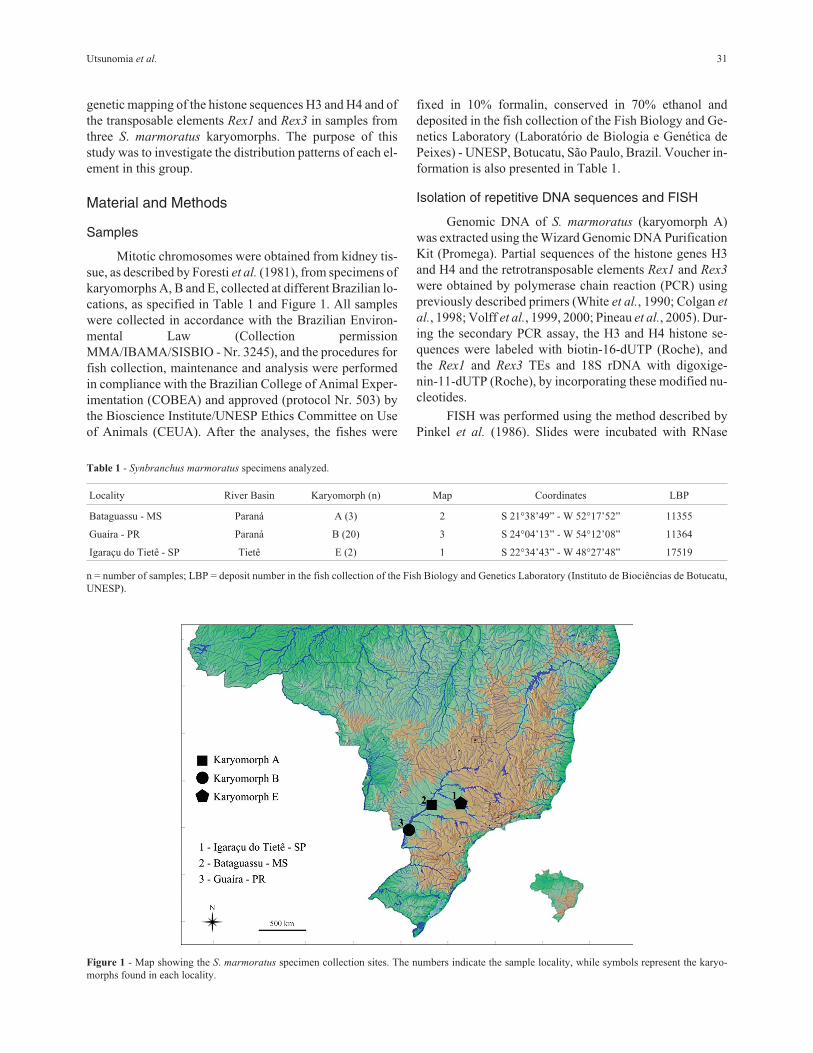
Figure 1 - Map showing the S. marmoratus specimen collection sites. The numbers indicate the sample locality, while symbols represent the karyo-
morphs found in each locality.
Table 1 - Synbranchus marmoratus specimens analyzed.
Locality River Basin Karyomorph (n) Map Coordinates LBP
Bataguassu - MS Paraná A (3) 2 S 21°38’49” - W 52°17’52” 11355
Guaíra - PR Paraná B (20) 3 S 24°04’13” - W 54°12’08” 11364
Igaraçu do Tietê - SP Tietê E (2) 1 S 22°34’43” - W 48°27’48” 17519
n = number of samples; LBP = deposit number in the fish collection of the Fish Biology and Genetics Laboratory (Instituto de Biociências de Botucatu,
UNESP).

(50 �g/mL) for 1 h at 37 °C. Then, the chromosomal DNA
was denatured in 70% formamide in 2x SSC for 5 min at
70 °C. For each slide, 30 �L of hybridization solution (con-
taining 200 ng of each labeled probe, 50% formamide, 2x
SSC and 10% dextran sulphate) were denatured for 10 min
at 95 °C, dropped onto the slides and hybridized overnight
at 37 °C in a 2x SSC moist chamber. After hybridization,
the slides were washed in a 0.2x SSC solution with 15%
formamide for 20 min at 42 °C, followed by a second wash
in 0.1x SSC for 15 min at 60 °C, and a final wash in 4x SSC
with 0.5% Tween for 10 min at room temperature. Probe
detection was carried out with Avidin-FITC (Sigma) or
anti-digoxigenin-rhodamine (Roche), the chromosomes
were counterstained with DAPI (4’,6-diamidino-2-phenyl-
indole, Vector Laboratories), and the FISH images were
captured by an optical photomicroscope (Olympus, BX61)
with the Image Pro Plus 6.0 software (Media Cybernetics).
Results
Cytogenetic analysis
All repetitive probes used here were clearly visual-
ized in the mitotic chromosomes. Both H3 and H4 histone
sequences appeared to be clustered together and were dis-
tributed in a general pattern with dispersed signals on all
chromosomes. Additionally, both sequences were accumu-
lated in one acrocentric pair (Figure 2a-f).
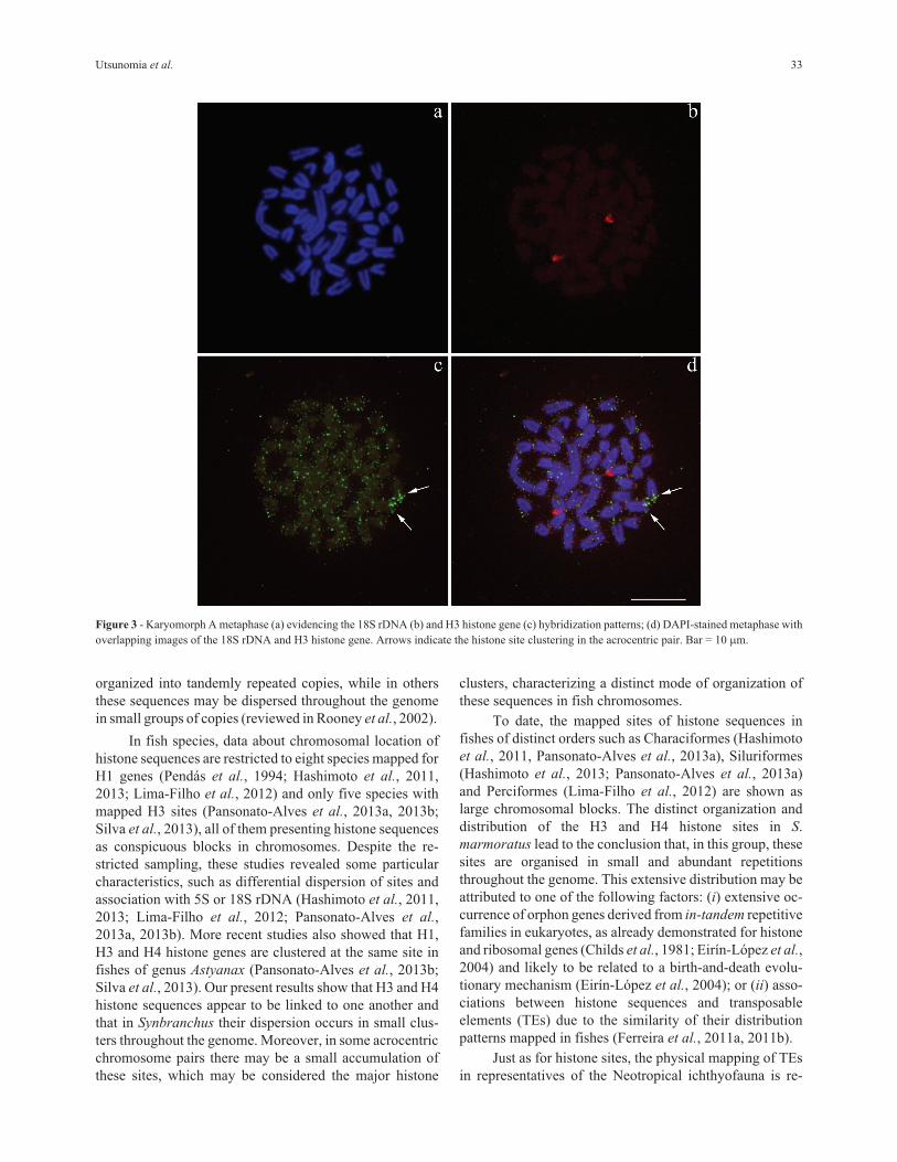
Since the histone sites mapped until now in fishes
were found in conspicuous blocks, we used the double-
FISH technique (18S rDNA + H3 histone sequences), in or-
der to compare the hybridization patterns of both probes
and check the veracity of the dispersed signal pattern of the
histone sequences. Double-FISH confirmed that, as ex-
pected, in Synbranchus the histone sites are dispersed
throughout the genome, while the 18S rDNA sites are only
present in one big cluster (Figure 3a-d).
Similarly, the Rex1 and Rex3 TEs are arranged in
small clusters that are also dispersed throughout the ge-
nome (Figure 4a-f). However, in the individuals belonging
to karyomorph A collected at Bataguassu, only the Rex3 el-
ements demonstrated significant accumulation in chromo-
some pair 3 (Figure 4d).
Discussion
Histone genes constitute a complex multigene family
and may show variations in copy number and organization
within the genome (Kedes, 1979). Thus, some species may
present up to a few thousand histone gene copies, usually
32 Repetitive DNA in Synbranchus
Figure 2 - Metaphases after FISH treatment using probes of H3 and H4 histone sequences, respectively, in karyomorphs A (a, d), B (b, e), and E (c, f). Ar-
rows indicate the histone site clustering in the acrocentric pair. Bar = 10 �m.
organized into tandemly repeated copies, while in others
these sequences may be dispersed throughout the genome
in small groups of copies (reviewed in Rooney et al., 2002).
In fish species, data about chromosomal location of
histone sequences are restricted to eight species mapped for
H1 genes (Pendás et al., 1994; Hashimoto et al., 2011,
2013; Lima-Filho et al., 2012) and only five species with
mapped H3 sites (Pansonato-Alves et al., 2013a, 2013b;
Silva et al., 2013), all of them presenting histone sequences
as conspicuous blocks in chromosomes. Despite the re-
stricted sampling, these studies revealed some particular
characteristics, such as differential dispersion of sites and
association with 5S or 18S rDNA (Hashimoto et al., 2011,
2013; Lima-Filho et al., 2012; Pansonato-Alves et al.,
2013a, 2013b). More recent studies also showed that H1,
H3 and H4 histone genes are clustered at the same site in
fishes of genus Astyanax (Pansonato-Alves et al., 2013b;
Silva et al., 2013). Our present results show that H3 and H4
histone sequences appear to be linked to one another and
that in Synbranchus their dispersion occurs in small clus-
ters throughout the genome. Moreover, in some acrocentric
chromosome pairs there may be a small accumulation of
these sites, which may be considered the major histone
clusters, characterizing a distinct mode of organization of
these sequences in fish chromosomes.
To date, the mapped sites of histone sequences in
fishes of distinct orders such as Characiformes (Hashimoto
et al., 2011, Pansonato-Alves et al., 2013a), Siluriformes
(Hashimoto et al., 2013; Pansonato-Alves et al., 2013a)
and Perciformes (Lima-Filho et al., 2012) are shown as
large chromosomal blocks. The distinct organization and
distribution of the H3 and H4 histone sites in S.
marmoratus lead to the conclusion that, in this group, these
sites are organised in small and abundant repetitions
throughout the genome. This extensive distribution may be
attributed to one of the following factors: (i) extensive oc-
currence of orphon genes derived from in-tandem repetitive
families in eukaryotes, as already demonstrated for histone
and ribosomal genes (Childs et al., 1981; Eirín-López et al.,
2004) and likely to be related to a birth-and-death evolu-
tionary mechanism (Eirín-López et al., 2004); or (ii) asso-
ciations between histone sequences and transposable
elements (TEs) due to the similarity of their distribution
patterns mapped in fishes (Ferreira et al., 2011a, 2011b).
Just as for histone sites, the physical mapping of TEs
in representatives of the Neotropical ichthyofauna is re-
Utsunomia et al. 33
Figure 3 - Karyomorph A metaphase (a) evidencing the 18S rDNA (b) and H3 histone gene (c) hybridization patterns; (d) DAPI-stained metaphase with
overlapping images of the 18S rDNA and H3 histone gene. Arrows indicate the histone site clustering in the acrocentric pair. Bar = 10 �m.

stricted to a small number of species and to the non-LTR
retrotransposons Rex1, Rex3 and Rex6 (Gross et al., 2009;
Cioffi et al., 2010; Valente et al., 2011; Ferreira et al.,
2011a,b; Pansonato-Alves et al., 2013a). The overlap of
signals generated by FISH among TEs and other repetitive
sequences raises questions about their role in the dispersion
of repetitive DNA sequences (Mandrioli et al., 2001;
Mandrioli and Manicardi, 2001; Cioffi et al., 2010). In S.
marmoratus, the Rex1 and Rex3 elements are found in
small clusters dispersed over all chromosomes. Notably, an
accentuated accumulation of repetitions in the centromeric
region of pair 3 was found in samples from Bataguassu
(karyomorph A); however, this seems to be only a local am-
plification because the individuals of karyomorph B, which
is believed to be recently derived from karyomorph A (un-
published data), did not present such blocks in this chromo-
some pair.
It is further worth noting that these elements may
present variable modes of chromosomal distribution in dif-
ferent species, but tend to be distributed in a similar manner
in close groups. The Rex1, Rex3 and Rex6 elements, for ex-
ample, are primarily compartmentalized in the pericentro-
meric heterochromatic regions in Cichlid fishes (Teixeira
et al., 2009; Valente et al., 2011) and dispersed throughout
the genome in Loricariidae, Bathydraconidae and Artedi-
draconidae species (Ozouf-Costaz et al., 2004; Ferreira et
al., 2011a; Pansonato-Alves et al., 2013a). However, in
Characiform species, Rex3 elements may also be compart-
mentalized (Cioffi et al., 2010; Pansonato-Alves et al.,
2013b; Silva et al., 2013), indicating that those elements are
highly dynamic and their type of genomic organization
does not reflect phylogenetic relationships among species.
Although S. marmoratus presents a remarkable varia-
tion in karyotype macrostructure, the physical mapping of
histone sequences and transposable elements revealed that
these sequences are all dispersed in different karyomorphs,
and this seems to be a conserved feature. Thus, we stress the
importance of further studies regarding the physical map-
ping of H1, H3 and H4 histone genes and other repetitive
DNAs, which may be useful in determining the organiza-
tion of these genes in eukaryote genomes. Similarly, the
mapping of transposable elements can bring new perspec-
tives on the genomic organization, dispersion of genes and
speciation driven by those sequences.
Acknowledgments
This study was supported by grants from the Brazil-
ian agencies Conselho Nacional de Desenvolvimento Cien-
tífico e Tecnológico (CNPq) and Fundação de Amparo à
34 Repetitive DNA in Synbranchus
Figure 4 - Metaphases after FISH treatment using Rex1 and Rex3 probes, respectively, in karyomorphs A (a, d), B (b, e), and E (c, f). Arrows indicate the
clustering of Rex3 sites in the centromere of a submetacentric pair. Bar = 10 �m.
Pesquisa do Estado de São Paulo (FAPESP). Ricardo Utsu-
nomia had a scholarship from FAPESP (2011/01370-0)
References
Charlesworth B, Snlegowski P and Stephan W (1994) The evolu-
tionary dynamics of repetitive DNA in eukaryotes. Nature
371:215-220.
Childs G, Maxson R, Cohn RH and Kedes L (1981) Orphons: Dis-
persed genetic elements derived from tandem repetitive
genes of eukaryotes. Cell 23:651-663.
Cioffi MB, Martins C and Bertollo LAC (2010) Chromosome
spreading of associated transposable elements and ribo-
somal DNA in the fish Erythrinus erythrinus. Implications
for genome change and karyoevolution in fish. BMC Evol
Biol 10:271-280.
Colgan DJ, McLauchlan A and Wilson GDF (1998) Histone H3
and U2 snRNA DNA sequences and arthropod molecular
evolution. Aust J Zool 46:419-437.
Eirin-Lopez JM, González-Tizón AM, Martínez A and Méndez J
(2004) Birth-and-death evolution with strong purifying se-
lection in the Histone H1 multigene family and the origin of
orphon H1 genes. Mol Biol Evol 21:1992-2003.
Ferreira DC, Oliveira C and Foresti F (2011a) Elements Rex1 and
Rex3 in three fish species in the subfamily Hypoptopoma-
tinae (Teleostei, Siluriformes, Loricariidae). Cytogenet Ge-
nome Res 132:64-70.
Ferreira DC, Oliveira C and Foresti F (2011b) A new dispersed el-
ement in the genome of the catfish Hisonotus leucofrenatus
(Teleostei, Siluriformes, Hypoptopomatinae). Mob Genet
Elements 1:103-106.
Foresti F, Almeida-Toledo LF and Toledo-Filho SA (1981) Poly-
morphic nature of nucleolus organizer regions on fishes.
Cytogenet Cell Gen 31:137-144.
Foresti F, Oliveira C and Tien OS (1992) Cytogenetic studies of
the genus Synbranchus (Pisces, Synbranchiformes,
Synbranchidae). Naturalia 17:129-138.
Gross MC, Schneider CH, Valente GT, Porto JIR, Martins C and
Feldberg E (2009) Comparative cytogenetic analysis of the
genus Symphysodon (Discus fishes, Cichlidae): Chromo-
somal characteristics of retrotransposons and minor ribo-
somal DNA. Cytogenet Genome Res 127:43-53.
Gursel I, Gursel M, Yamada H, Ishii KJ, Takeshita F and Klinman
DM (2003) Repetitive elements in mammalian telomeres
suppress bacterial DNA-induced immune activation. J Im-
munol 171:1393-1400.
Haff T, Schmid M, Steinlein C, Galetti Jr PM, Willard H (1993)
Organization and molecular cytogenetics of a satellite DNA
family from Hoplias malabaricus (Pisces, Erythrinidae).
Chromosome Res 1:77-86.
Hashimoto DT, Ferguson-Smith MA, Rens W, Foresti F and
Porto-Foresti F (2011) Chromosome mapping of H1 histone
and 5S rRNA genes clusters in three species of Astyanax
(Teleostei, Characiformes). Cytogen Genome Res 134:64-
71.
Hashimoto DT, Ferguson-Smith MA, Rens W, Prado FD, Foresti
F and Porto-Foresti F (2013) Cytogenetic mapping of H1
histone and ribosomal RNA genes in hybrids between cat-
fish species Pseudoplatystoma corruscans and Pseudo-
platystoma reticulatum. Cytogen Genome Res 139:102-106.
Kedes LH (1979) Histone genes and histone messengers. Annu
Rev Biochem 48:837-870.
Lima-Filho PA, Cioffi MB, Bertollo LAC and Molina WF (2012)
Chromosomal and morphological divergences in Atlantic
populations of the frillfin Bathygobius soporator (Gobiidae,
Perciformes). J Exp Mar Biol Ecol 434:63-70.
Mandrioli M, Manicardi GC, Machella N and Caputo V (2001)
Molecular and cytogenetic analysis of the goby Gobius
niger (Teleostei, Gobiidae). Genetica 110:73-78.
Mandrioli M and Manicardi GC (2001) Cytogenetics and molecu-
lar analysis of the pufferfish Tetraodon fluviatilis
(Osteichthyes). Genetica 111:433-438.
Martins C and Galetti Jr PM (1999) Chromosomal localization of
5S rDNA genes in Leporinus fish (Anostomidae, Chara-
ciformes). Chromosome Res 7:363-367.
Melilo IFM, Foresti F and Oliveira C (1996) Additional cyto-
genetic studies on local populations of Synbranchus
marmoratus (Pisces, Synbranchiformes, Synbranchidae).
Naturalia 21:201-208.
Nagoda N, Fukuda A, Nakashima Y and Matsuo Y (2005) Molec-
ular characterization and evolution of the repeating units of
histone genes in Drosophila americana: Coexistence of
quartet and quintet units in a genome. Insect Mol Biol
14:713-717.
Ozouf-Costaz C, Brandt J, Korting C, Pisano E, Bonillo C,
Coutanceau JP and Volff JN (2004) Genome dynamics and
chromosomal localization of the non-LTR retrotransposons
Rex1 and Rex3 in Antartic fish. Antartic Science 16:51-57.
Pansonato-Alves JC, Serrano EA, Utsunomia R, Scacchetti PC,
Oliveira C and Foresti F (2013a) Mapping five repetitive
DNA classes in sympatric species of Hypostomus (Teleos-
tei, Siluriformes, Loricariidae): Analysis of chromosomal
variability. Rev Fish Biol Fisher 23:477-489
Pansonato-Alves JC, Hilsdorf AWS, Utsunomia R, Silva DMZA,
Oliveira C and Foresti F (2013b) Chromosomal mapping of
repetitive DNA and cytochrome C oxidase I sequence analy-
sis reveal differentiation among sympatric samples of
Astyanax fasciatus (Characiformes, Characidae). Cytogenet
Genome Res 141:133-142.
Pendás AM, Morán P and García-Vázquez E (1994) Organization
and chromosomal location of the major histone cluster in
brown trout, Atlantic salmon and rainbow trout. Chromo-
soma 103:147-152.
Pineau P, Henry M, Suspène R, Marchio A, Dettai A, Debruyne R,
Petit T, Lécu A, Moisson P, Dejean A, et al. (2005) A uni-
versal primer set for PCR amplification of nuclear histone
H4 genes from all animal species. Mol Biol Evol 22:582-
588.
Pinkel D, Straume T and Gray JW (1986) Cytogenetic analysis us-
ing quantitative, high-sensitivity, fluorescence hybridiza-
tion. Proc Natl Acad Sci USA 83:2934-2938.
Sanchez S and Fenocchio AS (1996) Karyotypic analysis in three
populations of the South-American eel like fish
Synbranchus marmoratus. Caryologia 49:65-71.
Silva DMZA, Pansonato-Alves JC, Utsunomia R, Daniel SN,
Hashimoto DT, Oliveira C, Porto-Foresti F and Foresti F
(2013) Chromosomal organization of repetitive DNA se-
quences in Astyanax bockmanni (Teleostei, Characiformes):
Dispersive location, association and co-localization in the
genome. Genetica 141:329-336.
Utsunomia et al. 35
Sumner AT (2003) Chromosomes: Organization and Function.
Blackwell Publishing Company, London, 287 pp.
Teixeira WG, Ferreira IA, Cabral-de-Melo DC, Mazzuchelli J,
Valente GT, Pinhal D, Poletto AB, Venere PC and Martins C
(2009) Organization of repeated DNA elements in the ge-
nome of the cichlid fish Cichla kelberi and its contributions
to the knowledge of fish genomes. Cytogenet Genome Res
125:224-234.
Torres RA, Roper JJ, Foresti F and Oliveira C (2005) Surprising
genomic diversity in the Neotropical Fish Synbranchus
marmoratus (Teleostei, Synbranchidae): How many spe-
cies? Neotrop Ichthyol 3:277-284.
Valente GT, Mazzuchelli J, Ferreira IA, Poletto AB, Fantinatti
BEA and Martins C (2011) Cytogenetic Mapping of the
retroelements Rex1, Rex3 and Rex6 among cichlid fish: New
insights on the chromosomal distribution of transposable el-
ements. Cytogenet Genome Res 133:34-42.
Vicari MR, Nogaroto V, Noleto RB, Cestari MM, Cioffi MB,
Almeida MC, Moreira-Filho O and Artoni RF (2010) Satel-
lite DNA and chromosomes in Neotropical fishes: Methods,
applications and perspectives. J Fish Biol 76:1094-1116.
Volff JN, Körting C, Sweeney K and Schartl M (1999) The
non-LTR retrotransposon Rex3 from the fish Xiphophorus is
widespread among teleosts. Mol Biol Evol 16:1427-1438.
Volff JN, Körting C and Schartl M (2000) Multiple lineages of the
non-LTR retrotransposon Rex1 with varying success in in-
vading fish genomes. Mol Biol Evol 17:1673-1684.
Wagner RP, Maguire MP and Stallings RL (1993) Chromosomes:
A synthesis. Wiley-Liss Inc., New York pp. 523.
White TJ, Bruns T, Lee S and Taylor J (1990) Amplification and
direct sequencing of fungal ribosomal RNA genes for
phylogenetics. In: Innis MA, Gelfand DH, Sninsky JJ and
White TJ (eds) PCR Protocols: A Guide to Methods and Ap-
plications. Academic Press Inc., New York, pp 315-322.
Associate Editor: Igor Schneider
All the content of the journal, except where otherwise noted, is licensed under a CreativeCommons License CC BY-NC.
36 Repetitive DNA in Synbranchus