Embed Size (px)
Citation preview
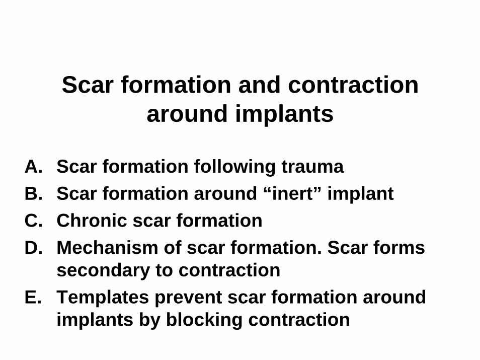
Scar formation and contraction around implants
A. Scar formation following traumaB. Scar formation around “inert” implantC. Chronic scar formationD. Mechanism of scar formation. Scar forms
secondary to contractionE. Templates prevent scar formation around
implants by blocking contraction
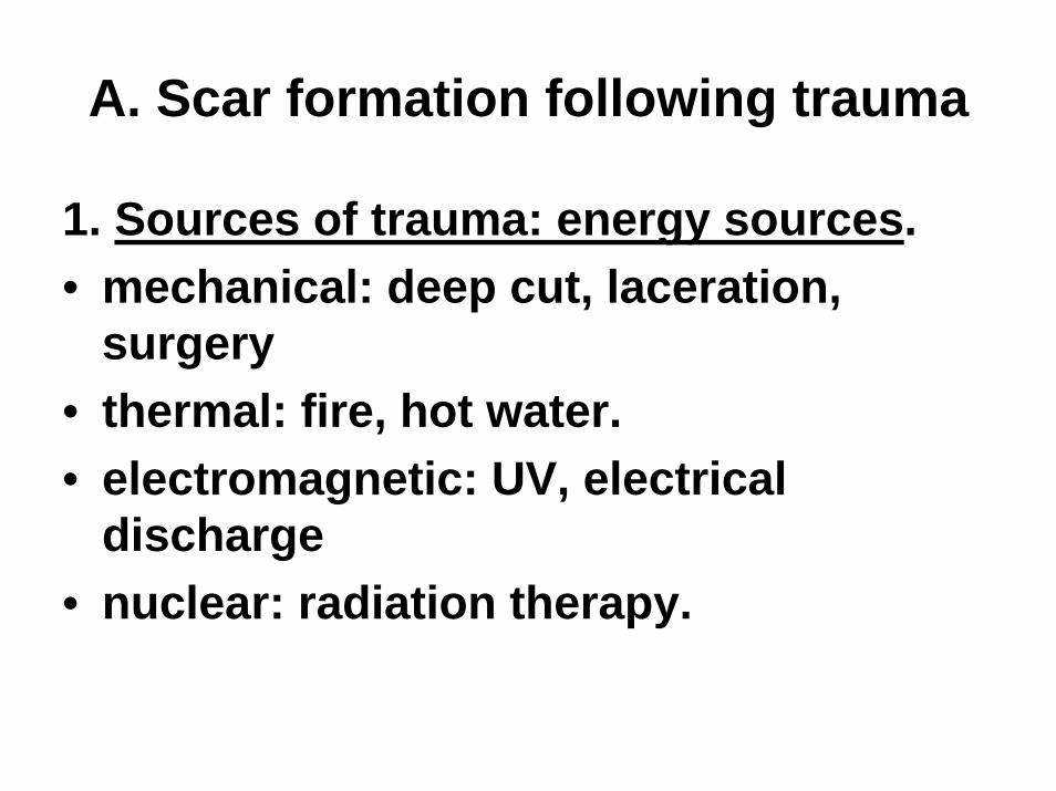
A. Scar formation following trauma
1. Sources of trauma: energy sources.• mechanical: deep cut, laceration,
surgery• thermal: fire, hot water.• electromagnetic: UV, electrical
discharge• nuclear: radiation therapy.
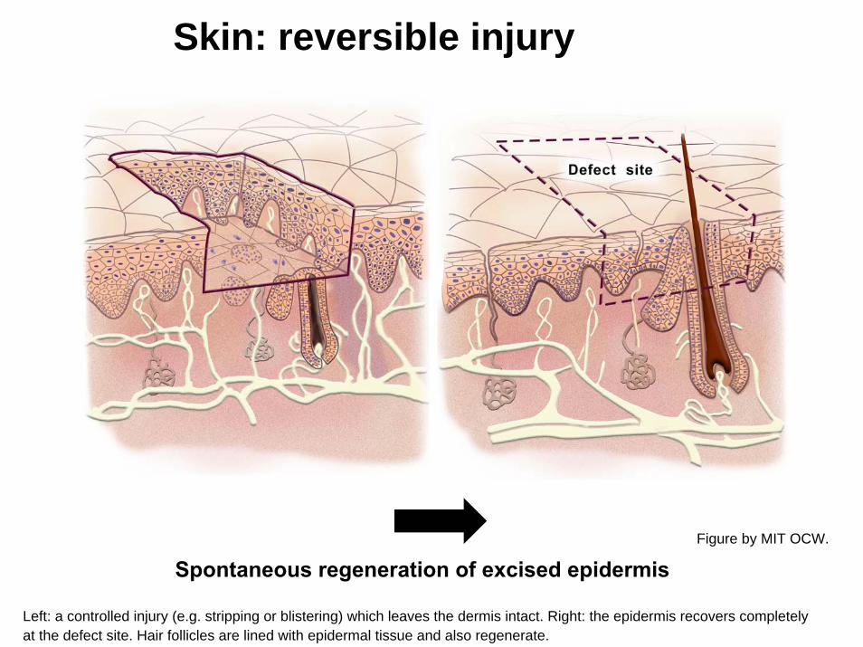
Skin: reversible injury
Spontaneous regeneration of excised epidermisFigure by MIT OCW.
Left: a controlled injury (e.g. stripping or blistering) which leaves the dermis intact. Right: the epidermis recovers completelyat the defect site. Hair follicles are lined with epidermal tissue and also regenerate.
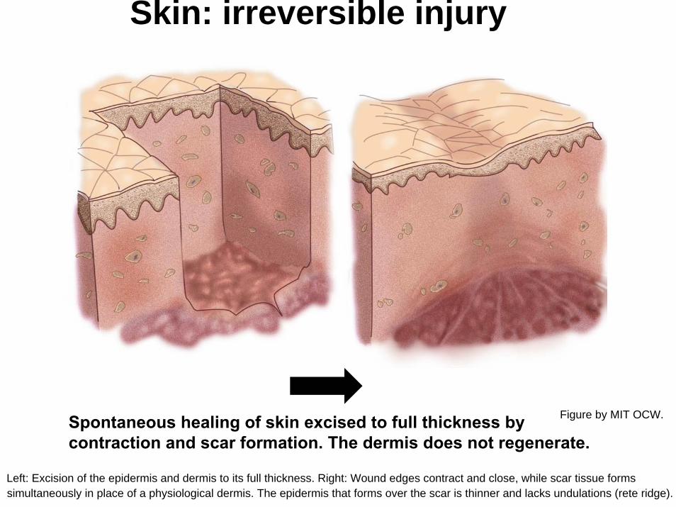
Skin: irreversible injury
Spontaneous healing of skin excised to full thickness by contraction and scar formation. The dermis does not regenerate.
Figure by MIT OCW.
Left: Excision of the epidermis and dermis to its full thickness. Right: Wound edges contract and close, while scar tissue forms simultaneously in place of a physiological dermis. The epidermis that forms over the scar is thinner and lacks undulations (rete ridge).
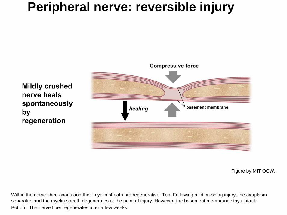
Peripheral nerve: reversible injury
Mildly crushed nerve heals spontaneously by regeneration
Figure by MIT OCW.
Within the nerve fiber, axons and their myelin sheath are regenerative. Top: Following mild crushing injury, the axoplasm separates and the myelin sheath degenerates at the point of injury. However, the basement membrane stays intact.Bottom: The nerve fiber regenerates after a few weeks.
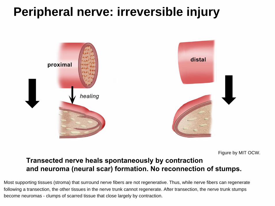
Peripheral nerve: irreversible injury
Transected nerve heals spontaneously by contraction and neuroma (neural scar) formation. No reconnection of stumps.
Figure by MIT OCW.
Most supporting tissues (stroma) that surround nerve fibers are not regenerative. Thus, while nerve fibers can regenerate following a transection, the other tissues in the nerve trunk cannot regenerate. After transection, the nerve trunk stumps become neuromas - clumps of scarred tissue that close largely by contraction.
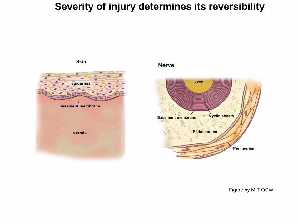
Severity of injury determines its reversibility
Figure by MIT OCW.
Scar formation following trauma (cont.)
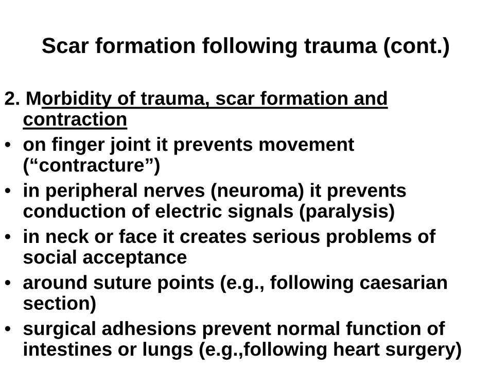
2. Morbidity of trauma, scar formation and contraction
• on finger joint it prevents movement (“contracture”)
• in peripheral nerves (neuroma) it prevents conduction of electric signals (paralysis)
• in neck or face it creates serious problems of social acceptance
• around suture points (e.g., following caesarian section)
• surgical adhesions prevent normal function of intestines or lungs (e.g.,following heart surgery)
Images removed due to copyright restrictions.
Poster warning NFL football players not to tackle with the crown of their helmet ("Play Heads-Up!)Diagram describing angioplasty.
B. Scar formation around “inert” implant
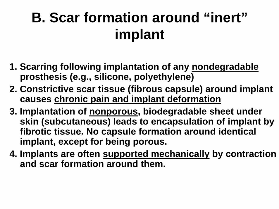
1. Scarring following implantation of any nondegradableprosthesis (e.g., silicone, polyethylene)
2. Constrictive scar tissue (fibrous capsule) around implant causes chronic pain and implant deformation
3. Implantation of nonporous, biodegradable sheet under skin (subcutaneous) leads to encapsulation of implant by fibrotic tissue. No capsule formation around identical implant, except for being porous.
4. Implants are often supported mechanically by contraction and scar formation around them.
NORMAL IMPLANT
Images removed due to copyright restrictions.
Diagrams of human breast - normal and with implant.
C. Chronic scar formation
• Scar formation and contraction result from chronic trauma; or from acute or chronic inflammation caused by various agents
• Scar takes different names depending on medical specialty
• Examples: 1. Scarred heart valve due to incidence of rheumatic fever leads, e.g., to valve stenosis or to leakage.2. Necrosis (death) of myocardium (infarct)due to interruption of oxygen supply (clogged arteries) interferes with electrical conduction of heart muscle3. Obstruction of intestinal tract, due to chronic inflammation, leads to digestive problems (e.g., duodenal ulcer with gastric outlet obstruction)4. Fibrotic liver (cirrhosis) prevents liver function
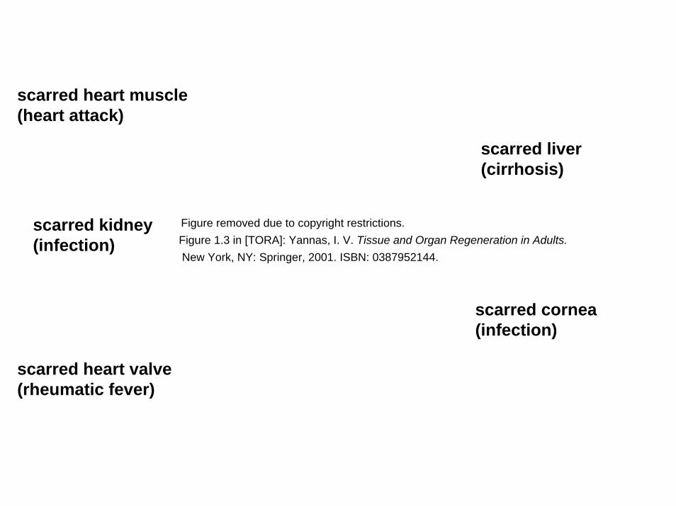
scarred heart muscle(heart attack)
scarred liver(cirrhosis)
scarred cornea(infection)
scarred kidney(infection)
scarred heart valve(rheumatic fever)
Figure 1.3 in [TORA]: Yannas, I. V. Tissue and Organ Regeneration in Adults. New York, NY: Springer, 2001. ISBN: 0387952144.
Figure removed due to copyright restrictions.
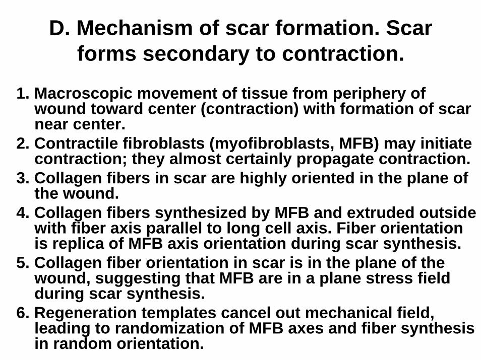
D. Mechanism of scar formation. Scar forms secondary to contraction.
1. Macroscopic movement of tissue from periphery of wound toward center (contraction) with formation of scar near center.
2. Contractile fibroblasts (myofibroblasts, MFB) may initiate contraction; they almost certainly propagate contraction.
3. Collagen fibers in scar are highly oriented in the plane of the wound.
4. Collagen fibers synthesized by MFB and extruded outside with fiber axis parallel to long cell axis. Fiber orientation is replica of MFB axis orientation during scar synthesis.
5. Collagen fiber orientation in scar is in the plane of the wound, suggesting that MFB are in a plane stress field during scar synthesis.
6. Regeneration templates cancel out mechanical field, leading to randomization of MFB axes and fiber synthesis in random orientation.
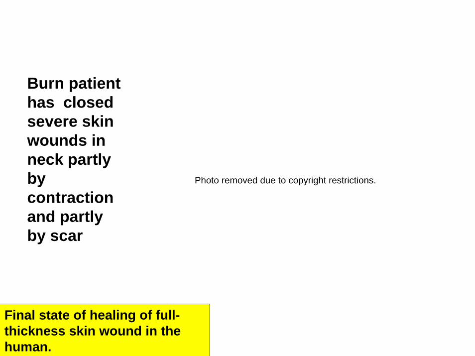
Burn patienthas closed severe skin wounds in neck partly by contractionand partly by scar
Final state of healing of full-thickness skin wound in the human.
Photo removed due to copyright restrictions.
Tomasek et al., 2000Spontaneous contraction and scar formation in burn victim
Photo removed due to copyright restrictions.
Contracting skin wound.Guinea pig model. Adipose tissue layerunderneathis greatlydeformedby contracting wound.
Natural light. Conventional histologicalview. Stained with H&E.S, scar. D, dermis.A, adipose tissue.
Viewed in polarized lightstage. Collagen fibers light up.
Photos removed due to copyright restrictions.
Edge of full-thickness skin wound in guinea pig. E, epidermis. F, fibroblasts.B, base of wound. Scale bar,
Photo removed due to copyright restrictions.
Measure C
Figure 4.1 in [TORA], illustrating contraction kinetics of dermis-free defect
Graph removed due to copyright restrictions.
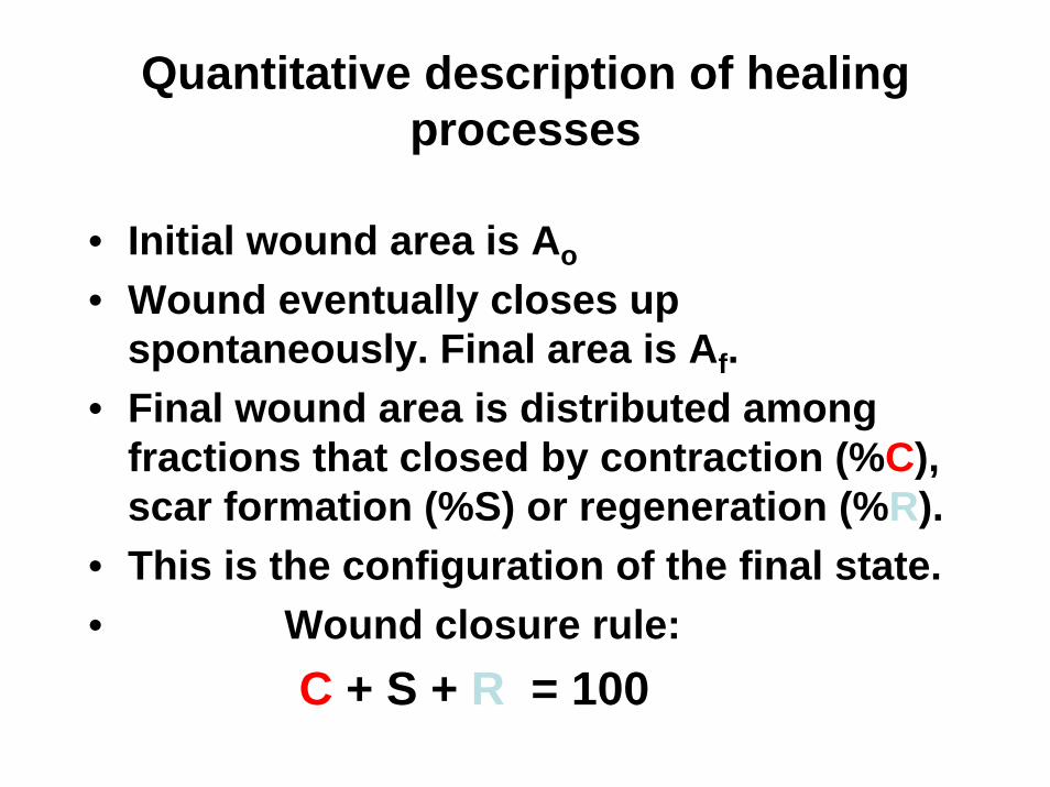
Quantitative description of healing processes
• Initial wound area is Ao
• Wound eventually closes up spontaneously. Final area is Af.
• Final wound area is distributed among fractions that closed by contraction (%C), scar formation (%S) or regeneration (%R).
• This is the configuration of the final state.• Wound closure rule:
C + S + R = 100
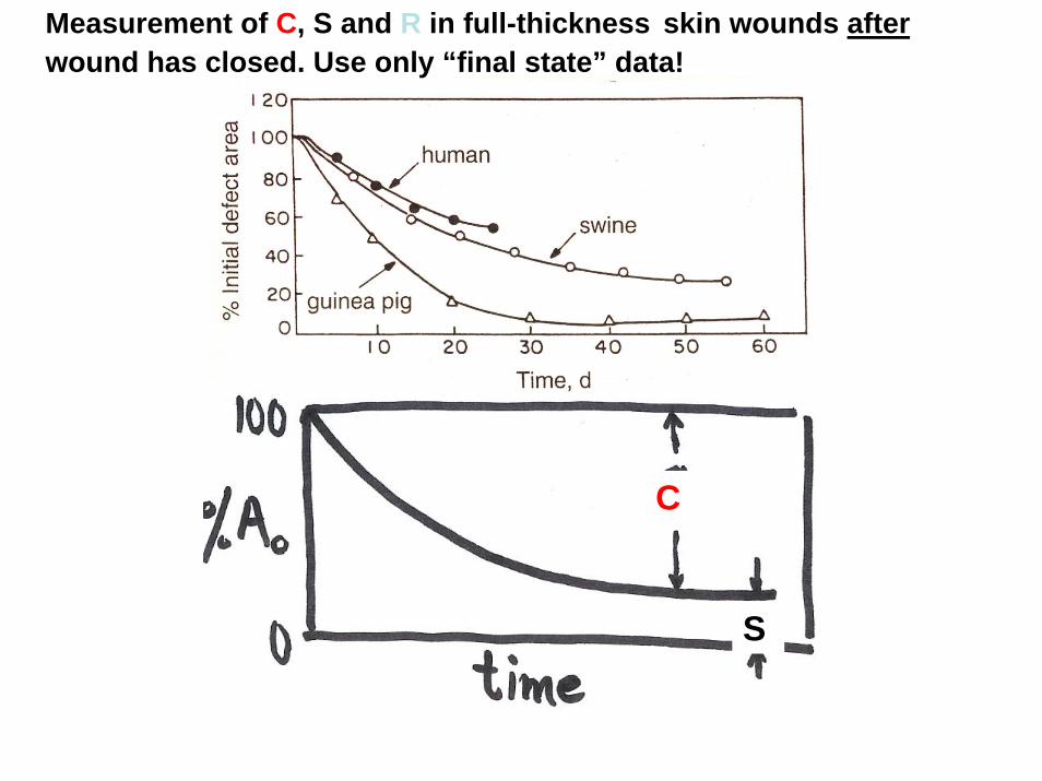
Measurement of C, S and R in full-thickness skin wounds afterwound has closed. Use only “final state” data!
C
S
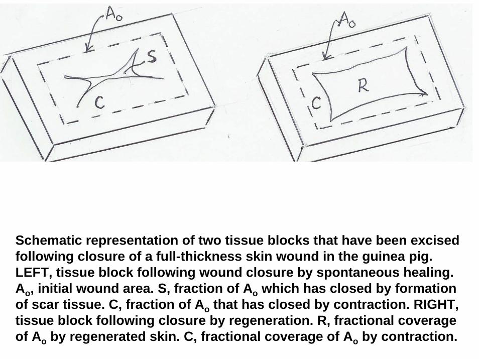
Schematic representation of two tissue blocks that have been excised following closure of a full-thickness skin wound in the guinea pig. LEFT, tissue block following wound closure by spontaneous healing. Ao, initial wound area. S, fraction of Ao which has closed by formation of scar tissue. C, fraction of Ao that has closed by contraction. RIGHT, tissue block following closure by regeneration. R, fractional coverage of Ao by regenerated skin. C, fractional coverage of Ao by contraction.
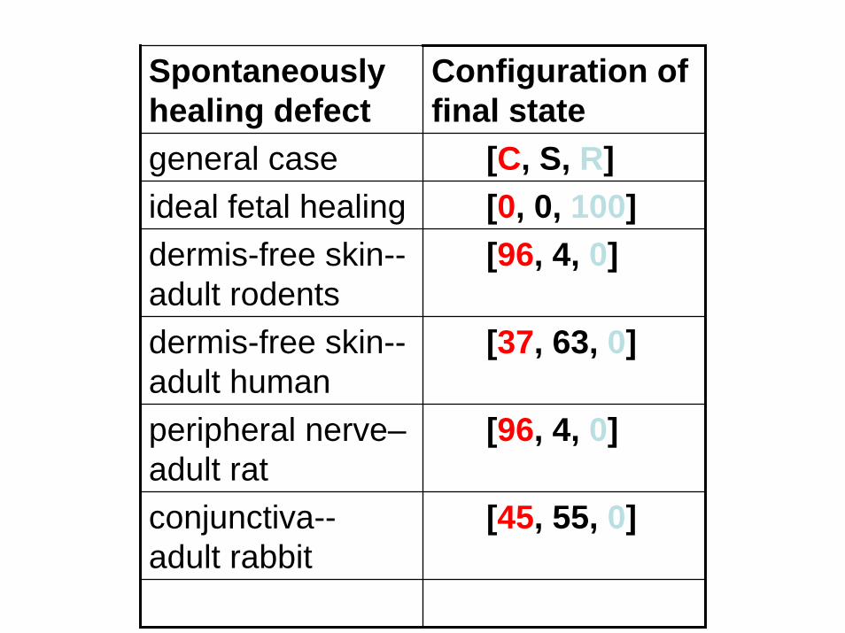
Spontaneously healing defect
Configuration of final state
general case [C, S, R]ideal fetal healing [0, 0, 100]dermis-free skin--adult rodents
[96, 4, 0]
dermis-free skin--adult human
[37, 63, 0]
peripheral nerve–adult rat
[96, 4, 0]
conjunctiva--adult rabbit
[45, 55, 0]
Orgill, 1983
Final state of healing of full-thickness skin wound in the guinea pig.
Photo removed due to copyright restrictions.
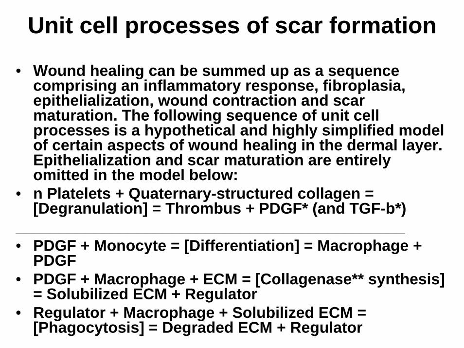
Unit cell processes of scar formation
• Wound healing can be summed up as a sequence comprising an inflammatory response, fibroplasia, epithelialization, wound contraction and scar maturation. The following sequence of unit cell processes is a hypothetical and highly simplified model of certain aspects of wound healing in the dermal layer. Epithelialization and scar maturation are entirely omitted in the model below:
• n Platelets + Quaternary-structured collagen = [Degranulation] = Thrombus + PDGF* (and TGF-b*)
_____________________________________________• PDGF + Monocyte = [Differentiation] = Macrophage +
PDGF• PDGF + Macrophage + ECM = [Collagenase** synthesis]
= Solubilized ECM + Regulator• Regulator + Macrophage + Solubilized ECM =
[Phagocytosis] = Degraded ECM + Regulator
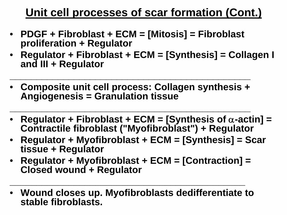
Unit cell processes of scar formation (Cont.)
• PDGF + Fibroblast + ECM = [Mitosis] = Fibroblast proliferation + Regulator
• Regulator + Fibroblast + ECM = [Synthesis] = Collagen I and III + Regulator
_____________________________________________• Composite unit cell process: Collagen synthesis +
Angiogenesis = Granulation tissue_____________________________________________• Regulator + Fibroblast + ECM = [Synthesis of α-actin] =
Contractile fibroblast ("Myofibroblast") + Regulator• Regulator + Myofibroblast + ECM = [Synthesis] = Scar
tissue + Regulator• Regulator + Myofibroblast + ECM = [Contraction] =
Closed wound + Regulator____________________________________________• Wound closes up. Myofibroblasts dedifferentiate to
stable fibroblasts.
Tomasek et al., 2000
Myofibroblast detected with antibody to α-SM actin
Figure removed due to copyright restrictions.
Measure S (quantitative assay)
Figure removed due to copyright restrictions.
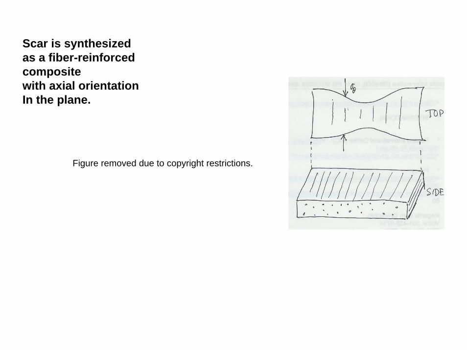
Scar is synthesized as a fiber-reinforced composite with axial orientationIn the plane.
Figure removed due to copyright restrictions.

Image removed due to copyright restrictions.
Table 4.1 in [TORA].
Developmental transition from scarlessfetal to scarring (adult-type) fetal healing
1. Study of wounded fetal rats, before and after developmental transition from scarless to scarring skin wound healing.
2. Scarless healing was accompanied by decreased and rapidly cleared levels of TGF-b1 and TGF-b2; also by increased and prolonged TGF-b3 levels.
3. Scarring healing was described by reversed appearance and duration of the three TGFbisomorphs.
[data by Soo et al., 2003]
E. Templates prevent scar formation around implants by blocking contraction
1. Differentiation of fibroblasts to the contractile phenotype requires a)TGF-b1, b) mechanical tension in the matrix, and c) presence of fibronectinfragments (ED-A fibronectin).
2. Contractile fibroblasts (myofibroblasts, MFB) contract skin wounds and nerve wounds.
3. Regeneration templates block contraction by downregulating MFB density and organization.
4. Contraction blockade by templates thwarts the adult healing response and hypothetically reactivates the dormant scarless fetal healing response (which appears to be the “default” response).
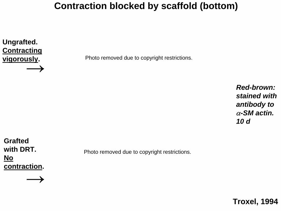
Ungrafted.Contractingvigorously.
Grafted with DRT.No contraction.
Troxel, 1994
→
Red-brown: stained with antibody to α-SM actin.10 d
Contraction blocked by scaffold (bottom)
→Photo removed due to copyright restrictions.
Photo removed due to copyright restrictions.
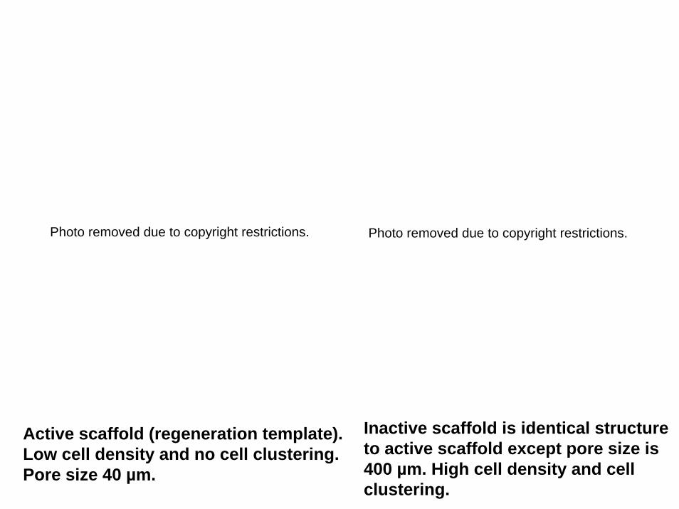
Active scaffold (regeneration template).Low cell density and no cell clustering. Pore size 40 µm.
Inactive scaffold is identical structure to active scaffold except pore size is 400 µm. High cell density and cell clustering.
Photo removed due to copyright restrictions. Photo removed due to copyright restrictions.
Closure of full-thickness skin wound in guinea pig using three protocols. KC, seeded keratinocytes. DRT, dermis regeneration template, a biologically active scaffold.
Figure removed due to copyright restrictions.
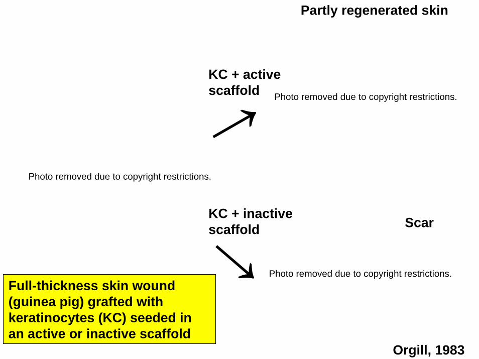
Partly regenerated skin
Orgill, 1983
KC + activescaffold
KC + inactive scaffold Scar
Full-thickness skin wound(guinea pig) grafted with keratinocytes (KC) seeded inan active or inactive scaffold
Photo removed due to copyright restrictions.
Photo removed due to copyright restrictions.
Photo removed due to copyright restrictions.
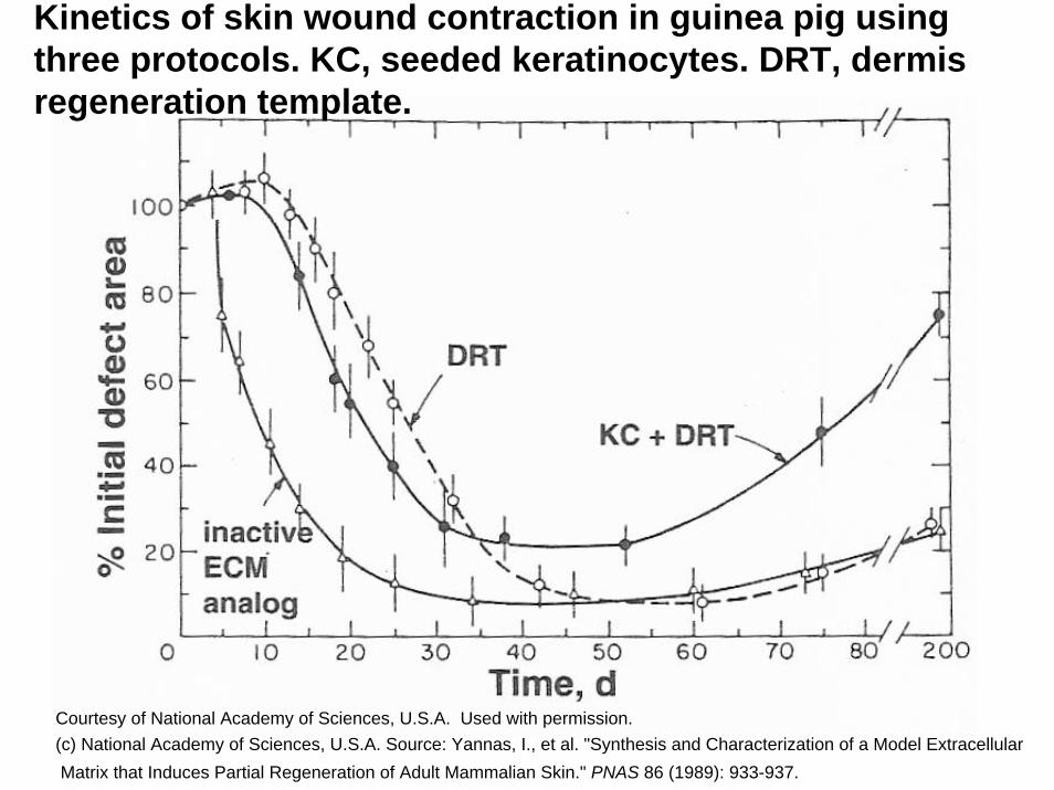
Kinetics of skin wound contraction in guinea pig using three protocols. KC, seeded keratinocytes. DRT, dermis regeneration template.
Courtesy of National Academy of Sciences, U.S.A. Used with permission.(c) National Academy of Sciences, U.S.A. Source: Yannas, I., et al. "Synthesis and Characterization of a Model Extracellular Matrix that Induces Partial Regeneration of Adult Mammalian Skin." PNAS 86 (1989): 933-937.
capillary loops
↑ ↑
rete ridges
capillary loops
Normal skin. Burkitt et al., 1992 Regenerated skin, swine. Compton et al., 1998
Images removed due to copyright restrictions.
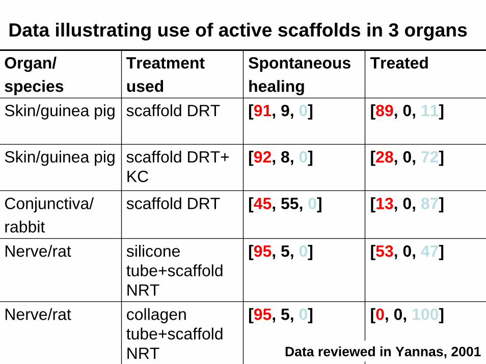
Organ/species
Treatmentused
Spontaneoushealing
Treated
Skin/guinea pig scaffold DRT [91, 9, 0] [89, 0, 11]
Skin/guinea pig scaffold DRT+ KC
[92, 8, 0] [28, 0, 72]
Conjunctiva/rabbit
scaffold DRT [45, 55, 0] [13, 0, 87]
Nerve/rat silicone tube+scaffoldNRT
[95, 5, 0] [53, 0, 47]
Nerve/rat collagen tube+scaffoldNRT
[95, 5, 0] [0, 0, 100]
Data illustrating use of active scaffolds in 3 organs
Data reviewed in Yannas, 2001