Embed Size (px)
Citation preview

Journal of Physics Conference Series
OPEN ACCESS
Scanning electron microscopy as a tool for theanalysis of colony architecture produced byphenotypic switching of a human pathogenic yeastCandida tropicalisTo cite this article M C Furlaneto et al 2012 J Phys Conf Ser 371 012022
View the article online for updates and enhancements
Related contentMicrostructural and analytical analysis ofplasma dissociated zirconE G Minnaar J H Neethling M E Lee et al
-
An electron microscopy study of the effectof Ce on plasma sprayed bronze coatingsLi Wensheng S C Wang Chao Ma et al
-
STEM mode in the SEM for the analysis ofcellular sections prepared byultramicrotome sectioningN Hondow J Harrington R Brydson et al
-
Recent citationsAntimycotic effects of a prodigiosinproducing Serratia marcescensrhizobacteriaC Jimtha John et al
-
Antifungal properties of prodigiosinproducing rhizospheric Serratia spC John Jimtha et al
-
Endophytic Nocardiopsis sp from Zingiberofficinale with both antiphytopathogenicmechanisms and antibiofilm activityagainst clinical isolatesRohini Sabu et al
-
This content was downloaded from IP address 1160145168 on 23092021 at 1736
Scanning electron microscopy as a tool for the analysis of
colony architecture produced by phenotypic switching of a
human pathogenic yeast Candida tropicalis
M C Furlaneto1 C G T J Andrade
2 P H A Aragatildeo
2 E J G Franccedila
1 A T P Moralez
1
and L C S Ferreira1
1Department of Microbiology Paranaacute State University at Londrina Brazil
2Electronic Microscopy and Microanalysis Laboratory Paranaacute State University at Londrina
Brazil
Abstract Candida tropicalis has been identified as one of the
most prevalent pathogenic yeast
species of the Candida-non-albicans group Phenotypic switching is a biological phenomenon related
to the occurrence of spontaneous emergence of colonies with different morphologies that provides
variability within colonizing populations in order to adapt to different environments Currently
studies of the microstructure of switching variant colonies are not subject of extensive research SEM
analysis was used to verify the architecture of whole Candida colonies The strain 4907 exhibited a
hemispherical shape character while the strain 33507 showed a volcano shape with mycelated-edge
colony The ring switch variant is characterized by a highly wrinkled centre and an irregular
periphery The rough phenotype exhibited a three-dimensional architecture and was characterized by
the presence of deep central and peripheral depressions areas The ultrastructural analysis also
allowed the observation of the arrangement of individual cells within the colonies The whole smooth
colony consisted entirely of yeast cells Differently aerial filaments were found all around the colony
periphery of the volcano shape colony For this colony type the mycelated-edge consisted mainly of
hyphae although yeast cells are also seen The ring and rough colonies phenotypes comprised
mainly yeast cells with the presence of extracellular material connecting neighbouring cells This
study has shown that SEM can be used effectively to examine the microarchitecture of colonies
morphotypes of the yeast C tropicalis and further our understanding of switching event in this
pathogen
1 Introduction
Candida tropicalis is an opportunistic yeast pathogen that causes superficial and systemic mycoses
[1-3] In Latin America C tropicalis accounts for the majority of non-albicans Candida species
associated with candidemia episodes [4]
Among several virulence factors that contribute to pathogenesis of Candida it has been
suggested that phenotypic switching provides variability within colonizing populations in order to
adapt to challenges at different environments including various anatomical sites in the human body
[5]
Phenotypic switching represents an epigenetic state that occurs in a small fraction of the
population is random and reversible For fungi phenotypic switching is defined as the spontaneous
emergence of colonies with altered colony morphology at rates higher than the somatic mutations
rates that enables the microorganism to undergo rapid microevolution [6] For Candida albicans a
relationship between colony shape of switching variants and the constituent cells (blastospores true
and psedo-hyphae) has been demonstrated by scanning electron microscopy analysis [7] According
Electron Microscopy and Analysis Group Conference 2011 (EMAG 2011) IOP PublishingJournal of Physics Conference Series 371 (2012) 012022 doi1010881742-65963711012022
Published under licence by IOP Publishing Ltd 1
to these authors the relationship between switched variant colonies and microstructure may help
elucidate the relationship between pathogenicity in vivo and colonial morphology in vitro [7]
In contrast to the species C albicans the study of switching in C tropicalis has not been the
subject of extensive research Our work focuses on using scanning electron microscopy for the
analysis of switched variant colonies of clinical isolates of C tropicalis An understanding of this
virulence determinant would provide insight into C tropicalis pathogenic mechanisms
2 Experimental techniques
21 Fungal strains
C tropicalis strains 4907 and 33507 included in this study were recovered from tracheal secretion
and belong to the Candida culture collection of the Fungal Genetics Laboratory The University of
Londrina-Brazil The switched variants ring and rough were obtained as previously described [8]
22 Scanning electron microscopy of intact colonies
To verify the architecture of C tropicalis colonies morphotypes whole yeast colonies were
removed from YPD (1 yeast extract 2 peptone 2 dextrose) agar plates using a scalpel blade
Colonies were fixed for 18 h at 4oC in 3 glutaraldehyde (Electron Microscopy Sciences) in 01 M
phosphate buffer pH 72 They were then immersed in liquid nitrogen for 30 sec and freeze-dried
for 90 min at 2x10⁻³
MPa (Juan LP3) Then colonies were coated with gold (BALTEC SDC 050
Sputter Coater) and viewed in a FEI Quanta 200 Scanning Electron Microscope at 30kV
3 Results and Discussion
31 Microarchitecture of whole Candida colony
Despite the pioneer study on colony variants from C tropicalis clinical strains [9] little information
is available concerning the microstructure of individual colonies Thus we employed SEM to verify
the architecture of C tropicalis morphotypes The preparation of colonies by a freeze-drying
technique allowed their architecture preservation (Fig 1) with maintenance of the phenotypes
observed at lower magnitude (data not shown) The strain 4907 exhibited a hemispherical shape
character (Fig 1A) while the strain 33507 showed a volcano shape with mycelated-edge colony
(Fig 1B) The ring switch variant is characterized by a highly wrinkled centre and an irregular
periphery (Fig 1C) The rough phenotype exhibited more complex architecture and was
characterized by the presence of deep central and peripheral depressions areas (Fig 1D) Franccedila et
al [8] were the first to describe the architecture of whole Candida colonies at ultrastructural level
Here we extend these observations of both non-variant colonies and switch variants produced by
phenotypic switching of C tropicalis
Electron Microscopy and Analysis Group Conference 2011 (EMAG 2011) IOP PublishingJournal of Physics Conference Series 371 (2012) 012022 doi1010881742-65963711012022
2
Figure 1 Scanning electron micrographs showing the architecture of C tropicalis colonies morphotypes
following 96 h incubation on YPD agar (A) smooth (B) volcano shape with myceliated-edge (C) ring and
(D) rough Scalebar = 1mm
32 Ultrastructural analysis of morphotypes
The ultrastructural analysis allowed the observation of the arrangement of individual cells within
the colonies After 4 days of colony development the whole smooth colony consisted entirely of
yeast cells (not shown) Aerial filaments were found all around the colony periphery of the volcano
shape colony (Fig 2A) Although the central area of this colony morphotype also comprises
blastoconidia the mycelated-edge consisted mainly of hyphae (Fig 2B) although yeast cells are
also seen
Figure 2 Electron micrographs of the C tropicalis volcano shape with myceliated-edge morphotype
following 96 h incubation on YPD agar Scalebar = 200microm (A) 50microm (B) (B) Insert shows detail of
myceliated edge that is composed of hyphae and blastoconidia at higher magnification (5000x)
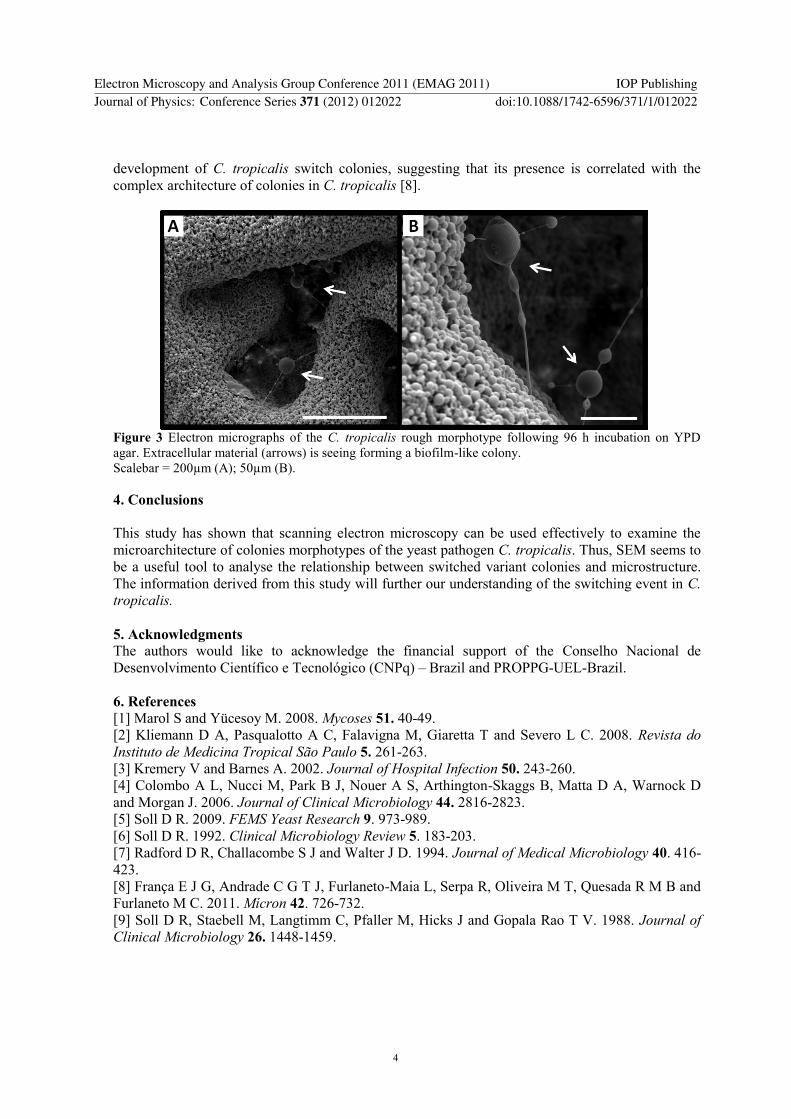
The ring and rough colonies phenotypes also comprised mainly yeast cells as observed at the
deep peripheral depressions areas (Fig 3) Most interesting is the presence of extracellular material
exclusively at these areas (Fig 3 A B) where many of the cells are almost hidden by this material
It was observed as fibrils with enlarged structures connecting neighbouring cells In a recent study
we report the presence of extracellular material resembling a biofilm-like colony throughout the
Electron Microscopy and Analysis Group Conference 2011 (EMAG 2011) IOP PublishingJournal of Physics Conference Series 371 (2012) 012022 doi1010881742-65963711012022
3
development of C tropicalis switch colonies suggesting that its presence is correlated with the
complex architecture of colonies in C tropicalis [8]
Figure 3 Electron micrographs of the C tropicalis rough morphotype following 96 h incubation on YPD
agar Extracellular material (arrows) is seeing forming a biofilm-like colony
Scalebar = 200microm (A) 50microm (B)
4 Conclusions
This study has shown that scanning electron microscopy can be used effectively to examine the
microarchitecture of colonies morphotypes of the yeast pathogen C tropicalis Thus SEM seems to
be a useful tool to analyse the relationship between switched variant colonies and microstructure
The information derived from this study will further our understanding of the switching event in C
tropicalis
5 Acknowledgments
The authors would like to acknowledge the financial support of the Conselho Nacional de
Desenvolvimento Cientiacutefico e Tecnoloacutegico (CNPq) ndash Brazil and PROPPG-UEL-Brazil
6 References
[1] Marol S and Yuumlcesoy M 2008 Mycoses 51 40-49
[2] Kliemann D A Pasqualotto A C Falavigna M Giaretta T and Severo L C 2008 Revista do
Instituto de Medicina Tropical Satildeo Paulo 5 261-263
[3] Kremery V and Barnes A 2002 Journal of Hospital Infection 50 243-260
[4] Colombo A L Nucci M Park B J Nouer A S Arthington-Skaggs B Matta D A Warnock D
and Morgan J 2006 Journal of Clinical Microbiology 44 2816-2823
[5] Soll D R 2009 FEMS Yeast Research 9 973-989
[6] Soll D R 1992 Clinical Microbiology Review 5 183-203
[7] Radford D R Challacombe S J and Walter J D 1994 Journal of Medical Microbiology 40 416-
423
[8] Franccedila E J G Andrade C G T J Furlaneto-Maia L Serpa R Oliveira M T Quesada R M B and
Furlaneto M C 2011 Miacutecron 42 726-732
[9] Soll D R Staebell M Langtimm C Pfaller M Hicks J and Gopala Rao T V 1988 Journal of
Clinical Microbiology 26 1448-1459
Electron Microscopy and Analysis Group Conference 2011 (EMAG 2011) IOP PublishingJournal of Physics Conference Series 371 (2012) 012022 doi1010881742-65963711012022
4
Scanning electron microscopy as a tool for the analysis of
colony architecture produced by phenotypic switching of a
human pathogenic yeast Candida tropicalis
M C Furlaneto1 C G T J Andrade
2 P H A Aragatildeo
2 E J G Franccedila
1 A T P Moralez
1
and L C S Ferreira1
1Department of Microbiology Paranaacute State University at Londrina Brazil
2Electronic Microscopy and Microanalysis Laboratory Paranaacute State University at Londrina
Brazil
Abstract Candida tropicalis has been identified as one of the
most prevalent pathogenic yeast
species of the Candida-non-albicans group Phenotypic switching is a biological phenomenon related
to the occurrence of spontaneous emergence of colonies with different morphologies that provides
variability within colonizing populations in order to adapt to different environments Currently
studies of the microstructure of switching variant colonies are not subject of extensive research SEM
analysis was used to verify the architecture of whole Candida colonies The strain 4907 exhibited a
hemispherical shape character while the strain 33507 showed a volcano shape with mycelated-edge
colony The ring switch variant is characterized by a highly wrinkled centre and an irregular
periphery The rough phenotype exhibited a three-dimensional architecture and was characterized by
the presence of deep central and peripheral depressions areas The ultrastructural analysis also
allowed the observation of the arrangement of individual cells within the colonies The whole smooth
colony consisted entirely of yeast cells Differently aerial filaments were found all around the colony
periphery of the volcano shape colony For this colony type the mycelated-edge consisted mainly of
hyphae although yeast cells are also seen The ring and rough colonies phenotypes comprised
mainly yeast cells with the presence of extracellular material connecting neighbouring cells This
study has shown that SEM can be used effectively to examine the microarchitecture of colonies
morphotypes of the yeast C tropicalis and further our understanding of switching event in this
pathogen
1 Introduction
Candida tropicalis is an opportunistic yeast pathogen that causes superficial and systemic mycoses
[1-3] In Latin America C tropicalis accounts for the majority of non-albicans Candida species
associated with candidemia episodes [4]
Among several virulence factors that contribute to pathogenesis of Candida it has been
suggested that phenotypic switching provides variability within colonizing populations in order to
adapt to challenges at different environments including various anatomical sites in the human body
[5]
Phenotypic switching represents an epigenetic state that occurs in a small fraction of the
population is random and reversible For fungi phenotypic switching is defined as the spontaneous
emergence of colonies with altered colony morphology at rates higher than the somatic mutations
rates that enables the microorganism to undergo rapid microevolution [6] For Candida albicans a
relationship between colony shape of switching variants and the constituent cells (blastospores true
and psedo-hyphae) has been demonstrated by scanning electron microscopy analysis [7] According
Electron Microscopy and Analysis Group Conference 2011 (EMAG 2011) IOP PublishingJournal of Physics Conference Series 371 (2012) 012022 doi1010881742-65963711012022
Published under licence by IOP Publishing Ltd 1
to these authors the relationship between switched variant colonies and microstructure may help
elucidate the relationship between pathogenicity in vivo and colonial morphology in vitro [7]
In contrast to the species C albicans the study of switching in C tropicalis has not been the
subject of extensive research Our work focuses on using scanning electron microscopy for the
analysis of switched variant colonies of clinical isolates of C tropicalis An understanding of this
virulence determinant would provide insight into C tropicalis pathogenic mechanisms
2 Experimental techniques
21 Fungal strains
C tropicalis strains 4907 and 33507 included in this study were recovered from tracheal secretion
and belong to the Candida culture collection of the Fungal Genetics Laboratory The University of
Londrina-Brazil The switched variants ring and rough were obtained as previously described [8]
22 Scanning electron microscopy of intact colonies
To verify the architecture of C tropicalis colonies morphotypes whole yeast colonies were
removed from YPD (1 yeast extract 2 peptone 2 dextrose) agar plates using a scalpel blade
Colonies were fixed for 18 h at 4oC in 3 glutaraldehyde (Electron Microscopy Sciences) in 01 M
phosphate buffer pH 72 They were then immersed in liquid nitrogen for 30 sec and freeze-dried
for 90 min at 2x10⁻³
MPa (Juan LP3) Then colonies were coated with gold (BALTEC SDC 050
Sputter Coater) and viewed in a FEI Quanta 200 Scanning Electron Microscope at 30kV
3 Results and Discussion
31 Microarchitecture of whole Candida colony
Despite the pioneer study on colony variants from C tropicalis clinical strains [9] little information
is available concerning the microstructure of individual colonies Thus we employed SEM to verify
the architecture of C tropicalis morphotypes The preparation of colonies by a freeze-drying
technique allowed their architecture preservation (Fig 1) with maintenance of the phenotypes
observed at lower magnitude (data not shown) The strain 4907 exhibited a hemispherical shape
character (Fig 1A) while the strain 33507 showed a volcano shape with mycelated-edge colony
(Fig 1B) The ring switch variant is characterized by a highly wrinkled centre and an irregular
periphery (Fig 1C) The rough phenotype exhibited more complex architecture and was
characterized by the presence of deep central and peripheral depressions areas (Fig 1D) Franccedila et
al [8] were the first to describe the architecture of whole Candida colonies at ultrastructural level
Here we extend these observations of both non-variant colonies and switch variants produced by
phenotypic switching of C tropicalis
Electron Microscopy and Analysis Group Conference 2011 (EMAG 2011) IOP PublishingJournal of Physics Conference Series 371 (2012) 012022 doi1010881742-65963711012022
2
Figure 1 Scanning electron micrographs showing the architecture of C tropicalis colonies morphotypes
following 96 h incubation on YPD agar (A) smooth (B) volcano shape with myceliated-edge (C) ring and
(D) rough Scalebar = 1mm
32 Ultrastructural analysis of morphotypes
The ultrastructural analysis allowed the observation of the arrangement of individual cells within
the colonies After 4 days of colony development the whole smooth colony consisted entirely of
yeast cells (not shown) Aerial filaments were found all around the colony periphery of the volcano
shape colony (Fig 2A) Although the central area of this colony morphotype also comprises
blastoconidia the mycelated-edge consisted mainly of hyphae (Fig 2B) although yeast cells are
also seen
Figure 2 Electron micrographs of the C tropicalis volcano shape with myceliated-edge morphotype
following 96 h incubation on YPD agar Scalebar = 200microm (A) 50microm (B) (B) Insert shows detail of
myceliated edge that is composed of hyphae and blastoconidia at higher magnification (5000x)
The ring and rough colonies phenotypes also comprised mainly yeast cells as observed at the
deep peripheral depressions areas (Fig 3) Most interesting is the presence of extracellular material
exclusively at these areas (Fig 3 A B) where many of the cells are almost hidden by this material
It was observed as fibrils with enlarged structures connecting neighbouring cells In a recent study
we report the presence of extracellular material resembling a biofilm-like colony throughout the
Electron Microscopy and Analysis Group Conference 2011 (EMAG 2011) IOP PublishingJournal of Physics Conference Series 371 (2012) 012022 doi1010881742-65963711012022
3
development of C tropicalis switch colonies suggesting that its presence is correlated with the
complex architecture of colonies in C tropicalis [8]
Figure 3 Electron micrographs of the C tropicalis rough morphotype following 96 h incubation on YPD
agar Extracellular material (arrows) is seeing forming a biofilm-like colony
Scalebar = 200microm (A) 50microm (B)
4 Conclusions
This study has shown that scanning electron microscopy can be used effectively to examine the
microarchitecture of colonies morphotypes of the yeast pathogen C tropicalis Thus SEM seems to
be a useful tool to analyse the relationship between switched variant colonies and microstructure
The information derived from this study will further our understanding of the switching event in C
tropicalis
5 Acknowledgments
The authors would like to acknowledge the financial support of the Conselho Nacional de
Desenvolvimento Cientiacutefico e Tecnoloacutegico (CNPq) ndash Brazil and PROPPG-UEL-Brazil
6 References
[1] Marol S and Yuumlcesoy M 2008 Mycoses 51 40-49
[2] Kliemann D A Pasqualotto A C Falavigna M Giaretta T and Severo L C 2008 Revista do
Instituto de Medicina Tropical Satildeo Paulo 5 261-263
[3] Kremery V and Barnes A 2002 Journal of Hospital Infection 50 243-260
[4] Colombo A L Nucci M Park B J Nouer A S Arthington-Skaggs B Matta D A Warnock D
and Morgan J 2006 Journal of Clinical Microbiology 44 2816-2823
[5] Soll D R 2009 FEMS Yeast Research 9 973-989
[6] Soll D R 1992 Clinical Microbiology Review 5 183-203
[7] Radford D R Challacombe S J and Walter J D 1994 Journal of Medical Microbiology 40 416-
423
[8] Franccedila E J G Andrade C G T J Furlaneto-Maia L Serpa R Oliveira M T Quesada R M B and
Furlaneto M C 2011 Miacutecron 42 726-732
[9] Soll D R Staebell M Langtimm C Pfaller M Hicks J and Gopala Rao T V 1988 Journal of
Clinical Microbiology 26 1448-1459
Electron Microscopy and Analysis Group Conference 2011 (EMAG 2011) IOP PublishingJournal of Physics Conference Series 371 (2012) 012022 doi1010881742-65963711012022
4
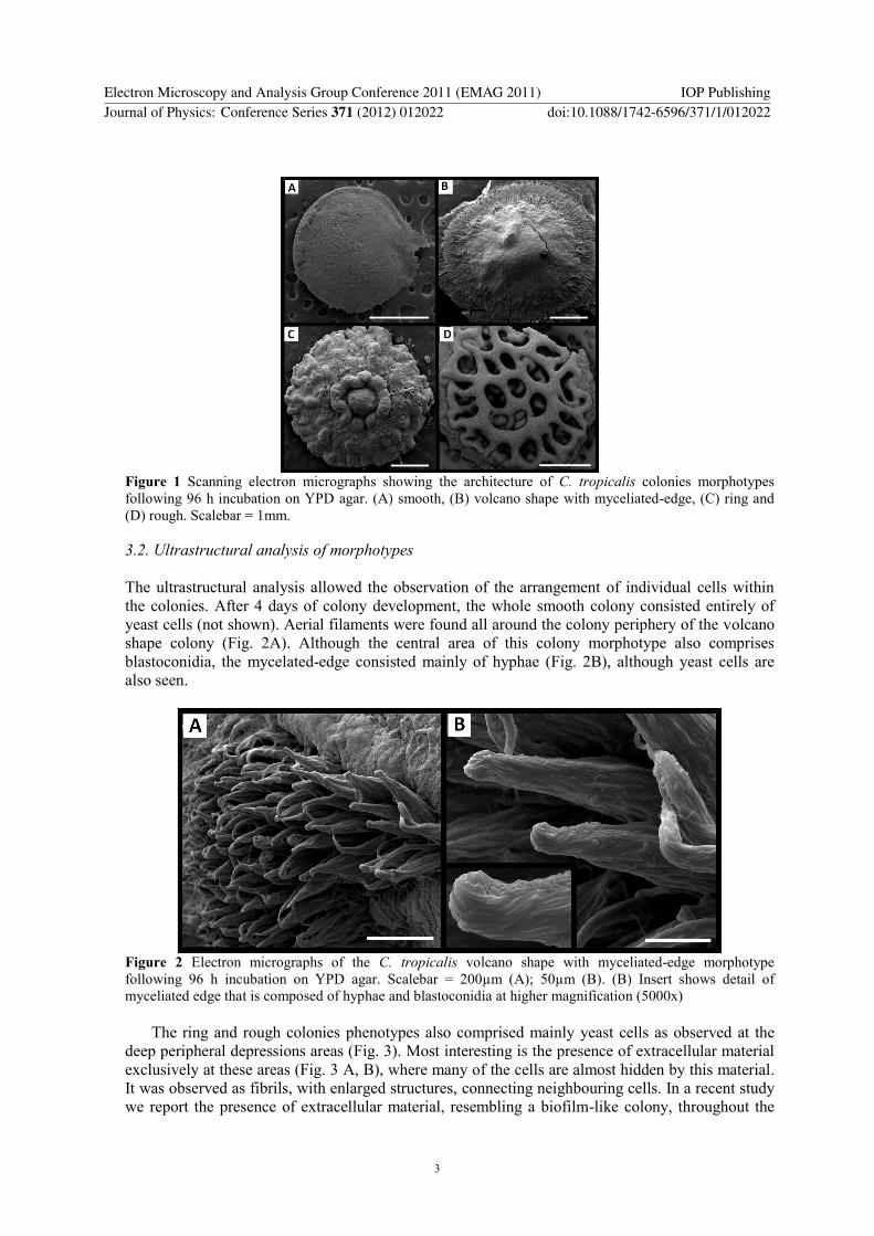