Embed Size (px)
Citation preview
Saudi Journal of Biological Sciences 27 (2020) 935–946
Contents lists available at ScienceDirect
Saudi Journal of Biological Sciences
journal homepage: www.sciencedirect .com
Original article
Biological control of yeast contamination of industrial foods by propolis
https://doi.org/10.1016/j.sjbs.2020.01.0231319-562X/� 2020 The Authors. Published by Elsevier B.V. on behalf of King Saud University.This is an open access article under the CC BY-NC-ND license (http://creativecommons.org/licenses/by-nc-nd/4.0/).
⇑ Corresponding author.E-mail address: [email protected] (E.F. Abd_Allah).
Peer review under responsibility of King Saud University.
Production and hosting by Elsevier
Mashail Fahd S. Alsayed a, Abeer Hashema,b, Amal A. Al-Hazzani a, Elsayed Fathi Abd_Allah c,⇑aBotany and Microbiology Department, College of Science, King Saud University, P.O. Box 2460, Riyadh 11451, Saudi ArabiabMycology and Plant Disease Survey Department, Plant Pathology Research Institute, ARC, Giza 12511, EgyptcPlant Production Department, College of Food and Agricultural Sciences, King Saud University, P.O. Box 2460, Riyadh 11451, Saudi Arabia
a r t i c l e i n f o a b s t r a c t
Article history:Received 14 September 2019Revised 17 January 2020Accepted 18 January 2020Available online 27 January 2020
Keywords:PropolisAntifungalAntioxidantProteomicsElectron microscopy
Bee glue (Propolis, PR), mixture of beeswax and resin is collected from honeybee (Apis mellifera) of differ-ent plant parts. The antimicrobial potential of PR against food borne yeast was reported. The experimentwas designed to examine the way of antimicrobial impact of PR on food borne yeasts (Cryptococcus lau-rentii and Candida famata) and its usage use as biological strategy for the preservation of soft foodsagainst microbial spoilage. The study also highlights, the ability of ethanol and water- PR extracts, dis-couraged growth of tested yeast. Antifungal properties were also determined using electron microscopewhile biochemical analysis was determined using free and proteinic amino acid technique and oxidativeenzymes were determined using HPLC analysis. Antioxidant enzymes were determined using ELISAassay. The highest effect was recorded on C. laurentii however, the lowest effect shows on C. famata.The electron microscopic studies clearly disclosed the effect of water PR distillate on the external shapeand internal organs of some tested yeast e.g. C. laurentii and C. famata. The result indicated some differ-ences on concentrations of bio-chemical analyses for these tested yeasts treated with 70% water- PRextracts of different food materials. Moreover, biochemical analysis results also reported that the treatedyeast indicated natural preservative to food products and considered as best alternative to the (chemical)preservatives currently employed.� 2020 The Authors. Published by Elsevier B.V. on behalf of King Saud University. This is an open access
article under the CC BY-NC-ND license (http://creativecommons.org/licenses/by-nc-nd/4.0/).
1. Introduction and the metabolic compounds of honeybee and is used to stabilize
Bee glue (Propolis), (PR) is a combination of a Greek term ‘‘Pro”meaning ‘‘opposite of entry”, and ‘‘polis” meaning ‘‘the city or com-munity” represents as ingredient which acts in protection of thehoard (Salatino et al., 2005). PR is a combination of beeswax andresins collected by Apis mellifera (honeybee) from different plantparts e.g., flower, buds, nectar and other plant exudates. PR fromdifferent geographic locations has been found to possess variousbiological happenings such as uncontaminated, antiviral, and anti-fungal. PR (bee glue) is a resinous waxbee hive by part produced bybees (Apis mellifera). It possessed various pharmaceutical proper-ties and till now has been used in folk medicines as bio-cosmetics and health foods (Bankova, 2005). PR is produced byhoneybees from the collected plant parts mixed with bee-resin
the honeycombs cells; act as defense against invaders and coldweather (Golder, 2004). PR composed of enzymes and salivarysecretions significant for fill cracks, cover hives or gaps to protectthemselves from microorganism’s entry, fungi and bacteria intothe hive (Bankova, 2000). Moreover, the PR can be used to linethe honeycomb, facilitate the smooth laying of eggs by the queen,and to preserve and petrifact the decay organisms (beetles andinsects) that commonly not part the hive and used by honeybees.(Castaldo and Capasso, 2002). Moreover, PR chemistry is viscousin nature includes essential oil (10%), beeswax (30%), resin (50%),pollen (5%) and organic and mineral compound (5%) (Fokt et al.,2010). Many scientists reported that (Bankova et al., 2000;Teixeira et al., 2010; Valencia et al., 2012), about 300 compoundsare present include phenolic acids, cinnamic acid, caffeic acid, ter-penes, flavonoids, esters, amino acids, sugar, sterols, steroid hydro-carbons, minerals, aliphatic hydrocarbons, sesquiterpene andtriterpene hydrocarbons in PR. PR is basically a lipophilic substancei.e., in colder environment, it’s hard, brittle while as in warm con-ditions, it0s soft, flexible and very sticky, hence called ”beeswax‘‘(Marcucci, 1995). It has a distinct odor, its oil shows adhesiveproperties and it has a strong reaction with skin proteins(Sforcin, 2007). Its composition is very complex (Boyanova,
936 M.F.S. Alsayed et al. / Saudi Journal of Biological Sciences 27 (2020) 935–946
2006). Moreover, vegetal origin of collected material affected bycollection time as well as chemical composition of resinous mate-rial (Fernandes et al., 2007). The color of PR varies from yellowishgreen to dark brown, depending upon the location viz; savannah,tropical forests, desert, coastal and mountainous regions, whereit is produced (Piccinelli et al., 2011). PR is attracting and morepopular as a natural preserving material in food industry. However,it has been added to foods and drinks as bioactive compounds toincrease life standard (Mishima et al., 2005; Moreira et al., 2008).
‘Hurdle technology’ planned by Leistner and Gorris (1995),advocates an intellectual use of mixtures of different preservationsmethods to realize multi-target, mild but preservation effectsshould be more safe, nutritious and economical foods. An extractof PR acts as successful antifungal agent in minute quantitiesagainst spoiled fruit juice yeasts. Focus in its antifungal propertieswas targeted on human health (Cafarchia et al., 1999). Na+ benzoatepreservative system is less efficient than PR as it more activeagainst yeast (Moreira et al., 2008). The antifungal mechanism ofPR was examined for the growth, aflatoxins production, lipids anddigestion (Hashem et al., 2012). It has also been reported byTakaisi-Kikuni and Schilcher (1994) cell division restriction wascaused by PR and might have inhibited the DNA replication of cells.Gas chromatographic analysis results of cellular fatty acids revealedthat PR increased the saturated fatty acids accumulation and sug-gested the defensemechanismof fungalmembrane by reducing cellflexibility and resistance (Hashem et al., 2012). Han and Park (2002)have reported that PR and its various products have been mainlyused for health benefits but not for fruit juice processing andpreservation yet. Currently, the focus of the use of PR in developedcountries has been on its utilization as a consumer suited healthsupplement. This is the reason it has nowadays been recognizedas a natural, healthy and beneficial product for human use(Espina et al., 2012). Several studies have been conducted pertain-ing to the composition and antifungal and antioxidant activitiesof PR from many topographical regions, such as Brazil, Bulgaria,Greece, Cyprus, France, Italy, and Croatia. (Marcucci et al., 2001;Prytzyk et al., 2003; Salomão et al., 2004; Bastos et al., 2008;Kalogeropoulos et al., 2009). However, little is known about SaudiPR, and its extracts. In current study, we explored the chemicalcomposition, antioxidant activities, and the anti-yeast propertiesof Saudi PR extracts. So, the current study was designed to charac-terize the bioactive properties of Saudi PR (regarding phenolic con-tent and antioxidant activity) and to evaluate the potentialeffectiveness of PR extract in different concentration against food-borne yeast, for biochemical analysis of treated yeast and indicatedthe application of natural preservative to food products as an alter-native to the (chemical) preservatives currently employed. The PRantimicrobial potential against food borne yeast were reported.Moreover, it was also investigating the mechanism of antimicrobialimpact of PR on food borne yeast and possibility to use PR as analternative biological strategy to preserve soft foods against yeast.
2. Materials and methods
2.1. Collecting and extract of PR
Raw Saudi PR was purchased from local bee farm (Al Soudah,Abha, Kingdom of Saudi Arabia). PR was collected manually fromthe beehives during the period of the dry season (May–July2017). After collection, PR was desiccated in dark and kept at atemperature of 4 �c until its processing.
2.2. Ethanolic extraction of PR (EEP)
PR material was crushed into very small pieces and 80% ethanolwas added to it by 1:10 (weight/volume) ratio. Afterwards the
blend was put in a closed flask at 25 �C for 48 h. The materials wereshake from time to time and distillate was then clean using What-man No. 1 filter paper type 4, and the distillate was a very densematerial like alcoholic extract (yaghobi et al., 2007).
2.3. Water extraction of PR (WEP)
PR samples were grinded using mortar and pestle and werethen diverse with purified water (1 g of PR per 10 ml of purifiedwater). PR water blend was then heated at 40 �C temperature ona hot plate till properly dissolved. The solution was then kept forcooling at room temperature and vortexed for 15 min. Afterwards,the solution was later filtered using Whatman filter paper no. 6.The filtrate was then evaporated at 40 �C using a hot oven andremaining filtrate solution was finally at 4 �C in the dark untiltested for antifungal activity (Siqueira et al., 2009). Different con-centrations (30, 50, 70, 100%) of water extract PR were preparedaccording to.
2.4. Chemical analysis of PR
2.4.1. Flavonoids assayFor the preparation of Solution, A dissolving 10 g of powder PR
was liquified in 50 ml of 95% ETOH solution followed by its filtra-tion. Solution B was prepared by adding 10 ml of ETOH (50%) to10 ml of NaOH (50%) followed by mixing equal volumes of bothsolutions A & B, the appearance of yellow color is the evidence offlavonoids (Kosalic et al., 2005).
2.4.2. Phenolic compounds assayPhenolic compound assay was prepared by adding 3 ml of PR
extract to 2 ml of ferric chloride solution (1%). The presence of abluish-green hue is evidence of the presence of phenols.
2.4.3. Resins assayFor the preparation of resin assay 10 g of powder PR was lique-
fied in 50 ml of 95% ETOH solution and left for two minutes in awater bath at 100 �C. The solution was then filtered, and 100 mlof water was added to solution followed by acidification withhydrochloric acid 4%. The appearance of turbidity in the solutionwas evidence of resins (Orsi et al., 2007).
2.4.4. Antifungal activity of PR extractThe agar well diffusion methods determined the antifungal
activities of PR extract as described by (Cafarchia et al., 1999).
2.4.5. Agar well diffusion methodYeast samples were isolated from food spoilage strains and
maintained at 30 �C on Sabouraud dextrose agar (SDA). The threeculture plates seeded with tested yeasts could solidify and werepunched to make open wells with a sterile cork borer (7.0 mmdiameter). 0.05 ml of PR extract were then filled with these openwells. The culture plates were then heated at 30 �C for 72 h andinhibition zones were observed. Evaluation of theFungistatic and Fungicidal Aspect after Exposure to Ethanolic andWater extraction of PR on Sabouraud dextrose Agar. The clear zoneof inhibition was swabbed using a sterile cotton swab. The sameswab was used to spread over the new plates containing SDA med-ium in order to figure out fungistatic and fungicidal aspect of theexposed PR to ethanolic and water extraction.
2.5. Evaluation of the antifungal properties of PR
2.5.1. Electron microscope studiesPreparation of isolated yeast after growing in PR extract accord-
ing to Afrikian et al., (1973). Scanning Electron Microscopes (SEM):
M.F.S. Alsayed et al. / Saudi Journal of Biological Sciences 27 (2020) 935–946 937
The effect of PR extract on structure of vegetation yeast cells aftergrowing in PR extract according to Bozzola and Russell (1999).Examination of the yeast cells was carried out by using JEOLJSM-7610F Scanning Electron Microscope at magnification of900�, 8000�, 10,000�, 16,000� and 20,000�.
Transmission Electron Microscopes (TEM): The Effect of PRextract on structure of vegetation yeast cells after growing in PRextract (Bozzola and Russell 1999). Examination of the yeast cellswas carried out by using JEOL JEM-1400plus TEM at magnificationof 20000�, 30000�, 40000�, 50000� and 60000�.
2.5.2. Extraction and analysis of amino acidsThe cell free extract after thoroughly washed with 70% ethanol
was use in small column (0.8 � 12 cm) of dowex 50 (H form) tohold the free amino acids. The free amino acids were eluted with25 ml ammonia in 75 ml of 75% ethanol. The amino acid was dis-solved in 0.2 ml distilled water immediately after drying them invacuum in a rotary evaporator. Proteinic Amino acid HPLC analysiswas carried according to.
For proteinic amino acid analysis, 1 g yeast cells were digestedwith 6 N Hydrochloric acid, then dried and dissolved in methanoland filtered through 0.45um membrane filter before HPLC injec-tion. The samples were finally redissolved in borate (pH 8.8) buffer(for derivatization with AQC (6-aminoquinolyl-N-hydroxysuccinimidyl carbamat). GBC HPLC system was used for the quantificationof hydrolyzed amino acids. This system consists of two GBC LC1110 pumps, a helium solvent degassing system, a 150 � 3.8 mmRP18 steel column, using fluorescence detector, a Win ChromChromatography Ver. 1.3 and having software for data procure-ment and a temperature control module. For the separation ofAQC derivative amino acids, eluent mobile phase cetonitrile: waterration (72:28; v/v) were used and 1.6 ml/min flow rate was appliedat 37 �C temperature. Excitation and emanation wavelengths wereset at 250 and 395 nm, respectively.
2.5.3. Determination of oxidative enzymesThe harvested yeast cells were washed double with sterile
double-distilled water. For oxidative enzymes, 1 g from the yeastcells was suspended in 2ml phosphate buffer (pH 7). The yeast cellssuspension was moved into an extraction buffer, contained 20 mMof a potassium phosphate buffer (pH 7) and a protease inhibitorcocktail. The cells were sonicated in ice-cold normal saline (1/9,w/v) in Virsonic� ultrasonic cell disruptor for 10 min., then cen-trifuged at 5000 rpm for 5 min at 4�C and the supernatant (celllysate) was stored at �80�C until the assays were performed (Timuret al., 2005). Superoxide dismutase (SOD) [using ELISA kit purchasedfrom Cayman company USA cat No. 706002] determined at 450 nm.Catalase [using ELISA kit Cayman company USA cat No. 707002]determined at 540 nm. Glutathione Reductase [using OxiSelectTM
ELISA kit purchased from Cell Biolabs company San Diego, CA, USAcatNo. STA-812] determined at 405nm.Ascorbate Peroxidase activ-ity were determined at 340 nm [using ELISA kit Cohesion Bio-sciences company USA cat No. CAK1052]. The said activity wasaccording to instruction manuals and the absorbance were mea-sured using 800 TS Microplate Reader Bio-Tek company, USA.
3. Results
3.1. Antifungal potential of ethanolic and water PR extract
For the determination of the antifungal effects of ethanolic andwater extract PR on the tested yeasts, the zone of inhibition wasdivided according to (Saubolle and Hoeprich., 1978) and the scaleof the results used in the current study was shown in Table 1.The weekly effect for ethanolic and water PR extracts for yeastwere in range of (�16 mm) in diameter, intermediate effect was
in range between (>17 to 23 mm) in diameter and highly effectfor ethanol and water propels extract were in range of (>24 mm)in diameter. According to size of inhibition zone, the highest effectof PR extract was on C. laurentii while as the least impact of PRextract was on C. famata. The analysis of variance (ANOVA) tablefor zone size inhibition based on error mean square value wasdescribe in the Table 2. All the zone size showed significant effectat 5% probability.
3.2. Scanning electron microscope
After examining different yeast using scanning electron micro-scope, the micrograph in Fig. 1 (A: B) showing effect of non-treatedand treated water PR on C. laurentii which indicated decreasingnumber of yeast cells when treated with the said extract. The scan-ning electron micrograph of non-treated C. laurentii cells in (Fig. 2A, B, C). showed regular oval shape yeast cells. On the other hand,crease on some cell walls with some deformities in the externalshape of cells were observed after treatment of C. laurentii by waterPR extract, shown in (Fig. 2 D, E, F). Again, after the treatment of C.laurentii by water PR extract, the micrograph was shown in (Fig. 3A, B) indicated some appendages on yeast cell wall, some deformi-ties in the external shape of cells and exit of the components ofsome cells. Also, the scanning electron micrograph of non-treatedC. famata yeast cells was shown in (Fig. 4 A), while as the micro-graph of C. famata after treated by water PR extract, showed somedecreasing number of cells was shown in (Fig. 4 B). Finally, thescanning electron micrograph of non-treated C. famata cells repre-sented by (Fig. 5 A) showed Oval to Globose shape of yeast cells. Onthe other hand, C. famata cells after treatment by water PR extractobserved deformities in the external shape of cells and someappendages on yeast cell wall shown in (Fig. 5 B).
3.3. Fungistatic and fungicidal of PR (ethanolic and water extraction)
The data was recorded according to growth (fungistatic, FS) ornon-growth (fungicidal, FC) on S.D.A and shown in (Table 3).Results discovered that of all the established isolates, PR waterextract gave FS effect against the isolates of Candida famata,C. parapsilosis and Cryptococcus terreus and FC effect against therest of the isolates. While as the PR ethanol extract gave FS effectagainst the isolates of Rhodotorula mucilaginosa, C. famata, C. pel-liculosa, C. parapsilosis and Cryptococcus terreus and FC effectagainst the rest of isolates. The data regarding mean value of fun-gistatic and fungicidal effects of ethanolic and water extraction ofPR against yeast cells isolated from industrial foods was pre-sented in the Table 4. The maximum fungicidal effect wasrecorded in EPE 30 Saccharomyces cerevisiae, Trichosporonmucoides, Rhodotorula mucilaginosa recorded 16.0 which was fol-lowed by the yeast Candida parapsilosis recorded (15.50). Theminimum fungicidal effect (12.0) was recorded for CandidaFamata for zone size 30. While for EPE 50 the maximum fungici-dal effect was (25.0) recorded against yeast Candida magnoliaeand Cryptococcus laurentii which was followed by the yeastsSaccharomyces cerevisiae, Rhodotorula mucilaginosa, C. zeylanoides,C. pelliculosa, C. sphaerica and C. lusitaniae recorded (24.0) usingEPE50. The maximum fungicidal effect for EPE70 was recorded28.0 under Cryptococcus laurentii which was followed by Candidazeylanoides, Candida pelliculosa and Kloeckera spp, recorded 27.0.The minimum was (18.0) recorded under Candida famatausing EPE 70 zone size. The maximum Cryptococcus laurentiirecorded (32.0) using zone size 100 which was followed by the30.5 recorded C. magnoliae and C. zeylanoides. The minimumfungicidal effect was 24.5 recorded using C. famata yeast cellsunder EPE 100.
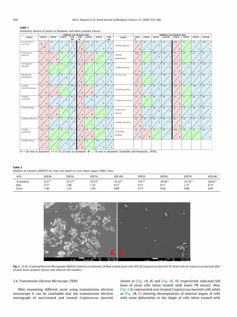
Table 1Sensitivity pattern of yeasts to Ethanolic and water propolis extract.
S= > 24 mm in diameter: I = 17 to 23 mm in diameter: R= � 16 mm in diameter (Saubolle and Hoeprich., 1978).
Fig. 1. (A-B). Scanning Electron Micrograph (SEM)of Cryptococcus laurentii, (A)Non treated yeast cells (NT) of Cryptococcus laurentii (B) Yeast cells of Cryptococcus laurentii aftertreated water propolis extract and reduced cell numbers.
Table 2Analysis of variance (ANOVA) for zone size based on error mean square (EMS) value.
SOV EPE30 EPE50 EPE70 EPE100 EPE30 EPE50 EPE70 EPE100
Treatment 4.27** 37.57** 18.33** 16.32** 3.07** 39.94** 23.78** 28.02**Rep 2.37 1.46 1.35 0.17 0.11 0.17 1.27 0.75Error 1.40 1.23 1.04 0.89 0.72 0.94 0.60 0.95
938 M.F.S. Alsayed et al. / Saudi Journal of Biological Sciences 27 (2020) 935–946
3.4. Transmission Electron Microscope (TEM)
After examining different yeast using transmission electronmicroscope it can be concluded that the transmission electronmicrograph of non-treated and treated Cryptococcus laurentii
shown in (Fig. 6A, B) and (Fig. 6C, D) respectively indicated fulllyses of yeast cells when treated with water PR extract. Also,(Fig. 6 A) represented non-treated Cryptococcus laurentii cells whileas (Fig. 6B, C) showing decomposition of internal organs of cellswith some deformities in the shape of cells when treated with
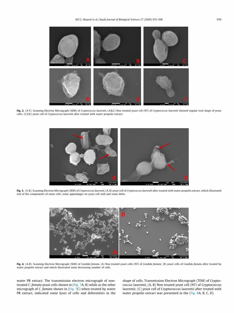
Fig. 2. (A-F). Scanning Electron Micrograph (SEM) of Cryptococcus laurentii, (A,B,C) Non treated yeast cell (NT) of Cryptococcus laurentii showed regular oval shape of yeastcells; (C,D,E) yeast cell of Cryptococcus laurentii after treated with water propolis extract.
Fig. 3. (A-B). Scanning Electron Micrograph (SEM) of Cryptococcus laurentii, (A, B) yeast cell of Cryptococcus laurentii after treated with water propolis extract, which illustratedexit of the components of some cells, some appendages on yeast cell wall and some defor.
Fig. 4. (A-B). Scanning Electron Micrograph (SEM) of Candida famata, (A) Non treated yeast cells (NT) of Candida famata; (B) yeast cells of Candida famata after treated bywater propolis extract and which illustrated some decreasing number of cells.
M.F.S. Alsayed et al. / Saudi Journal of Biological Sciences 27 (2020) 935–946 939
water PR extract. The transmission electron micrograph of non-treated C. famata yeast cells shown in (Fig. 7A, B) while as the othermicrograph of C. famata shown in (Fig. 7C) when treated by waterPR extract, indicated some lyses of cells and deformities in the
shape of cells. Transmission Electron Micrograph (TEM) of Crypto-coccus laurentii, (A, B) Non treated yeast cell (NT) of Cryptococcuslaurentii; (C) yeast cell of Cryptococcus laurentii after treated withwater propolis extract was presented in the (Fig. 8A, B, C, D).
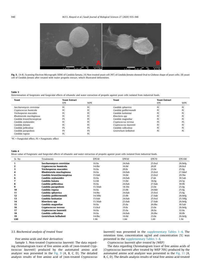
Fig. 5. (A-B). Scanning Electron Micrograph (SEM) of Candida famata, (A) Non treated yeast cell (NT) of Candida famata showed Oval to Globose shape of yeast cells; (B) yeastcell of Candida famata after treated with water propolis extract, which illustrated deformities.
Table 3Determination of fungistatic and fungicidal effects of ethanolic and water extraction of propolis against yeast cells isolated from industrial foods.
Yeast Yeast Extract Yeast Yeast ExtractEPE WPE EPE WPE
Saccharomyces cerevisiae FC FC Candida sphaerica FC FCCryptococcus humicola FC FC Candida guilliermondii FC FCTrichosporon mucoides FC FC Candida lusitaniae FC FCRhodotorula mucilaginosa FC FC Kloeckera spp FC FCCandida krusei/inconspicua FS FC Candida magnoliae FC FCCandida zeylanoides FC FC Cryptococcus terreus FS FSCandida famata FC FC Cryptococcus laurentii FC FCCandida pelliculosa FS FS Candida colliculosa FC FCCandida parapsilosis FS FS Geotrichum kelbahnii FC FCCandida rugosa FC FC
*FC = Fungicidal effect, FS = Fungistatic effect
Table 4Mean value of Fungistatic and fungicidal effects of ethanolic and water extraction of propolis against yeast cells isolated from industrial foods.
Sr. No Treatments EPE30 EPE50 EPE70 EPE100
1 Saccharomyces cerevisiae 16.0a 24.0ab 25.0cd 26.0efg2 Cryptococcus humicola 14.0bc 16.0d 20.0f 28.0cd3 Trichosporon mucoides 16.0a 20.0c 23.0e 25.0g4 Rhodotorula mucilaginosa 16.0a 24.0ab 25.0cd 27.0def5 Candida krusei/inconspicua 15.0ab 16.0d 23.0cd 29.5bc6 Candida zeylanoides 16.0a 24.0ab 27ab 30.5ab7 Candida famata 12.0d 15.0d 18.0g 24.5g8 Candida pelliculosa 16.0a 24.0ab 27.0ab 30.0b9 Candida parapsilosis 15.50ab 18.50c 23.0e 25.0g10 Candida rugosa 16.0a 23.0b 24.0de 25.0g11 Candida sphaerica 14.0bc 24.0ab 25.0cd 25.0g12 Candida guilliermondii 14.50bc 23.0b 27.0ab 26.0efg13 Candida lusitaniae 15.0ab 24.0ab 25.0cd 25.50fg14 Kloeckera spp 15.50ab 23.0ab 27.0ab 26.0efg15 Candida magnoliae 16.0a 25.0a 26.0bc 30.5ab16 Cryptococcus terreus 13.0cd 19.0c 23.0e 26.0efg17 Cryptococcus laurentii 16.0a 25.0a 28.0a 32.0a18 Candida colliculosa 16.0a 24.0ab 26.0bc 30.0b19 Geotrichum kelbahnii 14.0bc 16.0d 23.0e 26.0efgLSD 1.96 1.84 1.69 1.56
940 M.F.S. Alsayed et al. / Saudi Journal of Biological Sciences 27 (2020) 935–946
3.5. Biochemical analysis of treated Yeast
Free amino acids and their derivativesSample 1. Non-treated Cryptococcus laurentii: The data regard-
ing chromatogram trace of free amino acids of (non-treated Cryp-tococcus laurentii) produced by the automated amino acidanalyzer was presented in the Fig. 9 (A, B, C, D). The detailedanalysis results of free amino acid of (non-treated Cryptococcus
laurentii) was presented in the supplementary Tables 1–4. Theretention time, concentration ug/ml and concentration (%) waspresented in the supplementary Tables 1–4.
Cryptococcus laurentii after treated by (WEP)The data regarding Chromatogram trace of free amino acids of
(Cryptococcus laurentii after treated by WEP 70%) produced by theautomated amino acid analyzer was presented in the Fig. 11 (A,B, C, D). The details analysis results of total free amino acid treated
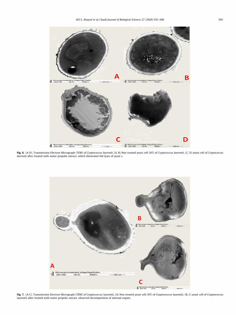
Fig. 6. (A-D). Transmission Electron Micrograph (TEM) of Cryptococcus laurentii, (A, B) Non treated yeast cell (NT) of Cryptococcus laurentii; (C, D) yeast cell of Cryptococcuslaurentii after treated with water propolis extract, which illustrated full lyses of yeast c.
Fig. 7. (A-C). Transmission Electron Micrograph (TEM) of Cryptococcus laurentii, (A) Non treated yeast cell (NT) of Cryptococcus laurentii; (B, C) yeast cell of Cryptococcuslaurentii after treated with water propolis extract, observed decomposition of internal organs.
M.F.S. Alsayed et al. / Saudi Journal of Biological Sciences 27 (2020) 935–946 941
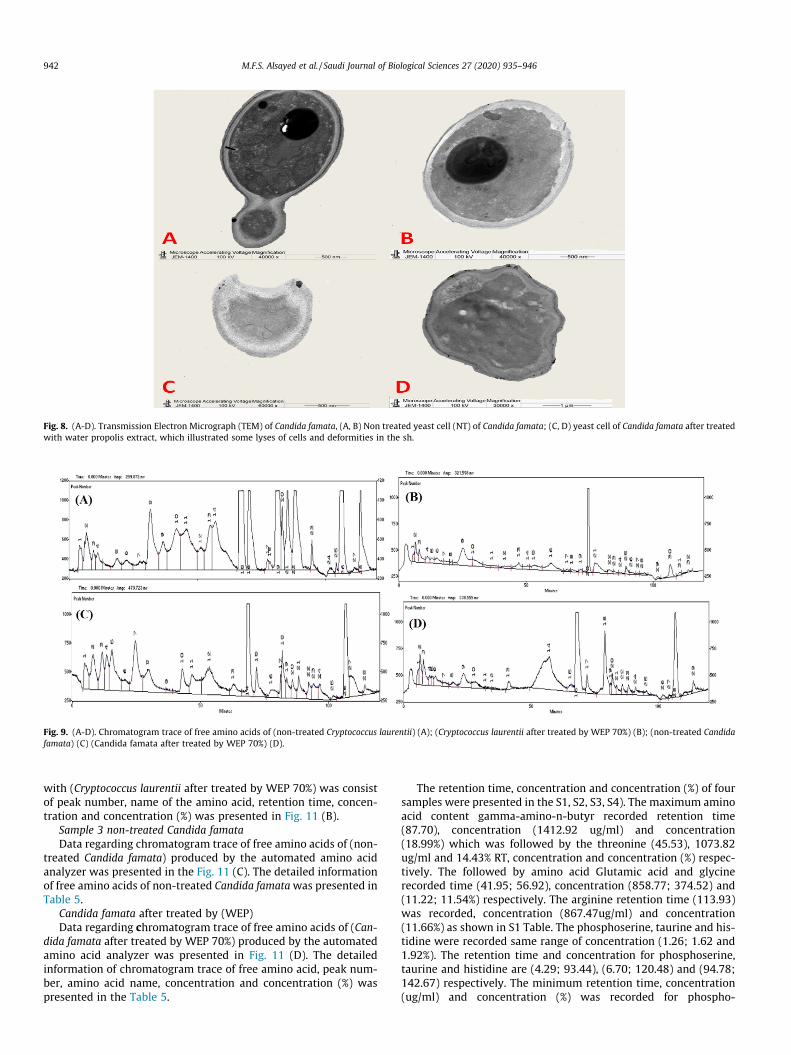
Fig. 8. (A-D). Transmission Electron Micrograph (TEM) of Candida famata, (A, B) Non treated yeast cell (NT) of Candida famata; (C, D) yeast cell of Candida famata after treatedwith water propolis extract, which illustrated some lyses of cells and deformities in the sh.
Fig. 9. (A-D). Chromatogram trace of free amino acids of (non-treated Cryptococcus laurentii) (A); (Cryptococcus laurentii after treated by WEP 70%) (B); (non-treated Candidafamata) (C) (Candida famata after treated by WEP 70%) (D).
942 M.F.S. Alsayed et al. / Saudi Journal of Biological Sciences 27 (2020) 935–946
with (Cryptococcus laurentii after treated by WEP 70%) was consistof peak number, name of the amino acid, retention time, concen-tration and concentration (%) was presented in Fig. 11 (B).
Sample 3 non-treated Candida famataData regarding chromatogram trace of free amino acids of (non-
treated Candida famata) produced by the automated amino acidanalyzer was presented in the Fig. 11 (C). The detailed informationof free amino acids of non-treated Candida famatawas presented inTable 5.
Candida famata after treated by (WEP)Data regarding chromatogram trace of free amino acids of (Can-
dida famata after treated by WEP 70%) produced by the automatedamino acid analyzer was presented in Fig. 11 (D). The detailedinformation of chromatogram trace of free amino acid, peak num-ber, amino acid name, concentration and concentration (%) waspresented in the Table 5.
The retention time, concentration and concentration (%) of foursamples were presented in the S1, S2, S3, S4). The maximum aminoacid content gamma-amino-n-butyr recorded retention time(87.70), concentration (1412.92 ug/ml) and concentration(18.99%) which was followed by the threonine (45.53), 1073.82ug/ml and 14.43% RT, concentration and concentration (%) respec-tively. The followed by amino acid Glutamic acid and glycinerecorded time (41.95; 56.92), concentration (858.77; 374.52) and(11.22; 11.54%) respectively. The arginine retention time (113.93)was recorded, concentration (867.47ug/ml) and concentration(11.66%) as shown in S1 Table. The phosphoserine, taurine and his-tidine were recorded same range of concentration (1.26; 1.62 and1.92%). The retention time and concentration for phosphoserine,taurine and histidine are (4.29; 93.44), (6.70; 120.48) and (94.78;142.67) respectively. The minimum retention time, concentration(ug/ml) and concentration (%) was recorded for phospho-
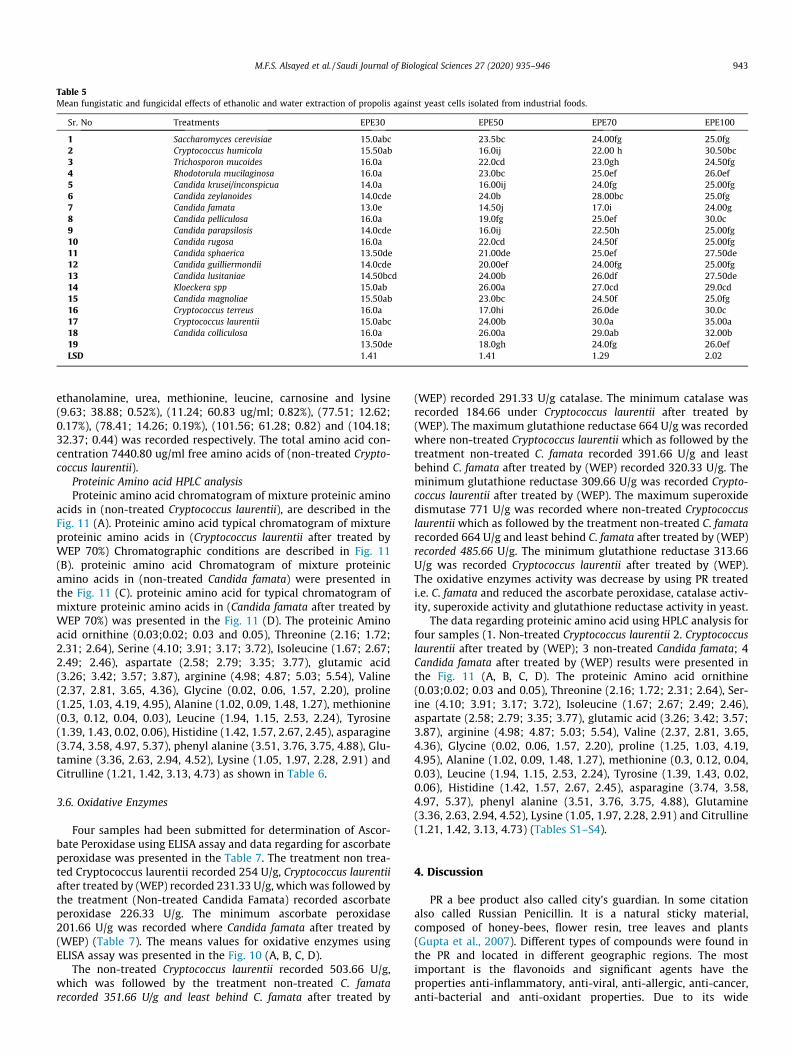
Table 5Mean fungistatic and fungicidal effects of ethanolic and water extraction of propolis against yeast cells isolated from industrial foods.
Sr. No Treatments EPE30 EPE50 EPE70 EPE100
1 Saccharomyces cerevisiae 15.0abc 23.5bc 24.00fg 25.0fg2 Cryptococcus humicola 15.50ab 16.0ij 22.00 h 30.50bc3 Trichosporon mucoides 16.0a 22.0cd 23.0gh 24.50fg4 Rhodotorula mucilaginosa 16.0a 23.0bc 25.0ef 26.0ef5 Candida krusei/inconspicua 14.0a 16.00ij 24.0fg 25.00fg6 Candida zeylanoides 14.0cde 24.0b 28.00bc 25.0fg7 Candida famata 13.0e 14.50j 17.0i 24.00g8 Candida pelliculosa 16.0a 19.0fg 25.0ef 30.0c9 Candida parapsilosis 14.0cde 16.0ij 22.50h 25.00fg10 Candida rugosa 16.0a 22.0cd 24.50f 25.00fg11 Candida sphaerica 13.50de 21.00de 25.0ef 27.50de12 Candida guilliermondii 14.0cde 20.00ef 24.00fg 25.00fg13 Candida lusitaniae 14.50bcd 24.00b 26.0df 27.50de14 Kloeckera spp 15.0ab 26.00a 27.0cd 29.0cd15 Candida magnoliae 15.50ab 23.0bc 24.50f 25.0fg16 Cryptococcus terreus 16.0a 17.0hi 26.0de 30.0c17 Cryptococcus laurentii 15.0abc 24.00b 30.0a 35.00a18 Candida colliculosa 16.0a 26.00a 29.0ab 32.00b19 13.50de 18.0gh 24.0fg 26.0efLSD 1.41 1.41 1.29 2.02
M.F.S. Alsayed et al. / Saudi Journal of Biological Sciences 27 (2020) 935–946 943
ethanolamine, urea, methionine, leucine, carnosine and lysine(9.63; 38.88; 0.52%), (11.24; 60.83 ug/ml; 0.82%), (77.51; 12.62;0.17%), (78.41; 14.26; 0.19%), (101.56; 61.28; 0.82) and (104.18;32.37; 0.44) was recorded respectively. The total amino acid con-centration 7440.80 ug/ml free amino acids of (non-treated Crypto-coccus laurentii).
Proteinic Amino acid HPLC analysisProteinic amino acid chromatogram of mixture proteinic amino
acids in (non-treated Cryptococcus laurentii), are described in theFig. 11 (A). Proteinic amino acid typical chromatogram of mixtureproteinic amino acids in (Cryptococcus laurentii after treated byWEP 70%) Chromatographic conditions are described in Fig. 11(B). proteinic amino acid Chromatogram of mixture proteinicamino acids in (non-treated Candida famata) were presented inthe Fig. 11 (C). proteinic amino acid for typical chromatogram ofmixture proteinic amino acids in (Candida famata after treated byWEP 70%) was presented in the Fig. 11 (D). The proteinic Aminoacid ornithine (0.03;0.02; 0.03 and 0.05), Threonine (2.16; 1.72;2.31; 2.64), Serine (4.10; 3.91; 3.17; 3.72), Isoleucine (1.67; 2.67;2.49; 2.46), aspartate (2.58; 2.79; 3.35; 3.77), glutamic acid(3.26; 3.42; 3.57; 3.87), arginine (4.98; 4.87; 5.03; 5.54), Valine(2.37, 2.81, 3.65, 4.36), Glycine (0.02, 0.06, 1.57, 2.20), proline(1.25, 1.03, 4.19, 4.95), Alanine (1.02, 0.09, 1.48, 1.27), methionine(0.3, 0.12, 0.04, 0.03), Leucine (1.94, 1.15, 2.53, 2.24), Tyrosine(1.39, 1.43, 0.02, 0.06), Histidine (1.42, 1.57, 2.67, 2.45), asparagine(3.74, 3.58, 4.97, 5.37), phenyl alanine (3.51, 3.76, 3.75, 4.88), Glu-tamine (3.36, 2.63, 2.94, 4.52), Lysine (1.05, 1.97, 2.28, 2.91) andCitrulline (1.21, 1.42, 3.13, 4.73) as shown in Table 6.
3.6. Oxidative Enzymes
Four samples had been submitted for determination of Ascor-bate Peroxidase using ELISA assay and data regarding for ascorbateperoxidase was presented in the Table 7. The treatment non trea-ted Cryptococcus laurentii recorded 254 U/g, Cryptococcus laurentiiafter treated by (WEP) recorded 231.33 U/g, which was followed bythe treatment (Non-treated Candida Famata) recorded ascorbateperoxidase 226.33 U/g. The minimum ascorbate peroxidase201.66 U/g was recorded where Candida famata after treated by(WEP) (Table 7). The means values for oxidative enzymes usingELISA assay was presented in the Fig. 10 (A, B, C, D).
The non-treated Cryptococcus laurentii recorded 503.66 U/g,which was followed by the treatment non-treated C. famatarecorded 351.66 U/g and least behind C. famata after treated by
(WEP) recorded 291.33 U/g catalase. The minimum catalase wasrecorded 184.66 under Cryptococcus laurentii after treated by(WEP). The maximum glutathione reductase 664 U/g was recordedwhere non-treated Cryptococcus laurentii which as followed by thetreatment non-treated C. famata recorded 391.66 U/g and leastbehind C. famata after treated by (WEP) recorded 320.33 U/g. Theminimum glutathione reductase 309.66 U/g was recorded Crypto-coccus laurentii after treated by (WEP). The maximum superoxidedismutase 771 U/g was recorded where non-treated Cryptococcuslaurentii which as followed by the treatment non-treated C. famatarecorded 664 U/g and least behind C. famata after treated by (WEP)recorded 485.66 U/g. The minimum glutathione reductase 313.66U/g was recorded Cryptococcus laurentii after treated by (WEP).The oxidative enzymes activity was decrease by using PR treatedi.e. C. famata and reduced the ascorbate peroxidase, catalase activ-ity, superoxide activity and glutathione reductase activity in yeast.
The data regarding proteinic amino acid using HPLC analysis forfour samples (1. Non-treated Cryptococcus laurentii 2. Cryptococcuslaurentii after treated by (WEP); 3 non-treated Candida famata; 4Candida famata after treated by (WEP) results were presented inthe Fig. 11 (A, B, C, D). The proteinic Amino acid ornithine(0.03;0.02; 0.03 and 0.05), Threonine (2.16; 1.72; 2.31; 2.64), Ser-ine (4.10; 3.91; 3.17; 3.72), Isoleucine (1.67; 2.67; 2.49; 2.46),aspartate (2.58; 2.79; 3.35; 3.77), glutamic acid (3.26; 3.42; 3.57;3.87), arginine (4.98; 4.87; 5.03; 5.54), Valine (2.37, 2.81, 3.65,4.36), Glycine (0.02, 0.06, 1.57, 2.20), proline (1.25, 1.03, 4.19,4.95), Alanine (1.02, 0.09, 1.48, 1.27), methionine (0.3, 0.12, 0.04,0.03), Leucine (1.94, 1.15, 2.53, 2.24), Tyrosine (1.39, 1.43, 0.02,0.06), Histidine (1.42, 1.57, 2.67, 2.45), asparagine (3.74, 3.58,4.97, 5.37), phenyl alanine (3.51, 3.76, 3.75, 4.88), Glutamine(3.36, 2.63, 2.94, 4.52), Lysine (1.05, 1.97, 2.28, 2.91) and Citrulline(1.21, 1.42, 3.13, 4.73) (Tables S1–S4).
4. Discussion
PR a bee product also called city’s guardian. In some citationalso called Russian Penicillin. It is a natural sticky material,composed of honey-bees, flower resin, tree leaves and plants(Gupta et al., 2007). Different types of compounds were found inthe PR and located in different geographic regions. The mostimportant is the flavonoids and significant agents have theproperties anti-inflammatory, anti-viral, anti-allergic, anti-cancer,anti-bacterial and anti-oxidant properties. Due to its wide
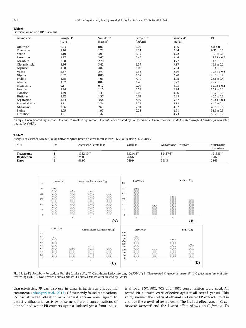
Table 6Proteinic Amino acid HPLC analysis.
Amino acids Sample 1*(mg/gm)
Sample 2*(mg/gm)
Sample 3*(mg/gm)
Sample 4*(mg/gm)
RT
Ornithine 0.03 0.02 0.03 0.05 8.8 ± 0.1Threonine 2.16 1.72 2.31 2.64 9.35 ± 0.1Serine 4.10 3.91 3.17 3.72 10.1 ± 0.1Isoleucine 1.67 2.67 2.49 2.46 13.52 ± 0.2Aspartate 2.58 2.79 3.35 3.77 14.9 ± 0.3Glutamic acid 3.26 3.42 3.57 3.87 16.8 ± 0.2Arginine 4.98 4.87 5.03 5.54 18.8 ± 0.1Valine 2.37 2.81 3.65 4.36 19.01 ± 0.1Glycine 0.02 0.06 1.57 2.20 23.3 ± 0.8Proline 1.25 1.03 4.19 4.95 25.6 ± 0.4Alanine 1.02 0.09 1.48 1.27 29.4 ± 0.3Methionine 0.3 0.12 0.04 0.03 32.75 ± 0.1Leucine 1.94 1.15 2.53 2.24 35.9 ± 0.1Tyrosine 1.39 1.43 0.02 0.06 38.2 ± 0.1Histidine 1.42 1.57 2.67 2.45 40.5 ± 0.1Asparagine 3.74 3.58 4.97 5.37 42.83 ± 0.1Phenyl alanine 3.51 3.76 3.75 4.88 44.7 ± 0.1Glutamine 3.36 2.63 2.94 4.52 48.1 ± 0.5Lysine 1.05 1.97 2.28 2.91 51.3 ± 0.3Citrulline 1.21 1.42 3.13 4.73 56.2 ± 0.7
*Sample 1 non treated Cryptococcus laurentii *Sample 2 Cryptococcus laurentii after treated by (WEP) *Sample 3 non treated Candida famata *Sample 4 Candida famata aftertreated by (WEP).
Table 7Analysis of Variance (ANOVA) of oxidative enzymes based on error mean square (EMS) value using ELISA assay.
SOV Df Ascorbate Peroxidase Catalase Glutathione Reductase Superoxidedismutase
Treatments 3 1382.89** 53214.3** 82437.6** 121535**Replication 2 25.08 266.6 1573.1 1287Error 6 90.97 749.9 565.3 2866
Fig. 10. (A-D). Ascorbate Peroxidase U/g; (B) Catalase U/g; (C) Glutathione Reductase U/g; (D) SOD U/g 1. (Non-treated Cryptococcus laurentii; 2. Cryptococcus laurentii aftertreated by (WEP) 3. Non-treated Candida famata 4. Candida famata after treated by (WEP).
944 M.F.S. Alsayed et al. / Saudi Journal of Biological Sciences 27 (2020) 935–946
characteristics, PR can also use in canal irrigation as endodontictreatments (Ahangari et al., 2018). Of the newly foundmedications,PR has attracted attention as a natural antimicrobial agent. Todetect antibacterial activity of some different concentrations ofethanol and water PR extracts against isolated yeast from indus-
trial food, 30%, 50%, 70% and 100% concentration were used. Alltested PR extracts were effective against all tested yeasts. Thisstudy showed the ability of ethanol and water PR extracts, to dis-courage the growth of tested yeast. The highest effect was on Cryp-tococcus laurentii and the lowest effect shows on C. famata. To
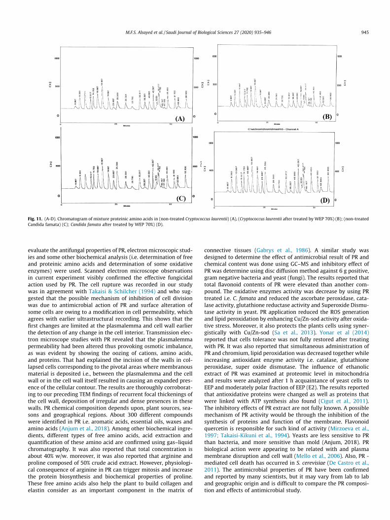
Fig. 11. (A-D). Chromatogram of mixture proteinic amino acids in (non-treated Cryptococcus laurentii) (A), (Cryptococcus laurentii after treated by WEP 70%) (B); (non-treatedCandida famata) (C); Candida famata after treated by WEP 70%) (D).
M.F.S. Alsayed et al. / Saudi Journal of Biological Sciences 27 (2020) 935–946 945
evaluate the antifungal properties of PR, electron microscopic stud-ies and some other biochemical analysis (i.e. determination of freeand proteinic amino acids and determination of some oxidativeenzymes) were used. Scanned electron microscope observationsin current experiment visibly confirmed the effective fungicidalaction used by PR. The cell rupture was recorded in our studywas in agreement with Takaisi & Schilcher (1994) and who sug-gested that the possible mechanism of inhibition of cell divisionwas due to antimicrobial action of PR and surface alteration ofsome cells are owing to a modification in cell permeability, whichagrees with earlier ultrastructural recording. This shows that thefirst changes are limited at the plasmalemma and cell wall earlierthe detection of any change in the cell interior. Transmission elec-tron microscope studies with PR revealed that the plasmalemmapermeability had been altered thus provoking osmotic imbalance,as was evident by showing the oozing of cations, amino acids,and proteins. That had explained the incision of the walls in col-lapsed cells corresponding to the pivotal areas where membranousmaterial is deposited i.e., between the plasmalemma and the cellwall or in the cell wall itself resulted in causing an expanded pres-ence of the cellular contour. The results are thoroughly corroborat-ing to our preceding TEM findings of recurrent focal thickenings ofthe cell wall, deposition of irregular and dense presences in thesewalls. PR chemical composition depends upon, plant sources, sea-sons and geographical regions. About 300 different compoundswere identified in PR i.e. aromatic acids, essential oils, waxes andamino acids (Anjum et al., 2018). Among other biochemical ingre-dients, different types of free amino acids, acid extraction andquantification of these amino acid are confirmed using gas–liquidchromatography. It was also reported that total concentration isabout 40% w/w. moreover, it was also reported that arginine andproline composed of 50% crude acid extract. However, physiologi-cal consequence of arginine in PR can trigger mitosis and increasethe protein biosynthesis and biochemical properties of proline.These free amino acids also help the plant to build collagen andelastin consider as an important component in the matrix of
connective tissues (Gabrys et al., 1986). A similar study wasdesigned to determine the effect of antimicrobial result of PR andchemical content was done using GC–MS and inhibitory effect ofPR was determine using disc diffusion method against 6 g positive,gram negative bacteria and yeast (fungi). The results reported thattotal flavonoid contents of PR were elevated than another com-pound. The oxidative enzymes activity was decrease by using PRtreated i.e. C. famata and reduced the ascorbate peroxidase, cata-lase activity, glutathione reductase activity and Superoxide Dismu-tase activity in yeast. PR application reduced the ROS generationand lipid peroxidation by enhancing Cu/Zn-sod activity after oxida-tive stress. Moreover, it also protects the plants cells using syner-gistically with Cu/Zn-sod (Sa et al., 2013). Yonar et al (2014)reported that cells tolerance was not fully restored after treatingwith PR. It was also reported that simultaneous administration ofPR and chromium, lipid peroxidation was decreased together whileincreasing antioxidant enzyme activity i.e. catalase, glutathioneperoxidase, super oxide dismutase. The influence of ethanolicextract of PR was examined at proteomic level in mitochondriaand results were analyzed after 1 h acquaintance of yeast cells toEEP and moderately polar fraction of EEP (E2). The results reportedthat antioxidative proteins were changed as well as proteins thatwere linked with ATP synthesis also found (Cigut et al., 2011).The inhibitory effects of PR extract are not fully known. A possiblemechanism of PR activity would be through the inhibition of thesynthesis of proteins and function of the membrane. Flavonoidquercetin is responsible for such kind of activity (Mirzoeva et al.,1997; Takaisi-Kikuni et al., 1994). Yeasts are less sensitive to PRthan bacteria, and more sensitive than mold (Anjum, 2018). PRbiological action were appearing to be related with and plasmamembrane disruption and cell wall (Mello et al., 2006). Also, PR -mediated cell death has occurred in S. cerevisiae (De Castro et al.,2011). The antimicrobial properties of PR have been confirmedand reported by many scientists, but it may vary from lab to laband geographic origin and is difficult to compare the PR composi-tion and effects of antimicrobial study.
946 M.F.S. Alsayed et al. / Saudi Journal of Biological Sciences 27 (2020) 935–946
Acknowledgement
The authors would like to extend their sincere appreciation tothe Researchers Supporting Project Number (RSP-2019/134), KingSaud University, Riyadh, Saudi Arabia.
Appendix A. Supplementary material
Supplementary data to this article can be found online athttps://doi.org/10.1016/j.sjbs.2020.01.023.
References
Ahangari, Z., Naseri, M., Vatandoost, F., 2018. Propolis: chemical composition and itsapplications in endodontics. Iranian Endod. J. 13 (3), 285–292.
Anjum, S.I., Ullah, A., Khan, K.A., Attaullah, M., Khan, H., Ali, H., Bashir, M.A., Tahir,M., Ansari, M.J., Ghramh, H.A., Adgaba, N., 2018. Composition and functionalproperties of propolis (bee glue): a review. Saudi J. Biolog. Sci. 26 (7), 1695–1703.
Bankova, V., 2005. Recent trends and important developments in propolis research.Evidence-Based Complement. Alternat. Med. 2 (1), 29–32.
Bankova, V.S., de Castro, S.L., Marcucci, M.C., 2000. Propolis: recent advances inchemistry and plant origin. Apidologie 31 (1), 3–15.
Bastos, E.M., Simone, M., Jorge, D.M., Soares, A.E., Spivak, M., 2008. In vitro study ofthe antimicrobial activity of Brazilian propolis against Paenibacillus larvae. J.Invertebr. Pathol. 97 (3), 273–281.
Boyanova, L., Kolarov, R., Gergova, G., Mitov, I., 2006. In vitro activity of Bulgarianpropolis against 94 clinical isolates of anaerobic bacteria. Anaerobe 12 (4), 173–177.
Bozzola, J.J., Russell, L.D., 1999. Electron Microscopy: Principles and Techniques forBiologists. Jones and Bartlett, Boston, p. 670.
Cafarchia, C., De Laurentis, N., Milillo, M.A., Losacco, V., Puccini, V., 1999. Antifungalactivity of Apulia region propolis. Parassitologia 41 (4), 587–590.
Castaldo, S., Capasso, F., 2002. Propolis, an old remedy used in modern medicine.Fitoterapia 73, S1–S6.
Cigut, T., Polak, T., Gašperlin, L., Raspor, P., Jamnik, P., 2011. Antioxidative activity ofpropolis extract in yeast cells. J. Agric. Food. Chem. 59 (21), 11449–11455.
De Castro, P.A., Savoldi, M., Bonatto, D., Barros, M.H., Goldman, M.H., Berretta, A.A.,Goldman, G.H., 2011. Molecular characterization of propolis-induced cell deathin Saccharomyces cerevisiae. Eukaryot. Cell 10 (3), 398–411.
Espina, L., Somolinos, M., Ouazzou, A.A., Condón, S., García-Gonzalo, D., Pagán, R.,2012. Inactivation of Escherichia coli O157: H7 in fruit juices by combinedtreatments of citrus fruit essential oils and heat. Int. J. Food Microbiol. 159 (1),9–16.
Fernandes, F.F., Dias, A.L., Ramos, C.L., Ikegaki, M., Siqueira, A.M., Franco, M.C., 2007.The‘‘ in vitro” antifungal activity evaluation of propolis G12 ethanol extract onCryptococcus neoformans. Revista do Instituto de Medicina Tropical de São Paulo49 (2), 93–95.
Fokt, H., Pereira, A., Ferreira, A.M., Cunha, A., Aguiar, C., 2010. How do bees preventhive infections? The antimicrobial properties of propolis. Curr. Res. Technol.Educat. Top. Appl. Microbiol. Microbial. Biotechnol. 1, 481–493.
Gabrys, J., Konecki, J., Krol, W., Scheller, S., Shani, J., 1986. Free amino acids in beehive product (propolis) as identified and quantified by gas-liquidchromatography. Pharmacol. Res. Commun. 18 (6), 513–518.
Golder, W., 2004. Propolis. The bee glue as presented by the Graeco-Romanliterature. Wurzburger Medizinhist. Mitteilung. 23, 133–145.
Gupta, S., Kundabala, M., Acharya, S.R., Ballal, V., 2007. A comparative evaluation ofthe antibacterial efficacy of propolis 30% sodium hypochlorite and 02%chlorhexidine gluconate against Enterococcus faecalis-An in vitro study.Endodontology 19 (2), 31–38.
Han, S.K., Park, H.K., 2002. Accumulation of thiobarbituric acid – reactive substancesin cured pork sausages treated with propolis extracts. Journal of the Science ofFood and Agriculture 82 (13), 1487–1489. https://doi.org/10.1002/jsfa.1216.
Hashem A., Abd_Allah E.F., Alwathnani H.A. (2012). Effect of propolis on growth,aflatoxins production and lipid metabolism in Aspergillus parasiticus spear.Pakistan J. Botany, 44(3):1153-1158.
Kalogeropoulos, N., Konteles, S.J., Troullidou, E., Mourtzinos, I., Karathanos, V.T.,2009. Chemical composition, antioxidant activity and antimicrobial propertiesof propolis extracts from Greece and Cyprus. Food Chem. 116 (2), 452–461.
Leistner, L., Gorris, L.G., 1995. Food preservation by hurdle technology. Trends FoodSci. Technol. 6 (2), 41–46.
Marcucci, M.C., Ferreres, F., Garcıa-Viguera, C., Bankova, V.S., De Castro, S.L., Dantas,A.P., Valente, P.H., Paulino, N., 2001. Phenolic compounds from Brazilianpropolis with pharmacological activities. J. Ethnopharmacol. 74 (2), 105–112.
Marcucci, M.C., 1995. Propolis: chemical composition, biological properties andtherapeutic activity. Apidologie 26 (2), 83–99.
Mello, A., Gomes, R.T., Lara, S., Silva, G., Alves, B., Cortés, M.E., Abreu, S.L., Santos, V.R., 2006. The effect of Brazilian propolis on the germ tube formation and cellwall of Candida albicans. Pharmacologyonline 3, 352–358.
Mirzoeva, O.K., Grishanin, R.N., Calder, P.C., 1997. Antimicrobial action of propolisand some of its components: the effects on growth, membrane potential andmotility of bacteria. Microbiol. Res. 152 (3), 239–246.
Mishima, S., Inoh, Y., Narita, Y., Ohta, S., Sakamoto, T., Araki, Y., Suzuki, K.M., Akao,Y., Nozawa, Y., 2005. Identification of caffeoylquinic acid derivatives fromBrazilian propolis as constituents involved in induction of granulocyticdifferentiation of HL-60 cells. Bioorg. Med. Chem. 13 (20), 5814–5818.
Moreira, L., Dias, L.G., Pereira, J.A., Estevinho, L., 2008. Antioxidant properties, totalphenols and pollen analysis of propolis samples from Portugal. Food Chem.Toxicol. 46 (11), 3482–3485.
Orsi, R.O.I., Sforcin, J.M., Funari, S.R.C., Fernandes, J.R.A., Rodrigues, P., Bankova, V.,2007. Effects of propolis from Brazil and Bulgaria on Salmonella serovars.Journal of Venomous Animals and Toxins including Tropical Diseases 13 (4),748–757. https://doi.org/10.1590/S1678-91992007000400006.
Piccinelli, A.L., Lotti, C., Campone, L., Cuesta-Rubio, O., Campo, Fernandez M.,Rastrelli, L., 2011. Cuban and Brazilian red propolis: botanical origin andcomparative analysis by high-performance liquid chromatography–photodiodearray detection/electrospray ionization tandem mass spectrometry. J. Agric.Food. Chem. 59 (12), 6484–6491.
Prytzyk, E., Dantas, A.P., Salomão, K., Pereira, A.S., Bankova, V.S., De Castro, S.L., Neto,F.R., 2003. Flavonoids and trypanocidal activity of Bulgarian propolis. J.Ethnopharmacol. 88 (2–3), 189–193.
Sá, R.A., de Castro, F.A., Eleutherio, E.C., Souza, R.M., da Silva, J.F., Pereira, M.D., 2013.Brazilian propolis protects Saccharomyces cerevisiae cells against oxidativestress. Brazil. J. Microbiol. 44 (3), 993–1000.
Salatino, A., Teixeira, É.W., Negri, G., 2005. Origin and chemical variation of Brazilianpropolis. Evidence-Based Complement. Alternat. Med. 2 (1), 33–38.
Salomão, K., Dantas, A.P., Borba, C.M., Campos, L.C., Machado, D.G., Aquino Neto, F.R.,De Castro, S.L., 2004. Chemical composition and microbicidal activity of extractsfrom Brazilian and Bulgarian propolis. Lett. Appl. Microbiol. 38 (2), 87–92.
Sforcin, J.M., 2007. Propolis and the immune system: a review. J. Ethnopharmacol.113 (1), 1–4.
Siqueira, A.B.S., Gomes, B.S., Cambuim, I., Maia, R., Abreu, S., Souza-Motta, C.M., DeQueiroz, L.A., Porto, A.L.F., 2009. Trichophyton species susceptibility to greenand red própolis from Brazil. Letters in Applied Microbiology 48, 90–96.
Takaisi-Kikuni, N.B., Schilcher, H., 1994. Electron microscopic and microcalorimetricinvestigations of the possible mechanism of the antibacterial action of a definedpropolis provenance. Planta Med. 60 (03), 222–227.
Teixeira, E.W., Negri, G., Salatino, A., Stringheta, P.C., 2010. Seasonal variation,chemical composition and antioxidant activity of Brazilian propolis samples.Evidence-Based Complement. Alternat. Med. 7 (3), 307–315.
Valencia, D., Alday, E., Robles-Zepeda, R., Garibay-Escobar, A., Galvez-Ruiz, J.C.,Salas-Reyes, M., Jiménez-Estrada, M., Velazquez-Contreras, E., Hernandez, J.,Velazquez, C., 2012. Seasonal effect on chemical composition and biologicalactivities of Sonoran propolis. Food Chem. 131 (2), 645–651.
Yaghoubi, S.M.J., Ghorbani, G.R., Soleimanian, Z.S., Satari, R., 2007. Antimicrobialactivity of Iranian propolis and its chemical composition. DARU. 15 (1), 45–48.
Yonar, M.E., Yonar, S.M., Çoban, M.Z., Eroglu, M., 2014. Antioxidant effect of propolisagainst exposure to chromium in Cyprinus carpio. Environ. Toxicol. 2, 155–164.