Embed Size (px)
Citation preview
SARITA AHORANTA BIOOXIDATION OF IRON AND SULPHUR IN BIOHEAP LEACH LIQUORS Master of Science Thesis
Examiners: Professor Jaakko Puhakka and Doctor Minna Peltola Examiners and topic approved in the council meeting of the Faculty of Science and Environmental Engineering on April 4th, 2012
i
ABSTRACT TAMPERE UNIVERSITY OF TECHNOLOGY Faculty of Natural Sciences, Department of Chemistry and Bioengineering AHORANTA, SARITA: Biooxidation of iron and sulphur in bioheap leach liquors Master of Science Thesis: 96 pages, 5 appendix pages January 2013 Major subject: Environmental Biotechnology Examiners: Professor Jaakko Puhakka and Doctor Minna Peltola Key words: Bioleaching, iron oxidation, sulphur oxidation, acidophilic microor-ganism, inhibition, nutrient sufficiency Ferrous iron oxidation and sulphur oxidation are two of the main metabolic functions in acidophilic microorganisms present in bioleaching environments. Metals recovery from sulphide ores is based on oxidation of an insoluble metal sulphide to a water-soluble and leachable metal sulphate. Biological oxidation reactions produce oxidising agents such as ferric ions (Fe3+) and protons, after which the metal is leached chemically. The activi-ty of iron- and sulphur-oxidisers is affected by several microbiological, physicochemi-cal, mineral, and processing parameters. In this thesis, several bioheap leach liquors originating from a mine site (Talvivaara Mining Company Plc, Sotkamo, Finland) were examined. The aim was to study the process liquors that were inhibiting biooxidation processes, and explore the microbiological and physicochemical factors responsible for their impaired growth conditions for the indigenous microorganisms. The indigenous microorganisms present in leach liquors were enriched by supplement-ing the cultures with nutrients and either ferrous iron (Fe2+; enrichment of iron-oxidisers) or elemental sulphur (S0; enrichment of sulphur-oxidisers). The enrichment cultures were then used as inoculants in shake flask experiments (100 mL, 150 rpm, pH 1.7, 27 °C) performed to study Fe2+ resistance, aluminium (Al3+) toxicity, and nutrient availability in the leaching environment. Different process liquors (PL1 - PL6) were also compared in terms of their bacterial community composition and growth conditions for the microorganisms. Incubation of the process liquors (95 % v/v) with iron- or sulphur-oxidising microorgan-isms (5 % v/v) showed that PL4 limited both oxidation mechanisms more than other liquors. This was the case even though the indigenous bacterial community in PL4 was diverse, containing iron-oxidisers (Leptospirillum ferrodiazotrophum), sulphur-oxidisers (Acidithiobacillus thiooxidans), and bacteria capable of both metabolic func-tions (Acidithiobacillus ferrooxidans, Acidiferrobacter thiooxydans, Sulfobacillus aci-dophilus). A dilution series of PL4 in mineral salts medium (MSM) demonstrated that 100 % (v/v) of the liquor had a very low iron oxidation rate (27 mg/L/h), whereas addi-tion of 4 % (v/v) of MSM and 1 % (v/v) of TES (trace elements solution) distinctly in-creased the oxidation activity (to 156 mg/L/h). It was concluded that PL4 was either
ii
lacking essential nutrients, or contained inhibiting components. Compared to other pro-cess liquors, it was found that PL4 contained the highest concentrations of several liquor components and in addition, nutrient concentrations in all the process liquors were largely unknown. Thus, further examination was required. Inoculated PL4 was incubated with nutrients, iron-oxidising microorganisms, and in-creasing Fe2+ concentrations (5, 8, 12, and 16 g/L) in order to study its Fe2+ tolerance. High Fe2+ content in the culture did not lengthen the oxidation lag phase and increasing iron concentrations even enhanced oxidation rates, demonstrating that the iron-oxidising microorganisms in PL4 were well adapted to high Fe2+ concentrations. This result was expected, as some of the other process liquors with effective oxidation activities had a distinctly higher Fe2+ content than in PL4. The results of a similar aluminium toxicity experiment with different Al3+ concentrations (3, 6, 9, and 12 g/L) revealed that increas-ing Al3+ concentrations slightly lengthened the lag phase and decreased iron oxidation rates. However, as the iron oxidation rate in a culture containing 12 g/L of Al3+ (almost double the concentration of PL4) remained quite high (119 mg/L/h), it was concluded that aluminium inhibition was not responsible for the impaired growth conditions in PL4. Incubation of non-inoculated PL4 (50 % v/v) with MSM (49 % v/v) and TES (1 % v/v) resulted in a high iron oxidation rate (175 mg/L/h). However, without MSM or TES the oxidation rate was significantly lower (18 mg/L/h), suggesting that nutrient deficiency was inhibiting the activity of bioleaching microorganisms. Iron oxidation in PL4 sup-plemented with sodium (Na), potassium (K), phosphorus (P), boron (B), molybdenum (Mo), or selenium (Se), or with a combination of some of these nutrients, was very slow with oxidation rates in the range of 11 - 17 mg/L/h. However, addition of 3900 mg/L of nitrogen (N; a concentration present in 49 % v/v of MSM) resulted in a distinctly higher oxidation activity, with an iron oxidation rate of 154 mg/L/h. Total organic and inorgan-ic N determination performed for PL4 showed that the liquor’s N content was under the detection limit (1 mg/L). It was concluded that the iron oxidation activity in PL4 was N limited. As previously shown in the dilution series, PL4 supplemented with 4 % (v/v) of MSM had a high iron oxidation activity, indicating that PL4 required 319 mg/L or less of N. This thesis work demonstrated that nitrogen supplementation to PL4 would increase iron oxidation rates and leaching efficiencies in bioheaps. In future studies, the N con-centration required by the indigenous microorganisms in PL4 should be examined. Nu-trient deficiency rather than high ion concentration was demonstrated to be the limiting factor for microbial activity. The limiting concentrations of some of the most high-content anions and cations in the prosess liquors, such as that of sulphate (SO4
2-), re-main to be determined.
iii
TIIVISTELMÄ TAMPEREEN TEKNILLINEN YLIOPISTO Luonnontieteiden tiedekunta, Kemian ja biotekniikan laitos AHORANTA, SARITA: Biokasaliuotuksen prosessivesien raudan ja rikin biolo-ginen hapetus Diplomityö: 96 sivua, 5 liitesivua Tammikuu 2013 Pääaine: Ympäristöbiotekniikka Tarkastajat: Professori Jaakko Puhakka ja tohtori Minna Peltola Avainsanat: Bioliuotus, raudan hapetus, rikin hapetus, asidofiilinen mikro-organismi, inhibitio, ravinteiden riittävyys Perinteisiin metallien erotusmenetelmiin verrattuna bioliuotus on yksinkertainen, halpa ja ympäristöystävällinen prosessi, johon tarvittavia mikrobeja esiintyy synnynnäisesti kaivosmaaperässä. Asidofiiliset mikro-organismit osallistuvat bioliuotusprosesseihin joko muuntamalla liukenemattoman metallisulfidin liukoiseksi -sulfaatiksi, tai muutta-malla mineraalin rakennetta jolloin sen kemiallinen liuotus tehostuu. Prosessiin vaadi-taan mikrobien lisäksi vain vettä ja ilmaa. Bioliuotettavan mineraalin tulee joko sisältää rautaa tai pelkistettyä rikkiä, tai olla suorassa yhteydessä näihin komponentteihin. Eri-laisia bioliuotusmenetelmiä on useita, joista kasaliuotusta on käytetty maailmanlaajui-sesti mm. kuparin liuottamiseen köyhistä sulfidimalmeista. Kasan olosuhteiden pitämi-nen happamina ja hapellisina voi kuitenkin olla haasteellista. Lisäksi kasaliuotuksen liuotusajat ovat pitkiä ja happamien jätevesien käsittely ongelmallista. Ferroraudan (Fe2+) ja rikin hapetus ovat kaksi tärkeintä metabolista toimintoa bioliuot-tavissa mikrobeissa. Hapetusreaktioiden johdosta metalli vapautuu liuokseen, josta se voidaan ottaa talteen. Raudanhapettajabakteerien tärkein tehtävä on muuttaa ferrorauta ferriraudaksi (Fe3+). Ferrirauta on vahva hapetin, joka osallistuu useiden eri sulfidimine-raalien liuotukseen ilman hapen tai mikrobien läsnäoloa. Metallisulfidien ominaisuuk-sista riippuen mineraalit hapetetaan joko tiosulfaatti- (happoon liukenemattomat sulfi-dit) tai polysulfidimekanismin (happoon liukenevat sulfidit) avulla. Jälkimmäisessä myös protonit osallistuvat mineraalin liuotukseen ferriraudan lisäksi. Tiosulfaatti- ja polysulfidimekanismit tuottavat erilaisia pelkistyneitä rikkiyhdisteitä, jotka rikinhapetta-jabakteerit hapettavat sulfaatiksi ja protoneiksi. Näistä syntyvä rikkihappo (H2SO4) pitää bioliuotusympäristön happamana ja samalla raudan liukoisessa muodossa, kun taas pro-tonit osallistuvat liuotusreaktioihin. Kaivosten jätevedet voivat aiheuttaa ympäristöongelmia niiden korkeiden metalli- ja sulfaattipitoisuuksien sekä matalan pH:n takia. Erilaisia biohydrometallurgisia teknii-koita kuten bioremediaatio, biosorptio ja bioakkumulaatio on kehitetty vesien epäpuhta-uksien poistamiseksi. Fysikaalisiin käsittelymenetelmiin lukeutuvat mm. sedimentaatio
iv
sekä laskeutus ja kemiallisiin menetelmiin sorptio, adsorptio, saostus ja metallien hape-tus. Raudan- ja rikinhapettajamikrobit ovat useimmiten aerobisia, asidofiilisia ja kemolito-autotrofisia bakteereja ja arkkeja, käyttäen ferrorautaa tai pelkistettyjä epäorgaanisia rikkiyhdisteitä (kemiallisen sulfidihapetuksen tuotteita) elektronien luovuttajana, happea elektronien vastaanottajana ja hiilidioksidia hiilen lähteenä. Useimmat bioliuotusbaktee-rit ovat joko mesofiileja tai termofiileja eli ne kasvavat mieluiten 15 - 60 °C lämpöti-loissa. Osa mikrobeista hapettaa sekä ferrorautaa että rikkiyhdisteitä (Acidithiobacillus ferrooxidans, Acidiferrobacter thiooxydans, Sulfobacillus acidophilus), osa on pelkkiä raudan- (Acidimicrobium ferrooxidans, Leptospirillum ferriphilum) tai rikinhapettajia (Acidiphilium cryptum, Acidithiobacillus thiooxidans). Näiden lisäksi sekundääriset bioliuotusmikrobit hyödyntävät orgaanisia yhdisteitä, jotka saattaisivat olla haitallisia autotrofeille. Asidofiiliset raudan- ja rikinhapettajat ovat kehittäneet erilaisia mekanis-meja suojautuakseen bioliuotusympäristöjen korkeita metallipitoisuuksia vastaan. Näi-hin mekanismeihin kuuluvat mm. metallien entsymaattinen konversio ja toksisen metal-lin pumppaus ulos solusta. Monet fysikaalis-kemialliset, mikrobiologiset, mineralogiset ja prosessitekijät vaikutta-vat mineraalien biologiseen hapetukseen. Fysikaalis-kemiallisista tekijöistä tärkeimpiä ovat liuoksen pH, redox-potentiaali, lämpötila, happipitoisuus, hiilidioksidin määrä, rauta- ja rikkiyhdisteet, ravinteiden saatavuus, erilaiset ionit sekä raskasmetallien esiin-tyvyys. Liuoksen pH (1.5 - 3.0) on tärkeä sekä hapettajabakteerien toiminnan että ferri-raudan ja metallien liukoisuuden kannalta. Bioliuotusympäristön liian korkea pH voi johtaa ferriraudan saostumiseen jarosiittina ja mineraalin passivoitumiseen. Myös liian suuri alkuainerikin pitoisuus voi aiheuttaa mineraalin passivoitumista. Mikrobisolujen kasvun kannalta tärkeimmät ravinteet ovat hiili (ilman hiilidioksidista), typpi ja fosfori (lannoitteista). Lämpötila taas vaikuttaa mikrobiyhteisön rakenteeseen ja kemiallisten reaktioiden nopeuteen. Tässä työssä tutkittiin Talvivaaran Kaivososakeyhtiö Oyj:n Sotkamon biokasaliuotuk-sesta peräisin olevia prosessivesiä ja niiden vaikutusta kaivoksen luontaisen mikrobiyh-teisön aktiivisuuden laskuun. Aluksi prosessivesien raudan- ja rikinhapettajabakteereja rikastettiin lisäämällä liuoksiin substraatiksi joko ferrorautaa tai alkuainerikkiä. Näitä rikastusviljelmiä käytettiin myöhemmissä 100 ml ravistelupullokokeissa, jotka suoritet-tiin 27 °C lämpötilassa, pH:ssa 1,7 ja 150 rpm sekoitusnopeudella. Ravistelupulloko-keilla tutkittiin biokasabakteerien käyttäytymistä kasvavissa ferrorauta- ja alumiinipitoi-suuksissa (Al3+) sekä tarkasteltiin ravinteiden riittävyyttä prosessivesissä. Lisäksi halut-tiin tutkia korkeiden sulfaattipitoisuuksien (SO4
2-) vaikutusta bakteerien raudanhape-tusaktiivisuuteen. Eri prosessivesiä (PL1 - PL6) myös vertailtiin niiden kasvuolosuhtei-den ja bakteeriyhteisöjen osalta.
v
Prosessivesien (95 % v/v) inkubointi raudan- tai rikinhapettajaviljelmien (5 % v/v) kanssa osoitti, että PL4-liuoksen kasvuolosuhteet olivat molemmille hapetusmekanis-meille epäsuotuisammat kuin muiden prosessivesien. PL4 rajoitti hapettajabakteerien toimintaa siitäkin huolimatta, että liuoksen bakteeristo oli moninaisin koostuen sekä raudanhapettajista (Leptospirillum ferrodiazotrophum), rikinhapettajista (Acidithiobacil-lus thiooxidans) että molempia substraatteja hyödyntävistä bakteereista (Acidithiobacil-lus ferrooxidans, Acidiferrobacter thiooxydans, Sulfobacillus acidophilus). PL4-liuoksen laimennossarja mineraalisuolaliuoksessa (MSM; mineral salts medium) osoitti, että 100 % (v/v) prosessiveden raudanhapetusnopeus oli hyvin alhainen (27 mg/L/h). Ravistelupullossa, jossa 95 %:iin (v/v) PL4-liuosta lisättiin 4 % (v/v) MSM-liuosta ja 1 % (v/v) hivenaineliuosta (TES; trace elements solution) raudanhapetus kuitenkin selväs-ti tehostui saavuttaen 156 mg/L/h hapetusnopeuden. Koe osoitti, että joko PL4-liuoksen ravinnepitoisuus oli riittämätön rautaa hapettaville bakteereille tai prosessivesi sisälsi bakteereja inhiboivia aineosia. Muihin prosessivesiin verrattuna PL4 sisälsi suuren pi-toisuuden mm. alumiinia, klooria (Cl), sulfaattia ja useita liuotettavia metalleja. Proses-sivesien ravinnepitoisuuksia ei tunnettu. Tästä syystä inhibition taustalla olevaa tekijää ei voitu suoraan päätellä, vaan lisäkokeet olivat välttämättömiä. PL4-liuosta inkuboitiin rautaa hapettavan bakteeriviljelmän, ravinteiden (MSM- ja TES-liuokset) ja kasvavan ferrorautapitoisuuden (5, 8, 12 ja 16 g/l) kanssa rautapitoisuuden vaikutuksen määrittämiseksi raudanhapetusaktiivisuuteen. Ravistelupullokoe osoitti, että kasvava ferrorautapitoisuus nopeutti raudanhapetusta. Koska raudanhapetuksen lag-vaihe ei pidentynyt pitoisuuden kasvaessa, PL4-liuoksen bakteeristo sieti biokasojen korkeita ferrorautapitoisuuksia. Tulos oli odotettu, koska osassa prosessivesistä oli PL4-liuosta selvästi korkeammat ferrorautapitoisuudet, mutta paremmat olosuhteet raudan-hapettajille. Alumiinin toksisuutta tutkittiin ravistelupullokokein käyttäen alumiinipitoisuuksia 3, 6, 9 ja 12 g/l. Kasvava alumiinipitoisuus pidensi hieman raudanhapetuksen lag-vaihetta ja laski raudanhapetusnopeuksia, osoittaen että PL4-liuoksen bakteerikanta ei ollut sopeu-tunut korkeisiin alumiinipitoisuuksiin yhtä hyvin kuin ferroraudan tapauksessa. Rau-danhapetusnopeus oli melko suuri (119 mg/l/h) 12 g/l alumiinipitoisuudessa. PL4 sisälsi 6,1 g/l alumiinia, joka ei inhiboinut liuoksen raudanhapettajia. PL4-liuoksen sulfaattipitoisuus oli yli 130 g/l ja ioni on mikrobi-inhibiittori. Sulfaatin toksisuutta aiottiin tutkia lisäämällä prosessiveteen eri määriä bariumkloridia (BaCl2 · 2 H2O) ja siten pienentämällä liukoisen sulfaatin pitoisuutta saostamalla sitä bariumsul-faattina. Koska kloridi-ioni (Cl-) inhiboi raudanhapetusta jo pienissä pitoisuuksissa (yli 5 g/l), koetta ei voitu suorittaa. Muita harkittuja pullokoevaihtoehtoja olivat sulfaatin lisääminen pulloihin joko ammonium- tai natriumsulfaattina tai sulfaatin lisäys rikki-happona, jolloin pH olisi säädetty 1,7:ään NaOH:lla tai KOH:lla. Näissä koejärjestelyis-
vi
sä ongelmaksi muodostui kationien konsentroituminen liuokseen ja sitä kautta raudan-hapettajabakteerien inhiboituminen. Ravistelupullokokeessa, jossa 50 % (v/v) PL4-liuosta inkuboitiin 49 % (v/v) MSM-liuoksen ja 1 % (v/v) TES:in kanssa, raudanhapetusnopeus oli suuri (175 mg/l/h). Tämä osoitti, että prosessiveden bakteerit olivat aktiivisia. Samalla PL4-pitoisuudella, mutta ilman ravinnelisäystä hapetusnopeus kuitenkin laski merkittävästi (18 mg/l/h) osoittaen, että ravinteiden puute rajoitti PL4-liuoksen bakteerien aktiivisuutta. Kun liuokseen lisät-tiin selektiivisesti natriumia (Na), kaliumia (K), fosforia (P), booria (B), molybdeenia (Mo), seleeniä (Se) tai näiden yhdistelmiä, raudanhapetusnopeudet olivat pieniä (11 - 17 mg/l/h). Lisäämällä PL4-liuokseen 3900 mg/l typpeä (N; pitoisuus 49 %:ssa v/v MSM-liuosta), raudanhapetusnopeus kasvoi merkittävästi (154 mg/l/h). Orgaanisen ja epäor-gaanisen kokonaistypen määritys osoitti, että kaikkien prosessivesien typpipitoisuudet olivat alhaiset (alle 3 mg/l) ja PL4-liuoksen pitoisuus oli alle määritysraja-arvon (1 mg/l). Tämä osoitti, että PL4-liuoksessa tapahtuva raudanhapetus on typpirajoittunutta. PL4-liuoksen laimennossarjan perusteella prosessiveteen tarvitaan 4 % (v/v) MSM-liuosta (< 319 mg/l typpeä). Tässä diplomityössä osoitettiin, että typen lisäys biokasaliuotusprosessiin tehostaisi pro-sessivesien bakteeriyhteisön raudanhapetusta ja sitä kautta metallien liuottamista. Pro-sessivesien typpipitoisuudet olivat alhaiset. Tulevissa kokeissa olisi syytä selvittää, kuinka paljon PL4-liuoksen mikrobit tarvitsevat typpeä toimiakseen aktiivisesti. Jatkos-sa typpipitoisuudet tulee määrittää tuoreista näytteistä. Alhaisissa ravinnepitoisuuksissa suuret metalli- ja ionikonsentraatiot (esimerkiksi sulfaatti) saattavat laskea hapetusno-peuksia.

vii
PREFACE This Master of Science thesis concentrates on the microbial oxidation of iron and sul-phur in bioheap leach liquors originating from Sotkamo, Finland. Research work was done at Tampere University of Technology, in the Department of Chemistry and Bioen-gineering. I wish to thank Talvivaara Mining Company Plc for providing and funding this study. I would like to express my gratitude to Professor Jaakko Puhakka for his excellent guid-ance and endless encouragement throughout the making of this thesis, and for giving me the opportunity to work in the department. I also sincerely want to thank Dr. Minna Pel-tola for proofreading my research plans and finally this thesis, and for being such an inspirational and supportive supervisor. I wish to thank Lic. Marja Riekkola-Vanhanen and Dr. Pauliina Saari at Talvivaara Mining Company Plc for their cooperation and val-uable opinions. I also want to thank Dr. Kathryn Wakeman, M.Sc. Toni Jaatinen, and M.Sc. Hanna Hynynen for their knowledge and guidance at the beginning of my labora-tory work. The help with DGGE by Dr. Aino-Maija Lakaniemi and with nitrogen de-termination by Chief Laboratory Technician Tarja Ylijoki-Kaiste is highly appreciated. Antti, thanks for your friendship. Finally I wish to thank everyone working in the de-partment for the wonderfully helpful and cosy atmosphere. Special and biggest thanks go to my parents, brother, and sister for their love, humour, and support throughout my life and my studies. Olli-Pekka, thank you for getting me through Mathematics, Mechanics, and Physics quite painlessly, and for your encour-agement during my thesis work. Tampere, 18th January 2013 Sarita Ahoranta
viii
TABLE OF CONTENTS 1 Introduction........................................................................................................... 1 2 Iron and sulphur oxidation in bioleaching .............................................................. 3
2.1 Iron oxidation ................................................................................................ 3 2.2 Sulphur oxidation .......................................................................................... 6 2.3 Iron- and sulphur-oxidising microorganisms .................................................. 8
2.3.1 Diversity and different species .......................................................... 9 2.3.2 Resistance towards high metal concentrations ................................. 15 2.3.3 Cell growth and its effect on bioleaching ........................................ 17
3 Physicochemical parameters affecting the activity of oxidation processes............ 18 3.1 Solution pH and redox potential................................................................... 18 3.2 Temperature ................................................................................................ 19 3.3 Dissolved oxygen and carbon dioxide .......................................................... 20 3.4 Substrates and residues ................................................................................ 21 3.5 Nutrients...................................................................................................... 22 3.6 Anions and cations ...................................................................................... 23 3.7 Heavy metals ............................................................................................... 23
4 Control of inhibiting factors ................................................................................ 28 4.1 Filtration...................................................................................................... 28 4.2 Ion exchange ............................................................................................... 29 4.3 Biological treatment .................................................................................... 29 4.4 Chemical precipitation ................................................................................. 30 4.5 Reverse osmosis .......................................................................................... 32 4.6 Electrodialysis ............................................................................................. 32
5 Materials and methods......................................................................................... 33 5.1 Process liquors from the mine site ............................................................... 33 5.2 Growth media .............................................................................................. 33
5.2.1 MSM and TES ................................................................................ 33 5.2.2 Substrates ....................................................................................... 34
5.3 Enrichment cultures ..................................................................................... 34 5.3.1 Iron-oxidising enrichment cultures .................................................. 34 5.3.2 Sulphur-oxidising enrichment culture ............................................. 35
5.4 Analytical solutions ..................................................................................... 35 5.5 Analytical methods ...................................................................................... 36
5.5.1 Measurement of pH and redox ........................................................ 36 5.5.2 Ferrous iron concentration .............................................................. 36 5.5.3 Sulphate analysis ............................................................................ 36 5.5.4 Total cell count ............................................................................... 37 5.5.5 Bacterial community analysis ......................................................... 37
5.6 Shake flask experiments .............................................................................. 38 5.6.1 Comparison of different process liquors .......................................... 41
ix
5.6.2 Ferrous iron resistance .................................................................... 41 5.6.3 Sulphate inhibition .......................................................................... 41 5.6.4 Toxicity of aluminium .................................................................... 42 5.6.5 Nutrient availability ........................................................................ 42
5.7 Sampling ..................................................................................................... 43 6 Results ................................................................................................................ 44
6.1 Enrichment of sulphur-oxidisers .................................................................. 44 6.2 Inhibition and toxicity ................................................................................. 45
6.2.1 Different process liquors ................................................................. 45 6.2.2 Process liquor 4 .............................................................................. 50 6.2.3 Ferrous iron .................................................................................... 51 6.2.4 Sulphate.......................................................................................... 52 6.2.5 Aluminium ..................................................................................... 53
6.3 Nutrient availability ..................................................................................... 54 6.3.1 Process liquor 4 .............................................................................. 54 6.3.2 Phosphorus, nitrogen, sodium, and potassium ................................. 57 6.3.3 Boron, molybdenum, and selenium ................................................. 59
6.4 Bacterial community composition................................................................ 60 7 Discussion ........................................................................................................... 64
7.1 Physicochemical parameters affecting biooxidation ..................................... 64 7.1.1 Substrate appearance and dosage .................................................... 64 7.1.2 Inhibitory anions, cations, and metals ............................................. 66 7.1.3 Nutrient sufficiency ........................................................................ 67
7.2 Microorganisms present in heap bioleaching ............................................... 69 8 Conclusions......................................................................................................... 71 References .................................................................................................................. 72 Appendix 1: Chemicals ............................................................................................... 86 Appendix 2: Bacterial community analysis ................................................................. 87
x
ABBREVIATIONS AMD acid mine drainage APHA American Public Health Association BLAST basic local alignment search tool CDF cation diffusion facilitator DGGE denaturing gradient gel electrophoresis DMSO dimethyl sulphoxide DNA deoxyribonucleic acid EPS extracellular polymeric substance FBR fluidised-bed reactor IC ion chromatography MSM mineral salts medium NCBI National Center for Biotechnology Information ORP oxidation reduction potential PCR polymerase chain reaction PL process liquor PLS pregnant leach solution RISC reduced inorganic sulphur compound RNA ribonucleic acid RND resistance, nodulation, cell division SFS Suomen standardisoimisliitto (Finnish Standard Associa-
tion) SRB sulphate-reducing bacteria TES trace elements solution v/v volume/volume w/v weight/volume
1
1 INTRODUCTION
In bioleaching, components readily present in the environment (water, air, and microor-ganisms) are used to solubilise and recover metals from sulphide ores (reviewed by Shagufta 2007). Microorganisms such as bacteria and fungi catalyse the bioleaching processes either by converting insoluble metal sulphides or oxides to water-soluble sul-phates, or by changing the mineral structure and thus enhancing its chemical solubilisa-tion (for a review, see Rawlings 2005). Copper, cobalt, nickel, zinc, and uranium are extracted from insoluble ores using bioleaching methods, whereas ores containing silver and gold are merely pretreated with biooxidation before chemical solubilisation (re-viewed by Rohwerder et al. 2003). Bioleaching is possible for minerals that either con-tain iron and reduced sulphur, or are in close contact with this kind of mineral (reviewed by Rawlings 2005). Ferrous iron oxidation, sulphur oxidation, and the fixation of carbon dioxide are the main metabolic functions in microorganisms used for bioleaching (re-viewed by Shagufta 2007). Bioleaching has many significant advantages compared to traditional ways of metal extraction. The bioleaching process is simple to expand, operate, and maintain: it uses naturally occurring components and does not require high pressure or temperature (re-viewed by Shagufta 2007). Harmful gaseous emissions, such as sulphur dioxide, typi-cally present in physico-chemical mining processes are absent in bioleaching applica-tions (for a review, see Rawlings et al. 2003). The extraction of metals from minerals by the use of microorganisms is also beneficial due to the vast variety of useful iron- and sulphur-oxidisers, a strong positive selection towards the most efficient microorgan-isms, and the fact that process acepticity is not required (reviewed by Rawlings 2007). Commercial biomining applications in base metal leaching, gold extraction, and copper extraction are well established (reviewed by Brierley 2008). Dump bioleaching, heap bioleaching, and stirred tank bioleaching are different methods commercially applied to base metal biomining. Heap bioleaching, the focus of this the-sis, is used worldwide for instance to extract copper from sulphide ores. Crushed and agglomerated ore is stacked, and the conditions inside the heap are maintained acidic and aerated for a successful growth of indigenous bioleaching bacteria. The heap tem-perature is increased and decreased simultaneously with mineral oxidation and deple-tion, respectively (reviewed by Brierley 2008). Heap bioreactors are especially useful in the leaching of low-grade ores, as they are cheap to build and operate (reviewed by Rawlings 2005). However, although appearing to be a simple process, heap bioleaching
2
has several sub-processes such as flows of solution, gas, and heat (for a review, see Ghorbani et al. 2011). Thus, maintaining the conditions inside the heap favourable for microorganisms can be a challenge, with insufficient aeration and gradients of pH or nutrients being problematic (reviewed by Rawlings 2005). Other disadvantages include low recoveries, lengthy pilot tests and leaching cycles, large footprint, and the possible release of pregnant leach solution (PLS) into the environment (reviewed by Ghorbani et al. 2011). There are several physicochemical, microbiological, mineral, and processing factors affecting the microbial mineral oxidation (reviewed by Shagufta 2007). The microbial communities present in bioleaching processes are affected and modified by temperature, pH, Fe3+/Fe2+ ratio, dissolved oxygen, carbon dioxide, and concentrations of sulphate and metal ions. Consequently, the changes in microbiology have an effect on leaching efficiency (Bowei et al. 2009). In this thesis, microbial oxidation in heap leaching liquors originating from Talvivaara Mining Company Plc mine site (in Sotkamo, Finland) was studied. Different physico-chemical factors affecting iron and sulphur oxidation such as nutrient sufficiency, heavy metal inhibition, and the impact of several ions were explored using shake flasks. The experiments were performed at low pH (1.7) to simulate the microbial growth condi-tions at the mine site liquors, and at a temperature of 27 °C. Additionally, microbial community compositions in the process liquors were examined in order to compare the liquors’ growth conditions. The aim of this work was to reveal the factors affecting de-creased oxidation activity in the bioheaps, and to offer suggestions for its improvement.
3
2 IRON AND SULPHUR OXIDATION IN BI-OLEACHING
Microorganisms mobilise metals by either forming organic or inorganic acids, through oxidation and reduction reactions, or by excreting complexing agents that increase metal solubilisation (reviewed by Mishra et al. 2005). Metals recovery from sulphide-containing ores using the bioleaching process is based on microbial oxidation of a biva-lent insoluble metal sulphide to a water-soluble and thus leachable metal sulphate (for a review, see Mohapatra 2006). Bioleaching processes involve both chemical and biologi-cal reactions. The metal is leached chemically, although ferric iron (Fe3+) and protons needed in the process are produced by microorganisms. Ferric ions play an important role as oxidising agents, and oxidation of sulphur compounds maintains the bioleaching environment acidic (Bhatti et al. 2012b). Additionally, the leaching space is generated by microorganisms (reviewed by Rawlings 2005). Leached into a solution, the metal is ready for extraction (for a review, see Rawlings et al. 2003).
2.1 Iron oxidation
In bioleaching systems, Fe2+ oxidation is an acid-consuming step, while sulphur oxida-tion decreases the culture pH (Fu et al. 2008). For years, it was believed that as an alter-native to an “indirect mechanism”, bioleaching processes may comprise of a so-called “direct mechanism”: enzymatic oxidation of the sulphur content in metal sulphides. In the direct leaching mechanism, microorganisms are thought to interact with the mineral and oxidise metal sulphides by obtaining electrons directly from the reduced mineral (for a review, see Bosecker 1997). There are almost as many ways to present the mech-anism as there are articles about the theory of it; however, reviews by Mishra et al. (2005) and Sand et al. (2001) both explained the direct leaching of pyrite (FeS2) using the following equations:
24
2222 225.3 SOHFeOHOFeS (1)
OHFeHOFe 23
22 225.02 (2)
The direct leaching mechanism is independent of the indirect mechanism and requires physical contact between microorganisms and the sulphide. Leaching of non-iron metal sulphides, such as covellite (CuS) and sphalerite (ZnS), to their respective sulphates is possible through direct contact leaching. However, there has been a lot of debate and
4
research about whether the direct mechanism does exist, and nowadays the indirect mechanism is widely accepted as the only plausible alternative (for reviews, see Rohw-erder et al. 2003; Sand & Gehrke 2006). Indirect leaching involves the ferric-ferrous cycle, consisting of microbial oxidation of ferrous ions to ferric ions (Breed & Hansford 1999). Using pyrite as an example, the indirect leaching mechanism may be presented using the following equations (Bosecker 1997; Sand et al. 2001):
24
22
32 21615814 SOHFeOHFeFeS (3)
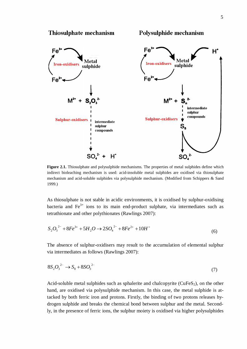
2023 22 FeSMFeMS (4) Elemental sulphur (S0) produced in indirect leaching is further oxidised to protons and sulphate (sulphuric acid, H2SO4) by sulphur-oxidising microorganisms (for reviews, see Mishra et al. 2005; Sand et al. 2001). In the indirect leaching mechanism, no physical contact between the microbes and mineral surface is required: the microorganisms cata-lytically accelerate the reoxidation of Fe2+ to Fe3+ ions, a reaction that chemically is very slow (Breed & Hansford 1999; for a review, see Bosecker 1997). As a strong oxi-dising agent, ferric ion is capable of dissolving a variety of different metal sulphide minerals in the absence of microorganisms and oxygen (Córdoba et al. 2008c). The role of iron-oxidising microorganisms is thus to produce more ferric iron than is consumed in the leaching reactions (reviewed by du Plessis et al. 2007). However, increased Fe3+ ion concentrations produce chemical instability and favour precipitation, consequently passivating the mineral (Córdoba et al. 2008c). Indirect leaching is often divided into two sub-mechanisms: “contact” and “non-contact” mechanism. The non-contact mechanism comprises of Fe2+ to Fe3+ oxidation by planktonic bacteria, after which the ferric ions are reduced in contact with the ore surface and re-introduced into the iron cycle. In the contact mechanism, mineral dissolu-tion takes place in an extracellular polymeric substance (EPS) or slime between bacteri-al cell walls and the mineral surface (reviewed by Sand & Gehrke 2006). There are two different groups of metal sulphides: acid-insoluble and acid-soluble. Thus, two different (indirect) oxidation mechanisms have been proposed (Figure 2.1; for a review, see Schippers 2007). When oxidising acid-insoluble metal sulphides such as pyrite, molybdenite (MoS2), or tungstenite (WS2), the mineral dissolution proceeds via thiosulphate mechanism. The mechanism is named after its main intermediate, S2O3
2-, which is the first free sulphur compound released after many electron-removal steps that finally break the chemical bond between the metal and sulphur. The metal sulphide (in this case, pyrite) is solubilised by the attack of ferric iron, not by the attack of protons (Schippers & Sand 1999):
HFeOSOHFeFeS 6736 22322
32 (5)
5
Figure 2.1. Thiosulphate and polysulphide mechanisms. The properties of metal sulphides define which indirect bioleaching mechanism is used: acid-insoluble metal sulphides are oxidised via thiosulphate mechanism and acid-soluble sulphides via polysulphide mechanism. (Modified from Schippers & Sand 1999.)
As thiosulphate is not stable in acidic environments, it is oxidised by sulphur-oxidising bacteria and Fe3+ ions to its main end-product sulphate, via intermediates such as tetrathionate and other polythionates (Rawlings 2007):
HFeSOOHFeOS 108258 2242
3232 (6)
The absence of sulphur-oxidisers may result to the accumulation of elemental sulphur via intermediates as follows (Rawlings 2007):
238
232 88 SOSOS (7)
Acid-soluble metal sulphides such as sphalerite and chalcopyrite (CuFeS2), on the other hand, are oxidised via polysulphide mechanism. In this case, the metal sulphide is at-tacked by both ferric iron and protons. Firstly, the binding of two protons releases hy-drogen sulphide and breaks the chemical bond between sulphur and the metal. Second-ly, in the presence of ferric ions, the sulphur moiety is oxidised via higher polysulphides
6
to elemental sulphur (for reviews, see Rawlings 2007; Rawlings 2005). The reactions may be presented as follows (Schippers & Sand 1999):
22
23 5.0 FeSHMHFeMS n (n 2) (8) HFeSFeSH n
28
32 125.05.0 (9)
Elemental sulphur is the main intermediate of the polysulphide mechanism, and alt-hough quite stable, it can be further oxidised to sulphate by sulphur-oxidising microor-ganisms (reviewed by Rawlings et al. 2003). Ferrous iron produced in the reactions can be reoxidised to ferric iron by iron-oxidising microorganisms (for reviews, see Holmes & Bonnefoy 2007; Rawlings 2005).
2.2 Sulphur oxidation
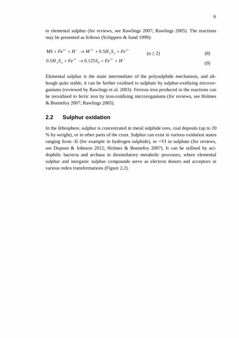
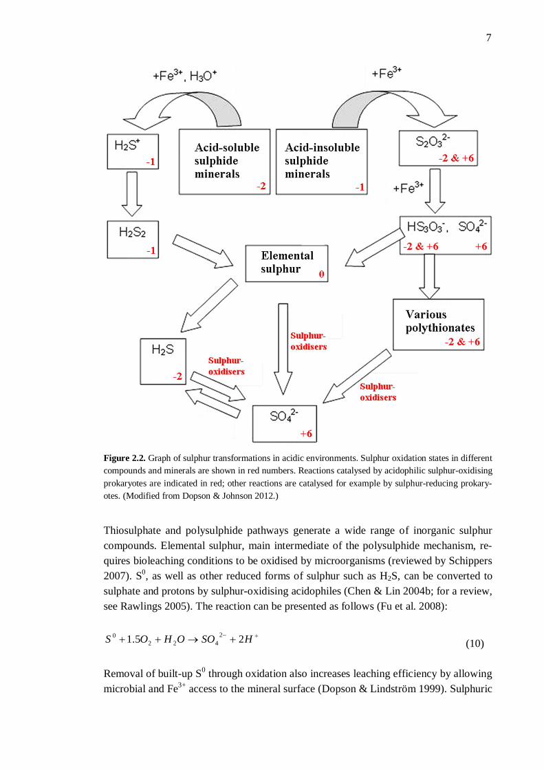
In the lithosphere, sulphur is concentrated in metal sulphide ores, coal deposits (up to 20 % by weight), or in other parts of the crust. Sulphur can exist in various oxidation states ranging from -II (for example in hydrogen sulphide), to +VI in sulphate (for reviews, see Dopson & Johnson 2012; Holmes & Bonnefoy 2007). It can be utilised by aci-dophilic bacteria and archaea in dissimilatory metabolic processes, where elemental sulphur and inorganic sulphur compounds serve as electron donors and acceptors in various redox transformations (Figure 2.2).
7
Figure 2.2. Graph of sulphur transformations in acidic environments. Sulphur oxidation states in different compounds and minerals are shown in red numbers. Reactions catalysed by acidophilic sulphur-oxidising prokaryotes are indicated in red; other reactions are catalysed for example by sulphur-reducing prokary-otes. (Modified from Dopson & Johnson 2012.)
Thiosulphate and polysulphide pathways generate a wide range of inorganic sulphur compounds. Elemental sulphur, main intermediate of the polysulphide mechanism, re-quires bioleaching conditions to be oxidised by microorganisms (reviewed by Schippers 2007). S0, as well as other reduced forms of sulphur such as H2S, can be converted to sulphate and protons by sulphur-oxidising acidophiles (Chen & Lin 2004b; for a review, see Rawlings 2005). The reaction can be presented as follows (Fu et al. 2008):
HSOOHOS 25.1 2422
0 (10)
Removal of built-up S0 through oxidation also increases leaching efficiency by allowing microbial and Fe3+ access to the mineral surface (Dopson & Lindström 1999). Sulphuric
8
acid (H2SO4) produced by sulphur-oxidising microorganisms may also bring forth the polysulphide mechanism in the absence of Fe3+ ions (reviewed by Rawlings 2007). H2SO4 is the most common inorganic acid found in bioleaching environments (reviewed by Shagufta 2007). It maintains the bioleaching environment acidic and takes part in metal sulphide and oxide mineral leaching by providing protons to the process (Bhatti et al. 2012b). Oxidation of sulphide moiety in minerals is the main acid-generating phase, although protons are also produced through jarosite ([K, Na, NH4] Fe3(SO4)2(OH)6) formation and the hydrolysis of ferric iron generated in the process (Fu et al. 2008). Pyrite, being acid-insoluble, is an exception and can only be dissolved by ferric ion at-tack. The leaching of tenorite (CuO) and uranium-trioxide (UO3), on the other hand, happens via proton attack and can be presented as follows (Schippers 2007):
OHCuSOSOHCuO 24422 (11) HOHSOUOSOHUO 4)(3 23442423 (12)
As an example, the oxidation of reduced pyrite to an oxy-hydroxide mineral, schwert-mannite, is presented as follows (Dopson & Johnson 2012):
244688222 1530)(18308 SOHSOOHOFeOHOFeS (13)
Unless a sufficient amount of basic minerals is present, the acidic waste produced through sulphur oxidation is enriched with metal cations, thus generating acid mine drainage (AMD) (reviewed by Dopson & Johnson 2012). In summary, the microbial generation of sulphuric acid is important in bioleaching op-erations for two reasons: proton attack, and to keep iron in an oxidised state (reviewed by Rawlings 2005). Conversion of sulphur to sulphate is also the most important heat-generating reaction in heap bioleaching (reviewed by du Plessis et al. 2007).
2.3 Iron- and sulphur-oxidising microorganisms
Bioleaching microorganisms oxidise the products of chemical metal sulphide oxidation, including Fe2+ ions and different sulphur compounds. The microbes also provide Fe3+ ions and protons for sulphide attack, and keep the leaching environment acidic by pro-ducing sulphuric acid. This low pH environment keeps iron in solution (for reviews, see Rawlings et al. 2003; Schippers 2007). As the temperature inside a bioheap changes with mineral oxidation and depletion, different groups of indigenous microorganisms activate and deactivate in succession (reviewed by Brierley 2008). The most common bioleaching microorganisms are presented in Table 2.1.
9
2.3.1 Diversity and different species
Bacteria and archaea oxidising Fe2+ ions and intermediate sulphur compounds are aero-bic, generally strict or moderate acidophiles, and usually either obligatory or facultative chemolithotrophs (Bowei et al. 2009; for reviews, see Holmes & Bonnefoy 2007; Schippers 2007). Chemolithotrophs use ferrous iron, reduced inorganic sulphur com-pounds, or both as an electron donor, and oxygen as the terminal electron acceptor (re-viewed by Rawlings et al. 2003). Microorganisms capable of both iron and sulphur oxi-dation have shown to be able to recover their Fe2+ oxidation ability when exposed to the substrate, even after months of having S0 as the sole energy source (Córdoba et al. 2008c). While some iron- and sulphur-oxidising microorganisms are obligate aerobes (Acidithi-obacillus [At.] thiooxidans, At. caldus), others are able to use Fe3+ as an electron accep-tor instead of oxygen and thus, grow in anaerobic environments. These microorganisms are called facultative anaerobes and include bacteria such as At. ferrooxidans (reviewed by Dopson & Johnson 2012). Mineral-oxidising microorganisms are mostly capable of CO2 fixation (autotrophs such as Acidithiobacillus and Leptospirillum spp.), although obligate heterotrophs require organic carbon for growth. Some bioleaching prokaryotes are able to use both organic and inorganic carbon as a carbon source (reviewed by John-son & Hallberg 2007). This group of facultative auto- or heterotrophs contains microor-ganisms such as Sulfobacillus spp. and Acidiphilium acidophilum (for a review, see Schippers 2007). Most metal leaching microbes are mesophilic or moderately thermophilic bacteria, with optimal growth temperatures of 15 - 40 and 40 - 60 °C, respectively. Bioleaching ar-chaea are mainly extreme thermophiles (optimal temperature for growth being 60 - 80 °C; for reviews, see Plumb et al. 2007; Schippers 2007). Psychrophilic (Topt < 15 °C) bioleaching acidophiles have not been identified (reviewed by Plumb et al. 2007), alt-hough psychrotolerant mixed and pure cultures have successfully been used in bioleach-ing (Dopson et al. 2007). Hyperthermophilic (Topt > 80 °C) metal leaching microbes are rare (for reviews, see Plumb et al. 2007; Schippers 2007). It should however be noted that some acidophiles can grow at temperatures higher or lower than their optimal tem-perature for growth, and the ranges for each temperature-based grouping are varying (for reviews, see Johnson & Hallberg 2007; Norris 2007). Especially with heap bi-oleaching, time of the year during sampling is important as dominating bacteria in summer greatly differ from winter-dominating communities (Bowei et al. 2009). Sub-strate availability and increasing concentrations of inhibitory ions, such as sulphate, also affect the community composition (Demergasso et al. 2005). So called secondary bioleaching microorganisms interact with iron- and sulphur-oxidising microorganisms and thus, have an impact on the overall bioleaching process.
10
Some of these mostly heterotrophic prokaryotes or eukaryotes may also directly impact the mineral oxidation. Many Acidiphilium species, for instance, are capable of reduced sulphur compound oxidation but unlike autotrophic prokaryotes, require organic carbon (reviewed by Johnson & Hallberg 2007). Organic compounds produced in oxidation processes may be harmful to the autotrophic microorganisms. Heterotrophic Ferrimi-crobium acidiphilum, Ferroplasma acidiphilum, and Sulfobacillus spp. utilise and con-sequently eliminate the toxicity of organic matter produced by autotrophs such as Acidi-thiobacillus and Leptospirillum spp. These species are also capable of anaerobic ferric iron reduction. Thus, the heterotrophs provide electron donors (Fe2+) for the autotrophic iron-oxidising microorganisms (Bowei et al. 2009).
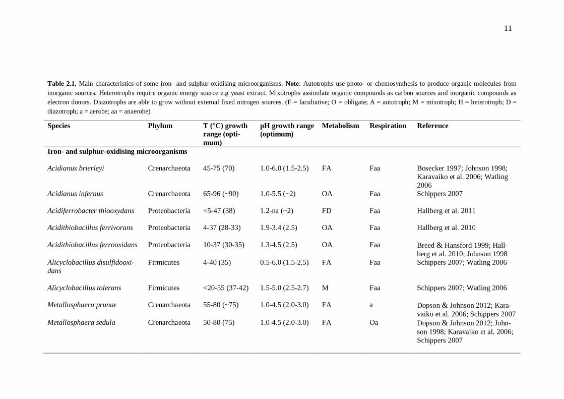
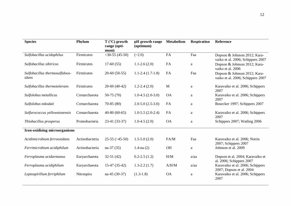
11
Table 2.1. Main characteristics of some iron- and sulphur-oxidising microorganisms. Note: Autotrophs use photo- or chemosynthesis to produce organic molecules from inorganic sources. Heterotrophs require organic energy source e.g yeast extract. Mixotrophs assimilate organic compounds as carbon sources and inorganic compounds as electron donors. Diazotrophs are able to grow without external fixed nitrogen sources. (F = facultative; O = obligate; A = autotroph; M = mixotroph; H = heterotroph; D = diazotroph; a = aerobe; aa = anaerobe)
Species Phylum T (°C) growth range (opti-mum)
pH growth range (optimum)
Metabolism Respiration Reference
Iron- and sulphur-oxidising microorganisms Acidianus brierleyi Crenarchaeota 45-75 (70) 1.0-6.0 (1.5-2.5) FA Faa Bosecker 1997; Johnson 1998;
Karavaiko et al. 2006; Watling 2006
Acidianus infernus Crenarchaeota 65-96 (~90) 1.0-5.5 (~2) OA Faa Schippers 2007
Acidiferrobacter thiooxydans Proteobacteria <5-47 (38) 1.2-na (~2) FD Faa Hallberg et al. 2011
Acidithiobacillus ferrivorans Proteobacteria 4-37 (28-33) 1.9-3.4 (2.5) OA Faa Hallberg et al. 2010
Acidithiobacillus ferrooxidans Proteobacteria 10-37 (30-35) 1.3-4.5 (2.5) OA Faa Breed & Hansford 1999; Hall-berg et al. 2010; Johnson 1998
Alicyclobacillus disulfidooxi-dans
Firmicutes 4-40 (35) 0.5-6.0 (1.5-2.5) FA Faa Schippers 2007; Watling 2006
Alicyclobacillus tolerans
Firmicutes <20-55 (37-42) 1.5-5.0 (2.5-2.7) M Faa Schippers 2007; Watling 2006
Metallosphaera prunae Crenarchaeota 55-80 (~75) 1.0-4.5 (2.0-3.0) FA a Dopson & Johnson 2012; Kara-vaiko et al. 2006; Schippers 2007
Metallosphaera sedula Crenarchaeota 50-80 (75) 1.0-4.5 (2.0-3.0) FA Oa Dopson & Johnson 2012; John-son 1998; Karavaiko et al. 2006; Schippers 2007
12
Species Phylum T (°C) growth range (opti-mum)
pH growth range (optimum)
Metabolism Respiration Reference
Sulfobacillus acidophilus Firmicutes <30-55 (45-50) (~2.0) FA Faa Dopson & Johnson 2012; Kara-vaiko et al. 2006; Schippers 2007
Sulfobacillus sibiricus Firmicutes 17-60 (55) 1.1-2.6 (2.0) FA a Dopson & Johnson 2012; Kara-vaiko et al. 2006
Sulfobacillus thermosulfidoox-idans
Firmicutes 20-60 (50-55) 1.1-2.4 (1.7-1.8) FA Faa Dopson & Johnson 2012; Kara-vaiko et al. 2006; Schippers 2007
Sulfobacillus thermotolerans Firmicutes 20-60 (40-42) 1.2-2.4 (2.0) M a Karavaiko et al. 2006; Schippers 2007
Sulfolobus metallicus Crenarchaeota 50-75 (70) 1.0-4.5 (2.0-3.0) OA a Karavaiko et al. 2006; Schippers 2007
Sulfolobus tokodaii
Crenarchaeota 70-85 (80) 2.0-5.0 (2.5-3.0) FA a Bosecker 1997; Schippers 2007
Sulfurococcus yellowstonensis Crenarchaeota 40-80 (60-65) 1.0-5.5 (2.0-2.4) FA a Karavaiko et al. 2006; Schippers 2007
Thiobacillus prosperus
Proteobacteria 23-41 (33-37) 1.0-4.5 (2.0) OA a Schippers 2007; Watling 2006
Iron-oxidising microorganisms Acidimicrobium ferrooxidans Actinobacteria 25-55 (~45-50) 1.5-5.0 (2.0) FA/M Faa Karavaiko et al. 2006; Norris
2007; Schippers 2007 Ferrimicrobium acidiphilum
Actinobacteria na-37 (35) 1.4-na (2) OH a Johnson et al. 2009
Ferroplasma acidarmanus Euryarchaeota 32-51 (42) 0.2-2.5 (1.2) H/M a/aa Dopson et al. 2004; Karavaiko et al. 2006; Schippers 2007
Ferroplasma acidiphilum Euryarchaeota 15-47 (35-42) 1.3-2.2 (1.7) A/H/M a/aa Karavaiko et al. 2006; Schippers 2007; Dopson et al. 2004
Leptospirillum ferriphilum Nitrospira na-45 (30-37) (1.3-1.8) OA a Karavaiko et al. 2006; Schippers 2007
13
Species Phylum T (°C) growth range (opti-mum)
pH growth range (optimum)
Metabolism Respiration Reference
Leptospirillum ferrooxidans Nitrospira 2-37 (28-30) 1.1-2.5 (2.0) OA a Bosecker 1997; Karavaiko et al. 2006
Leptospirillum thermoferroox-idans
Nitrospira 30-55 (45-50) 1.3-na (1.65-1.90) OA a Karavaiko et al. 2006; Schippers 2007
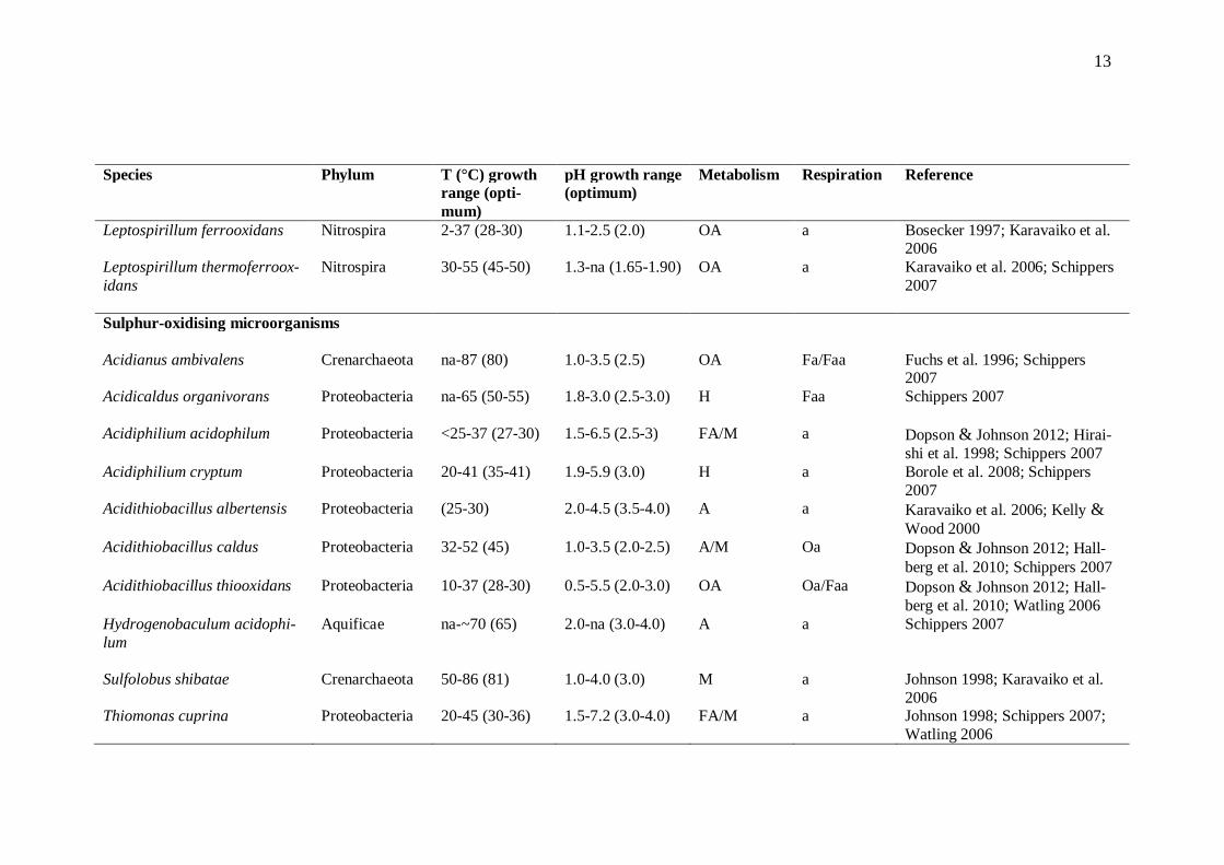
Sulphur-oxidising microorganisms Acidianus ambivalens Crenarchaeota na-87 (80) 1.0-3.5 (2.5) OA Fa/Faa Fuchs et al. 1996; Schippers
2007 Acidicaldus organivorans
Proteobacteria na-65 (50-55) 1.8-3.0 (2.5-3.0) H Faa Schippers 2007
Acidiphilium acidophilum Proteobacteria <25-37 (27-30) 1.5-6.5 (2.5-3) FA/M a Dopson & Johnson 2012; Hirai-shi et al. 1998; Schippers 2007
Acidiphilium cryptum Proteobacteria 20-41 (35-41) 1.9-5.9 (3.0) H a Borole et al. 2008; Schippers 2007
Acidithiobacillus albertensis Proteobacteria (25-30) 2.0-4.5 (3.5-4.0) A a Karavaiko et al. 2006; Kelly & Wood 2000
Acidithiobacillus caldus Proteobacteria 32-52 (45) 1.0-3.5 (2.0-2.5) A/M Oa Dopson & Johnson 2012; Hall-berg et al. 2010; Schippers 2007
Acidithiobacillus thiooxidans Proteobacteria 10-37 (28-30) 0.5-5.5 (2.0-3.0) OA Oa/Faa Dopson & Johnson 2012; Hall-berg et al. 2010; Watling 2006
Hydrogenobaculum acidophi-lum
Aquificae na-~70 (65) 2.0-na (3.0-4.0) A a Schippers 2007
Sulfolobus shibatae Crenarchaeota 50-86 (81) 1.0-4.0 (3.0) M a Johnson 1998; Karavaiko et al. 2006
Thiomonas cuprina Proteobacteria 20-45 (30-36) 1.5-7.2 (3.0-4.0) FA/M a Johnson 1998; Schippers 2007; Watling 2006
14
At. ferrooxidans, previously known as Thiobacillus ferrooxidans, is a chemolithoauto-trophic and mesophilic bioleaching microorganism and the most widely studied aci-dophilic bacterium. It uses inorganic substrates as energy source, is able to fix carbon, and leaches metals from both oxide and sulphide ores (reviewed by Harvey & Bath 2007). At. ferrooxidans derives its energy for growth from oxidative respiration (Ohmu-ra et al. 2002) and is able to oxidise both ferrous iron to ferric iron, and sulphur com-pounds to sulphuric acid (Chen et al. 2011). The bacterium is also capable of anaerobic iron respiration with Fe3+, S0, or reduced sulphur compounds as electron acceptors and H2 as an electron donor (Bowei et al. 2009; Ohmura et al. 2002). At. ferrooxidans is widely used in bioleaching processes as it has been found to oxidise chalcopyrite at a higher rate than many other metal leaching microorganisms, such as Leptospirillum (L.) ferrooxidans or At. thiooxidans (Akcil et al. 2007). As At. ferrooxidans is easy to cultivate, it was previously believed that sulphidic miner-al environments were mainly occupied by this bacterium. However, due to the use of molecular phylogenetic techniques (16S rRNA gene amplification, fluorescent in situ hybridisation), the importance of other bacterial species has been discovered (reviewed by Dopson et al. 2003). Several other bacteria and archaea have been identified espe-cially at higher process temperatures of 45 - 80 °C (reviewed by Holmes & Bonnefoy 2007). Other microorganisms known to be involved in bioleaching operations are the mesophilic bacteria At. thiooxidans and L. ferrooxidans, as well as thermophilic archaea such as Sulpholobus, Ferroplasma, and Acidianus species (Bowei et al. 2009; for a re-view, see Harvey & Bath 2007). However, their oxidation rates as pure cultures are usually quite low. Mixed microbial cultures in general have shown higher oxidations activities than pure cultures (Akcil et al. 2007). Sulphur-oxidising mesophiles and moderate thermophiles are exclusively bacteria, while the extreme thermophiles are solely crenarchaeotes with the exception of Hy-drogenobaculum acidophilum, an autotrophic sulphur-oxidising bacterium. Although none of the acidophilic microorganisms oxidising merely sulphur compounds are psy-chrophiles, acidophiles capable of oxidising both iron and sulphur such as At. ferrivo-rans, At. ferrooxidans, and Alicyclobacillus disulfidooxidans grow at temperatures of 10 °C or lower (reviewed by Dopson & Johnson 2012). At. thiooxidans is a Gram-negative, strictly acidophilic proteobacterium and an obligately autotrophic sulphur-oxidiser (re-viewed by Holmes & Bonnefoy 2007). Although At. ferrooxidans is capable of oxidis-ing elemental sulphur to sulphuric acid, At. thiooxidans converts it much faster. Simul-taneously, it creates acidity and favourable conditions for iron-oxidising bacteria such as At. ferrooxidans and L. ferrooxidans (reviewed by Bosecker 1997). Another sulphur-oxidiser of the Acidithiobacillus genus is At. caldus, which has a higher thermotolerance than At. thiooxidans and has been shown to have a more important role in sulphur oxi-dation than previously thought (Bouchez et al. 2006; for a review, see Norris 2007).
15
2.3.2 Resistance towards high metal concentrations
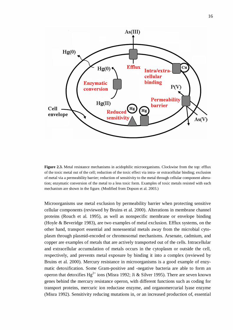
Although many metals have important biochemical roles as catalysts, enzyme co-factors, and stabilisers, they can become toxic via constitutively expressed transport systems that result in metal accumulation inside the cells (reviewed by Dopson et al. 2003). By growing in bioleaching environments, acidophilic microorganisms have de-veloped several mechanisms to resist high metal ion concentrations. This is due to the fact that many metal ions are released in environments that contain ferric iron and a low pH (for reviews, see Dopson et al. 2003; Rawlings 2007). It has been found that bacteria resistant to one metal may also decrease the toxic effect of other metals by exploiting their existing resistance mechanisms (Ahemad & Malik 2011). Metals such as silver, aluminium, cadmium, gold, lead, and mercury have no function in cells at any concen-trations (for a review, see Bruins et al. 2000) and thus, resistance towards these metals is important in bioleaching microorganisms. The heavy metal resistance in different bacte-rial species has been widely explored, an example being the resistance mechanisms in Ralstonia metallidurans reviewed by Nies (2003). Heavy metal ions need to enter the cell before they can have any physiological or toxic effect (reviewed by Nies 1999). Microorganisms use different chromosomal, transpos-on, and plasmid-mediated systems to resist high metal concentrations by decreasing sensitivity to them. Metal uptake mechanisms, metabolic role of the metal, and gene location all have an effect on microbial metal resistance (reviewed by Bruins et al. 2000). There are five different mechanisms that increase the metal resistance of aci-dophilic microorganisms: efflux, or active transport, of the toxic metal out of the cell or microorganism; enzymatic conversion of the metal to a less toxic form; intra- or extra-cellular binding of the metal; exclusion of the metal by a permeability barrier; and re-duction of sensitivity in a cellular component (Figure 2.3; reviewed by Dopson et al. 2003). Intra- and extracellular sequestration are sometimes separated into two mecha-nisms (reviewed by Bruins et al. 2000). Different metals are excluded through different mechanisms. As an example, mercury is resisted through enzymatic conversion, arsenic through efflux, and copper via cellular binding (for a review, see Dopson et al. 2003).
16
Figure 2.3. Metal resistance mechanisms in acidophilic microorganisms. Clockwise from the top: efflux of the toxic metal out of the cell; reduction of the toxic effect via intra- or extracellular binding; exclusion of metal via a permeability barrier; reduction of sensitivity to the metal through cellular component altera-tion; enzymatic conversion of the metal to a less toxic form. Examples of toxic metals resisted with each mechanism are shown in the figure. (Modified from Dopson et al. 2003.)
Microorganisms use metal exclusion by permeability barrier when protecting sensitive cellular components (reviewed by Bruins et al. 2000). Alterations in membrane channel proteins (Rouch et al. 1995), as well as nonspecific membrane or envelope binding (Hoyle & Beveridge 1983), are two examples of metal exclusion. Efflux systems, on the other hand, transport essential and nonessential metals away from the microbial cyto-plasm through plasmid-encoded or chromosomal mechanisms. Arsenate, cadmium, and copper are examples of metals that are actively transported out of the cells. Intracellular and extracellular accumulation of metals occurs in the cytoplasm or outside the cell, respectively, and prevents metal exposure by binding it into a complex (reviewed by Bruins et al. 2000). Mercury resistance in microorganisms is a good example of enzy-matic detoxification. Some Gram-positive and -negative bacteria are able to form an operon that detoxifies Hg2+ ions (Misra 1992; Ji & Silver 1995). There are seven known genes behind the mercury resistance operon, with different functions such as coding for transport proteins, mercuric ion reductase enzyme, and organomercurial lyase enzyme (Misra 1992). Sensitivity reducing mutations in, or an increased production of, essential
17
cellular components are ways of some microorganisms to adapt to high toxic metal con-centrations (Rouch et al. 1995; for a review, see Bruins et al. 2000).
2.3.3 Cell growth and its effect on bioleaching
Effective mineral degradation is dependent on the activity of iron- and sulphur-oxidising microorganisms (Akcil et al. 2007). The activity and cell growth of iron- and sulphur-oxidising chemolithotrophs is mainly controlled by the availability of three fac-tors: iron and reduced sulphur (substrates), oxygen (electron acceptor), and carbon diox-ide (carbon source). In addition, a wide range of other parameters such as macro- and micronutrients affect the microbial growth (reviewed by du Plessis et al. 2007). Since redox transformations are important in mineral leaching environments, the activity of microorganisms can be detected by measuring oxidation and reduction rates of iron and sulphur elements. Oxygen consumption is also associated with oxidation reactions (for a review, see Johnson & Hallberg 2007). Many physicochemical parameters also affect microbial cell growth. For instance an optimal solution pH results in increased microbi-al growth rates, even though it appears not to increase the biomass concentration (Breed & Hansford 1999). Physicochemical parameters affecting iron and sulphur oxidation will be discussed in more detail in Chapter 3.
18
3 PHYSICOCHEMICAL PARAMETERS AF-FECTING THE ACTIVITY OF OXIDATION PRO-CESSES
Bacterial mineral oxidation and metal mobilisation are affected by several factors and parameters. In addition to microbiological, mineral, and processing parameters, physi-cochemical factors play an important role in the rate and efficiency of oxidation pro-cesses (for a review, see Brandl 2001).
3.1 Solution pH and redox potential
A suitable pH value is necessary for the growth and activity of bioleaching microorgan-isms, as well as for the solubilisation of metals (reviewed by Bosecker 1997). Although some acidophilic archaea are able to grow at pH 0, most mineral-oxidising bacteria re-quire a pH value higher than 1. Extreme and moderate acidophiles grow optimally at pH < 3 and pH 3 - 5, respectively (reviewed by Johnson & Hallberg 2007). Chemical and biological oxidation of metal sulphides requires an acidic environment in order to keep a sufficient amount of metal ions in solution. The most important one is ferric iron, as its solubility is required for the ion to be readily available in bioleaching reactions. A low pH environment is thus required for a successful iron cycle (Bhatti et al. 2012a). As iron precipitation may also prevent the leaching agent from getting in contact with the mineral, a suitable pH controls the overall process of metal solubilisation (Ahonen & Tuovinen 1995). Fe3+ solubility is beneficial especially in heaps where oxygen deficien-cy might occur, as some bacteria are capable of anaerobic respiration with Fe3+ as an electron acceptor and hydrogen as an electron donor (Bowei et al. 2009; Ohmura et al. 2002). Solution pH also largely affects whether chemical or biological oxidation is the dominating mechanism, and consequently the leaching efficiency since bacterial leach-ing has been found to be clearly faster than chemical leaching (Ahonen & Tuovinen 1995). In a study by Meruane & Vargas (2003), chemical Fe2+ oxidation dominated at pH over 5.0 whereas below pH 5.0, bacterial oxidation was relevant. Microbial growth and oxidation of minerals however mostly take place at a pH value of 1.5 - 2.0 (Dorado et al. 2012). Highest microbial cell counts have also been obtained in that pH range (Ahonen & Tuovinen 1995). Each bioleaching process should be optimised based on the sulphide ore, as sensitivity to acidity has been shown to be highly metal-dependent (Cameron et al. 2009).
19
Microbial bioleaching is based on redox reactions, where ferrous ions (Fe2+) and re-duced sulphur compounds are electron donors and molecular oxygen serves as the final electron acceptor in the overall bioleaching process (for reviews, see Belzile et al. 2004; Schippers 2007). The Fe2+/Fe3+ couple has a relatively high redox potential in low-pH liquors (+ 0.77 V), meaning that in order to maintain the growth of Fe2+-oxidising mi-croorganisms, large amounts of ferrous iron need to be oxidised and consequently, many electrons have to be transferred to oxygen (reviewed by Holmes & Bonnefoy 2007). Due to this very positive standard electrode potential, only oxygen may be used as an electron acceptor. Additionally, since Fe2+/Fe3+ and O2/H2O redox couples have similar redox potentials, relatively little cell mass is produced by ferrous iron oxidation (reviewed by Rawlings 2007). Redox potential is affected by oxidation reactions and increases due to Fe2+ oxidation (Bhatti et al. 2012a; Xia et al. 2008), sulphur oxidation, or when the process is aerated (Chen & Lin 2004b). Increased ORP is a result of de-creased free electron concentration in the system (Couillard et al. 1994). The effect of redox potential on bioleaching kinetics is ore- and metal-dependent (Ahonen & Tuovinen 1995; Gericke & Govender 2011). For instance, copper leaching from chalco-pyrite is most effective at low redox potential levels of 400 - 430 mV (Ahmadi et al. 2011; Gericke & Govender 2011), whereas efficient Cu dissolution from pentlandite requires a high 600 mV redox potential (Gericke & Govender 2011). Intermediate redox potential of 500 mV has been shown to result in highest cell counts (Ahonen & Tuovinen 1995). Similar to process pH, redox potential is responsible for iron solubility (Ahonen & Tuovinen 1995) as high initial ORP (oxidation reduction potential) favours ferric ion precipitation and thus mineral passivation in chalcopyrite leaching (Córdoba et al. 2008b). Formation of passive layers may thus be prevented by decreasing ORP (Ahmadi et al. 2011).
3.2 Temperature
Biological oxidation of mineral sulphides produces free energy and generates heat. While chemical reaction rates increase along with temperature and can be described using the Arrhenius equation (Peleg et al. 2012), the relationship of temperature and microbial growth follows different kinetics (reviewed by Plumb et al. 2007). Ratkowsky et al. (1983) described the bacterial growth rate of any strain throughout a temperature range in Kelvin degrees:
)1)(( )(min
maxTTceTTbr (14)
In the equation, Tmin and Tmax define the minimum and maximum temperatures, respec-tively, at which no cell growth occurs. Parameter r is a growth rate constant, b a regres-sion coefficient, c an additional parameter which enables the use of temperatures higher than optimal, and T the absolute temperature.
20
It has been stated that a 10 °C increase in temperature doubles the rate of chemical reac-tions (reviewed by Rawlings et al. 2003). d’Hugues et al. (2008) found that with micro-organisms, no significant effect on bioleaching activity was discovered as the process temperature was increased from 35 to 45 °C. Different minerals can however have quite varying optimal temperatures for fastest decomposition. Since passivating oxidation products such as elemental sulphur are reduced in higher temperatures, metal yields may consequently improve (reviewed by Rawlings et al. 2003). Microbial response of pure cultures to temperature changes is well studied in laboratory conditions; however, a better understanding of the effects of temperature in heap environments is required (reviewed by Plumb et al. 2007). It has been stated that temperature has no significant effect on the rate of ferrous iron oxidation (Abdel-Fattah & Abdel-Fattah 2002), alt-hough low temperatures have been shown to decrease leaching rates (Ahonen & Tuovinen 1995). This is debatable, as low temperatures have also been shown to de-crease Fe3+ precipitation (Dopson et al. 2007), resulting in increased availability of the ion for mineral oxidation. Following temperature changes in the process, different microbial populations take over (Bowei et al. 2009). Microorganisms have been grouped according to their optimal tem-peratures for growth: psychrophiles, mesophiles, moderate thermophiles, thermophiles, and hyperthermophiles grow optimally at temperatures of < 15, 15 - 40, 40 - 60, 60 - 80, and > 80 °C, respectively. Acidophilic microorganisms are mostly mesophiles, moder-ate thermophiles, and thermophiles, although some hyperthermophilic acidophiles have also been found (reviewed by Plumb et al. 2007).
3.3 Dissolved oxygen and carbon dioxide
Although some of the metal leaching microbes can tolerate anaerobic conditions, the cells require dissolved oxygen (O2) for active growth (reviewed by Mohapatra 2006). O2 acts as an electron acceptor in bioleaching reactions (reviewed by Ojumu et al. 2006). In the solution phase, O2 demand is generated by different microbial and chemi-cal oxidation reactions involved in mineral dissolution (for a review, see du Plessis et al. 2007). Sufficient O2 supply in bioleaching processes is usually achieved by aeration, stirring, sparging, or shaking (for reviews, see Bosecker 1997; du Plessis et al. 2007). Temperature of the leaching environment affects the gas mass transfer (reviewed by du Plessis et al. 2007), with higher temperatures requiring more effective and energy-consuming aeration techniques due to the reduced solubility of O2 (for a review, see Brierley 2008). Iron- and sulphur-oxidizing microorganisms present in bioleaching processes are obli-gate autotrophs: they use carbon dioxide (CO2) as a sole carbon source and do not re-quire organic substrates (reviewed by Mohapatra 2006). There are several different CO2 fixation mechanisms used by bioleaching microbes (Bryan et al. 2012). Microbial cul-
21
tures obtain CO2 either from air (contains 0.033 % of CO2 by volume) or from car-bonate-containing mineral concentrates that release the gas in acidic environments (re-viewed by du Plessis et al. 2007). CO2 concentration of normal air has been found to be more than enough for the optimal cell growth of iron-oxidising microorganisms. How-ever, mass transfer gradients within heap bioleaching operations create a challenge in aeration (Bryan et al. 2012). CO2 insufficiency has been shown to limit biomass for-mation and therefore might have a more inhibiting impact than oxygen deficiency (d’Hugues et al. 2008).
3.4 Substrates and residues
Iron and sulphur serve as electron donors for the iron- and sulphur-oxidising microor-ganisms during metal leaching processes. Fe2+ ion acts as an electron donor when oxi-dised to Fe3+ during aerobic respiration (reviewed by Rawlings 2005). Fe2+ ions also slow down Fe3+ precipitation and nucleation, thus delaying the passivation of the sul-phide mineral (Córdoba et al. 2008a). At high concentrations, Fe2+ ions may however inhibit biological iron oxidation (reviewed by Nemati et al. 1998). Fe3+ on the other hand is a chemical solvent of sulphide minerals (Ahonen & Tuovinen 1995). Its pres-ence chemically oxidises elemental sulphur and metal sulphides (Chen et al. 2003b). Bacterial generation of Fe3+ ensures high redox potential and thus, fast leaching rates (Ahonen & Tuovinen 1995). High concentrations of Fe3+ ions may inhibit biological iron oxidation through Fe3+ precipitation as jarosite and by, consequently, covering the ore surface (Córdoba et al. 2008c; Harahuc et al. 2000; for reviews, see Brandl 2001; Nemati et al. 1998). Iron- and sulphur-oxidisers favour jarosite formation by producing ferric and sulphate ions, respectively (Córdoba et al. 2008c). It has been indicated that when additional Fe3+ is added to a copper recovery process, sulphur oxidation rate be-comes the limiting bioleaching step (Dorado et al. 2012). Simultaneously it was found that microorganisms re-oxidised iron faster than Fe3+ ions oxidised sulphur, concluding that Fe3+ regeneration is not limiting the bioleaching process. Reduced inorganic sulphur compounds (RISCs) serve as electron donors during biologi-cal sulphur oxidation, producing sulphuric acid responsible for low pH environment (Bouchez et al. 2006; for a review, see Rawlings 2005). Oxygen usually acts as the elec-tron acceptor, being energetically the most favourable one (reviewed by Rawlings 2005). Sulphur availability depends on ore mineralogy and the properties of the mineral particles (deportment, physical characteristics). Availability of sulphur for microbial oxidation is process-wise more important than its total content in the ore (reviewed by du Plessis et al. 2007). Rather than its particle size, the number of microspores on S0 particles defines the substrate’s specific surface area. The form of sulphur (i.e. pastilles, powder) also affects the bioleaching performance (Chen et al. 2003a). Bacteria oxidise sulphur by attaching on the S0 particles via adsorption, indicating that a high substrate concentration favours S0 oxidation and consequently metal leaching (Chen & Lin 2001).
22
Similarly to jarosite, another secondary solid phase of bioleaching, high concentrations of S0 may however passivate minerals (Bhatti et al. 2012a; reviewed by Rawlings et al. 2003). On the other hand, it has been found that the porosity of S0 may prevent it from forming a passivating layer on the mineral surface (Córdoba et al. 2008b). As not all of S0 is utilised in bioleaching processes, the remaining substrate may cause further acidi-fication (Chen & Lin 2010).
3.5 Nutrients
Microorganisms responsible for metal sulphide leaching are chemolithoautotrophs. Thus, they require only inorganic compounds for growth and acquire mineral nutrients from the bioleaching environment (reviewed by Bosecker 1997). Sulphide ores usually contain adequate amounts of micronutrients; however, macronutrients such as ammoni-um (NH4
+) and potassium need to be supplied (reviewed by du Plessis et al. 2007). Af-ter carbon, nitrogen is the second most important element for biomass formation and the demand for it depends on the quality of cell growth (reviewed by Rawlings 2007). Ni-trogen source has an effect on Fe3+ precipitation which, as has been shown, may have a role in cell attachment to the mineral surface (d’Hugues et al. 2008). Assuring that enough nitrogen is present for iron- and sulphur-oxidisers is problematic for two rea-sons: firstly, oxygen inhibits nitrogen fixation (although in heaps aeration is usually heterogenous), and secondly, even addition of nitrogen-containing fertiliser to the vast heaps is difficult (reviewed by Rawlings 2007). NH4
+ limitation has clearly been shown to inhibit bacterial growth and bioleaching efficiency, the extent of the negative effects being dependent on the microbial community composition. For example, L. ferriphilum is incapable of nitrogen fixation from air (d’Hugues et al. 2008). As traces of NH4
+ may be absorbed into the bioleaching solution from air due to the ion’s high solubility in acidic environments, the estimation of nitrogen requirement is difficult. Nitrogen, both organic and inorganic, may also inhibit iron oxidation in high concentrations (reviewed by Rawlings 2007). Especially nitrate (NO3
-) has been shown to have a negative effect on biooxidation at high concentrations (Harahuc et al. 2000; Sarcheshmehpour et al. 2009; Suzuki et al. 1999). Unlike with nitrogen and carbon, bioleaching microorganisms cannot acquire phosphate (PO4
3-) or trace elements from air; therefore these nutrients need to be added as fertilis-ers (for a review, see Rawlings 2007). Low PO4
3- concentrations (10 mM) are usually sufficient for maximal iron and sulphur oxidation, although iron-oxidisers are more in-hibited by high PO4
3- concentrations than sulphur-oxidising microorganisms (Harahuc et al. 2000). Trace metals are potentially toxic to bioleaching microorganisms at high concentrations, although at reasonable levels they enhance oxidation rates (Abdel-Fattah & Abdel-Fattah 2002). Other nutrients such as potassium and calcium have been shown to have a significant effect on ferrous iron oxidation (Abdel-Fattah & Abdel-Fattah 2002; Sarcheshmehpour et al. 2009). The main function of magnesium in microorgan-
23
isms is to activate enzymes and sustain the membrane stability. However, when Mg2+ concentration becomes excessive, osmotic pressure in the cells increases and conse-quently eliminates microbial growth (Zhen et al. 2009).
3.6 Anions and cations
Anions that inhibit the growth of bioleaching microorganisms, such as chloride (Cl-), nitrate (NO3
-), and fluoride (F-), may be derived from the ore minerals, or be present in the process waters (reviewed by du Plessis et al. 2007). Salinity has an adverse effect on biological metal extraction and the extent of the negative effect is dependent on Cl- con-centration and microbial strain. However, the solubilisation of some metals, such as that of lead, may increase with increasing Cl- levels due to the formation of complexes (Deveci 2002). At low concentrations (below 0.1 M), addition of salts has been shown to increase sulphur oxidation activity by neutralising the surface charge of colloid parti-cles. Salt concentrations of 0.2 M, however, lengthen the oxidation lag phase and inhibit microorganisms due to increased osmotic pressure (Suzuki et al. 1999). Changes in os-motic pressure affect sulphur-oxidisers more than iron-oxidising microorganisms. This is demonstrated in a study by Harahuc et al. (2000), in which increased concentrations (over 0.2 M) of sulphate slightly inhibited iron oxidation while drastically decreasing sulphur oxidation activity, and in studies by Gómez et al. (1999) and Harahuc et al. (2000) which showed that Cl- ions inhibit iron oxidation more than sulphur oxidation. The inhibiting effect of F- ions is highly dependent on the bioleaching conditions. For instance jarosite is able to include small amounts of F- by exchanging hydroxide ions with fluoride (Gunneriusson et al. 2009). It has been discussed that interaction of some ions, such as that of molybdate or sodium (Na+), with non-toxic ions of nutritional value may result in inhibited oxidation activity (Abdel-Fattah & Abdel-Fattah 2002). Na+ and other monovalent cations may also pro-mote the formation of harmful ferric precipitates (Deveci 2002; Deveci et al. 2004). During copper leaching, chloride (as NaCl) has been shown to inhibit the cell growth of S. rivotincti even at low concentrations (0.1 M NaCl). However, the simultaneous pH increase probably accelerated bacterial death (Gómez et al. 1999). Bioleaching in the presence of several metallic cations such as aluminium (Al3+), at concentrations below their previously detected toxicity thresholds (Ballester et al. 1992; Mier et al. 1995; Mier et al. 1996), has also been studied in order to unveil the possible positive inter-change between the cations and bioleaching substrates (Gómez et al. 1999).
3.7 Heavy metals
Metals that have a density of 6 g/cm3 or above are called heavy metals (reviewed by Akpor & Muchie 2010). Due to their incomplete d-orbitals, heavy metal cations are capable of forming both redox-active and redox-inactive complexes and thus, have an
24
important role in biochemical reactions or as trace elements. However, if the intracellu-lar heavy metal concentrations become too high, unspecific and toxic complexes are formed (reviewed by Nies 1999). Heavy metals are also very persistent in the environ-ment and therefore can affect oxidation processes for lengthened periods (for a review, see Akpor & Muchie 2010). Heavy metals Sn, Ce, Ga, Zr, and Th have a very low solu-bility and thus, have little influence on microbial oxidation (reviewed by Nies 1999). Others are reviewed here. Iron (Fe), molybdenum (Mo), and manganese (Mn) are important trace elements and have a low toxic effect towards microorganisms. Fe is the only heavy metal cation with a macro-bioelement status. Thus, it is biologically the most important of heavy metals and a crucial growth requirement (reviewed by Nies 1999). Fe takes part in redox pro-cesses, as do other essential transition metals (for a review, see Bruins et al. 2000). The bioavailability of Fe in aerobic conditions is usually very low due to the chemical oxida-tion of soluble Fe2+ to quite insoluble Fe3+. However, the solubility of Fe3+ increases in acidic environments (reviewed by Dopson et al. 2003). Being similar to Mg2+ ion, Fe2+ is transported and accumulated in bacterial cells through the same transport system as magnesium. This uptake system is especially important to bacteria living in anaerobic environments (reviewed by Nies 1999). Acidophiles are usually more tolerant to Fe than microorganisms living in environments of higher pH, but the resistance mechanisms are unknown (for a review, see Dopson et al. 2003). Mo is biologically the most important heavy metal oxyanion. It occurs primarily as mo-lybdate, Mo(VI). Mo is capable of oxyanion catalysis and acts as an essential trace ele-ment (reviewed by Nies 1999). However, interaction of Mo(VI) with non-toxic ions of nutritional value may result in inhibited oxidation activity (Abdel-Fattah & Abdel-Fattah 2002). Mn is an essential heavy metal for oxygen production during photosyn-thesis. It has a very low toxic effect compared to other heavy metals. Although the cati-on Mn2+ is the most common state of manganese, the metal can exist in various oxida-tion states. Mn may be able to perform as a substitute for magnesium since it forms complexes that are fairly inert as redox compounds (reviewed by Nies 1999). Zinc (Zn), nickel (Ni), copper (Cu), vanadium (V), cobalt (Co), tungsten (W), and chromium (Cr) are heavy metals that have toxic effects towards microorganisms, but also moderate to high importance as trace elements. Zn has a similar toxicity to Cu, Ni, and Co (reviewed by Nies 1999), although bacteria resistant to Zn have also been shown to resist cadmium (Ahemad & Malik 2011). Zn is transported through the cell mem-brane via several different protein families. One example of Zn detoxification system in neutrophilic bacteria is P-type efflux ATPase; however, the genetics of acidophile zinc resistance are widely unknown. Zinc cation is not capable of redox changes under bio-logical conditions due to its completely filled d orbitals. Despite this, Zn is present in various enzymes as the divalent cation Zn2+. The heavy metal has a very important
25
function as a component in enzymes and DNA-binding proteins, as well as in forming complex polypeptide chains. The ability of Zn2+ to form complexes with cellular com-ponents is also the reason for its toxicity (for a review, see Nies 1999). The toxicity of Ni is quite similar to that of Co. As Ni3+ is very unstable, free nickel is mainly found as Ni2+ (for a review, see Nies 1999). Ni is used in a few important reac-tions in the cells, such as in forming complexes with polypeptide chains (reviewed by Dopson et al. 2003). The metal is also similar to transition metals Fe and Cu, which are all involved in cellular redox processes (reviewed by Bruins et al. 2000). Complex rear-rangements may be catalysed by Ni in a process where the cation binds small molecules and then splits, or by fusing small molecules or atoms together. Enzymes such as hy-drogenase and urease may have Ni bound in their active sites. Cells presumably uptake Ni by chemiosmotic gradient, and the heavy metal is detoxified by sequestration and/or transport (reviewed by Nies 1999). Copper is required by many microorganisms. It is an essential transition metal that takes part in redox processes in cells (reviewed by Bruins et al. 2000). Cu has a radical char-acter, making it easily reactive with other radicals such as molecular oxygen and thus, the metal can be very toxic. The Cu2+/Cu+ pair has a standard electrochemical potential of - 268 mV, making Cu2+ the main ionic form of copper (reviewed by Nies 1999). Aci-dophilic microorganisms are usually more resistant to high Cu2+ concentrations than other microbes (Dew et al. 1999; Johnson et al. 1992). Cu acts in terminal oxygen ac-ceptors such as cytochrome-c oxidase and thus, has a function in the respiratory chain of many organisms (for reviews, see Dopson et al. 2003; Nies 1999). Another important function is the formation of copper/zinc superoxide dismutases (reviewed by Nies 1999). Vanadium is mostly toxic to microorganisms. It exists mainly as the trivalent oxyanion vanadate, V(V), a structure similar to phosphate (PO4
3-). Microorganisms are very rarely able to use V for their advantage, and microbial V(V) resistance mechanisms are widely unknown. Co has medium toxicity towards microorganisms. As Co3+ is stable in com-plexes only, Co2+ is the main form of the heavy metal. Co takes part in several catalyses as the cofactor B12 (for a review, see Nies 1999). Gram-negative bacteria resist high Co concentrations by transenvelope efflux through a RND (resistance, nodulation, cell divi-sion) transporter (Saier et al. 1994). Eukaryotes and gram-positive bacteria detoxify Co2+ by CDF (cation diffusion facilitator) transporters (Paulsen & Saier 1997). W is the heaviest beneficial element. As WS is more available in anaerobic environments than MoS, anaerobic microorganisms such as bacteria and archaea have tungsten-containing enzymes. The heavy metal may also act as a component of a tungsten cofactor, much like with Mo (reviewed by Nies 1999). Chromium is not usually beneficial for microor-ganisms. It occurs mainly as two biologically important ions, the divalent oxyanion chromate (Cr(VI), and the trivalent cation, Cr(III). Cr(III) has less toxic effects than
26
Cr(VI). Structurally Cr(VI) resembles PO43-, SO4
2-, and arsenate (As(V)). It has been suspected that the chromate resistance mechanisms in microorganisms are based on the interaction of Cr(VI) reduction and efflux (reviewed by Nies 1999). Heavy metals that are toxic with limited beneficial function for microorganisms include arsenic (As), silver (Ag), antimony (Sb), cadmium (Cd), mercury (Hg), lead (Pb), and uranium (U) (reviewed by Nies 1999). Arsenic is often found in acidic environments due to its presence in minerals such as arsenical pyrite (reviewed by Dopson et al. 2003). It exists in bioleaching solutions mainly as arsenate, AsO4
3- (As(V)), and arse-nite, AsO2
- (As(III)) (for review, see Dopson et al. 2003; Nies 1999). Due to the very similar structures of As(V) and PO4
3-, arsenate may interfere with the metabolism of phosphorus by replacing it in cellular processes and thus, become toxic to the cell (re-viewed by Nies 1999). Although acidophilic microbes have proved to be more tolerant towards As than other microorganisms (Suzuki et al. 1997; Harvey & Crundwell 1996), studies have shown that especially As(III) inhibits the growth of bioleaching microbes (reviewed by Dopson et al. 2003). Its toxic effect may occur via formation of extracellu-lar sulphur, as has been noticed with At. caldus (Hallberg et al. 1996). The main ionic form of silver is the monovalent cation Ag+ due to the standard electro-chemical potential of Ag2+/Ag+ pair (1.56 V at pH 7) (reviewed by Nies 1999). Oxida-tion of refractory sulphide ores releases the fine heavy metal particles from the mineral matrix (for a review, see Dopson et al. 2003). Ag resistance mechanism in gram-negative bacteria may be based on RND-driven transenvelope efflux, while gram-positive bacteria resist the heavy metal using efflux by P-type ATPases (Gupta et al. 1999). Resistance may also be based on additional complexation by intracellular com-pounds (reviewed by Nies 1999). It has been shown that neutrophilic microorganisms resist Ag+ through efflux, while the mechanisms in acidophiles are more unknown (for a review, see Dopson et al. 2003). The toxic effects of cadmium towards microorganisms have been studied and its toxici-ty has been demonstrated, but few specific mechanisms have been found (reviewed by Nies 1999). It is however known that Cd is more toxic to At. ferrooxidans than zinc (Ragan & Mast 1990) and copper (Mahapatra et al. 2002). The inhibiting effect of Cd2+ may be due to interaction with calcium and zinc metabolism, membrane damage, pro-tein denaturation, binding to thiol groups, or loss of a protective function (reviewed by Dopson et al. 2003). Microorganisms use Cd efflux to resist the heavy metal, and RNA- and P-type transport systems may play an important role in cyanobacteria resistance. Mercury has a very high affinity of Hg2+ to thiol groups (stronger than the affinity of Cd2+ to sulphide). Therefore, it has the highest toxicity of all heavy metals and no bene-ficial effects for microorganisms (reviewed by Nies 1999). The cation Hg2+ is produced along with RISCs by oxidation of cinnabar, HgS (for a review, see Dopson et al. 2003). Gram-negative bacteria transport Hg2+ via specific uptake systems and consequently

27
prevent its toxicity. Lead is not very soluble and thus, has a low toxic effect on microor-ganisms due to its decreased availability. Metal ion efflux has been considered as the main mechanism of Pb resistance. The toxicity of uranium for bacteria is low as U(VI) (as it occurs in UO2
2-) (reviewed by Nies 1999). Because the heavy metal is capable of forming various ionic forms, it may be used in anaerobic respiration as a substrate (Lovley et al. 1991). Otherwise, U has not proven biologically beneficial (reviewed by Nies 1999).
28
4 CONTROL OF INHIBITING FACTORS
Acid mine drainage (AMD) may cause environmental problems due to its high soluble metal concentrations, high sulphate content, and low pH. Remediation of AMD contam-inated waters concentrates on all of these factors (Aelion et al. 2009). The term biohy-drometallurgy contains several technologies where microbes and metals interact with each other. In addition to bioleaching - the dissolution of metals from their mineral sources - biohydrometallurgical technologies include bioremediation, biosorption, bio-accumulation, and other ways of reducing contaminants in mine waste waters (reviewed by Mishra et al. 2005). Of physical contaminant removal, settling and sedimentation are two examples. Chemical removal processes include sorption and adsorption, precipita-tion, metal oxidation and hydrolysis, and carbonate or sulphide formation (reviewed by Sheoran & Sheoran 2006).
4.1 Filtration
Filtration is often used prior to, combined with, or after a more selective remediation method such as adsorption, precipitation, coagulation, and oxidation. With filtration, any particulate matter that might plug, deactivate, or otherwise disturb the purification process is removed from the contaminated water (reviewed by Malik et al. 2009). The remediation efficiency of filtration techniques is highly dependent on the size of con-taminant-bearing particles (reviewed by Hashim et al. 2011). For example Leupin & Hug (2005) studied arsenic removal combining iron filing (oxidant) and quartz sand (filtrate) to repeatedly pass contaminated water through. Membrane and filtration tech-nologies concentrate the contaminants in waste water, thus reducing the volume of wa-ter to be treated. Electrodialytic, liquid, polymer, ultrafiltration, and nanofibre mem-branes are a few membrane types used in remediation (reviewed by Hashim et al. 2011). Microfiltration and ultrafiltration are membrane processes that have been used for water purification from contaminants such as arsenic. Cu(II), Cr(VI), and As(V) have success-fully been removed from waste water using metal-complexation followed by ultrafiltra-tion (Clifford 1986). Nano-filtration membranes have been effective in removing dis-solved contaminants (for a review, see Hashim et al. 2011). Membrane filtration tech-niques are not used as often as other remediation methods (precipitation, adsorption, ion exchange) due to their sensitivity, high volumes of formed residuals, and costs (re-viewed by Malik et al. 2009).
29
4.2 Ion exchange
Ion exchange is an adsorption method that can be used to remove specific cations or anions from waste water. In this remediation technique, an ion in waste water is ex-changed to another ion that has a similar charge (for reviews, see Akpor & Muchie 2010; Zhuang 2009). An ion exchange resin can be regenerated in situ (reviewed by Malik et al. 2009). Compared to precipitation, electrolytic remediation, evaporation, and adsorption, ion exchange is very efficient and inexpensive. Ion exchange also produces cleaner and a lower volume of sludge than chemical precipitation, and the process re-covers metal value (Rengaraj et al. 2001). Ion exchange, as well as adsorption, is often used when only one contaminant (such as arsenic) requires treatment. Dissolved solids, sulphates, and nitrates may disturb the adsorption efficiency. The ion exchange resin may also become saturated with the contaminant (reviewed by Malik et al. 2009). The efficiency of an ion exchange resin is pH-independent. As an example, removal of nick-el is optimal at pH 4, whereas zinc is most efficiently removed at a higher pH of 6. The adsorption rates of different metals are also varying (Alyüz & Veli 2009).
4.3 Biological treatment
The use of microorganisms in the treatment of metal-contaminated waters is cost-effective, and new technologies such as biosorption, biological oxidation, and biological reduction have been applied (reviewed by Zhuang 2009). Advantages of microbial re-mediation include environmental safety, absence of waste, and self-sustainability. How-ever, the suitability of microbial remediation requires a biodegradable contaminant, and the processes are slow and highly specific (reviewed by Akpor & Muchie 2010). Biosorption, as well as bioaccumulation, is used in environments where heavy metals accumulate (reviewed by Mishra et al. 2005). In biosorption, the various functional groups of living or nonliving biomass (carboxyl-, hydroxyl-, sulphate-, phosphate-, and amino-groups) bind dissolved metals through reversible physicochemical mechanisms. The application of biopolymers as an adsorbent has several advantages compared to conventional organic and inorganic adsorbents, such as lower costs, non-hazardous ma-terials and wastes, and specificity (for a review, see Zhuang 2009). Chemically modi-fied plants, such as Ficus carica, have been shown to adsorb metals from aqueous sys-tems, but the presence of some ions (i.e. Ca2+, Mg2+) may reduce the removal efficiency (Gupta et al. 2012). In addition, zinc resistant bacteria have been shown to accumulate zinc and copper and the mechanism could be applied to heavy metal removal from con-taminated soils (Ahemad & Malik 2011). Biological oxidation in waste water treatment is based on microbial formation of insol-uble metal hydroxides and carbonates, such as MnO2 or Fe(OH)3 (reviewed by Zhuang 2009). Iron and sulphate have for instance been precipitated from bioleaching waste
30
streams by combining an iron-oxidising fluidised-bed reactor (FBR) with a gravity set-tler (Nurmi et al. 2010). Biological reduction, on the other hand, applies sulphate to sul-phide reduction (reviewed by Zhuang 2009). Sulphate-reducing biofilm and suspension processes for the treatment of sulphate-, zinc-, and iron-containing waste waters have been studied (Kaksonen et al. 2003), concluding that SRB (sulphate-reducing bacteria) treatment is a promising alternative for chemical remediation methods, precipitating the metals as sulphides and neutralising the acidity of the waste water. Wakeman et al. (2010) studied the use of SRB in the treatment of metals-containing waste water further, demonstrating that silage can be used as an inexpensive carbon and electron source for sulphate reduction. As a conclusion: biological oxidation and reduction form same kind of products as chemical hydroxide and sulphide precipitation (see Chapter 4.4), but without the use of high temperatures or commercial, possibly expensive or harmful chemicals. Phytoremediation, the use of plants in removal of selected contaminants from soil, sludge, sediment, or water, may also be categorised as biological treatment. Metals can be removed from waste water using phytoextraction (absorption, translocation, and stor-age of contaminants in the plant roots), rhizofiltration (breakdown of contaminants us-ing the microbes in plant rhizosphere), or phytostabilisation (limiting metal mobility and availability by absorption, accumulation, adsorption, or precipitation by plant roots) (reviewed by Akpor & Muchie 2010). Zinc, arsenic, mercury, silver, iron, manganese, molybdenum, magnesium, copper, nickel, chromium, cadmium, and lead removal are a few examples of successful phytoremediation (reviewed by Brar et al. 2006).
4.4 Chemical precipitation
Dissolved heavy metals in waste waters have primarily been removed by chemical pre-cipitation. Precipitation methods such as hydroxide-, sulphide-, carbonate-, and dithio-carbamate-precipitation as well as sodium borohydride (SBH) treatment have been used (for a review, see Zhuang 2009). In chemical precipitation, dissolved contaminants are transformed into insoluble solids using coagulants and flocculants, therefore making them easier to remove. Water pH and alkalinity define the chemical concentration need-ed, and pH also affects the solubility of precipitates and the process kinetics (reviewed by Akpor & Muchie 2010). After the process, the effluent has lower concentrations of metals and inorganics due to further coagulation, flocculation, settlement, clarification, or filtering (reviewed by Zhuang 2009). Chemical precipitation may be combined with other remediation techniques; for instance, precipitation followed by ion exchange has been proven effective (Feng et al. 2000). Hydroxide precipitation is a simple and inexpensive method for metal removal. Caustic soda, hydrated lime, and magnesium hydroxide are the most common precipitating agents, lime neutralisation being the most often used method (low costs, removes acidi-
31
ty). The pH is an important factor in metal hydroxide precipitation, dissolution, and sludge stability, depending on the precipitating agent used. Hydroxide precipitation is usually involved in other precipitation methods as well, due to the ability of heavy met-als to form hydroxide precipitates (for a review, see Zhuang 2009). Fe3+, Cr3+, and Al3+ may be precipitated as hydroxides (reviewed by Neculita et al. 2007). However, hydrox-ide precipitation requires time for effective settling and filtration, and the leaching of hydroxide concentrates consumes chemicals and energy (Pakarinen & Paatero 2011). Carbonate precipitation using soda ash (Na2CO3) or sodium bicarbonate (NaHCO3) may be performed instead of hydroxide precipitation; however the process is slower than when using lime or caustic soda. The resulting sludge is also difficult to handle (re-viewed by Zhuang 2009). Fe2+ and Mn2+ metal cations are two examples precipitated as carbonates (for a review, see Neculita et al. 2007). Sulphide precipitation is an efficient and selective precipitation method (Akpor & Muchie 2010). It is usually performed near neutral pH by adding soluble salts such as Na2S, NaHS, or FeS into the waste water (for a review, see Zhuang 2009). Metal cations that can be removed using sulphide precipitation are Pb2+, Co2+, Cd2+, Cu2+, Ni2+, Fe2+, and Zn2+ (reviewed by Neculita et al. 2007). Because they are not stable in oxidising environments, the metal sulphides require further stabilisation (reviewed by Zhuang 2009). Precipitation using sodium sulphide salts may also release toxic compounds in acidic environments (reviewed by Akpor & Muchie 2010). As with hydroxide precipita-tion, sulphide precipitation requires a long residence time and the concentrate leaching is energy consuming (Pakarinen & Paatero 2011). SBH reduction is also a selective precipitation treatment resulting in a low sludge vol-ume and therefore easier waste disposal (reviewed by Zhuang 2009). Precipitation using sodium dithiocarbamate is based on metal complex formation as insoluble ionic col-loids, resulting in high metal removal efficiency. It is a good alternative for sulphide precipitation (for reviews, see Akpor & Muchie 2010; Zhuang 2009). For the precipita-tion of manganese and iron from sulphate solutions, soda ash is an effective and inex-pensive alternative (Pakarinen & Paatero 2011). Pakarinen & Paatero also found that by combining air oxidation and iron precipitation with limestone, simultaneous Mn recov-ery and Fe separation is cost effective. The sludge produced in chemical precipitation usually requires further stabilisation for instance by using phosphate (due to the low solubility of most metal phosphates), dif-ferent flocculants (for instance ferric sulphate), or adsorbents. Stabilisation reduces the possibility of metal leaching back into solution (reviewed by Zhuang 2009).
32
4.5 Reverse osmosis
Reverse osmosis has generally been applied in desalination, but the process is also use-ful in removing pollutants, minerals, and microorganisms from water. The opposite of osmosis, reverse osmosis is based on forced water flow through a semi-permeable membrane from high-solute region to a region of lower solute concentration. The pro-cess requires high pressure to overcome the natural osmotic pressure in the solution (reviewed by Rao 2007). Reverse osmosis is an effective method to remove ionic con-tents from solution. In the process, contaminated water is passed through a membrane, leaving dissolved and particulate minerals behind. However, production and operation of membranes is sensitive and expensive due to the required high pressure (reviewed by Akpor & Muchie 2010).
4.6 Electrodialysis
Contaminants present in low hydraulic permeability soils and sediments may be diffi-cult to remove using primary remediation techniques. Electrodialysis, the application of low-level direct current through electrodes into the contaminated soil, may however become useful. Anode and cathode reactions produce H+ and OH- ions, respectively, making pH control an important process parameter. Electromigration (according to the electrical charge of ion contaminants) and electro-osmotic flow are responsible for pol-lutant movement in the soil. The distribution and association of metals with soil compo-nents (through ion exchange, adsorption, precipitation, or complexation) affect the suc-cess of electrokinetic remediation (for a review, see Peng et al. 2009). Once the metal anions and cations have accumulated at the anode and cathode, respectively, they are extracted using electroplating, precipitation, pumped water, or complexing with ion exchange (Krishna et al. 2001). In-situ chromium (VI) removal from soil using electro-dialysis has been studied, combining electromigration with ion exchange membranes to selectively control the migration of certain ions (Nieto Castillo et al. 2008).
33
5 MATERIALS AND METHODS
5.1 Process liquors from the mine site
Talvivaara Mining Company Plc provided and sampled six (1 - 6) different process liq-uors (PL) for this thesis. The process liquors were collected from several parts of the heap leaching process and thus their component concentrations were varying. The sam-ples were stored at 4 C and used in the experiments unsterilised. PL4 was studied most thoroughly and its chemical composition at three different sampling dates was as pre-sented in Table 5.1 (not shown). The analyses were performed by Talvivaara Mining Company Plc unless otherwise stated. Process liquors 1, 2, 3, 5, and 6 were sampled only once. All six process liquors and their chemical compositions on the 3rd sampling date are presented in Table 6.1 (not shown).
5.2 Growth media
5.2.1 MSM and TES
Basic culture medium contained 10 % (v/v) of mineral salts medium (MSM) and 1 % (v/v) of trace elements solution (TES). However, in some shake flask experiments the percentages of these solutions varied (see Chapter 5.6). MSM consisted of 37.5 g/L (NH4)2SO4, 18.75 g/L Na2SO4 · 10 H2O, 1.25 g/L KCl, 0.625 g/L K2HPO4, 6.25 g/L MgSO4 · 7 H2O, and 0.175 g/L Ca(NO3)2 · 4 H2O. Manufacturers of the chemicals used in this thesis are presented in Appendix 1.
TES was prepared by adding 1.375 g/L FeCl3 · 6 H2O, 0.0625 g/L CuSO4 · 5 H2O, 0.25 g/L H3BO3, 0.319 g/L MnSO4 · 4 H2O, 0.1 g/L Na2MoO4 · 2 H2O, 0.075 g/L CoCl2 · 6 H2O, 0.1125 g/L ZnSO4 · 7 H2O, and 0.1125 g/L Na2SeO4. MSM was adjusted to pH 1.8 and TES to pH 1.5 by addition of concentrated H2SO4 or NaOH.
The final volume of each enrichment culture and shake flask experiment was filled up to 100 mL with deionised Milli-Q water. MSM, TES, and Milli-Q were sterilised in an autoclave at 121 C for 20 minutes.
34
5.2.2 Substrates
Ferrous iron concentration used in iron oxidation experiments was 5 g/L or more. If iron-oxidising cultures did not contain Fe2+-rich process liquors, they were supplement-ed with ferrous iron. The iron stock solution containing 22.5 g/L of ferrous iron was prepared by weighing 112 g of FeSO4 · 7 H2O per 1000 mL of Milli-Q. The solution pH was adjusted to 1.8 with concentrated H2SO4 in order to prevent Fe2+ oxidation to Fe3+ ions. Finally, the iron stock solution was sterile-filtered through 0.2 m polyethersul-fone membrane filters (Whatman, UK), protected from light, and stored at 4 C.
Sulphur-oxidising cultures were supplemented with 10 g/L of elemental sulphur. Sul-phur was sterilised by heating at 105 C over night and stored at room temperature. Both iron stock solution and elemental sulphur were added into the growth media after autoclaving.
5.3 Enrichment cultures
5.3.1 Iron-oxidising enrichment cultures
Iron-oxidising enrichment culture 1
This iron-oxidising culture was previously enriched from PL4 (1st sampling) given by Talvivaara Mining Company Plc. The culture was supplemented with ferrous iron in order to enrich indigenous iron-oxidising microorganisms from the mine site sample. Iron-oxidising enrichment culture 1 was used as an inoculant in the preliminary shake flask experiments.
Iron-oxidising enrichment culture 2
Iron-oxidising enrichment culture 2 was subcultured from an experiment consisting of 10 % (v/v) MSM, 1 % (v/v) TES, and 15 % (v/v) PL4 (from 2nd sampling) inoculated with 5 % (v/v) of iron-oxidising enrichment culture 1 (see Chapter 5.6.5). The aim was to enrich the culture with fresh microorganisms from the mine site. Enrichment culture 2 was used as an inoculant in most of the experiments.
Iron-oxidising enrichment culture 3
The third iron-oxidising culture consisted entirely of the indigenous microorganisms of PL4 on the 2nd sampling date. It was subcultured from a shake flask experiment contain-ing 40 % (v/v) of PL4, 59 % (v/v) of MSM, and 1 % (v/v) of TES (see Chapter 5.6.1). Iron-oxidising enrichment culture 3 was used in some of the shake flask experiments combined with enrichment culture 2.
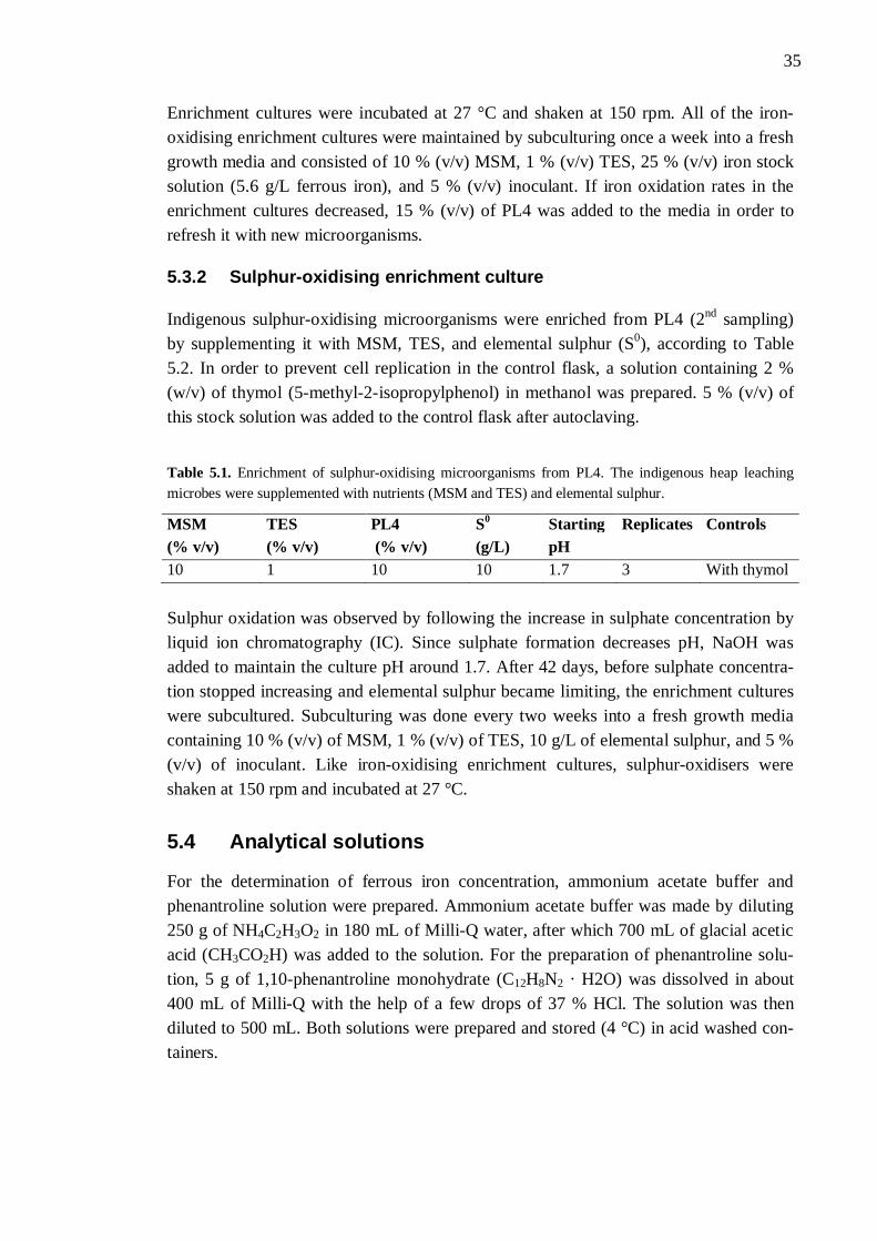
35
Enrichment cultures were incubated at 27 °C and shaken at 150 rpm. All of the iron-oxidising enrichment cultures were maintained by subculturing once a week into a fresh growth media and consisted of 10 % (v/v) MSM, 1 % (v/v) TES, 25 % (v/v) iron stock solution (5.6 g/L ferrous iron), and 5 % (v/v) inoculant. If iron oxidation rates in the enrichment cultures decreased, 15 % (v/v) of PL4 was added to the media in order to refresh it with new microorganisms.
5.3.2 Sulphur-oxidising enrichment culture
Indigenous sulphur-oxidising microorganisms were enriched from PL4 (2nd sampling) by supplementing it with MSM, TES, and elemental sulphur (S0), according to Table 5.2. In order to prevent cell replication in the control flask, a solution containing 2 % (w/v) of thymol (5-methyl-2-isopropylphenol) in methanol was prepared. 5 % (v/v) of this stock solution was added to the control flask after autoclaving.
Table 5.1. Enrichment of sulphur-oxidising microorganisms from PL4. The indigenous heap leaching microbes were supplemented with nutrients (MSM and TES) and elemental sulphur.
MSM (% v/v)
TES (% v/v)
PL4 (% v/v)
S0 (g/L)
Starting pH
Replicates Controls
10 1 10 10 1.7 3 With thymol Sulphur oxidation was observed by following the increase in sulphate concentration by liquid ion chromatography (IC). Since sulphate formation decreases pH, NaOH was added to maintain the culture pH around 1.7. After 42 days, before sulphate concentra-tion stopped increasing and elemental sulphur became limiting, the enrichment cultures were subcultured. Subculturing was done every two weeks into a fresh growth media containing 10 % (v/v) of MSM, 1 % (v/v) of TES, 10 g/L of elemental sulphur, and 5 % (v/v) of inoculant. Like iron-oxidising enrichment cultures, sulphur-oxidisers were shaken at 150 rpm and incubated at 27 °C.
5.4 Analytical solutions
For the determination of ferrous iron concentration, ammonium acetate buffer and phenantroline solution were prepared. Ammonium acetate buffer was made by diluting 250 g of NH4C2H3O2 in 180 mL of Milli-Q water, after which 700 mL of glacial acetic acid (CH3CO2H) was added to the solution. For the preparation of phenantroline solu-tion, 5 g of 1,10-phenantroline monohydrate (C12H8N2 · H2O) was dissolved in about 400 mL of Milli-Q with the help of a few drops of 37 % HCl. The solution was then diluted to 500 mL. Both solutions were prepared and stored (4 °C) in acid washed con-tainers.
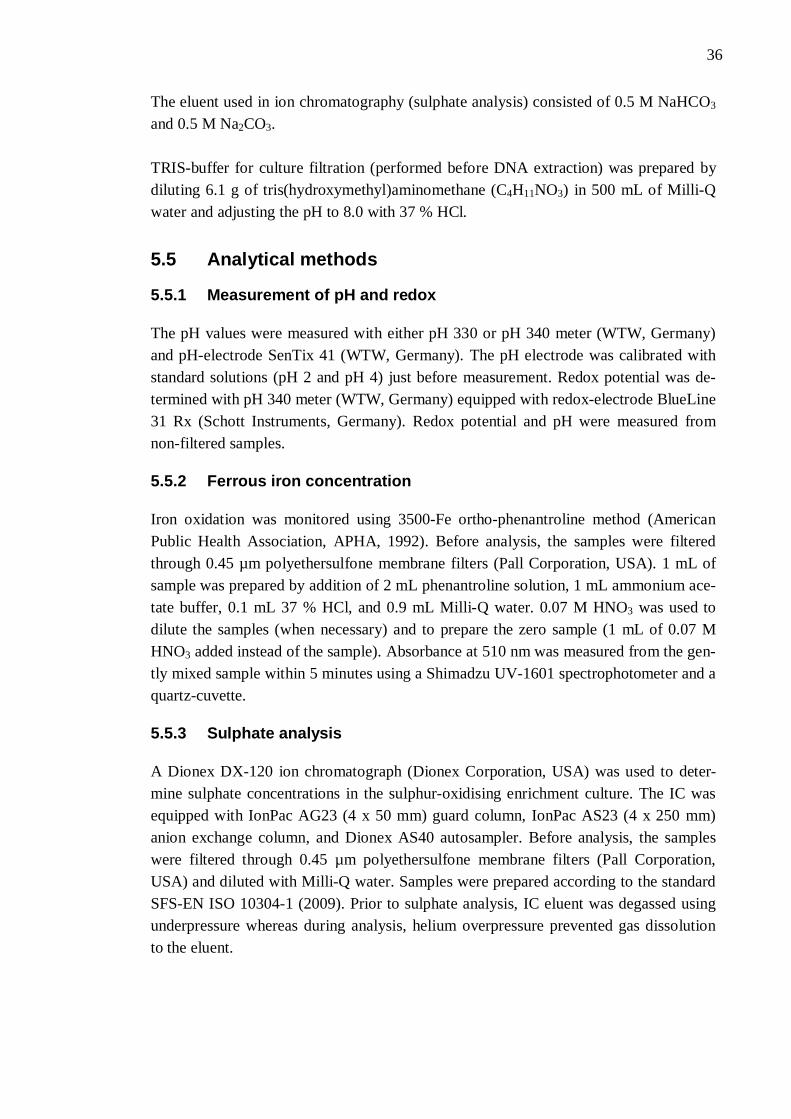
36
The eluent used in ion chromatography (sulphate analysis) consisted of 0.5 M NaHCO3 and 0.5 M Na2CO3. TRIS-buffer for culture filtration (performed before DNA extraction) was prepared by diluting 6.1 g of tris(hydroxymethyl)aminomethane (C4H11NO3) in 500 mL of Milli-Q water and adjusting the pH to 8.0 with 37 % HCl.
5.5 Analytical methods
5.5.1 Measurement of pH and redox
The pH values were measured with either pH 330 or pH 340 meter (WTW, Germany) and pH-electrode SenTix 41 (WTW, Germany). The pH electrode was calibrated with standard solutions (pH 2 and pH 4) just before measurement. Redox potential was de-termined with pH 340 meter (WTW, Germany) equipped with redox-electrode BlueLine 31 Rx (Schott Instruments, Germany). Redox potential and pH were measured from non-filtered samples.
5.5.2 Ferrous iron concentration
Iron oxidation was monitored using 3500-Fe ortho-phenantroline method (American Public Health Association, APHA, 1992). Before analysis, the samples were filtered through 0.45 µm polyethersulfone membrane filters (Pall Corporation, USA). 1 mL of sample was prepared by addition of 2 mL phenantroline solution, 1 mL ammonium ace-tate buffer, 0.1 mL 37 % HCl, and 0.9 mL Milli-Q water. 0.07 M HNO3 was used to dilute the samples (when necessary) and to prepare the zero sample (1 mL of 0.07 M HNO3 added instead of the sample). Absorbance at 510 nm was measured from the gen-tly mixed sample within 5 minutes using a Shimadzu UV-1601 spectrophotometer and a quartz-cuvette.
5.5.3 Sulphate analysis
A Dionex DX-120 ion chromatograph (Dionex Corporation, USA) was used to deter-mine sulphate concentrations in the sulphur-oxidising enrichment culture. The IC was equipped with IonPac AG23 (4 x 50 mm) guard column, IonPac AS23 (4 x 250 mm) anion exchange column, and Dionex AS40 autosampler. Before analysis, the samples were filtered through 0.45 µm polyethersulfone membrane filters (Pall Corporation, USA) and diluted with Milli-Q water. Samples were prepared according to the standard SFS-EN ISO 10304-1 (2009). Prior to sulphate analysis, IC eluent was degassed using underpressure whereas during analysis, helium overpressure prevented gas dissolution to the eluent.
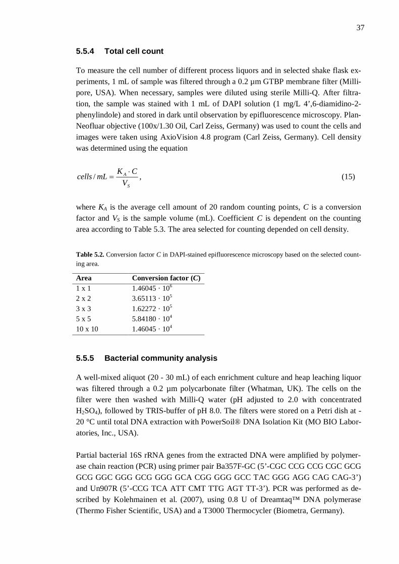
37
5.5.4 Total cell count
To measure the cell number of different process liquors and in selected shake flask ex-periments, 1 mL of sample was filtered through a 0.2 µm GTBP membrane filter (Milli-pore, USA). When necessary, samples were diluted using sterile Milli-Q. After filtra-tion, the sample was stained with 1 mL of DAPI solution (1 mg/L 4’,6-diamidino-2-phenylindole) and stored in dark until observation by epifluorescence microscopy. Plan-Neofluar objective (100x/1.30 Oil, Carl Zeiss, Germany) was used to count the cells and images were taken using AxioVision 4.8 program (Carl Zeiss, Germany). Cell density was determined using the equation
S
A
VCKmLcells / , (15)
where KA is the average cell amount of 20 random counting points, C is a conversion factor and VS is the sample volume (mL). Coefficient C is dependent on the counting area according to Table 5.3. The area selected for counting depended on cell density.
Table 5.2. Conversion factor C in DAPI-stained epifluorescence microscopy based on the selected count-ing area.
Area Conversion factor (C) 1 x 1 1.46045 · 106 2 x 2 3.65113 · 105 3 x 3 1.62272 · 105 5 x 5 5.84180 · 104 10 x 10 1.46045 · 104
5.5.5 Bacterial community analysis
A well-mixed aliquot (20 - 30 mL) of each enrichment culture and heap leaching liquor was filtered through a 0.2 µm polycarbonate filter (Whatman, UK). The cells on the filter were then washed with Milli-Q water (pH adjusted to 2.0 with concentrated H2SO4), followed by TRIS-buffer of pH 8.0. The filters were stored on a Petri dish at -20 °C until total DNA extraction with PowerSoil® DNA Isolation Kit (MO BIO Labor-atories, Inc., USA). Partial bacterial 16S rRNA genes from the extracted DNA were amplified by polymer-ase chain reaction (PCR) using primer pair Ba357F-GC (5’-CGC CCG CCG CGC GCG GCG GGC GGG GCG GGG GCA CGG GGG GCC TAC GGG AGG CAG CAG-3’) and Un907R (5’-CCG TCA ATT CMT TTG AGT TT-3’). PCR was performed as de-scribed by Kolehmainen et al. (2007), using 0.8 U of Dreamtaq™ DNA polymerase (Thermo Fisher Scientific, USA) and a T3000 Thermocycler (Biometra, Germany).
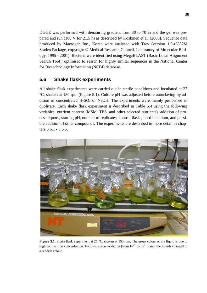
38
DGGE was performed with denaturing gradient from 30 to 70 % and the gel was pre-pared and run (100 V for 21.5 h) as described by Koskinen et al. (2006). Sequence data produced by Macrogen Inc., Korea were analysed with Trev (version 1.9-r2852M Staden Package, copyright © Medical Research Council, Laboratory of Molecular Biol-ogy, 1995 - 2001). Bacteria were identified using MegaBLAST (Basic Local Alignment Search Tool), optimised to search for highly similar sequences in the National Center for Biotechnology Information (NCBI) database.
5.6 Shake flask experiments
All shake flask experiments were carried out in sterile conditions and incubated at 27 °C, shaken at 150 rpm (Figure 5.1). Culture pH was adjusted before autoclaving by ad-dition of concentrated H2SO4 or NaOH. The experiments were mainly performed in duplicate. Each shake flask experiment is described in Table 5.4 using the following variables: nutrient content (MSM, TES, and other selected nutrients), addition of pro-cess liquors, starting pH, number of replicates, control flasks, used inoculum, and possi-ble addition of other compounds. The experiments are described in more detail in chap-ters 5.6.1 - 5.6.5.
Figure 5.1. Shake flask experiment at 27 °C, shaken at 150 rpm. The green colour of the liquid is due to high ferrous iron concentration. Following iron oxidation (from Fe2+ to Fe3+ ions), the liquids changed to a reddish colour.
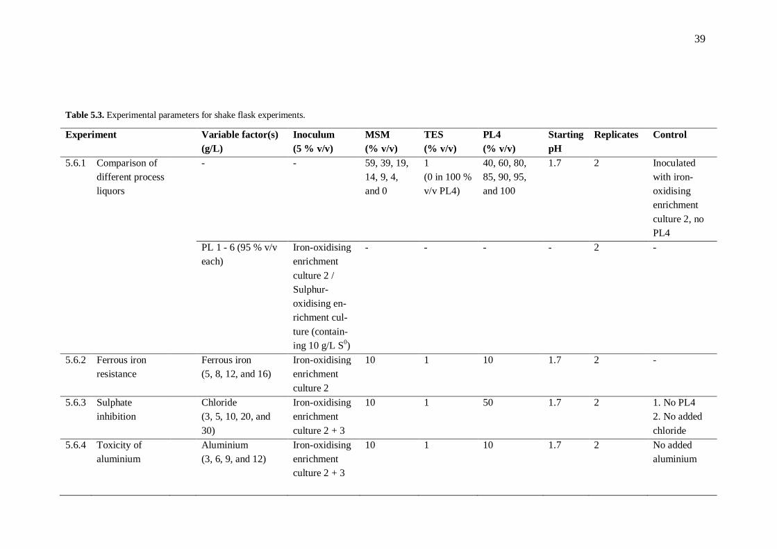
39
Table 5.3. Experimental parameters for shake flask experiments.
Experiment Variable factor(s) (g/L)
Inoculum (5 % v/v)
MSM (% v/v)
TES (% v/v)
PL4 (% v/v)
Starting pH
Replicates Control
5.6.1 Comparison of different process liquors
- - 59, 39, 19, 14, 9, 4, and 0
1 (0 in 100 % v/v PL4)
40, 60, 80, 85, 90, 95, and 100
1.7 2 Inoculated with iron-oxidising enrichment culture 2, no PL4
PL 1 - 6 (95 % v/v each)
Iron-oxidising enrichment culture 2 / Sulphur-oxidising en-richment cul-ture (contain-ing 10 g/L S0)
- - - - 2 -
5.6.2 Ferrous iron resistance
Ferrous iron (5, 8, 12, and 16)
Iron-oxidising enrichment culture 2
10 1 10 1.7 2 -
5.6.3 Sulphate inhibition
Chloride (3, 5, 10, 20, and 30)
Iron-oxidising enrichment culture 2 + 3
10 1 50 1.7 2 1. No PL4 2. No added chloride
5.6.4 Toxicity of aluminium
Aluminium (3, 6, 9, and 12)
Iron-oxidising enrichment culture 2 + 3
10 1 10 1.7 2 No added aluminium
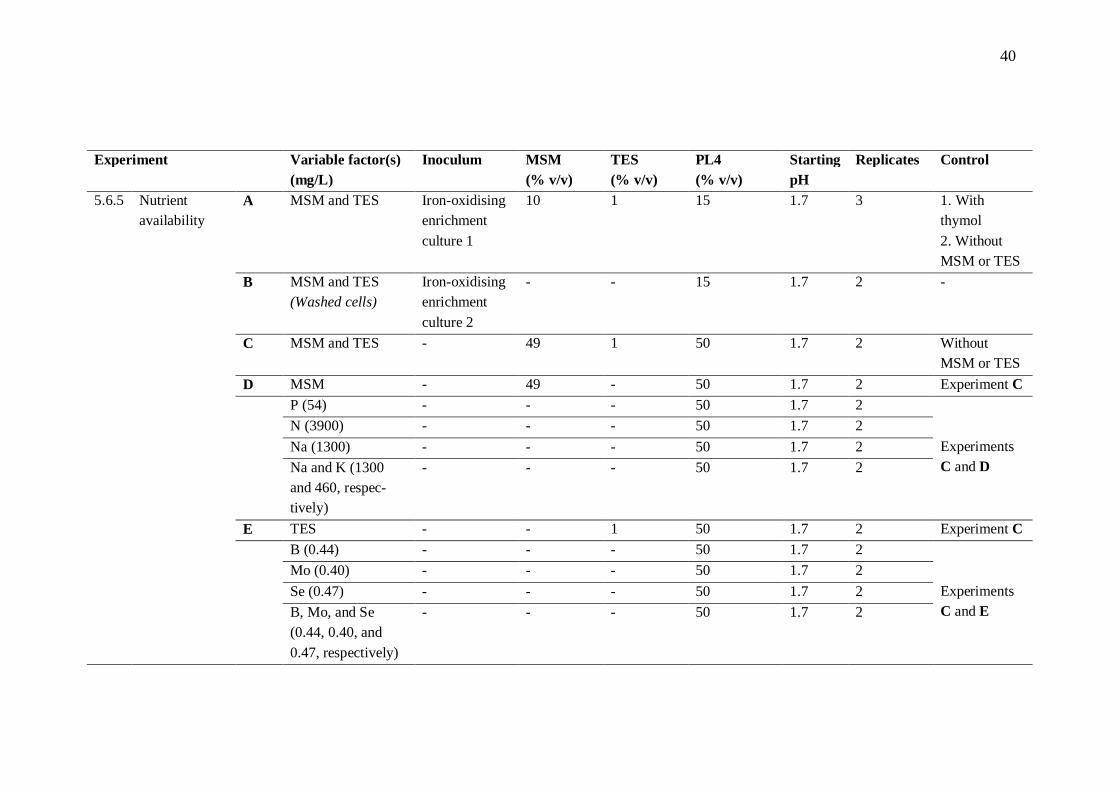
40
Experiment Variable factor(s) (mg/L)
Inoculum MSM (% v/v)
TES (% v/v)
PL4 (% v/v)
Starting pH
Replicates Control
5.6.5 Nutrient availability
A MSM and TES Iron-oxidising enrichment culture 1
10 1 15 1.7 3 1. With thymol 2. Without MSM or TES
B MSM and TES (Washed cells)
Iron-oxidising enrichment culture 2
- - 15 1.7 2 -
C MSM and TES - 49 1 50 1.7 2 Without MSM or TES
D MSM - 49 - 50 1.7 2 Experiment C P (54) - - - 50 1.7 2
Experiments C and D
N (3900) - - - 50 1.7 2 Na (1300) - - - 50 1.7 2 Na and K (1300
and 460, respec-tively)
- - - 50 1.7 2
E TES - - 1 50 1.7 2 Experiment C B (0.44) - - - 50 1.7 2
Experiments C and E
Mo (0.40) - - - 50 1.7 2 Se (0.47) - - - 50 1.7 2 B, Mo, and Se
(0.44, 0.40, and 0.47, respectively)
- - - 50 1.7 2
41
5.6.1 Comparison of different process liquors
The process liquors originated from different parts of the bioleaching process, therefore their component concentrations and physicochemical properties varied significantly. Since the plausible inhibitory effects of PL4 were of most interest, iron oxidation in a non-inoculated dilution series of this liquor in MSM was studied. The experiments were carried out according to Table 5.4, containing 40 - 100 % (v/v) of PL4, 1 % (v/v) of TES, and MSM so that the final culture volume was 100 mL. To define the best possi-ble iron oxidation rate, an inoculated control without PL4 addition was made. The possible inhibitory effects of different process liquors (1 - 6) towards any microor-ganisms they might contain were studied by incubating 95 % (v/v) of each process liq-uor with 5 % (v/v) of either iron-oxidising enrichment culture 2 (to study iron oxida-tion), or the sulphur-oxidising enrichment culture (Table 5.4). Sulphur-oxidising cul-tures were also supplemented with 10 g/L of elemental sulphur. The activity of sulphur-oxidisers was observed by measuring pH, which decreases following sulphate for-mation. Total cell counts were carried out from both raw process liquor samples at the start and end of the experiment. The oxidation end point for sulphur-oxidisers was after 2 - 3 days of unchanged pH.
5.6.2 Ferrous iron resistance
To study the resistance of iron-oxidising microorganisms towards high ferrous iron con-centrations, PL4 was inoculated with iron-oxidisers, and incubated with nutrients and different Fe2+ concentrations (Table 5.4). Ferrous iron concentration of 5 g/L was nor-mally used in iron-oxidising control flasks, so no additional control flask was made. Fe2+ concentrations in Talvivaara leach liquors have usually been under 16 g/L.
5.6.3 Sulphate inhibition
Due to high sulphate concentrations in bioleaching liquors (especially in liquors 3 and 4), the possible inhibitive effect of SO4
2- ions towards iron-oxidising microorganisms was considered. Process liquors 3 and 4 contained over 120 g/L of sulphate, whereas liquors 1, 2, 5, and 6 contained less than 80 g/L. One method to study the effects of different SO4
2- concentrations was to precipitate sul-phate ions in PL4 with barium chloride (BaCl2 · 2 H2O). However, in order to reach a concentration of 70 g/L SO4
2- (a concentration unlikely to be toxic), chloride ion content in PL4 would increase to 30 g/L due to barium chloride addition. Therefore to study how chloride resistant the enriched iron-oxidisers and the indigenous microorganisms of PL4 were, they were incubated with nutrients and Cl- concentrations up to 30 g/L (Table 5.4). Chloride was added to the culture before sterilisation as a sodium chloride stock
42
solution containing 144 g NaCl per liter of Milli-Q (87 g/L Cl-). Two controls were car-ried out; one without PL4 (30 g/L of chloride) and one without added chloride. Other deliberated ways to study sulphate toxicity were addition of different volumes of SO4
2- stock solution (with (NH4)2SO4 and Na2SO4 · 10 H2O), or addition of sulphate as H2SO4 (after which culture pH was adjusted to 1.7 with NaOH or KOH). The problem with these techniques was the highly increased concentration of cations.
5.6.4 Toxicity of aluminium
Aluminium concentrations of different process liquors ranged between 71 and 9000 mg/L (around 6100 mg/L in PL4). Aluminium toxicity was studied by inoculating and incubating PL4 with different Al3+ concentrations (according to Table 5.4). No alumini-um was added to the control flask. Aluminium stock solution was prepared by dissolv-ing 216 g of Al2(SO4)3 · ~ 14 H2O into 1000 mL of Milli-Q, resulting in an Al3+ concen-tration of approximately 20 g/L. The solution was adjusted to pH 1.7 with H2SO4 and sterile-filtered using polyethersulfone membrane filters (0.2 m, Whatman, UK). Alu-minium stock solution was added to the culture after autoclave sterilisation.
5.6.5 Nutrient availability
Nutrient availability in PL4 was studied by incubating it with and without nutrients (MSM and TES). In experiment A (Table 5.4), 15 % (v/v) of PL4 was inoculated with iron-oxidising enrichment culture 1 and incubated with nutrients. Two controls were used: one with 5 % (v/v) of thymol solution (2 % w/v in methanol; to prevent cell repli-cation), and another without MSM or TES. A third control flask was later prepared us-ing washed iron-oxidising enrichment culture without nutrients (experiment B). The cells were washed thrice before inoculation with acidic Milli-Q (pH adjusted to 2.0 with concentrated H2SO4) to prevent any nutrient transfer from the enrichment culture. Washing was carried out by centrifuging the cells at 5000 rcf for 10 minutes, after which the supernatant was removed and exactly the same amount of Milli-Q was added. Since experiments A and B were both inoculated, experiment C was made to rule out possible nutrient remains from the enrichment cultures. Thus, 50 % (v/v) of PL4 was incubated with 49 % (v/v) of MSM and 1 % (v/v) of TES, in addition to a control flask containing only PL4 and Milli-Q (Table 5.4). Nutrients available in MSM but possibly lacking from PL4 (concentrations either very low or not measured) were phosphorus (P), nitrogen (N), sodium (Na), and potassium (K). In experiment D (Table 5.4), 50 % (v/v) of PL4 was incubated with 49 % (v/v) of MSM. In the absence of TES, the nutritional effects of MSM and TES could be com-pared. In addition, one or more of the selected nutrients were incubated with 50 % (v/v)
43
of PL4 according to Table 5.4. The concentration of each nutrient used was similar to its concentration in 49 % (v/v) of MSM. TES was studied in a similar way (experiment E in Table 5.4). Nutrients available in TES but potentially lacking from PL4 were boron (B), molybdenum (Mo), and selenium (Se). The selected nutrients were added to PL4 according to Table 5.4, at same concen-trations as were present in 1 % (v/v) of TES. Nutrients were added as compounds nor-mally present in MSM and TES (see Chapter 5.2.1). Total inorganic and organic nitrogen was determined from heap leaching liquors 1 - 6 with modified Kjeldahl method according to standard SFS 5505 (Suomen standardisoi-misliitto, SFS, 1988). The device used for sample distillation was a Kjeltec™ 2100 (FOSS Analytical, Denmark).
5.7 Sampling
To measure ferrous iron concentration, iron-oxidising shake flask experiments were usually sampled once a day during the lag phase of iron oxidation, and twice a day dur-ing the exponential growth phase. Sample volume (1 mL) was replaced with sterile Mil-li-Q water to maintain the culture volume at around 100 mL. Ferrous iron concentration was determined within 60 minutes. Sulphur-oxidising enrichment cultures were sampled in two different ways. To deter-mine sulphate concentration by ion chromatography, 1 mL of culture was sampled 3 - 5 times a week and stored at - 20 °C until filtration and analysis. For pH measurement, 2 mL of culture was sampled three times a week and the pH value was immediately de-termined. As with iron-oxidising cultures, sample volume was replaced with sterile Mil-li-Q. Process liquors 1 - 6 were sampled shortly after delivery to determine pH and redox potential. 2 mL of each liquor was sampled and immediately analysed. Unlike with iron- and sulphur-oxidising cultures, the sample volume was not replaced with Milli-Q water. In addition, process liquors and selected experimental flasks were sampled (1 mL) for analysis of total cell number. To determine bacterial community composition, enrich-ment cultures and process liquors were sampled once (20 - 30 mL). All iron- and sul-phur-oxidising cultures and different process liquors were sampled in a laminar flow cabinet to minimise contamination.
44
6 RESULTS
6.1 Enrichment of sulphur-oxidisers
In experiments regarding iron oxidation, an enrichment culture was ready to use. Indig-enous sulphur-oxidisers were enriched from PL4 by incubating the liquor with nutrients (MSM and TES) and 10 g/L of elemental sulphur. Sulphur oxidation was monitored by measuring sulphate concentration in the culture, and culture pH was maintained around 1.7 to ensure suitable conditions for cell growth. The increase in sulphate concentration was compared to a control flask, where cell replication was prevented by thymol addi-tion (Figure 6.1). As seen in Figure 6.1, sulphur oxidation was slow during the first 10 days of incubation, with a sulphate formation rate of 800 mg/L/d (from a linear part of the curve). Follow-ing this lag phase, SO4
2- formation more than doubled, reaching a rate of 1900 mg/L/d. Sulphur oxidation then remained steady for three weeks, until after 31 day cultivation the microorganisms started to generate sulphate at a higher rate. After this point, SO4
2- formation rate remained at 4700 mg/L/d until the end of the experiment. Figure 6.1 shows that the sulphur-oxidising microorganisms of PL4 required a few weeks to adjust to new culture conditions, after which their sulphur oxidation activity distinctly in-creased.
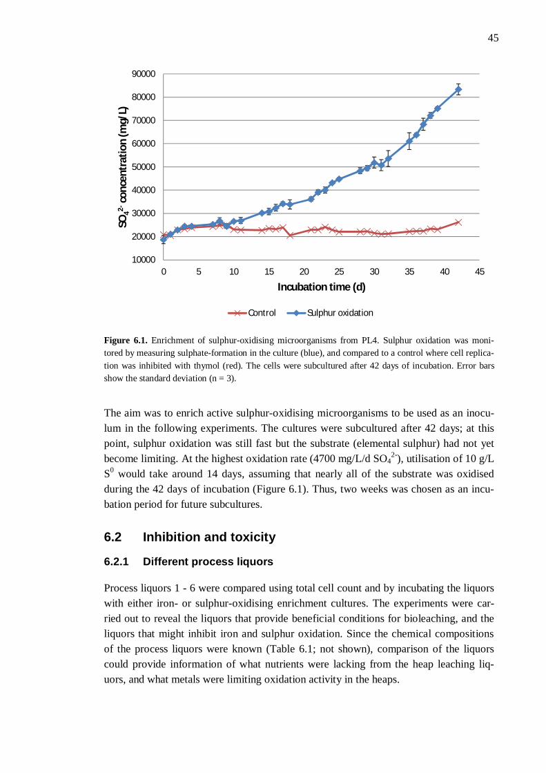
45
Figure 6.1. Enrichment of sulphur-oxidising microorganisms from PL4. Sulphur oxidation was moni-tored by measuring sulphate-formation in the culture (blue), and compared to a control where cell replica-tion was inhibited with thymol (red). The cells were subcultured after 42 days of incubation. Error bars show the standard deviation (n = 3).
The aim was to enrich active sulphur-oxidising microorganisms to be used as an inocu-lum in the following experiments. The cultures were subcultured after 42 days; at this point, sulphur oxidation was still fast but the substrate (elemental sulphur) had not yet become limiting. At the highest oxidation rate (4700 mg/L/d SO4
2-), utilisation of 10 g/L S0 would take around 14 days, assuming that nearly all of the substrate was oxidised during the 42 days of incubation (Figure 6.1). Thus, two weeks was chosen as an incu-bation period for future subcultures.
6.2 Inhibition and toxicity
6.2.1 Different process liquors
Process liquors 1 - 6 were compared using total cell count and by incubating the liquors with either iron- or sulphur-oxidising enrichment cultures. The experiments were car-ried out to reveal the liquors that provide beneficial conditions for bioleaching, and the liquors that might inhibit iron and sulphur oxidation. Since the chemical compositions of the process liquors were known (Table 6.1; not shown), comparison of the liquors could provide information of what nutrients were lacking from the heap leaching liq-uors, and what metals were limiting oxidation activity in the heaps.
10000
20000
30000
40000
50000
60000
70000
80000
90000
0 5 10 15 20 25 30 35 40 45
SO42-
conc
entr
atio
n (m
g/L)
Incubation time (d)
Control Sulphur oxidation
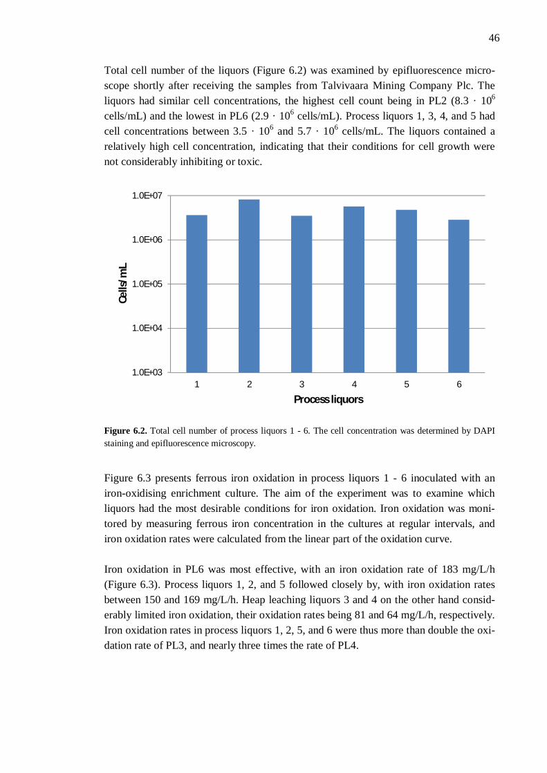
46
Total cell number of the liquors (Figure 6.2) was examined by epifluorescence micro-scope shortly after receiving the samples from Talvivaara Mining Company Plc. The liquors had similar cell concentrations, the highest cell count being in PL2 (8.3 · 106 cells/mL) and the lowest in PL6 (2.9 · 106 cells/mL). Process liquors 1, 3, 4, and 5 had cell concentrations between 3.5 · 106 and 5.7 · 106 cells/mL. The liquors contained a relatively high cell concentration, indicating that their conditions for cell growth were not considerably inhibiting or toxic.
Figure 6.2. Total cell number of process liquors 1 - 6. The cell concentration was determined by DAPI staining and epifluorescence microscopy.
Figure 6.3 presents ferrous iron oxidation in process liquors 1 - 6 inoculated with an iron-oxidising enrichment culture. The aim of the experiment was to examine which liquors had the most desirable conditions for iron oxidation. Iron oxidation was moni-tored by measuring ferrous iron concentration in the cultures at regular intervals, and iron oxidation rates were calculated from the linear part of the oxidation curve. Iron oxidation in PL6 was most effective, with an iron oxidation rate of 183 mg/L/h (Figure 6.3). Process liquors 1, 2, and 5 followed closely by, with iron oxidation rates between 150 and 169 mg/L/h. Heap leaching liquors 3 and 4 on the other hand consid-erably limited iron oxidation, their oxidation rates being 81 and 64 mg/L/h, respectively. Iron oxidation rates in process liquors 1, 2, 5, and 6 were thus more than double the oxi-dation rate of PL3, and nearly three times the rate of PL4.
1.0E+03
1.0E+04
1.0E+05
1.0E+06
1.0E+07
1 2 3 4 5 6
Cells
/mL
Process liquors
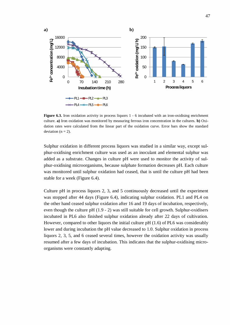
47
a) b)
Figure 6.3. Iron oxidation activity in process liquors 1 - 6 incubated with an iron-oxidising enrichment culture. a) Iron oxidation was monitored by measuring ferrous iron concentration in the cultures. b) Oxi-dation rates were calculated from the linear part of the oxidation curve. Error bars show the standard deviation (n = 2).
Sulphur oxidation in different process liquors was studied in a similar way, except sul-phur-oxidising enrichment culture was used as an inoculant and elemental sulphur was added as a substrate. Changes in culture pH were used to monitor the activity of sul-phur-oxidising microorganisms, because sulphate formation decreases pH. Each culture was monitored until sulphur oxidation had ceased, that is until the culture pH had been stable for a week (Figure 6.4). Culture pH in process liquors 2, 3, and 5 continuously decreased until the experiment was stopped after 44 days (Figure 6.4), indicating sulphur oxidation. PL1 and PL4 on the other hand ceased sulphur oxidation after 16 and 19 days of incubation, respectively, even though the culture pH (1.9 - 2) was still suitable for cell growth. Sulphur-oxidisers incubated in PL6 also finished sulphur oxidation already after 22 days of cultivation. However, compared to other liquors the initial culture pH (1.6) of PL6 was considerably lower and during incubation the pH value decreased to 1.0. Sulphur oxidation in process liquors 2, 3, 5, and 6 ceased several times, however the oxidation activity was usually resumed after a few days of incubation. This indicates that the sulphur-oxidising micro-organisms were constantly adapting.
0
4000
8000
12000
16000
0 70 140 210 280Fe2+
conc
entr
atio
n (m
g/L)
Incubation time (h)
PL1 PL2 PL3
PL4 PL5 PL6
0
50
100
150
200
1 2 3 4 5 6
Fe2+
oxid
atio
n (m
g/L/
h)
Process liquors
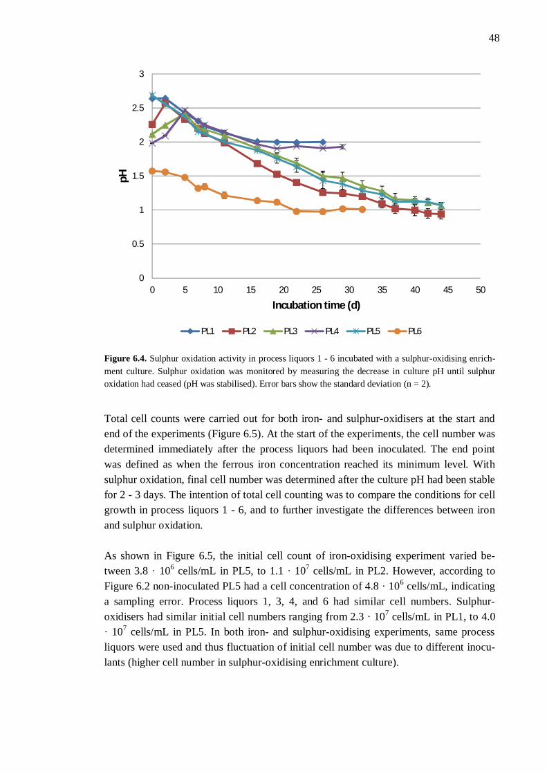
48
Figure 6.4. Sulphur oxidation activity in process liquors 1 - 6 incubated with a sulphur-oxidising enrich-ment culture. Sulphur oxidation was monitored by measuring the decrease in culture pH until sulphur oxidation had ceased (pH was stabilised). Error bars show the standard deviation (n = 2).
Total cell counts were carried out for both iron- and sulphur-oxidisers at the start and end of the experiments (Figure 6.5). At the start of the experiments, the cell number was determined immediately after the process liquors had been inoculated. The end point was defined as when the ferrous iron concentration reached its minimum level. With sulphur oxidation, final cell number was determined after the culture pH had been stable for 2 - 3 days. The intention of total cell counting was to compare the conditions for cell growth in process liquors 1 - 6, and to further investigate the differences between iron and sulphur oxidation. As shown in Figure 6.5, the initial cell count of iron-oxidising experiment varied be-tween 3.8 · 106 cells/mL in PL5, to 1.1 · 107 cells/mL in PL2. However, according to Figure 6.2 non-inoculated PL5 had a cell concentration of 4.8 · 106 cells/mL, indicating a sampling error. Process liquors 1, 3, 4, and 6 had similar cell numbers. Sulphur-oxidisers had similar initial cell numbers ranging from 2.3 · 107 cells/mL in PL1, to 4.0 · 107 cells/mL in PL5. In both iron- and sulphur-oxidising experiments, same process liquors were used and thus fluctuation of initial cell number was due to different inocu-lants (higher cell number in sulphur-oxidising enrichment culture).
0
0.5
1
1.5
2
2.5
3
0 5 10 15 20 25 30 35 40 45 50
pH
Incubation time (d)
PL1 PL2 PL3 PL4 PL5 PL6
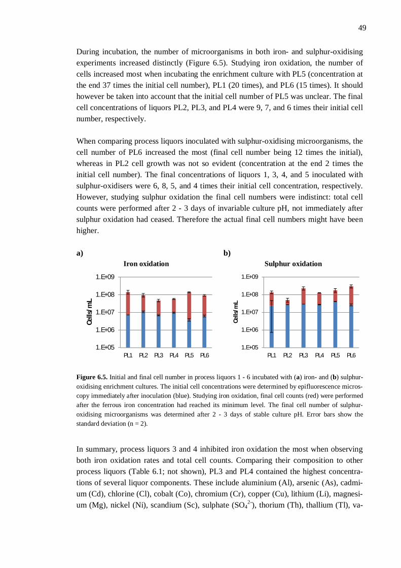
49
During incubation, the number of microorganisms in both iron- and sulphur-oxidising experiments increased distinctly (Figure 6.5). Studying iron oxidation, the number of cells increased most when incubating the enrichment culture with PL5 (concentration at the end 37 times the initial cell number), PL1 (20 times), and PL6 (15 times). It should however be taken into account that the initial cell number of PL5 was unclear. The final cell concentrations of liquors PL2, PL3, and PL4 were 9, 7, and 6 times their initial cell number, respectively. When comparing process liquors inoculated with sulphur-oxidising microorganisms, the cell number of PL6 increased the most (final cell number being 12 times the initial), whereas in PL2 cell growth was not so evident (concentration at the end 2 times the initial cell number). The final concentrations of liquors 1, 3, 4, and 5 inoculated with sulphur-oxidisers were 6, 8, 5, and 4 times their initial cell concentration, respectively. However, studying sulphur oxidation the final cell numbers were indistinct: total cell counts were performed after 2 - 3 days of invariable culture pH, not immediately after sulphur oxidation had ceased. Therefore the actual final cell numbers might have been higher. a) b)
Iron oxidation
Sulphur oxidation
Figure 6.5. Initial and final cell number in process liquors 1 - 6 incubated with (a) iron- and (b) sulphur-oxidising enrichment cultures. The initial cell concentrations were determined by epifluorescence micros-copy immediately after inoculation (blue). Studying iron oxidation, final cell counts (red) were performed after the ferrous iron concentration had reached its minimum level. The final cell number of sulphur-oxidising microorganisms was determined after 2 - 3 days of stable culture pH. Error bars show the standard deviation (n = 2).
In summary, process liquors 3 and 4 inhibited iron oxidation the most when observing both iron oxidation rates and total cell counts. Comparing their composition to other process liquors (Table 6.1; not shown), PL3 and PL4 contained the highest concentra-tions of several liquor components. These include aluminium (Al), arsenic (As), cadmi-um (Cd), chlorine (Cl), cobalt (Co), chromium (Cr), copper (Cu), lithium (Li), magnesi-um (Mg), nickel (Ni), scandium (Sc), sulphate (SO4
2-), thorium (Th), thallium (Tl), va-
1.E+05
1.E+06
1.E+07
1.E+08
1.E+09
PL1 PL2 PL3 PL4 PL5 PL6
Cells
/mL
1.E+05
1.E+06
1.E+07
1.E+08
1.E+09
PL1 PL2 PL3 PL4 PL5 PL6
Cells
/mL
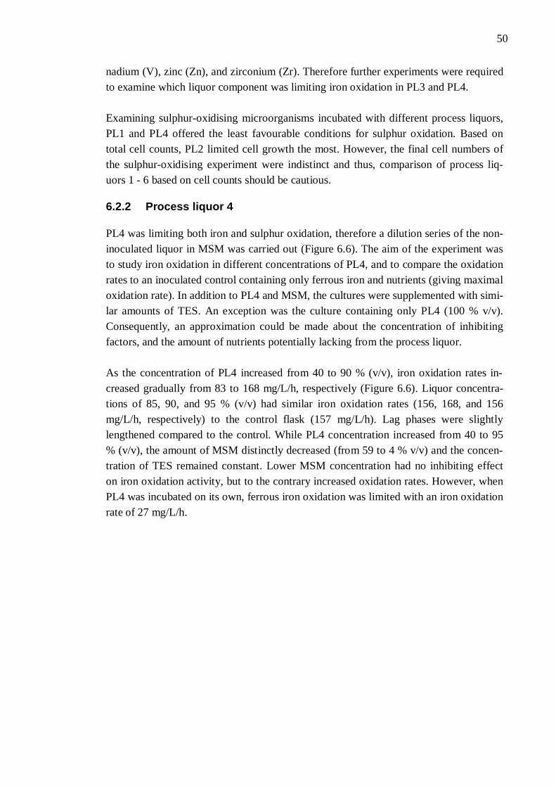
50
nadium (V), zinc (Zn), and zirconium (Zr). Therefore further experiments were required to examine which liquor component was limiting iron oxidation in PL3 and PL4. Examining sulphur-oxidising microorganisms incubated with different process liquors, PL1 and PL4 offered the least favourable conditions for sulphur oxidation. Based on total cell counts, PL2 limited cell growth the most. However, the final cell numbers of the sulphur-oxidising experiment were indistinct and thus, comparison of process liq-uors 1 - 6 based on cell counts should be cautious.
6.2.2 Process liquor 4
PL4 was limiting both iron and sulphur oxidation, therefore a dilution series of the non-inoculated liquor in MSM was carried out (Figure 6.6). The aim of the experiment was to study iron oxidation in different concentrations of PL4, and to compare the oxidation rates to an inoculated control containing only ferrous iron and nutrients (giving maximal oxidation rate). In addition to PL4 and MSM, the cultures were supplemented with simi-lar amounts of TES. An exception was the culture containing only PL4 (100 % v/v). Consequently, an approximation could be made about the concentration of inhibiting factors, and the amount of nutrients potentially lacking from the process liquor. As the concentration of PL4 increased from 40 to 90 % (v/v), iron oxidation rates in-creased gradually from 83 to 168 mg/L/h, respectively (Figure 6.6). Liquor concentra-tions of 85, 90, and 95 % (v/v) had similar iron oxidation rates (156, 168, and 156 mg/L/h, respectively) to the control flask (157 mg/L/h). Lag phases were slightly lengthened compared to the control. While PL4 concentration increased from 40 to 95 % (v/v), the amount of MSM distinctly decreased (from 59 to 4 % v/v) and the concen-tration of TES remained constant. Lower MSM concentration had no inhibiting effect on iron oxidation activity, but to the contrary increased oxidation rates. However, when PL4 was incubated on its own, ferrous iron oxidation was limited with an iron oxidation rate of 27 mg/L/h.
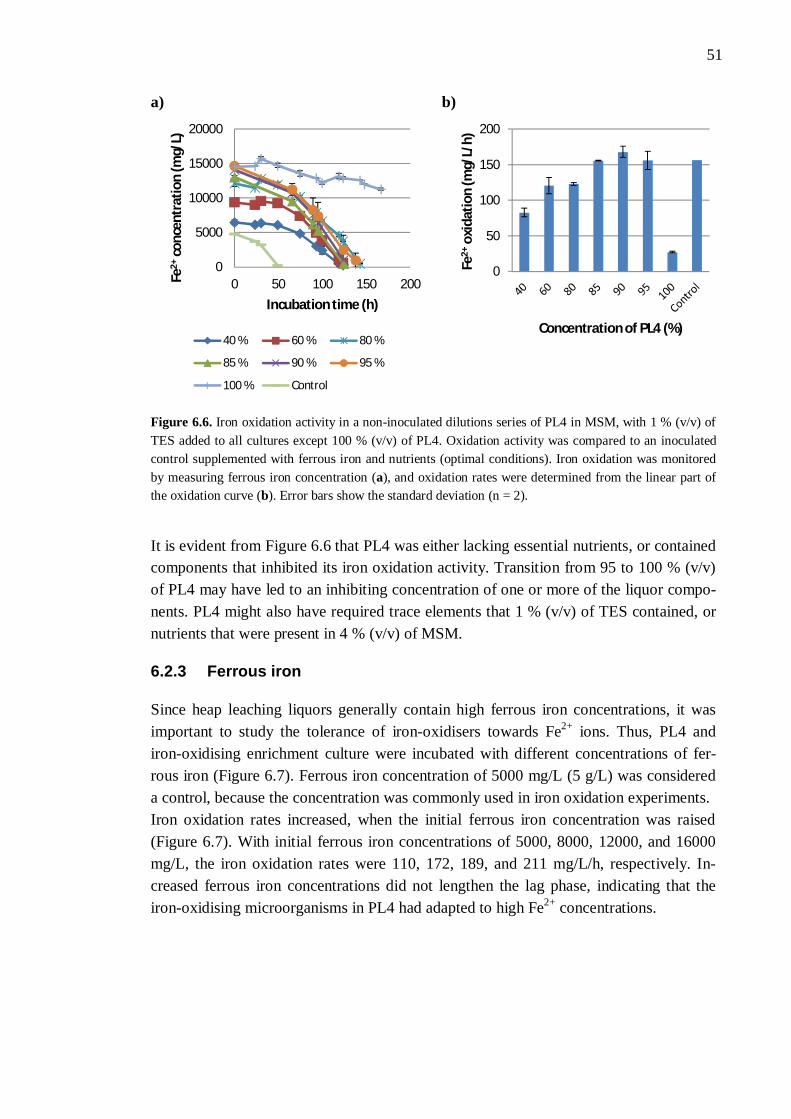
51
a) b)
Figure 6.6. Iron oxidation activity in a non-inoculated dilutions series of PL4 in MSM, with 1 % (v/v) of TES added to all cultures except 100 % (v/v) of PL4. Oxidation activity was compared to an inoculated control supplemented with ferrous iron and nutrients (optimal conditions). Iron oxidation was monitored by measuring ferrous iron concentration (a), and oxidation rates were determined from the linear part of the oxidation curve (b). Error bars show the standard deviation (n = 2).
It is evident from Figure 6.6 that PL4 was either lacking essential nutrients, or contained components that inhibited its iron oxidation activity. Transition from 95 to 100 % (v/v) of PL4 may have led to an inhibiting concentration of one or more of the liquor compo-nents. PL4 might also have required trace elements that 1 % (v/v) of TES contained, or nutrients that were present in 4 % (v/v) of MSM.
6.2.3 Ferrous iron
Since heap leaching liquors generally contain high ferrous iron concentrations, it was important to study the tolerance of iron-oxidisers towards Fe2+ ions. Thus, PL4 and iron-oxidising enrichment culture were incubated with different concentrations of fer-rous iron (Figure 6.7). Ferrous iron concentration of 5000 mg/L (5 g/L) was considered a control, because the concentration was commonly used in iron oxidation experiments. Iron oxidation rates increased, when the initial ferrous iron concentration was raised (Figure 6.7). With initial ferrous iron concentrations of 5000, 8000, 12000, and 16000 mg/L, the iron oxidation rates were 110, 172, 189, and 211 mg/L/h, respectively. In-creased ferrous iron concentrations did not lengthen the lag phase, indicating that the iron-oxidising microorganisms in PL4 had adapted to high Fe2+ concentrations.
0
5000
10000
15000
20000
0 50 100 150 200Fe2+
conc
entr
atio
n (m
g/L)
Incubation time (h)
40 % 60 % 80 %
85 % 90 % 95 %
100 % Control
0
50
100
150
200
Fe2+
oxid
atio
n (m
g/L/
h)
Concentration of PL4 (%)
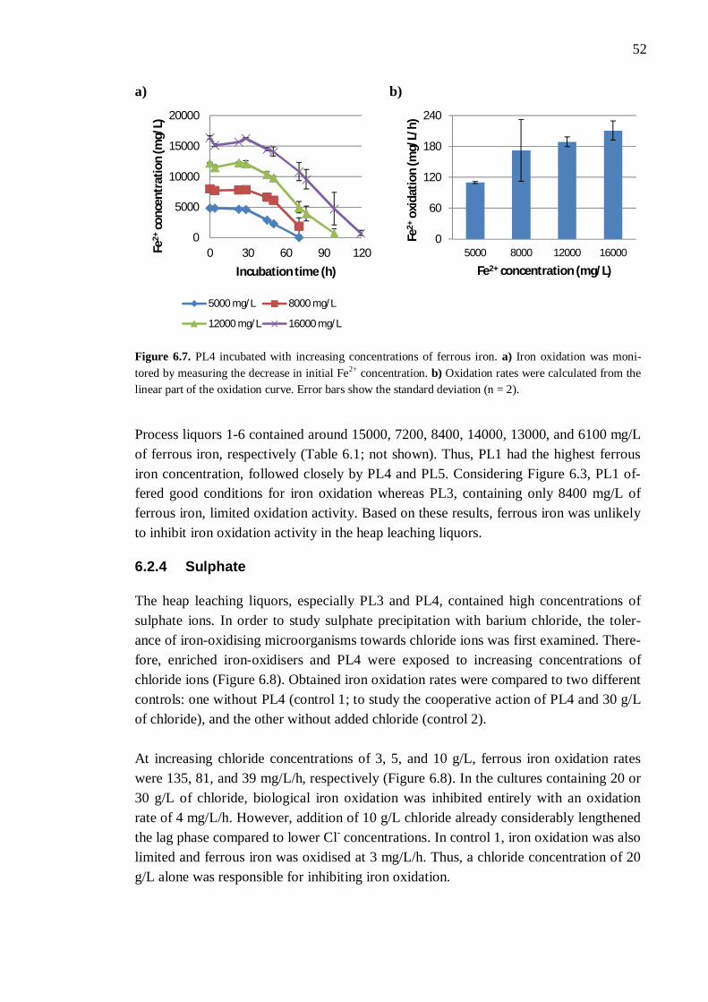
52
a) b)
Figure 6.7. PL4 incubated with increasing concentrations of ferrous iron. a) Iron oxidation was moni-tored by measuring the decrease in initial Fe2+ concentration. b) Oxidation rates were calculated from the linear part of the oxidation curve. Error bars show the standard deviation (n = 2).
Process liquors 1-6 contained around 15000, 7200, 8400, 14000, 13000, and 6100 mg/L of ferrous iron, respectively (Table 6.1; not shown). Thus, PL1 had the highest ferrous iron concentration, followed closely by PL4 and PL5. Considering Figure 6.3, PL1 of-fered good conditions for iron oxidation whereas PL3, containing only 8400 mg/L of ferrous iron, limited oxidation activity. Based on these results, ferrous iron was unlikely to inhibit iron oxidation activity in the heap leaching liquors.
6.2.4 Sulphate
The heap leaching liquors, especially PL3 and PL4, contained high concentrations of sulphate ions. In order to study sulphate precipitation with barium chloride, the toler-ance of iron-oxidising microorganisms towards chloride ions was first examined. There-fore, enriched iron-oxidisers and PL4 were exposed to increasing concentrations of chloride ions (Figure 6.8). Obtained iron oxidation rates were compared to two different controls: one without PL4 (control 1; to study the cooperative action of PL4 and 30 g/L of chloride), and the other without added chloride (control 2). At increasing chloride concentrations of 3, 5, and 10 g/L, ferrous iron oxidation rates were 135, 81, and 39 mg/L/h, respectively (Figure 6.8). In the cultures containing 20 or 30 g/L of chloride, biological iron oxidation was inhibited entirely with an oxidation rate of 4 mg/L/h. However, addition of 10 g/L chloride already considerably lengthened the lag phase compared to lower Cl- concentrations. In control 1, iron oxidation was also limited and ferrous iron was oxidised at 3 mg/L/h. Thus, a chloride concentration of 20 g/L alone was responsible for inhibiting iron oxidation.
0
5000
10000
15000
20000
0 30 60 90 120Fe2+
conc
entr
atio
n (m
g/L)
Incubation time (h)
5000 mg/L 8000 mg/L
12000 mg/L 16000 mg/L
0
60
120
180
240
5000 8000 12000 16000
Fe2+
oxid
atio
n (m
g/L/
h)
Fe2+ concentration (mg/L)
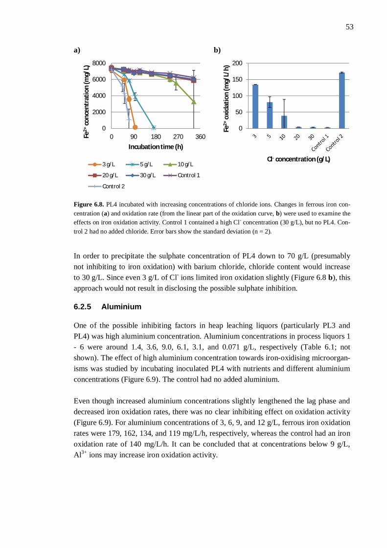
53
a) b)
Figure 6.8. PL4 incubated with increasing concentrations of chloride ions. Changes in ferrous iron con-centration (a) and oxidation rate (from the linear part of the oxidation curve, b) were used to examine the effects on iron oxidation activity. Control 1 contained a high Cl- concentration (30 g/L), but no PL4. Con-trol 2 had no added chloride. Error bars show the standard deviation (n = 2).
In order to precipitate the sulphate concentration of PL4 down to 70 g/L (presumably not inhibiting to iron oxidation) with barium chloride, chloride content would increase to 30 g/L. Since even 3 g/L of Cl- ions limited iron oxidation slightly (Figure 6.8 b), this approach would not result in disclosing the possible sulphate inhibition.
6.2.5 Aluminium
One of the possible inhibiting factors in heap leaching liquors (particularly PL3 and PL4) was high aluminium concentration. Aluminium concentrations in process liquors 1 - 6 were around 1.4, 3.6, 9.0, 6.1, 3.1, and 0.071 g/L, respectively (Table 6.1; not shown). The effect of high aluminium concentration towards iron-oxidising microorgan-isms was studied by incubating inoculated PL4 with nutrients and different aluminium concentrations (Figure 6.9). The control had no added aluminium. Even though increased aluminium concentrations slightly lengthened the lag phase and decreased iron oxidation rates, there was no clear inhibiting effect on oxidation activity (Figure 6.9). For aluminium concentrations of 3, 6, 9, and 12 g/L, ferrous iron oxidation rates were 179, 162, 134, and 119 mg/L/h, respectively, whereas the control had an iron oxidation rate of 140 mg/L/h. It can be concluded that at concentrations below 9 g/L, Al3+ ions may increase iron oxidation activity.
0
2000
4000
6000
8000
0 90 180 270 360Fe2+
conc
entr
atio
n (m
g/L)
Incubation time (h)
3 g/L 5 g/L 10 g/L
20 g/L 30 g/L Control 1
Control 2
0
50
100
150
200
Fe2+
oxid
atio
n (m
g/L/
h)
Cl- concentration (g/L)
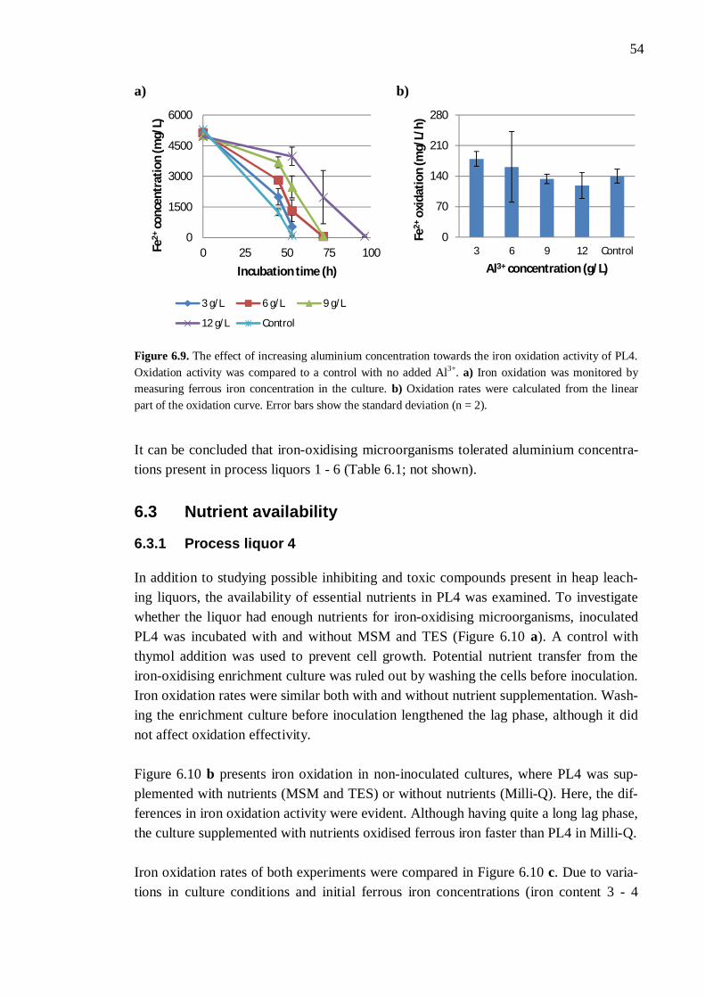
54
a) b)
Figure 6.9. The effect of increasing aluminium concentration towards the iron oxidation activity of PL4. Oxidation activity was compared to a control with no added Al3+. a) Iron oxidation was monitored by measuring ferrous iron concentration in the culture. b) Oxidation rates were calculated from the linear part of the oxidation curve. Error bars show the standard deviation (n = 2).
It can be concluded that iron-oxidising microorganisms tolerated aluminium concentra-tions present in process liquors 1 - 6 (Table 6.1; not shown).
6.3 Nutrient availability
6.3.1 Process liquor 4
In addition to studying possible inhibiting and toxic compounds present in heap leach-ing liquors, the availability of essential nutrients in PL4 was examined. To investigate whether the liquor had enough nutrients for iron-oxidising microorganisms, inoculated PL4 was incubated with and without MSM and TES (Figure 6.10 a). A control with thymol addition was used to prevent cell growth. Potential nutrient transfer from the iron-oxidising enrichment culture was ruled out by washing the cells before inoculation. Iron oxidation rates were similar both with and without nutrient supplementation. Wash-ing the enrichment culture before inoculation lengthened the lag phase, although it did not affect oxidation effectivity. Figure 6.10 b presents iron oxidation in non-inoculated cultures, where PL4 was sup-plemented with nutrients (MSM and TES) or without nutrients (Milli-Q). Here, the dif-ferences in iron oxidation activity were evident. Although having quite a long lag phase, the culture supplemented with nutrients oxidised ferrous iron faster than PL4 in Milli-Q. Iron oxidation rates of both experiments were compared in Figure 6.10 c. Due to varia-tions in culture conditions and initial ferrous iron concentrations (iron content 3 - 4
0
1500
3000
4500
6000
0 25 50 75 100Fe2+
conc
entr
atio
n (m
g/L)
Incubation time (h)
3 g/L 6 g/L 9 g/L
12 g/L Control
0
70
140
210
280
3 6 9 12 Control
Fe2+
oxid
atio
n (m
g/L/
h)
Al3+ concentration (g/L)
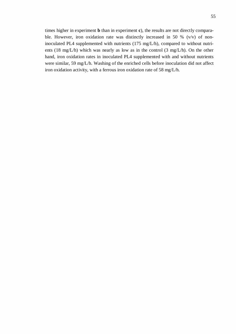
55
times higher in experiment b than in experiment c), the results are not directly compara-ble. However, iron oxidation rate was distinctly increased in 50 % (v/v) of non-inoculated PL4 supplemented with nutrients (175 mg/L/h), compared to without nutri-ents (18 mg/L/h) which was nearly as low as in the control (3 mg/L/h). On the other hand, iron oxidation rates in inoculated PL4 supplemented with and without nutrients were similar, 59 mg/L/h. Washing of the enriched cells before inoculation did not affect iron oxidation activity, with a ferrous iron oxidation rate of 58 mg/L/h.
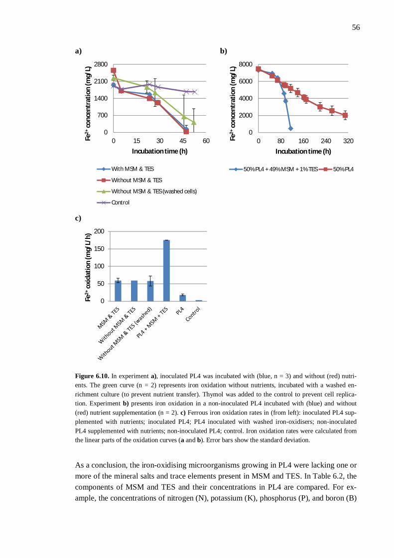
56
a) b)
c)
Figure 6.10. In experiment a), inoculated PL4 was incubated with (blue, n = 3) and without (red) nutri-ents. The green curve (n = 2) represents iron oxidation without nutrients, incubated with a washed en-richment culture (to prevent nutrient transfer). Thymol was added to the control to prevent cell replica-tion. Experiment b) presents iron oxidation in a non-inoculated PL4 incubated with (blue) and without (red) nutrient supplementation (n = 2). c) Ferrous iron oxidation rates in (from left): inoculated PL4 sup-plemented with nutrients; inoculated PL4; PL4 inoculated with washed iron-oxidisers; non-inoculated PL4 supplemented with nutrients; non-inoculated PL4; control. Iron oxidation rates were calculated from the linear parts of the oxidation curves (a and b). Error bars show the standard deviation.
As a conclusion, the iron-oxidising microorganisms growing in PL4 were lacking one or more of the mineral salts and trace elements present in MSM and TES. In Table 6.2, the components of MSM and TES and their concentrations in PL4 are compared. For ex-ample, the concentrations of nitrogen (N), potassium (K), phosphorus (P), and boron (B)
0
700
1400
2100
2800
0 15 30 45 60Fe2+
conc
entr
atio
n (m
g/L)
Incubation time (h)
With MSM & TES
Without MSM & TES
Without MSM & TES (washed cells)
Control
0
2000
4000
6000
8000
0 80 160 240 320Fe2+
conc
entr
atio
n (m
g/L)
Incubation time (h)
50% PL4 + 49% MSM + 1% TES 50% PL4
0
50
100
150
200
Fe2+
oxid
atio
n (m
g/L/
h)
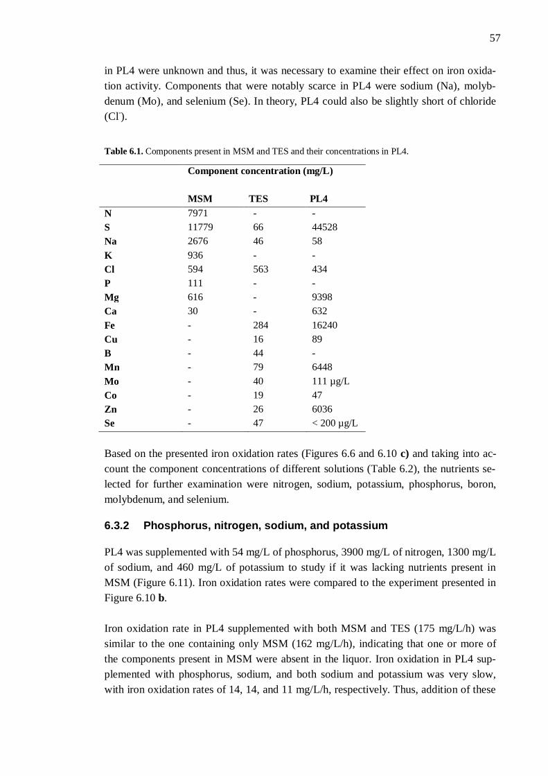
57
in PL4 were unknown and thus, it was necessary to examine their effect on iron oxida-tion activity. Components that were notably scarce in PL4 were sodium (Na), molyb-denum (Mo), and selenium (Se). In theory, PL4 could also be slightly short of chloride (Cl-).
Table 6.1. Components present in MSM and TES and their concentrations in PL4.
Component concentration (mg/L)
MSM TES PL4 N 7971 - - S 11779 66 44528 Na 2676 46 58 K 936 - - Cl 594 563 434 P 111 - - Mg 616 - 9398 Ca 30 - 632 Fe - 284 16240 Cu - 16 89 B - 44 - Mn - 79 6448 Mo - 40 111 µg/L Co - 19 47 Zn - 26 6036 Se - 47 < 200 µg/L Based on the presented iron oxidation rates (Figures 6.6 and 6.10 c) and taking into ac-count the component concentrations of different solutions (Table 6.2), the nutrients se-lected for further examination were nitrogen, sodium, potassium, phosphorus, boron, molybdenum, and selenium.
6.3.2 Phosphorus, nitrogen, sodium, and potassium
PL4 was supplemented with 54 mg/L of phosphorus, 3900 mg/L of nitrogen, 1300 mg/L of sodium, and 460 mg/L of potassium to study if it was lacking nutrients present in MSM (Figure 6.11). Iron oxidation rates were compared to the experiment presented in Figure 6.10 b. Iron oxidation rate in PL4 supplemented with both MSM and TES (175 mg/L/h) was similar to the one containing only MSM (162 mg/L/h), indicating that one or more of the components present in MSM were absent in the liquor. Iron oxidation in PL4 sup-plemented with phosphorus, sodium, and both sodium and potassium was very slow, with iron oxidation rates of 14, 14, and 11 mg/L/h, respectively. Thus, addition of these
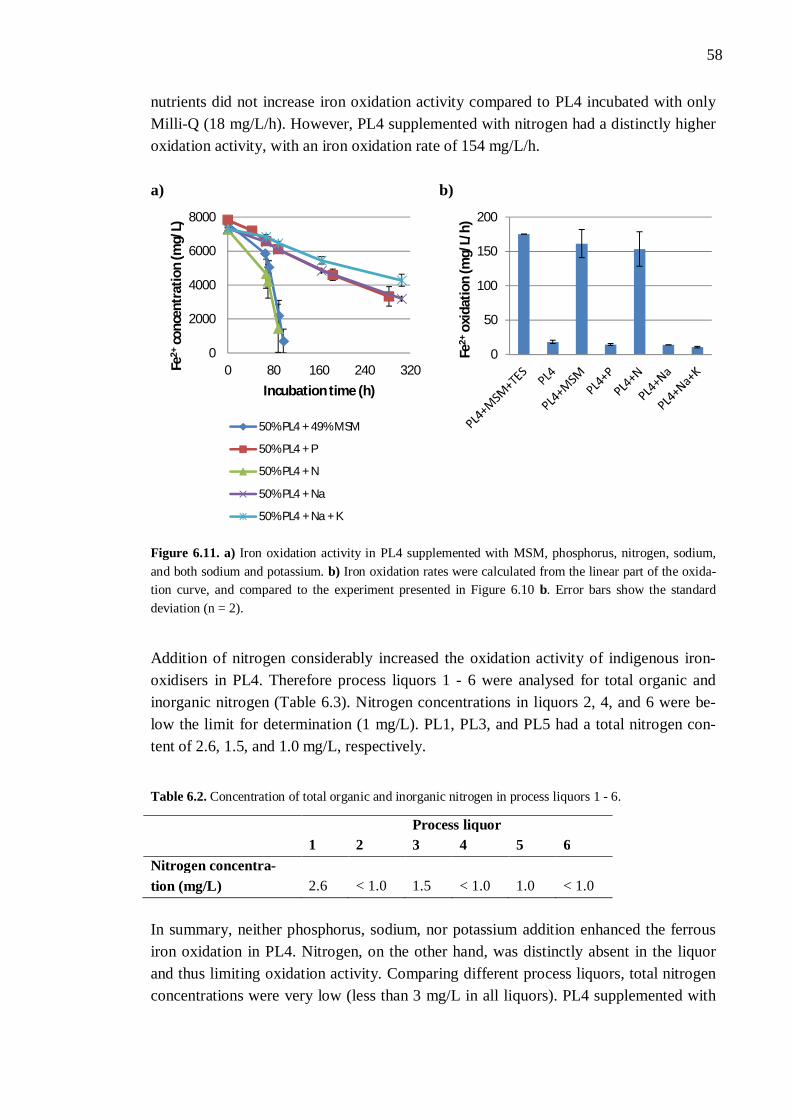
58
nutrients did not increase iron oxidation activity compared to PL4 incubated with only Milli-Q (18 mg/L/h). However, PL4 supplemented with nitrogen had a distinctly higher oxidation activity, with an iron oxidation rate of 154 mg/L/h. a) b)
Figure 6.11. a) Iron oxidation activity in PL4 supplemented with MSM, phosphorus, nitrogen, sodium, and both sodium and potassium. b) Iron oxidation rates were calculated from the linear part of the oxida-tion curve, and compared to the experiment presented in Figure 6.10 b. Error bars show the standard deviation (n = 2).
Addition of nitrogen considerably increased the oxidation activity of indigenous iron-oxidisers in PL4. Therefore process liquors 1 - 6 were analysed for total organic and inorganic nitrogen (Table 6.3). Nitrogen concentrations in liquors 2, 4, and 6 were be-low the limit for determination (1 mg/L). PL1, PL3, and PL5 had a total nitrogen con-tent of 2.6, 1.5, and 1.0 mg/L, respectively.
Table 6.2. Concentration of total organic and inorganic nitrogen in process liquors 1 - 6.
Process liquor 1 2 3 4 5 6 Nitrogen concentra-tion (mg/L) 2.6 < 1.0 1.5 < 1.0 1.0 < 1.0 In summary, neither phosphorus, sodium, nor potassium addition enhanced the ferrous iron oxidation in PL4. Nitrogen, on the other hand, was distinctly absent in the liquor and thus limiting oxidation activity. Comparing different process liquors, total nitrogen concentrations were very low (less than 3 mg/L in all liquors). PL4 supplemented with
0
2000
4000
6000
8000
0 80 160 240 320Fe2+
conc
entr
atio
n (m
g/L)
Incubation time (h)
50% PL4 + 49% MSM
50% PL4 + P
50% PL4 + N
50% PL4 + Na
50% PL4 + Na + K
0
50
100
150
200
Fe2+
oxid
atio
n (m
g/L/
h)
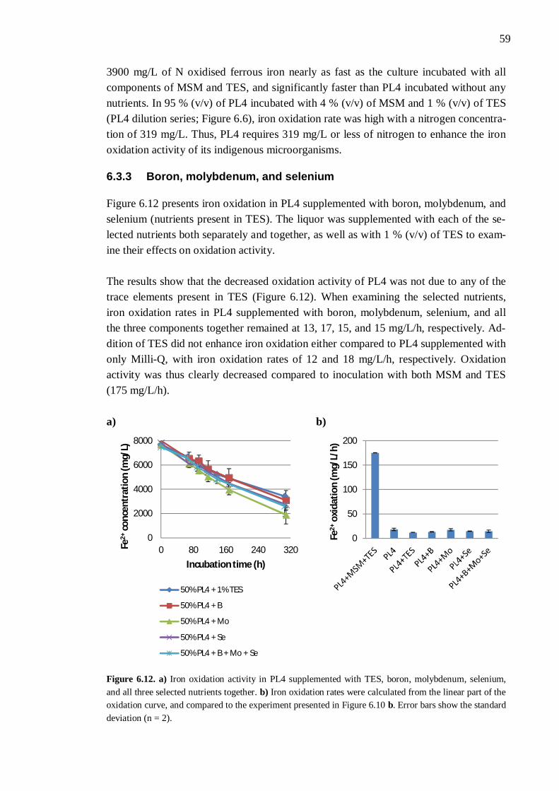
59
3900 mg/L of N oxidised ferrous iron nearly as fast as the culture incubated with all components of MSM and TES, and significantly faster than PL4 incubated without any nutrients. In 95 % (v/v) of PL4 incubated with 4 % (v/v) of MSM and 1 % (v/v) of TES (PL4 dilution series; Figure 6.6), iron oxidation rate was high with a nitrogen concentra-tion of 319 mg/L. Thus, PL4 requires 319 mg/L or less of nitrogen to enhance the iron oxidation activity of its indigenous microorganisms.
6.3.3 Boron, molybdenum, and selenium
Figure 6.12 presents iron oxidation in PL4 supplemented with boron, molybdenum, and selenium (nutrients present in TES). The liquor was supplemented with each of the se-lected nutrients both separately and together, as well as with 1 % (v/v) of TES to exam-ine their effects on oxidation activity. The results show that the decreased oxidation activity of PL4 was not due to any of the trace elements present in TES (Figure 6.12). When examining the selected nutrients, iron oxidation rates in PL4 supplemented with boron, molybdenum, selenium, and all the three components together remained at 13, 17, 15, and 15 mg/L/h, respectively. Ad-dition of TES did not enhance iron oxidation either compared to PL4 supplemented with only Milli-Q, with iron oxidation rates of 12 and 18 mg/L/h, respectively. Oxidation activity was thus clearly decreased compared to inoculation with both MSM and TES (175 mg/L/h). a) b)
Figure 6.12. a) Iron oxidation activity in PL4 supplemented with TES, boron, molybdenum, selenium, and all three selected nutrients together. b) Iron oxidation rates were calculated from the linear part of the oxidation curve, and compared to the experiment presented in Figure 6.10 b. Error bars show the standard deviation (n = 2).
0
2000
4000
6000
8000
0 80 160 240 320Fe2+
conc
entr
atio
n (m
g/L)
Incubation time (h)
50% PL4 + 1% TES
50% PL4 + B
50% PL4 + Mo
50% PL4 + Se
50% PL4 + B + Mo + Se
0
50
100
150
200
Fe2+
oxid
atio
n (m
g/L/
h)
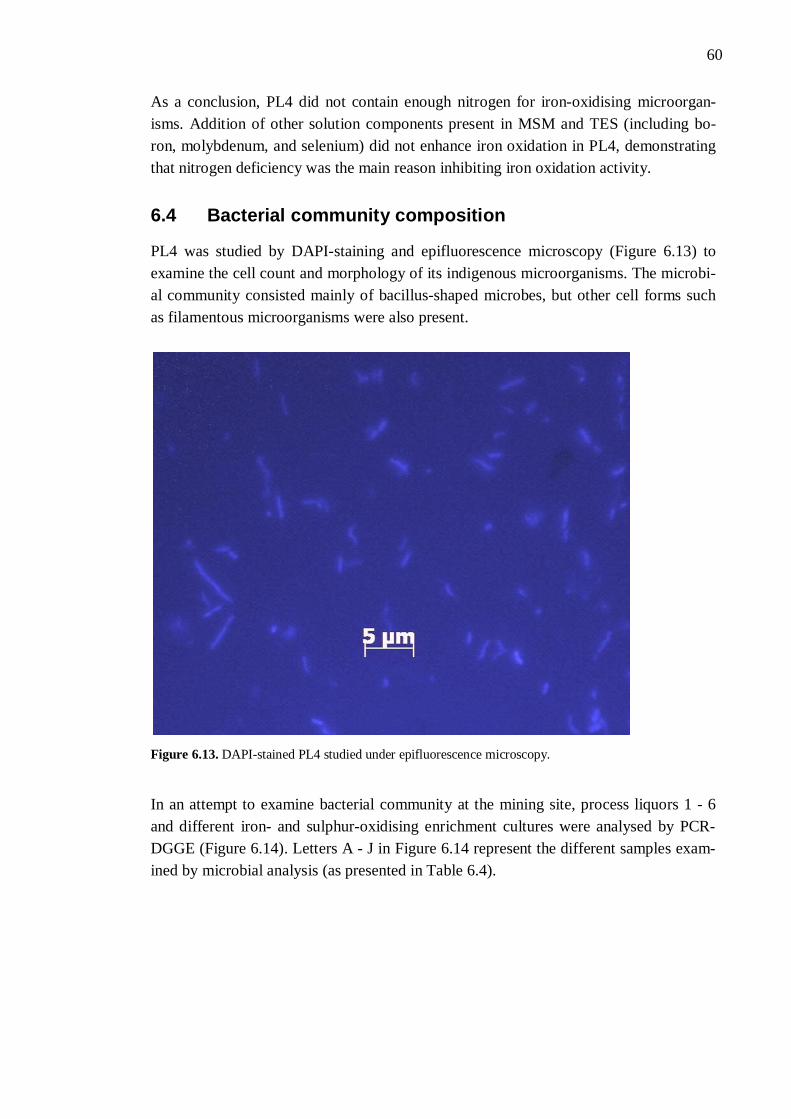
60
As a conclusion, PL4 did not contain enough nitrogen for iron-oxidising microorgan-isms. Addition of other solution components present in MSM and TES (including bo-ron, molybdenum, and selenium) did not enhance iron oxidation in PL4, demonstrating that nitrogen deficiency was the main reason inhibiting iron oxidation activity.
6.4 Bacterial community composition
PL4 was studied by DAPI-staining and epifluorescence microscopy (Figure 6.13) to examine the cell count and morphology of its indigenous microorganisms. The microbi-al community consisted mainly of bacillus-shaped microbes, but other cell forms such as filamentous microorganisms were also present.
Figure 6.13. DAPI-stained PL4 studied under epifluorescence microscopy.
In an attempt to examine bacterial community at the mining site, process liquors 1 - 6 and different iron- and sulphur-oxidising enrichment cultures were analysed by PCR-DGGE (Figure 6.14). Letters A - J in Figure 6.14 represent the different samples exam-ined by microbial analysis (as presented in Table 6.4).
61
Table 6.3. Samples analysed by PCR-DGGE and the letters representing them in Figure 6.14.
Letter in Figure 6.14 Sample A Process liquor 4 B Sulphur-oxidising enrichment culture C Iron-oxidising enrichment culture 1 D Iron-oxidising enrichment culture 2 E Process liquor 1 F Process liquor 2 G Process liquor 3 H Process liquor 5 I Process liquor 6 J Iron-oxidising enrichment culture 3 The mine site samples had a diverse bacterial community (Figure 6.14). Comparing process liquors 1 - 6, PL6 (I) had the distinctly lowest microbial diversity. Process liq-uors 1 - 5 on the other hand consisted of highly similar bacterial communities. Although iron- and sulphur-oxidising enrichment cultures were originally enriched from PL4 (A) associated with several types of bacteria, several weeks of subculturing significantly decreased their microbial diversity. Thus, the oldest iron-oxidising enrichment culture (C) contained only one distinguishable DNA fragment whereas the more recent enrich-ment cultures (D and J) had several.
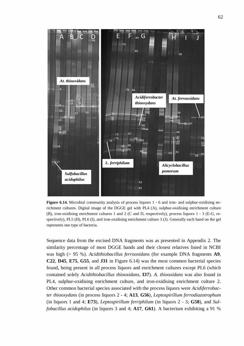
62
Figure 6.14. Microbial community analysis of process liquors 1 - 6 and iron- and sulphur-oxidising en-richment cultures. Digital image of the DGGE gel with PL4 (A), sulphur-oxidising enrichment culture (B), iron-oxidising enrichment cultures 1 and 2 (C and D, respectively), process liquors 1 - 3 (E-G, re-spectively), PL5 (H), PL6 (I), and iron-oxidising enrichment culture 3 (J). Generally each band on the gel represents one type of bacteria.
Sequence data from the excised DNA fragments was as presented in Appendix 2. The similarity percentage of most DGGE bands and their closest relatives listed in NCBI was high (> 95 %). Acidithiobacillus ferrooxidans (for example DNA fragments A9, C22, D45, E75, G55, and J31 in Figure 6.14) was the most common bacterial species found, being present in all process liquors and enrichment cultures except PL6 (which contained solely Acidithiobacillus thiooxidans, I37). A. thiooxidans was also found in PL4, sulphur-oxidising enrichment culture, and iron-oxidising enrichment culture 2. Other common bacterial species associated with the process liquors were Acidiferrobac-ter thiooxydans (in process liquors 2 - 4; A13, G56), Leptospirillum ferrodiazotrophum (in liquors 1 and 4; E73), Leptospirillum ferriphilum (in liquors 2 - 3; G58), and Sul-fobacillus acidophilus (in liquors 3 and 4; A17, G61). A bacterium exhibiting a 91 %
L. ferriphilum
At. ferrooxidans Acidiferrobacter thiooxydans
At. thiooxidans
Sulfobacillus acidophilus
Alicyclobacillus pomorum
63
identity with L. ferriphilum was also present in PL1. Less common bacterial species were Sulfobacillus benefaciens present in PL3 (G62), and Alicyclobacillus pomorum found in PL5 (H47). Some strains remained unrecognised, for instance bacteria similar to Acidiphilium spe-cies in iron-oxidising enrichment cultures 2 (91 %) and 3 (100 %), and strains similar to Leptospirillum species in PL4 (94 - 99 %), PL5 (88 %), and iron-oxidising enrichment culture 3 (99 %). Many of the excised DNA fragments undoubtedly originated from overlapping sequences.
64
7 DISCUSSION
Based on DAPI-staining and epifluorescence microscopy, process liquors 1 - 6 con-tained relatively high cell amounts. With cell concentrations ranging between 2.9 · 106 and 8.3 · 106 cells/mL, none of the liquors distinctly differed from the others concerning cell replication. Halinen et al. (2012) studied the microbial community of a low grade nickel ore bioheap and found similar bacterial cell numbers of 105 - 107 cells/mL. How-ever, when inoculating different liquors with an iron-oxidising enrichment culture, PL3 and PL4 had 51 and 61 % lower oxidation rates than the other process liquors, respec-tively. A similar experiment with sulphur-oxidising microorganisms showed that PL1 and PL4 offered the least favourable environment for sulphur oxidation. Taking into account that pH measurement was used to monitor sulphur oxidation, the initial pH val-ues of the process liquors may have affected the results. For instance, the pH value of PL6 stopped decreasing quite early (indicating that sulphur oxidation had ceased), but due to the liquor’s low initial (1.6) and final (1.0) pH, the activity of sulphur-oxidising microorganisms in that specific liquor was difficult to predict. It has however been re-ported that for At. thiooxidans, the only bacterium present in PL6, the lowest pH after growth on sulphur compounds is 0.5 - 0.8 (Kelly & Wood 2000). The bacterium has also been shown to oxidise sulphur optimally at pH 1.5 - 2.0 and be completely inhibit-ed at pH 0.5 (Plumb et al. 2008). This indicates that sulphur oxidation in PL6 was pos-sibly inhibited by something other than pH. It was however clear that PL4 inhibited both iron and sulphur oxidation more than other process liquors. The work presented in this thesis focused on finding the factor/factors in PL4 that were inhibiting the oxidation processes. A dilution series performed in nutrient solution showed that with 40 - 95 % (v/v) of PL4, ferrous iron oxidation rate gradually increased and became steady at around 160 mg/L/h (similar to the control flask). However, in 100 % (v/v) of PL4 the oxidation rate drastically decreased by 83 %. Two possible reasons for low oxidation activity were 1) inhibitory ion or metal concentrations, and/or 2) nu-trient insufficiency.
7.1 Physicochemical parameters affecting biooxidation
7.1.1 Substrate appearance and dosage
In the present work, substrate concentrations for iron- and sulphur-oxidising enrichment cultures were 5.6 g/L of Fe2+ and 10 g/L of S0, respectively. Several microorganisms, such as At. ferrooxidans, are able to utilise both substrates simultaneously (Chen et al.
65
2011); however, the enrichment of heap microorganisms growing on both Fe2+ and S0 and its effect on the enriched microbial community was not explored in the present study. It has been demonstrated that iron-oxidising bacteria often solubilise metals faster than sulphur-oxidisers, although sulphur-oxidising microorganisms may achieve higher metal leaching percentages due to their higher acid generation capacity (Chan et al. 2003). In the cultures growing on Fe2+, a 24-hour oxidation lag phase was usually de-tected (with a slight Fe2+ decrease due to chemical hydrolysis) before a sharp decrease in Fe2+ concentration. The lag phase was 1 day shorter than detected by Chen et al. (2008) in similar culture conditions. Complete oxidation was achieved during 2 to 3 days. The threshold Fe2+ concentration (after which no further oxidation occurred) in the present study was found to be around 1 % of the initial Fe2+ concentration. This finding is comparable to a study by Özkaya et al. (2007), which demonstrated that the threshold concentration linearly increases with increasing Fe2+ concentrations. Chen & Lin (2001) found that the activity of sulphur-oxidisers was enhanced with in-creasing S0 concentrations until above 0.5 % (w/v; 5 g/L), after which the substrate con-centration became inhibiting for sulphur oxidation. In 2004a Chen & Lin demonstrated that while increasing the concentration from 0.5 to 5 g/L of S0, acidification and SO4
2- production increased but were not improved after 3 g/L of S0. A later study by Chen & Lin (2010) optimised pH reduction and SO4
2- production rates and concluded that in the range of 0.1 - 1.0 % (w/v; 1 - 10 g/L) S0, sulphur oxidation was enhanced with increas-ing substrate dosage. As sulphur is insoluble, bacteria adsorb and grow on its surface and thus, sulphur is oxidised. Therefore, increase in sulphur concentration (increase in available surface area) is favourable for oxidation and consequently, for metal solubili-sation (Chen & Lin 2001). However, Liu et al. (2008) studied the effect of quite high S0 concentrations (0.5, 1, 2, 3, and 5 %; 5, 10, 20, 30, and 50 g/L) on heavy metal remobi-lisation by indigenous sulphur-oxidisers and concluded that a substrate concentration of 20 g/L was indicated to be the best for bacterial activity under the experimental condi-tions (28 °C, 180 rpm). These different studies indicate that an optimal S0 concentration for sulphur oxidation is difficult to predict and is largely dependent on the experimental conditions. It may however be deduced that the S0 concentration used in the present study (10 g/L) should have been adequate for the sulphur-oxidising microorganisms. A study by Suzuki et al. (1999) demonstrated that the type of sulphur is important, and found that sulphur dissolved in DMSO (dimethyl sulphoxide) made the microbial cells more responsive to various effects. In this thesis, S0 was added as non-dissolved pow-der. S0 oxidation is also important for producing acidic conditions, and Bouchez et al. (2006) demonstrated that a low S0 concentration (0.2 %; 2 g/L) might not be enough to decrease the solution pH. As the culture pH in sulphur-oxidising experiments presented in this thesis decreased efficiently, the S0 in the cultures was not too low. Chen et al. (2003a) found that the specific surface area available for bacterial adsorption does not necessarily depend on the particle size, but rather on the number of micropores in the
66
sulphur particles. The porosity of S0 has also been demonstrated to impact the formation of passivating S0 layer on the mineral surface (Córdoba et al. 2008b).
7.1.2 Inhibitory anions, cations, and metals
PL4 (along with PL3, which also inhibited iron oxidation) contained higher Al, As, Cd, Cl, Co, Cr, Cu, Li, Mg, Ni, Sc, SO4
2-, Th, Tl, V, Zn, and Zr concentrations than the oth-er process liquors. Although ferrous iron concentration in PL4 was not higher than in other liquors, the effect of increasing Fe2+ concentrations on iron oxidation was interest-ing owing to the fact that bioleaching environments often contain high iron concentra-tions. In the present work, Fe2+ concentrations of 5 - 16 g/L did not inhibit iron oxida-tion; in fact, increasing iron concentrations enhanced iron oxidation rates. Özkaya et al. (2007) studied the effect of different Fe2+ concentrations and found that 2 g/L of Fe2+ resulted in the highest specific iron oxidation rate, and at high concentrations the ion inhibited its own oxidation. These results are inconsistent with the work presented in this thesis. One possible reason is that the resistance towards Fe2+ ions is highly de-pendent on the microbial strains present in the bioleaching systems. Nemati & Harrison (2000) showed that the growth of Acidianus brierley was inhited at Fe2+ concentrations higher than 7.5 kg/m3 (7.5 g/L; non-growing cells were still able to utilise the substrate), whereas the growth of At. ferrooxidans was not affected by Fe2+ concentrations lower than 30 kg/m3 (30 g/L) and its iron oxidation rate was distinctly higher than that of Acidianus brierley. Metal leaching is usually improved by increasing Fe2+, and subse-quently increasing Fe3+, concentrations (Ahonen & Tuovinen 1995; Chen et al. 2008; Córdoba et al. 2008b). However, it has been shown that a high concentration of Fe3+ ions also creates instability in the bioleaching system and thus, favours precipitation and consequently mineral passivation (Córdoba et al. 2008c). At a higher pH (2.5) than stud-ied in this thesis, ferric oxide precipitation occurred and inhibited bacterial activity through adsorption on the cells (Meruane & Vargas 2003). Therefore at pH higher than 2.5, low Fe2+ concentrations are preferred. However, as dissolved Fe3+ concentrations were not measured in the present work, the possible effects of ferric iron cannot be fur-ther discussed. Due to its extremely high concentration in PL4, sulphate inhibition was discussed. Harahuc et al. (2000) selectively inhibited the oxidation of either Fe2+ or S0 by At. fer-rooxidans at pH 3 and found that at a concentration of 0.2 M (20 g/L), sulphate did not inhibit iron or sulphur oxidation. At higher concentrations sulphate had little effect on iron oxidation, but the activity of sulphur-oxidising microorganisms was distinctly de-creased, possibly due to changes in osmotic pressure. Several sulphate removal methods have been tested, such as barium carbonate precipitation (only if SO4
2- is associated with calcium; Hlabela et al. 2007) and pH-adjusted precipitation as jarosite or gypsum (Nurmi et al. 2010).
67
In the present study, the toxicity of chloride ions to iron-oxidising microorganisms was studied as a preliminary test before sulphate toxicity experiment. However, Cl- ions in-hibited iron oxidation at such low concentrations (slight inhibition at 3 g/L, total inhibi-tion at 20 g/L) that the sulphate experiment could not be performed as planned. Micro-organisms involved in bioleaching are often sensitive to Cl- ions (Sarcheshmehpour et al. 2009). A concentration of 5 g/L NaCl (3 g/L Cl-) has been found not to inhibit iron or sulphur oxidation (Vestola et al. 2010), although Harahuc et al. (2000) demonstrated that even 10 mM of chloride (0.4 g/L) inhibited iron oxidation. Sulphur oxidation on the other hand was prevented only at 0.4 M (14 g/L). These results indicate that iron-oxidisers are more affected by high chloride concentrations than sulphur-oxidisers, alt-hough with sulphate inhibition the results were contrary (Harahuc et al. 2000). In an experiment performed by Deveci et al. (2008), extreme thermophiles were efficient in ore bioleaching in the range of 10 - 40 g/L of Cl- whereas mesophiles (mostly present in bioheaps) were inhibited by salinity. This might explain the sensitivity of iron-oxidisers towards Cl- ions presented in this thesis. Mesophilic bioleaching microorganisms have however been found to resist higher Cl- concentrations (8 - 10 g/L) than moderate ther-mophiles (2 g/L) (Deveci 2002). Addition of high NaCl concentrations, as in this thesis, rapidly increases culture pH and consequently accelerates microbial death (Gómez et al. 1999). The presence of certain ions such as Na+ may also enhance the production of harmful ferric precipitates (Deveci 2002). In the future, it would be reasonable to study the inhibitory effect of SO4
2- towards the indigenous microorganisms present in Talvivaara bioheaps. Aluminium inhibition was selected for further studies due to its trivalent nature. Al3+ concentrations up to 12 g/L were not toxic to iron-oxidising microorganisms; however, increased Al3+ concentrations lengthened the oxidation lag phase and slightly decreased oxidation rates. It was also demonstrated that below 9 g/L, Al3+ may enhance iron oxi-dation. Gómez et al. (1999) studied the effect of Al on the bacterial growth of Sulfolo-bus rivotincti and found that at a non-toxic concentration, Al increased cell growth. The presence of Al has also been proven to overcome fluoride (F) toxicity in low-grade cop-per ores through complexation (Sicupira et al. 2011; Veloso et al. 2012). As PL4 had high concentrations of several ions and metals, complexation might explain why low Al3+ concentrations had a positive effect on iron oxidation.
7.1.3 Nutrient sufficiency
In terms of nutrient inadequacy, major nutrients nitrogen, potassium, and phosphorus, as well as minor nutrient boron required further investigation due to their unknown con-centrations in PL4. Minor nutrients sodium, molybdenum, and selenium were also ex-amined due to their very low concentrations in the process liquor. The experiments pre-sented in this thesis showed that K, P, B, Na, Mo, and Se concentrations in PL4 were adequate to support microbial growth. Harahuc et al. (2000) demonstrated that a low phosphate (PO4
3-) concentration (10 mM; 0.3 g/L of phosphorus) was enough to achieve
68
maximal iron and sulphur oxidation in an At. ferrooxidans culture. At 50 mM (2 g/L of P), PO4
3- decreased Fe2+ oxidation but increased S0 oxidation activity. At even higher concentration (100 mM; 3 g/L of P), the inhibitory effect of PO4
3- on iron oxidation was significant. Although the PO4
3- content of PL4 was not studied in the present study, the nutrient’s inhibitory effect even at 50 mM suggests that supplementing the bioheap leaching process with PO4
3- probably wouldn’t enhance the process. A limiting inorgan-ic phosphate concentration for the activity of At. thiooxidans has been shown to be 6 mg/L (Zheng & Zhou 2011). Gómez et al. (1999) studied the effect of Mo on the bacterial growth of Sulfolobus rivot-incti and found that at a non-toxic concentration, Mo distinctly decreased cell growth. Trace metal solution (containing Na, Ca, Fe, Mn, Zn, Co, Mo, and Cu) and potassium chloride (KCl) have proved to be negative parameters and therefore inhibiting to iron oxidation by At. ferrooxidans at high concentrations (Abdel-Fattah & Abdel-Fattah 2002). These results indicate that trace elements should be added to bioleaching pro-cesses with care. As potassium has been shown to have a positive effect on copper ore bioleaching (Sarcheshmehpour et al. 2009), the inhibitory effect of KCl was most defi-nitely due to Cl- ions. However, trace metal solution at low levels enhanced oxidation rates (Abdel-Fattah & Abdel-Fattah 2002). Nitrogen supplementation to PL4 distinctly increased iron oxidation rates in an experi-ment presented in this study, and based on PL4 dilution series the process liquor re-quired 319 mg/L or less of N. Sarcheshmehpour et al. (2009) studied the nutrient con-tent of four low-grade copper ore samples and found that concentrations of N, P, K, Ca, Mg, and S were higher in an acidic environment (pH 1.8) than at natural pH. For in-stance N concentrations at natural pH were in the range of 1.05 - 1.15 mg/L, whereas at pH 1.8 the concentrations were between 1.75 and 2.6 mg/L. This demonstrated higher solubility of the nutrients under acidic conditions. In comparison, process liquors PL1 - PL6 studied in the present work contained N concentrations in the range of < 1.0 - 2.6 mg/L, which, according to Sarcheshmehpour et al. (2009), were not adequate to support bacterial growth. It should however be noted that in freshly sampled process liquors, N concentrations might be higher. As other process liquors providing better biooxidation conditions than PL4 also contained very low N concentrations, it may be hypothesised that in addition to the low nutrient content in the process liquor, other factors might also be affecting the oxidation activity in PL4. The effect of different ions should be taken into account especially since iron oxidation in PL3 containing high SO4
2- and metal concentrations was also limited. It has been shown that the form of nitrogen has a large impact on the growth of bi-oleaching microorganisms. Examining different growth media, Sarcheshmehpour et al. found that (NH4)2SO4 and (NH4)2HPO4 enhanced Fe2+ oxidation, whereas nitrate (NO3
-) had a negative effect on bacterial activity. A study by Harahuc et al. (2000) reached
69
similar results, indicating that increased NO3- concentrations totally inhibited iron oxi-
dation by At. ferrooxidans. Sulphur-oxidising microorganisms proved to be more re-sistant to high NO3
- concentrations than iron-oxidisers; however, Suzuki et al. (1999) demonstrated that sulphur oxidation by At. thiooxidans was almost totally inhibited by NO3
- at 100 mM (6 g/L). The nutrient was also found to be more inhibitory at low pH (2.3) than at more alkaline conditions (pH 3; Suzuki et al. 1999), probably due to its increased solubility. Nitrogen should therefore be supplemented to the bioheap leach process as ammonium (NH4
+). Even though NH4+ may react with Fe3+ to produce jaro-
site precipitates (d’Hugues et al. 2002), its increased availability for microbial growth enhances the overall process. For example, decreasing the NH4
+ concentration in nutri-ent medium from 1000 to 270 mg/L significantly decreased bacterial growth and sub-strate oxidation in a pyrite bioleaching process (d’Hugues et al. 2008). Different micro-organisms were also differently affected by changing NH4
+ concentrations.
7.2 Microorganisms present in heap bioleaching
Experiments performed with different process liquors showed that iron- and sulphur-oxidising microorganisms were present in all mining waters, and that they could be en-riched. Iron- and sulphur-oxidising enrichment cultures originating from PL4 lost a lot of their initial microbial diversity after weeks of subculturing. Iron-oxidisers initially belonged to Acidithiobacillus (At. ferrooxidans, At. thiooxidans), Acidiphilium, and Leptospirillum species, but over time At. ferrooxidans become dominant. Halinen et al. (2012) studied the microbial community of a low grade nickel ore bioheap over a period of three years in Talvivaara, Finland, and found a similar pattern. In addition, Johnson and Hallberg (2007) reported that in a media containing ferrous sulphate (as in the pre-sent work), At. ferrooxidans rather than Leptospirillum spp. is enriched due to the bacte-rium’s faster growth on Fe2+. Sulphur-oxidising enrichment culture consisted solely of At. ferrooxidans and At. thiooxidans. Johnson & Hallberg (2007) reported that at 30 °C, sulphur-based media enriches At. thiooxidans, whereas at higher temperatures (45 °C) At. caldus is enriched. As the sulphur-oxidising enrichment culture was incubated at 27 °C, enrichment of At. thiooxidans was expected. In addition to PL4, At. ferrooxidans was present in all process liquors except PL6. The iron- and sulphur-oxidising bacterium is often present in bioheaps, as studied by De-mergasso et al. (2005), Goebel & Stackebrandt (1994), Halinen et al. (2012), and Ha-linen et al. (2009a). At high heap temperatures, Leptospirillum species might become dominating over At. ferrooxidans (Bowei et al. 2009; Halinen et al. 2009b). At. thiooxi-dans was the only bacterial species found in the very acidic (pH 1.6) PL6, and the bacte-rium has been found to have very limited bioleaching capabilities as a pure culture (Akcil et al. 2007). At. caldus, Acidiferrobacter thiooxydans, Leptospirillum species (L. ferrodiazotrophum, L. ferriphilum), Sulfobacillus species (S. acidophilus, S. bene-faciens), and Alicyclobacillus pomorum were also found in different process liquors. At.
70
caldus has, for example, been extensively found in a sulphur-incubated sludge bioleach-ing process at a pH range from 1 to 2, indicating that At. thiooxidans may not be the key microorganism in sulphur oxidation (Bouchez et al. 2006). Similar microbial bioheap communities have been reported in several studies (Bowei et al. 2009; Goebel and Stackebrandt 1994; Halinen et al. 2012; Halinen et al. 2009a; He et al. 2008). Zhou et al. (2010) studied a low pH (1.8), high SO4
2- (52 g/L), high Fe (82 g/L), and high arse-nide (21 g/L) AMD sample and found that it was dominated by At. ferrooxidans and L. ferrooxidans. Demergasso et al. (2005) found variation in dominant species during dif-ferent heap operation periods, indicating that the microbial community in Talvivaara leaching process might be different if sampled at several time intervals. Despite the demanding growth conditions (low pH, high SO4
2- and Fe2+ concentrations), the microbial community in PL4 was diverse. Several studies have shown that mixed cultures (two or more different bacterial cultures) are more efficient at leaching than pure cultures (Akcil et al. 2007; Fu et al. 2008; Xia et al. 2008; Zhang et al. 2008). However, Fu et al. (2008) found that mixed cultures of moderate thermophiles (L. fer-riphilum and At. caldus) were more effective in leaching than pure or mixed cultures of mesophilic At. ferrooxidans. Effective mineral degradation requires active sulphur oxi-dation in addition to iron-oxidising activities (Akcil et al. 2007; Fu et al. 2008; Zhang et al. 2008). This is due to the fact that iron-oxidisers, such as At. ferrooxidans, are respon-sible for Fe3+ production and maintaining high redox potential, whereas sulphur oxidis-ing microorganisms (i.e. At. thiooxidans) eliminate the passivation of elemental sulphur on mineral surface and provide the process with protons (Xia et al. 2008). Recent stud-ies (Zhou et al. 2009; Zhu et al. 2011) have demonstrated that the formation of jarosite or PbSO4 on mineral surface is the main passivation component, not elemental sulphur. The formation of leaching products is nonetheless accelerated and thus, microbial ac-tivity and leaching rates are increased (Zhu et al. 2011). Since several iron- and sulphur-oxidising microorganisms were indigenously present in Talvivaara heap leaching pro-cess, it can be hypothesised that by modifying their growth conditions, the bioleaching process could be improved.
71
8 CONCLUSIONS
The following conclusions can be drawn from this study:
1. Iron oxidation in PL4 is nitrogen limited and thus, nitrogen supplementation to PL4 will increase iron oxidation rates and leaching efficiencies in bioheaps. Based on PL4 dilution series, the liquor needs 319 mg/L or less of N.
2. In the presence of 40 - 95 % (v/v) PL4 in MSM, high-rate iron oxidation is
achieved. 3. Ferrous iron concentrations up to 16 g/L are not inhibitory for the iron-oxidising
bacteria in PL4. 4. Chloride inhibits iron oxidation at concentrations higher than 5 g/L. 5. Increasing Al3+ concentrations decrease iron oxidation rates slightly, but even 12
g/L of Al3+ does not inhibit iron oxidation. 6. PL3 and PL4 limit iron oxidation more than other process liquors. 7. PL1, PL4, and PL6 limit sulphur oxidation the most, while PL3 and PL5 offer
good conditions for sulphur oxidation. 8. Most of the process liquors have diverse bacterial communities. PL4 consists
mainly of At. ferrooxidans, At. thiooxidans, Acidiferrobacter thiooxydans, L. ferrodiazotrophum, and Sulfobacillus acidophilus. Other process liquors also contain L. ferriphilum, Sulfobacillus benefaciens, and Alicyclobacillus po-morum. PL6 contains only At. thiooxidans.
9. Iron-enriched cultures initially consist of At. ferrooxidans, At. thiooxidans, and
Acidiphilium and Leptospirillum species. Over time, At. ferrooxidans becomes the main species present. At. ferrooxidans and At. thiooxidans become enriched when the medium is supplemented with elemental sulphur.
72
REFERENCES Abdel-Fattah YR and Abdel-Fattah WR. 2002. Evaluation and optimization of culture conditions influencing ferrous-ion oxidation rate of Thiobacillus ferrooxidans ATCC 23270 through statistically designed experiments. Arab Journal of Biotechnology 5: 173-184. Aelion CM, Davis HT, Flora JRV, Kirtland BC, Amidon MB. 2009. Application of en-capsulation (pH-sensitive polymer and phosphate buffer macrocapsules): a novel ap-proach to remediation of acidic ground water. Environmental Pollution 157: 186-193. Ahemad M and Malik A. 2011. Bioaccumulation of heavy metals by zinc resistant bac-teria isolated from agricultural soils irrigated with wastewater. Bacteriology Journal 2: 12-21. Ahmadi A, Schaffie M, Petersen J, Schippers A, Ranjbar M. 2011. Conventional and electrochemical bioleaching of chalcopyrite concentrates by moderately thermophilic bacteria at high pulp density. Hydrometallurgy 106: 84-92. Ahonen L and Tuovinen OH. 1995. Bacterial leaching of complex sulfide ore samples in bench-scale column reactors. Hydrometallurgy 37: 1-21. Akcil A, Ciftci H, Deveci H. 2007. Role and contribution of pure and mixed cultures of mesophiles in bioleaching of a pyritic chalcopyrite concentrate. Minerals Engineering 20: 310-318. Akpor OB and Muchie M. 2010. Remediation of heavy metals in drinking water and wastewater treatment systems: processes and applications. International Journal of the Physical Sciences 5: 1807-1817. Alyüz B and Veli S. 2009. Kinetics and equilibrium studies for the removal of nickel and zinc from aqueous solutions by ion exchange resins. Journal of Hazardous Materials 167: 482-488. APHA. 3500-Fe. Washington, DC 1992. 1100 p. Ballester A, Gonzalez F, Blazquez ML, Gómez C, Mier JL. 1992. The use of catalytic ions in bioleaching. Hydrometallurgy 29: 151-160. Belzile N, Chen YW, Cai MF, Li Y. 2004. A review on pyrrhotite oxidation. Journal of Geochemical Exploration 84: 65-76.
73
Bhatti TM, Bigham JM, Vuorinen A, Tuovinen OH. 2012a. Chemical and bacterial leaching of metals from black schist sulfide minerals in shake flasks. International Jour-nal of Mineral Processing 110-111: 25-29. Bhatti TM, Vuorinen A, Tuovinen OH. 2012b. Dissolution of non-sulfide phases during the chemical and bacterial leaching of a sulfidic black schist. Hydrometallurgy 117-118: 32-35. Borole AP, O’Neill H, Tsouris C, Cesar S. 2008. A microbial fuel cell operating at low pH using the acidophile Acidiphilium cryptum. Biotechnology Letters 30: 1367-1372. Bosecker K. 1997. Bioleaching: metal solubilization by microorganisms. FEMS Micro-biology Reviews 20: 591-604. Bouchez T, Jacob P, d’Hugues P, Durand A. 2006. Acidophilic microbial communities catalyzing sludge bioleaching monitored by fluorescent in situ hybridization. Antonie van Leeuwenhoek 89: 435-442. Bowei C, Xingyu L, Wenyan L, Jiankang W. 2009. Application of clone library analysis and real-time PCR for comparison of microbial communities in a low-grade copper sul-fide ore bioheap leachate. Journal of Industrial Microbiology and Biotechnology 36: 1409-1416. Brandl H. 2001. Microbial leaching of metals. In: Biotechnology 10. Rehm HJ, editor. Wiley-VCH, Weinheim, Germany. 191-224 p. Brar SK, Verma M, Surampalli RY, Misra K, Tyagi RD, Meunier N, Blais JF. 2006. Bioremediation of hazardous wastes – a review. Practice Periodical of Hazardous, Tox-ic, and Radioactive Waste Management 10, Special Issue Bioremediation: 59-72. Breed AW and Hansford GS. 1999. Effect of pH on ferrous-iron oxidation kinetics of Leptospirillum ferrooxidans in continuous culture. Biochemical Engineering Journal 3: 193-201. Brierley CL. 2008. How will biomining be applied in future? The Transactions of Non-ferrous Metals Society of China 18: 1302-1310. Bruins MR, Kapil S, Oehme FW. 2000. Microbial resistance to metals in the environ-ment. Ecotoxicology and Environmental Safety 45: 198-207. Bryan CG, Davis-Belmar CS, van Wyk N, Fraser MK, Dew D, Rautenbach GF, Harri-son STL. 2012. The effect of CO2 availability on the growth, iron oxidation and CO2-
74
fixation rates of pure cultures of Leptospirillum ferriphilum and Acidithiobacillus fer-rooxidans. Biotechnology and Bioengineering. 11 p. DOI 10.1002/bit.24453 Cameron RA, Lastra R, Mortazavi S, Bedard PL, Morin L, Gould WD, Kennedy KJ. 2009. Bioleaching of a low-grade ultramafic nickel sulphide ore in stirred-tank reactors at elevated pH. Hydrometallurgy 97: 213-220. Chan LC, Gu XY, Wong JWC. 2003. Comparison of bioleaching of heavy metals from sewage sludge using iron- and sulfur-oxidizing bacteria. Advances in Environmental Research 7: 603-607. Chen J, Li SP, Gu XY. 2008. Effects of energy source concentration on bioleaching of heavy metals from undigested sewage sludge by using iron-oxidizing bacterium. Prac-tice Periodical of Hazardous, Toxic, and Radioactive Waste Management 12 (Special Issue: Metals Removal): 165-169. Chen P, Yan L, Leng F, Nan W, Yue X, Zheng Y, Feng N, Li H. 2011. Bioleaching of realgar by Acidithiobacillus ferrooxidans using ferrous iron and elemental sulfur as the sole and mixed energy sources. Bioresource Technology 102: 3260-3267. Chen SY, Chiu YC, Chang PL, Lin JG. 2003a. Assessment of recoverable forms of sul-fur particles used in bioleaching of contaminated sediments. Water Research 37: 450-458. Chen SY and Lin JG. 2001. Effect of substrate concentration on bioleaching of metal-contaminated sediment. Journal of Hazardous Material B82: 77-89. Chen SY and Lin JG. 2004a. Bioleaching of heavy metals from contaminated sediment by indigenous sulfur-oxidizing bacteria in an air-lift bioreactor: effects of sulfur concen-tration. Water Research 38: 3205-3214. Chen SY and Lin JG. 2004b. Bioleaching of heavy metals from livestock sludge by in-digenous sulfur-oxidizing bacteria: effects of sludge solids concentration. Chemosphere 54: 283-289. Chen SY, Lin JG, Lee CY. 2003b. Effects of ferric ion on bioleaching of heavy metals from contaminated sediment. Water Science and Technology 48: 151-158. Chen SY and Lin PL. 2010. Optimization of operating parameters for the metal bi-oleaching process of contaminated soil. Separation and Purification Technology 71: 178-185.
75
Clifford D. 1986. Removing dissolved inorganic contaminants from water. Environmen-tal Science and Technology 20: 1072-1080. Córdoba EM, Muñoz JA, Blázquez ML, Gonzáles F, Ballester A. 2008a. Leaching of chalcopyrite with ferric ion. Part I: General aspects. Hydrometallurgy 93: 81-87. Córdoba EM, Muñoz JA, Blázquez ML, Gonzáles F, Ballester A. 2008b. Leaching of chalcopyrite with ferric ion. Part II: Effect of redox potential. Hydrometallurgy 93: 88-96. Córdoba EM, Muñoz JA, Blázquez ML, González F, Ballester A. 2008c. Leaching of chalcopyrite with ferric ion. Part IV: The role of redox potential in the presence of mes-ophilic and thermophilic bacteria. Hydrometallurgy 93: 106-115. Couillard D, Chartier M, Mercier G. 1994. Major factors influencing bacterial leaching of heavy metals (Cu and Zn) from anaerobic sludge. Environmental Pollution 85: 175-184. Demergasso CS, Galleguillos P PA, Escudero G LV, Zepeda A VJ, Castillo D, Casa-mayor EO. 2005. Molecular characterization of microbial populations in a low-grade copper ore bioleaching test heap. Hydrometallurgy 80: 241-253. Deveci H. 2002. Effect of salinity on the oxidative activity of acidophilic bacteria dur-ing bioleaching of a complex Zn/Pb sulphide ore. The European Journal of Mineral Pro-cessing and Environmental Protection 2: 141-150. Deveci H, Akcil A, Alp I. 2004. Bioleaching of complex zinc sulphides using meso-philic and thermophilic bacteria: comparative importance of pH and iron. Hydrometal-lurgy 73: 293-303. Deveci H, Jordan MA, Powell N, Alp I. 2008. Effect of salinity and acidity on bioleach-ing activity of mesophilic and extremely thermophilic bacteria. Transactions of Nonfer-rous Metals Society of China 18: 714-721. Dew DW, Muhlbauer R, van Buuren C. 1999. Bioleaching of copper sulphide concen-trates with mesophiles and thermophiles. In: Alta Copper 99. Brisbane, Australia. d’Hugues P, Foucher S, Gallé-Cavalloni P, Morin D. 2002. Continuous bioleaching of chalcopyrite using a novel extremely thermophilic mixed culture. International Journal of Mineral Processing 66: 107-119.
76
d’Hugues P, Joulian C, Spolaore P, Michel C, Garrido F, Morin D. 2008. Continuous bioleaching of a pyrite concentrate in stirred reactors: Population dynamics and exopol-ysaccharide production vs. bioleaching performance. Hydrometallurgy 94: 34-41. Dopson M, Baker-Austin C, Hind A, Bowman JP, Bond PL. 2004. Characterization of Ferroplasma isolates and Ferroplasma acidarmanus sp. nov., extreme acidophiles from acid mine drainage and industrial bioleaching environments. Applied and Environmen-tal Microbiology 70: 2079-2088. Dopson M, Baker-Austin C, Koppineedi PR, Bond PL. 2003. Growth in sulfidic mineral environments: metal resistance mechanisms in acidophilic micro-organisms. Microbiol-ogy 149: 1959-1970. Dopson M, Halinen AK, Rahunen N, Özkaya B, Sahinkaya E, Kaksonen AH, Lindström EB, Puhakka JA. 2007. Mineral and iron oxidation at low temperatures by pure and mixed cultures of acidophilic microorganisms. Biotechnology and Bioengi-neering 97: 1205-1215. Dopson M and Johnson DB. 2012. Biodiversity, metabolism and applications of aci-dophilic sulfur-metabolizing microorganisms. Environmental Microbiology. 12 p. doi:10.1111/j.1462-2920.2012.02749.x Dopson M and Lindström EB. 1999. Potential role of Thiobacillus caldus in arsenopy-rite bioleaching. Applied and Environmental Microbiology 65: 36-40. Dorado AD, Solé M, Lao C, Alfonso P, Gamisans X. 2012. Effect of pH and Fe(III) ions on chalcopyrite bioleaching by an adapted consortium from biogas sweetening. Minerals Engineering 39: 36-38. du Plessis CA, Batty JD, Dew DW. 2007. Commercial applications of thermophile bi-oleaching. In: Biomining. Rawlings DE and Johnson DB, editors. Springer-Verlag, Ber-lin Heidelberg. 57-80 p. Feng D, Aldrich C, Tan H. 2000. Treatment of acid mine water by use of heavy metal precipitation and ion exchange. Minerals Engineering 13: 623-642. Fu B, Zhou H, Zhang R, Qiu G. 2008. Bioleaching of chalcopyrite by pure and mixed cultures of Acidithiobacillus spp. and Leptospirillum ferriphilum. International Biodete-rioration & Biodegradation 62: 109-115.
77
Fuchs T, Huber H, Burggraf S, Stetter KO. 1996. 16S rDNA-based phylogeny of the archaeal order Sulfolobales and reclassification of Desulfurolobus ambivalens as Acidi-anus ambivalens comb. nov. Systematic and Applied Microbiology 19: 56-60. Gericke M and Govender Y. 2011. Bioleaching strategies for the treatment of nickel-copper sulphide concentrates. Minerals Engineering 24: 1106-1112. Ghorbani Y, Becker M, Mainza A, Franzidis JP, Petersen J. 2011. Large particle effects in chemical/biochemical heap leach processes – A review. Minerals Engineering 24: 1172-1184. Goebel BM and Stackebrandt E. 1994. Cultural and phylogenetic analysis of mixed mi-crobial populations found in natural and commercial bioleaching environments. Applied and Environmental Microbiology 60: 1614-1621. Gómez E, Ballester A, González F, Blázquez ML. 1999. Leaching capacity of a new extremely thermophilic microorganism, Sulfolobus rivotincti. Hydrometallurgy 52: 349-366. Gunneriusson L, Sandström Å, Holmgren A, Kuzmann E, Kovacs K, Vértes A. 2009. Jarosite inclusion of fluoride and its potential significance to bioleaching of sulphide minerals. Hydrometallurgy 96: 108-116. Gupta A, Matsui K, Lo JF, Silver S. 1999. Molecular basis for resistance to silver in Salmonella. Nature Medicine 5: 183-188. Gupta VK, Pathania D, Sharma S. 2012. Remediation of noxious chromium (VI) utiliz-ing acrylic acid grafted lignocellulosic adsorbent. Journal of Molecular Liquids: http://dx.doi.org/10.1016/j.molliq.2012.10.017 Halinen AK, Beecroft NJ, Määttä K, Nurmi P, Laukkanen K, Kaksonen AH, Riekkola-Vanhanen M, Puhakka JA. 2012. Microbial community dynamics during a demonstra-tion-scale bioheap leaching operation. Hydrometallurgy 125-126: 34-41. Halinen AK, Rahunen N, Kaksonen AH, Puhakka JA. 2009a. Heap bioleaching of a complex sulfide ore. Part I: Effect of pH on metal extraction and microbial composition in pH controlled columns. Hydrometallurgy 98: 92-100. Halinen AK, Rahunen N, Kaksonen AH, Puhakka JA. 2009b. Heap bioleaching of a complex sulfide ore: Part II. Effect of temperature on base metal extraction and bacterial compositions. Hydrometallurgy 98: 101-107.
78
Hallberg KB, Dopson M, Lindström EB. 1996. Arsenic toxicity is not due to a direct effect on the oxidation of reduced sulfur compounds by Thiobacillus caldus. FEMS Microbiology Letters 145: 409-414. Hallberg KB, González-Toril E, Johnson DB. 2010. Acidithiobacillus ferrivorans, sp. nov.; facultatively anaerobic, psychrotolerant iron-, and sulfur-oxidizing acidophiles isolated from metal mine-impacted environments. Extremophiles 14: 9-19. Hallberg KB, Hedrich S, Johnson DB. 2011. Acidiferrobacter thiooxydans, gen. Nov. Sp. Nov.; an acidophilic, thermo-tolerant, facultatively anaerobic iron- and sulfur-oxidizer of the family Ectothiorhodospiraceae. Extremophiles 15: 271-279. Harahuc L, Lizama HM, Suzuki I. 2000. Selective inhibition of the oxidation of ferrous iron or sulfur in Thiobacillus farrooxidans. Applied and Environmental Microbiology 66: 1031-1037. Harvey PI and Crundwell FK. 1996. The effect of As(III) on the growth of Thiobacillus ferrooxidans in an electrolytic cell under controlled redox potential. Minerals Engineer-ing 9: 1059-1068. Harvey TJ and Bath M. 2007. The GeoBiotics GEOCOAT® technology – progress and challenges. In: Biomining. Rawlings DE and Johnson DB, editors. Springer-Verlag, Berlin Heidelberg. 97-112 p. Hashim MA, Mukhopadhyay S, Sahu JN, Sengupta B. 2011. Remediation technologies for heavy metal contaminated groundwater. Journal of Environmental Management 92: 2355-2388. He Z, Xiao S, Xie X, Hu Y. 2008. Microbial diversity in acid mineral bioleaching sys-tems of Dongxiang copper mine and Yinshan lead-zinc mine. Extremophiles 12: 225-234. Hiraishi A, Nagashima KVP, Matsuura K, Shimada K, Takaichi S, Wakao N, Katayama Y. 1998. Phylogeny and photosynthetic features of Thiobacillus acidophilus and related acidophilic bacteria: its transfer to the genus Acidiphilium as Acidiphilium acidophilum comb. nov. International Journal of Systematic Bacteriology 48: 1389-1398. Hlabela P, Maree J, Bruinsma D. 2007. Barium carbonate process for sulphate and met-al removal from mine water. Mine Water and the Environment 26: 14-22.
79
Holmes DS and Bonnefoy V. 2007. Genetic and bioinformatic insights into iron and sulfur oxidation mechanisms of bioleaching organisms. In: Biomining. Rawlings DE and Johnson DB, editors. Springer-Verlag, Berlin Heidelberg. 281-307 p. Hoyle B and Beveridge TS. 1983. Binding of metallic ions to the outer membrane of Escherichia coli. Applied and Environmental Microbiology 46: 749-752. Ji G and Silver S. 1995. Bacterial resistance mechanism for heavy metals of environ-mental concern. Journal of Industrial Microbiology 14: 61-75. Johnson DB. 1998. Biodiversity and ecology of acidophilic microorganisms. FEMS Microbiology Ecology 27: 307-317. Johnson DB, Bacelar-Nicolau P, Okibe N, Thomas A, Hallberg KB. 2009. Ferrimicro-bium acidiphilum gen. nov., sp. nov. and Ferrithrix thermotolerans gen. nov., sp. nov.: heterotrophic, iron-oxidizing, extremely acidophilic actinobacteria. International Journal of Systematic and Evolutionary Microbiology 59: 1082-1089. Johnson DB, Ghauri MA, Said MF. 1992. Isolation and characterization of an aci-dophilic, heterotrophic bacterium capable of oxidizing ferrous iron. Applied and Envi-ronmental Microbiology 58: 1423-1428. Johnson DB and Hallberg KB. 2007. Techniques for detecting and identifying aci-dophilic mineral-oxidizing microorganisms. In: Biomining. Rawlings DE and Johnson DB, editors. Springer-Verlag, Berlin Heidelberg. 237-261 p. Kaksonen AH, Riekkola-Vanhanen ML, Puhakka JA. 2003. Optimization of metal sul-phide precipitation in fluidized-bed treatment of acidic wastewater. Water Research 37: 255-266. Karavaiko GI, Dubinina GA, Kondrat’eva TF. 2006. Lithotrophic microorganisms of the oxidative cycles of sulfur and iron. Microbiology 75: 512-545. Kelly DP and Wood AP. 2000. Reclassification of some species of Thiobacillus to the newly designated genera Acidithiobacillus gen. nov., Halothiobacillus gen. nov. and Thermithiobacillus gen. nov. International Journal of Systematic and Evolutionary Mi-crobiology 50: 511-516. Kolehmainen RE, Langwaldt JH, Puhakka JA. 2007. Natural organic matter (NOM) removal and structural changes in the bacterial community during artificial groundwater recharge with humic lake water. Water Research 41: 2715-2725.
80
Koskinen PEP, Kaksonen AH, Puhakka JA. 2006. The relationship between instability of H2 production and compositions of bacterial communities within a dark fermentation fluidized-bed bioreactor. Biotechnology and Bioengineering 97: 742-758. Krishna RR, Xu CY, Supraja C. 2001. Assessment of electrokinetic removal of heavy metals from soils by sequential extraction analysis. Journal of Hazardous Materials 84: 279-296. Leupin OX and Hug SJ. 2005. Oxidation and removal of arsenic (III) from aerated groundwater by filtration through sand and zero-valent iron. Water Research 39: 1729-1740. Liu YG, Zhou M, Zeng GM, Wang X, Li X, Fan T, Xu WH. 2008. Bioleaching of heavy metals from mine tailings by indigenous sulfur-oxidizing bacteria: Effects of sub-strate concentration. Bioresource Technology 99: 4124-4129. Lovley DR, Phillips EJP, Gorby YA, Landa ER. 1991. Microbial reduction of uranium. Nature 350: 413-416. Mahapatra NR, Ghosh S, Deb C, Banerjee PC. 2002. Resistance to cadmium and zinc in Acidiphilium symbioticum KM2 is plasmid mediated. Current Microbiology 45: 180-186. Malik AH, Khan ZM, Mahmood Q, Nasreen S, Bhatti ZA. 2009. Perspectives of low cost arsenic remediation of drinking water in Pakistan and other countries. Journal of Hazardous Materials 168: 1-12. Meruane G and Vargas T. 2003. Bacterial oxidation of ferrous iron by Acidithiobacillus ferrooxidans in the pH range 2.5-7.0 Hydrometallurgy 71: 149-158. Mier JL, Ballester A, Blazquez ML, González F, Muñoz JA. 1995. Influence of metallic ions in the bioleaching of chalcopyrite by Sulfolobus BC: experiments using pneumati-cally stirred reactors and massive samples. Minerals Engineering 8: 949-965. Mier JL, Ballester A, González F, Blazquez ML, Gómez E. 1996. Influence of metallic ions on the activity of Sulfolobus BC. Journal of Chemical Technology and Biotechnol-ogy 65: 272-280. Mishra D, Kim DJ, Ahn JG, Rhee YH. 2005. Bioleaching: a microbial process of metal recovery; a review. Metals and Materials International 11: 249-256.
81
Misra TK. 1992. Bacterial resistance to inorganic mercury salts and organomercurials. Plasmid 27: 4-16. Mohapatra PK. 2006. Textbook of environmental biotechnology. IK Internatioal Pub-lishing House Pvt Ltd, New Delhi. 664 p. Neculita CM, Zagury GJ, Bussière B. 2007. Passive treatment of acid mine drainage in bioreactors using sulfate-reducing bacteria: critical review and research needs. Journal of Environmental Quality 36: 1-16. Nemati M and Harrison STL. 2000. A comparative study on thermophilic and meso-philic biooxidation of ferrous iron. Minerals Engineering 13: 19-24. Nemati M, Harrison STL, Hansford GS, Webb C. 1998. Biological oxidation of ferrous sulphate by Thiobacillus ferrooxidans: a review on the kinetic aspects. Biochemical Engineering Journal 1: 171-190. Nies DH. 1999. Microbial heavy-metal resistance. Applied Microbiology and Biotech-nology 51: 730-750. Nies DH. 2003. Efflux-mediated heavy metal resistance in prokaryotes. FEMS Micro-biology Reviews 27: 313-339. Nieto Castillo AM, Soriano JJ, García-Delgado RA. 2008. Changes in chromium distri-bution during the electrodialytic remediation of a Cr (VI)-contaminated soil. Environ-mental Geochemistry and Health 30: 153-157. Norris PR. 2007. Acidophile diversity in mineral sulfide oxidation. In: Biomining. Rawlings DE and Johnson DB, editors. Springer-Verlag, Berlin Heidelberg. 199-216 p. Nurmi P, Özkaya B, Sasaki K, Kaksonen AH, Riekkola-Vanhanen M, Tuovinen OH, Puhakka JA. 2010. Biooxidation and precipitation for iron and sulfate removal from heap bioleaching effluent streams. Hydrometallurgy 101: 7-14. Ohmura N, Sasaki K, Matsumoto N, Saiki H. 2002. Anaerobic respiration using Fe3+, S0, and H2 in the chemolithoautotrophic bacterium Acidithiobacillus ferrooxidans. Jour-nal of Bacteriology 184: 2081-2087. Ojumu TV, Petersen J, Searby GE, Hansford GS. 2006. A review of rate equations pro-posed for microbial ferrous-iron oxidation with a view to application to heap bioleach-ing. Hydrometallurgy 83: 21-28.
82
Pakarinen J and Paatero E. 2011. Recovery of manganese from iron containing sulfate solutions by precipitation. Minerals Engineering 24: 1421-1429. Paulsen IT and Saier MH Jr. 1997. A novel family of ubiquitous heavy metal ion transport proteins. Journal of Membrane Biology 156: 99-103. Peleg M, Normand MD, Corradini MG. 2012. The Arrhenius equation revisited. Critical Reviews in Food Science and Nutrition 52: 830-851. Peng JF, Song YH, Yuan P, Cui XY, Qiu GL. 2009. The remediation of heavy metals contaminated sediment. Journal of Hazardous Materials 161: 633-640. Plumb JJ, Hawkes RB, Franzmann PD. 2007. The microbiology of moderately thermo-philic and transiently thermophilic ore heaps. In: Biomining. Rawlings DE and Johnson DB, editors. Springer-Verlag, Berlin Heidelberg. 217-235 p. Plumb JJ, Muddle R, Franzmann PD. 2008. Effect of pH on rates of iron and sulfur oxi-dation by bioleaching organisms. Minerals Engineering 21: 76-82. Ragan HA and Mast TJ. 1990. Cadmium inhalation and male reproductive toxicity. Re-views of Environmental Contamination & Toxicology 114: 1-22. Rao SM. 2007. Reverse osmosis. Resonance 12: 37-40. Ratkowsky DA, Lowry RK, McMeekin TA, Stokes AN, Chandler RE. 1983. Model for bacterial culture growth rate throughout the entire biokinetic temperature range. Journal of Bacteriology 154: 1222-1226. Rawlings DE. 2005. Characteristics and adaptability of iron- and sulfur-oxidizing mi-croorganisms used for the recovery of metals from minerals and their concentrates. Mi-crobial Cell Factories 4. 15 p. Rawlings DE. 2007. Relevance of cell physiology and genetic adaptability of biomining microorganisms to industrial processes. In: Biomining. Rawlings DE and Johnson DB, editors. Springer-Verlag, Berlin Heidelberg. 177-198 p. Rawlings DE, Dew D, du Plessis C. 2003. Biomineralization of metal-containing ores and concentrates. TRENDS in Biotechnology 21: 38-44. Rengaraj S, Kyeong-Ho Y, Seung-Hyeon M. 2001. Removal of chromium from water and wastewater by ion exchange resins. Journal of Hazardous Materials B87: 273-287.
83
Rohwerder T, Gehrke T, Kinzler K, Sand W. 2003. Bioleaching review part A: Progress in bioleaching: fundamentals and mechanisms of bacterial metal sulfide oxidation. Ap-plied Microbiology and Biotechnology 63: 239-248. Rouch DA, Lee BTD, Morby AP. 1995. Understanding cellular responses to toxic agents: A model for mechanism choice in bacterial metal resistance. Journal of Industri-al Microbiology 14: 132-141. Saier MH, Tam R, Reizer A, Reizer J. 1994. Two novel families of bacterial membrane proteins concerned with nodulation, cell division and transport. Molecular Microbiology 11: 841-847. Sand W and Gehrke T. 2006. Extracellular polymeric substances mediate bioleach-ing/biocorrosion via interfacial processes involving iron (III) ions and acidophilic bacte-ria. Research in Microbiology 157: 49-56. Sand W, Gehrke T, Jozsa PG, Schippers A. 2001. (Bio)chemistry of bacterial leaching – direct vs. indirect bioleaching. Hydrometallurgy 59: 159-175. Sarcheshmehpour Z, Lakzian A, Fotovat A, Berenji AR, Haghnia GH, Bagheri SAS. 2009. Possibility of using chemical fertilizers instead of 9K medium in bioleaching pro-cess of low-grade sulfide copper ores. Hydrometallurgy 96: 264-267. Schippers A. 2007. Microorganisms involved in bioleaching and nucleic acid-based molecular methods for their identification and quantification. In: Microbial Processing of Metal Sulfides. Donati ER and Sand W, editors. Springer-Verlag, New York. 3-33 p. Schippers A and Sand W. 1999. Bacterial leaching of metal sulfides proceeds by two indirect mechanisms via thiosulfate or via polysulfides and sulfur. Applied and Envi-ronmental Microbiology 65: 319-321. SFS 5505. 1988. Jäteveden epäorgaanisen ja orgaanisen typen määritys. Modifioitu kjeldahlmenetelmä. Suomen standardisoimisliitto (Finnish Standards Association), Hel-sinki. 4 p. SFS-EN ISO 10304-1. 2009. Water quality. Determination of dissolved anions by liquid chromatography of ions. Part 1: Determination of bromide, chloride, fluoride, nitrate, nitrite, phosphate and sulfate. Suomen standardisoimisliitto (Finnish Standards Associa-tion), Helsinki. 15 p. Shagufta CJ. 2007. Environmental biotechnology. APH Publishing Corporation, New Delhi. 280 p.
84
Sheoran AS and Sheoran V. 2006. Heavy metal removal mechanism of acid mine drain-age in wetlands: a critical review. Minerals Engineering 19: 105-116. Sicupira L, Veloso T, Reis F, Leão V. 2011. Assessing metal recovery from low-grade copper ores containing fluoride. Hydrometallurgy 109: 202-210. Suzuki I, Lee D, MacKay B, Harahuc L, Oh JK. 1999. Effect of various ions, pH, and osmotic pressure on oxidation of elemental sulfur by Thiobacillus thiooxidans. Applied and Environmental Microbiology 65: 5163-5168. Suzuki K, Wakao N, Sakurai Y, Kimura T, Sakka K, Ohmiya K. 1997. Transformation of Escherichia coli with a large plasmid of Acidiphilium multivorum AIU 301 encoding arsenic resistance. Applied and Environmental Microbiology 63: 2089-2091. Veloso TC, Sicupira LC, Rodrigues ICB, Silva LAM, Leão VA. 2012. The effects of fluoride and aluminium ions on ferrous-iron oxidation and copper sulfide bioleaching with Sulfobacillus thermosulfidooxidans. Biochemical Engineering Journal 62: 48-55. Vestola EA, Kuusenaho MK, Närhi HM, Tuovinen OH, Puhakka JA, Plumb JJ, Kakso-nen AH. 2010. Acid bioleaching of solid waste materials from copper, steel and recy-cling industries. Hydrometallurgy 103: 74-79. Wakeman KD, Erving L, Riekkola-Vanhanen ML, Puhakka JA. 2010. Silage supports sulfate reduction in the treatment of metals- and sulfate-containing waste waters. Water Research 44: 4932-4939. Watling HR. 2006. The bioleaching of sulphide minerals with emphasis on copper sul-phides - A review. Hydrometallurgy 84: 81-108. Xia LX, Liu JS, Xiao L, Zeng J, Li BM, Geng MM, Qiu GZ. 2008. Single and coopera-tive bioleaching of sphalerite by two kinds of bacteria – Acidithiobacillus ferriooxidans and Acidithiobacillus thiooxidans. Transactions of Nonferrous Metals Society of China 18: 190-195. Zhang YS, Qin WQ, Wang J, Zhen SJ, Yang CR, Zhang JW, Nai SS, Qiu GZ. 2008. Bioleaching of chalcopyrite by pure and mixed culture. Transactions of Nonferrous Metals Society of China 18: 1491-1496. Zhen S, Yan Z, Zhang Y, Wang J, Campbell M, Qin W. 2009. Column bioleaching of a low grade nickel-bearing sulfide ore containing high magnesium as olivine, chlorite and antigorite. Hydrometallurgy 96: 337-341.
85
Zheng G and Zhou L. 2011. Supplementation of inorganic phosphate enhancing the removal efficiency of tannery sludge-borne Cr through bioleaching. Water Research 45: 5295-5301. Zhou HB, Zeng WM, Yang ZF, Xie YJ, Qiu GZ. 2009. Bioleaching of chalcopyrite concentrate by a moderately thermophilic culture in a stirred tank reactor. Bioresource Technology 100: 515-520. Zhou ZJ, Yin HQ, Liu Y, Xie M, Qiu GZ, Liu XD. 2010. Diversity of microbial com-munity at acid mine drainages from Dachang metals-rich mine, China. Transactions of Nonferrous Metals Society of China 20: 1097-1103. Zhu W, Xia JL, Yang Y, Nie ZY, Zheng L, Ma CY, Chang RY, Peng AA, Tang L, Qiu GZ. 2011. Sulfur oxidation activities of pure and mixed thermophiles and sulfur specia-tion in bioleaching of chalcopyrite. Bioresource Technology 102: 3877-3882. Zhuang JM. 2009. Acidic rock drainage treatment: a review. Recent Patents on Chemi-cal Engineering 2: 238-252. Özkaya B, Sahinkaya E, Nurmi P, Kaksonen AH, Puhakka JA. 2007. Kinetics of iron oxidation by Leptospirillum ferriphilum dominated culture at pH below one. Biotech-nology and Bioengineering 97: 1121-1127.
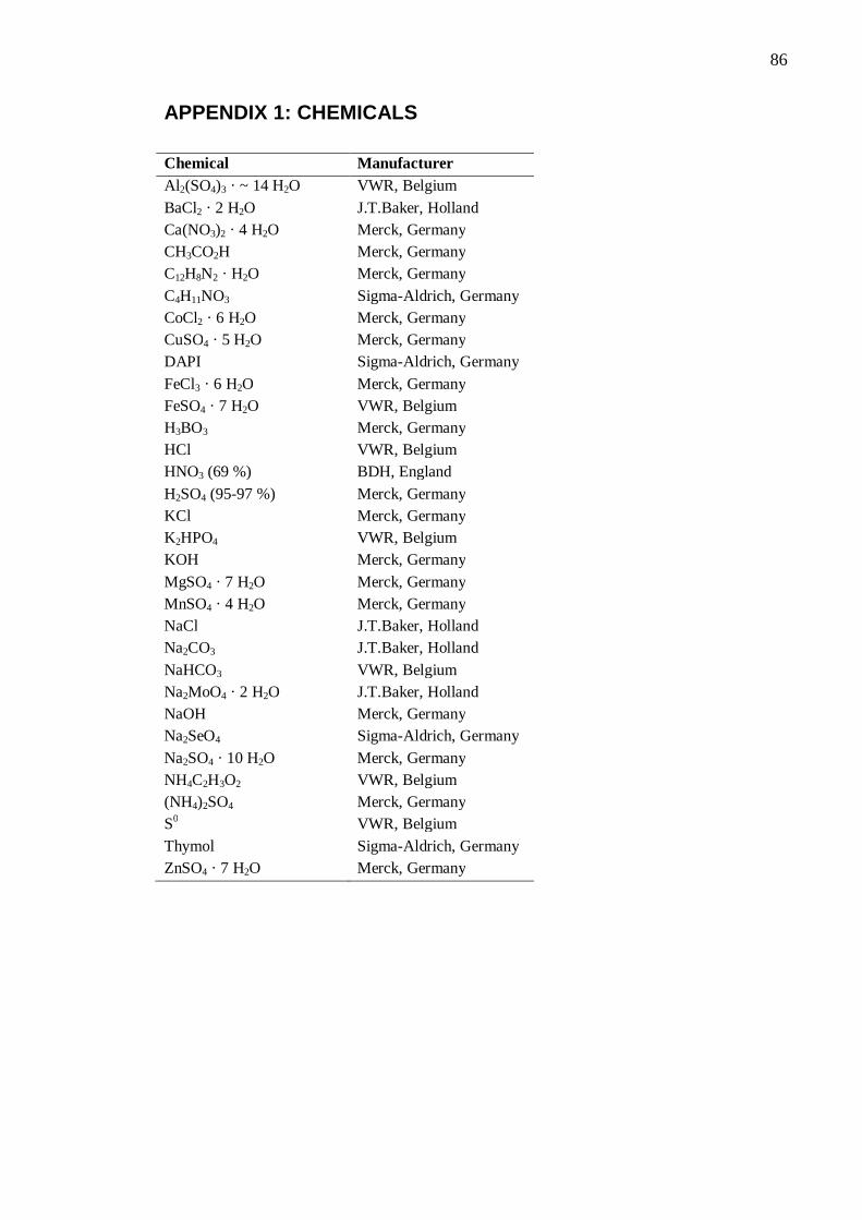
86
APPENDIX 1: CHEMICALS Chemical Manufacturer Al2(SO4)3 · ~ 14 H2O VWR, Belgium BaCl2 · 2 H2O J.T.Baker, Holland Ca(NO3)2 · 4 H2O Merck, Germany CH3CO2H Merck, Germany C12H8N2 · H2O Merck, Germany C4H11NO3 Sigma-Aldrich, Germany CoCl2 · 6 H2O Merck, Germany CuSO4 · 5 H2O Merck, Germany DAPI Sigma-Aldrich, Germany FeCl3 · 6 H2O Merck, Germany FeSO4 · 7 H2O VWR, Belgium H3BO3 Merck, Germany HCl VWR, Belgium HNO3 (69 %) BDH, England H2SO4 (95-97 %) Merck, Germany KCl Merck, Germany K2HPO4 VWR, Belgium KOH Merck, Germany MgSO4 · 7 H2O Merck, Germany MnSO4 · 4 H2O Merck, Germany NaCl J.T.Baker, Holland Na2CO3 J.T.Baker, Holland NaHCO3 VWR, Belgium Na2MoO4 · 2 H2O J.T.Baker, Holland NaOH Merck, Germany Na2SeO4 Sigma-Aldrich, Germany Na2SO4 · 10 H2O Merck, Germany NH4C2H3O2 VWR, Belgium (NH4)2SO4 Merck, Germany S0 VWR, Belgium Thymol Sigma-Aldrich, Germany ZnSO4 · 7 H2O Merck, Germany
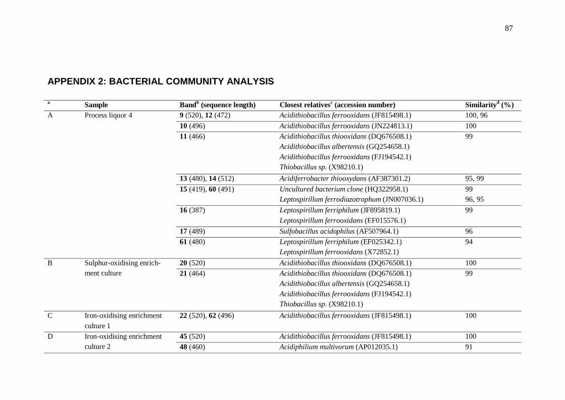
87
APPENDIX 2: BACTERIAL COMMUNITY ANALYSIS a Sample Bandb (sequence length) Closest relativesc (accession number) Similarityd (%) A Process liquor 4 9 (520), 12 (472) Acidithiobacillus ferrooxidans (JF815498.1) 100, 96
10 (496) Acidithiobacillus ferrooxidans (JN224813.1) 100 11 (466) Acidithiobacillus thiooxidans (DQ676508.1)
Acidithiobacillus albertensis (GQ254658.1) Acidithiobacillus ferrooxidans (FJ194542.1) Thiobacillus sp. (X98210.1)
99
13 (480), 14 (512) Acidiferrobacter thiooxydans (AF387301.2) 95, 99 15 (419), 60 (491) Uncultured bacterium clone (HQ322958.1)
Leptospirillum ferrodiazotrophum (JN007036.1) 99 96, 95
16 (387) Leptospirillum ferriphilum (JF895819.1) Leptospirillum ferrooxidans (EF015576.1)
99
17 (489) Sulfobacillus acidophilus (AF507964.1) 96 61 (480) Leptospirillum ferriphilum (EF025342.1)
Leptospirillum ferrooxidans (X72852.1) 94
B Sulphur-oxidising enrich-ment culture
20 (520) Acidithiobacillus thiooxidans (DQ676508.1) 100 21 (464) Acidithiobacillus thiooxidans (DQ676508.1)
Acidithiobacillus albertensis (GQ254658.1) Acidithiobacillus ferrooxidans (FJ194542.1) Thiobacillus sp. (X98210.1)
99
C Iron-oxidising enrichment culture 1
22 (520), 62 (496) Acidithiobacillus ferrooxidans (JF815498.1) 100
D Iron-oxidising enrichment culture 2
45 (520) Acidithiobacillus ferrooxidans (JF815498.1) 100 48 (460) Acidiphilium multivorum (AP012035.1) 91

88
a Sample Bandb (sequence length) Closest relativesc (accession number) Similarityd (%) Acidiphilium cryptum (FJ915152.1)
Acidiphilium organovorum (NR 025853.1)
52 (496), 53 (512) Acidithiobacillus ferrooxidans (JF815498.1) 99, 100 E Process liquor 1 72 (439) Acidithiobacillus caldus (CP002573.1)
Thiobacillus caldus (AF137369.1) Thermus sp. (EU368984.1)
99
73 (403) Uncultured Nitrospirae bacterium (GU120604.1) Leptospirillum ferrodiazotrophum (JN007036.1)
99 96
74 (451) Leptospirillum ferriphilum (DQ646517.1) 91 75 (488), 78 (473) Acidithiobacillus ferrooxidans (FJ913262.1) 99, 97 77 (462) Acidithiobacillus ferrivorans (CP002985.1)
Acidithiobacillus ferrooxidans (FN686788.1) 99
79 (452) Acidithiobacillus ferrooxidans (JN224813.1) 99 F Process liquor 2 65 (463) Acidithiobacillus ferrivorans (CP002985.1)
Acidithiobacillus ferrooxidans (FN686788.1) 99
67 (457) Leptospirillum ferrooxidans (GU168016.1) Leptospirillum ferriphilum (GU168013.1)
98
68 (417), 69 (455), 70 (493), 71 (449)
Uncultured Alicyclobacillaceae bacterium (FN870323.1) 95, 96, 96, 96
G Process liquor 3 53 (509), 54 (464), 63 (464) Acidithiobacillus ferrivorans (CP002985.1) Acidithiobacillus ferrooxidans (FN686788.1)
99
55 (462) Acidithiobacillus ferrooxidans (JN224813.1) 100 56 (441) Acidiferrobacter thiooxydans (AF387301.2) 99 57 (450) Acidithiobacillus ferrooxidans (AM502930.1) 99 58 (462) Leptospirillum ferriphilum (EF025342.1) 100 61 (461) Sulfobacillus sp. (AJ306701.1) 97
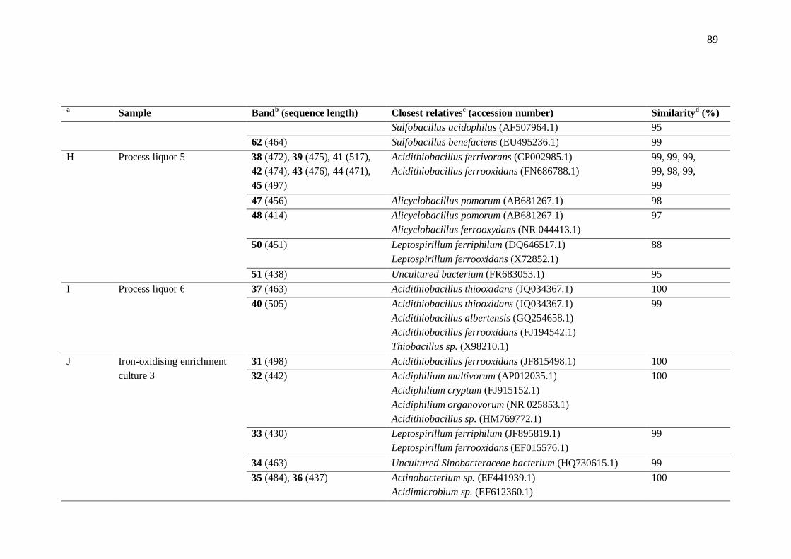
89
a Sample Bandb (sequence length) Closest relativesc (accession number) Similarityd (%) Sulfobacillus acidophilus (AF507964.1) 95
62 (464) Sulfobacillus benefaciens (EU495236.1) 99 H Process liquor 5 38 (472), 39 (475), 41 (517),
42 (474), 43 (476), 44 (471), 45 (497)
Acidithiobacillus ferrivorans (CP002985.1) Acidithiobacillus ferrooxidans (FN686788.1)
99, 99, 99, 99, 98, 99, 99
47 (456) Alicyclobacillus pomorum (AB681267.1) 98 48 (414) Alicyclobacillus pomorum (AB681267.1)
Alicyclobacillus ferrooxydans (NR 044413.1) 97
50 (451) Leptospirillum ferriphilum (DQ646517.1) Leptospirillum ferrooxidans (X72852.1)
88
51 (438) Uncultured bacterium (FR683053.1) 95 I Process liquor 6 37 (463) Acidithiobacillus thiooxidans (JQ034367.1) 100
40 (505) Acidithiobacillus thiooxidans (JQ034367.1) Acidithiobacillus albertensis (GQ254658.1) Acidithiobacillus ferrooxidans (FJ194542.1) Thiobacillus sp. (X98210.1)
99
J Iron-oxidising enrichment culture 3
31 (498) Acidithiobacillus ferrooxidans (JF815498.1) 100 32 (442) Acidiphilium multivorum (AP012035.1)
Acidiphilium cryptum (FJ915152.1) Acidiphilium organovorum (NR 025853.1) Acidithiobacillus sp. (HM769772.1)
100
33 (430) Leptospirillum ferriphilum (JF895819.1) Leptospirillum ferrooxidans (EF015576.1)
99
34 (463) Uncultured Sinobacteraceae bacterium (HQ730615.1) 99 35 (484), 36 (437) Actinobacterium sp. (EF441939.1)
Acidimicrobium sp. (EF612360.1) 100
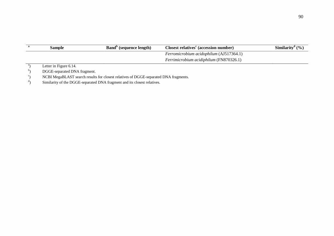
90
a Sample Bandb (sequence length) Closest relativesc (accession number) Similarityd (%) Ferromicrobium acidophilum (AJ517364.1)
Ferrimicrobium acidiphilum (FN870326.1)
a) Letter in Figure 6.14. b) DGGE-separated DNA fragment. c) NCBI MegaBLAST search results for closest relatives of DGGE-separated DNA fragments. d) Similarity of the DGGE-separated DNA fragment and its closest relatives.