Embed Size (px)
Citation preview
Case ReportSarcoidosis and Multiple Myeloma: A Case Report andLiterature Review
Joao Tiago Serra , Aurelia Martinho, Fernanda Paixao Duarte, and Fernando Aldomiro
Department of Internal Medicine II, Professor Doutor Fernando Fonseca Hospital, Amadora, Portugal
Correspondence should be addressed to Joao Tiago Serra; [email protected]
Received 30 July 2019; Accepted 14 October 2019; Published 30 October 2019
Academic Editor: Takashi Sonoki
Copyright © 2019 Joao Tiago Serra et al. )is is an open access article distributed under the Creative Commons Attribution License,which permits unrestricted use, distribution, and reproduction in any medium, provided the original work is properly cited.
)e existence of a sarcoidosis-lymphoma syndrome has been previously proposed since the relation between sarcoidosis and anincreased risk of lymphoproliferative disorders is well established. Multiple myeloma is a malignant multifocal proliferation ofclonal plasma cells within the bone marrow, and its association with sarcoidosis has been rarely described. We present aconcurrent diagnosis of sarcoidosis and multiple myeloma and make a brief analysis of the reported cases in the literature. A 65-year-old woman underwent surgery for the excision of a wrist mass that presented 3 years before. Histological analysis showedsarcoid-type epithelioid granulomas without necrosis, establishing soft tissue sarcoidosis. Further evaluation revealed markedinterstitial lung parenchyma lesions and large intrathoracic adenopathies. Bronchofibroscopy with transbronchial biopsyconfirmed lung sarcoidosis. In addition, blood analysis showed monoclonal IgG kappa gammopathy. A bone marrow biopsyconfirmed hypercellularity with 60% plasma cells and plasmocyte infiltration. )us, the diagnosis of systemic sarcoidosis andmultiple myeloma was established simultaneously. In a brief review of the literature, we identified 33 reports of cases with bothsarcoidosis and multiple myeloma.We point out the importance of a high level of suspicion for the association of sarcoidosis withmalignant haematological diseases such as multiple myeloma.
1. Introduction
Sarcoidosis is a T-cell-mediated immunological responseagainst an unknown environmental trigger in a susceptiblehost. Noncaseating granulomas, consisting of compact andcentrally organized collections of CD4+ T cells, macro-phages, and epithelioid cells, are the hallmark of sarcoidosisand the outcome of an unbalanced T-helper 1 immuneresponse [1, 2]. Multiple myeloma (MM) is a malignantmultifocal proliferation of clonal plasma cells within thebone marrow, associated with skeletal destruction, serummonoclonal gammopathy, and end-organ sequelae [3].Brincker [4] described the sarcoidosis-lymphoma syndromein 1986 for the particular association between sarcoidosisand lymphoproliferative disorders (LD); however, the as-sociation between sarcoidosis and MM has been rarely re-ported and the explanation for this relation remains unclearand controversial. We describe a case of sarcoidosis asso-ciated with MM and present a brief analysis of publishedcases in the literature.
2. Case Report
A 65-year-old Caucasian women, retired cook, was referredto our outpatient clinic due to a three-year history of apainless mass on the dorsal side of the right wrist. Hermedical background was positive for type 2 diabetes mel-litus, dyslipidaemia, bronchitis, and venous thrombosis ofthe right eye. )e wrist lesion had been growing graduallyand affected finger movement. )ere was no history of bluntor penetrating trauma.
An ultrasound, complemented with nuclear magneticresonance, identified a lesion on the dorsal plane of the rightwrist, extending to the inner face, but without vascular ortendinous invasion, measuring about 100× 60×18mm indiameter and having well-defined limits (Figure 1). Aftersurgery for lesion removal, histological analysis showed anextensive granulomatous process without necrosis, con-sisting of sarcoid-type epithelioid granulomas. )e patientalso complained of long-term intermittent nodular skinlesions on both legs, dry cough, and dyspnoea, for which she
HindawiCase Reports in HematologyVolume 2019, Article ID 4586265, 5 pageshttps://doi.org/10.1155/2019/4586265

had been previously prescribed bronchodilators with littlesymptomatic relief. Physical examination revealed painlessbilateral supraclavicular lymphadenopathies, bibasilarcoarse crackles, and nodular skin lesions scattered alongboth inferior limbs (erythema nodosum). )ere were noother palpable lymph nodes, hepatosplenomegaly, fever,night sweats, or constitutional symptoms.
Laboratory studies showed haemoglobin 9.7 g/dL (ref-erence range 11.5–16.5 g/dL), white blood cells count of4.6×109/L (reference range 4–11× 109/L), C-reactive pro-tein of 0.87mg/dL (reference range <0.30mg/dL), eryth-rocyte sedimentation rate of 83mm (reference range<20mm), serum angiotensin converting enzyme (ACE) of141.28U/L (reference range 12–68U/L), albumin of 3.66 g/dL (reference range 3.97–4.94 g/dL), and total serum proteinlevel of 8.40 g/dL (reference range 6.40–8.20 g/dL). Serumprotein electrophoresis revealed a monoclonal band, con-firmed by immunofixation to be immunoglobulin IgGkappa. Further quantification of serum immunoglobulinsshowed an elevated IgG level of 3120mg/dL (reference range70–1600mg/dL), with normal IgM and IgA, and beta-2microglobulin of 2.09mg/dL (reference range 0.67–1.31mg/dL). Mantoux assay and Bence Jones proteinuriaquantification were negative, and both renal function andserum calcium were within the normal range. )e screeningfor HIV and hepatitis B and C was negative.
A high-resolution computed tomography of the chestshowed large mediastinal and axillary adenopathies withextensive conglomerates, associated with marked and diffusepermeability of the entire pulmonary parenchyma, thick-ening of the interlobular septa, and fine bronchovascular andsubpleural micronodularity (Figure 2).
)e patient’s lung function testing revealed a mild re-strictive pattern with decreased diffusion capacity. Abronchofibroscopy with transbronchial biopsy and bron-chioalveolar lavage was performed. Respiratory tract fluid
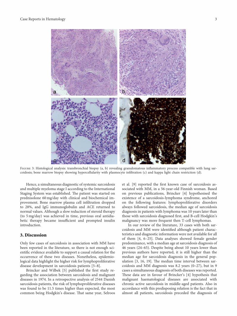
analysis showed an increased CD4/CD8 ratio of 9.02, and bothacid-fast staining (Ziehl–Neelsen coloration) and mycobacte-rial culture were negative. Bronchial biopsy histological resultsrevealed mild chronic inflammatory infiltrate with multiplenonnecrotizing sarcoid-type epithelioid granulomas and giantmultinucleated Langerhans cells (Figures 3(a) and 3(b)). )epatient also underwent a scintigraphy with gallium 67, whichdocumented pathological uptake in the lungs, axillary andinguinal regions, and several cutaneous foci on the lower limbs.Ophthalmologic evaluation was consistent with ocular sar-coidosis. For further evaluation of serum monoclonal IgGkappa gammopathy, a bone marrow aspirate and biopsy wereperformed, showing a hypercellular bone marrow with 60%plasma cells and plasmocyte infiltration with K light chainrestriction, respectively (Figures 3(c) and 3(d)).
(a)
(b) (c)
Figure 1: Magnetic resonance images in T1-weighted axial view (a), T2-weighted axial view (b), and T2-weighted sagital view (c),identifying a lesion on the dorsal plane of the right wrist, extending to the inner face but without vascular or tendinous invasion, measuringabout 100× 60×18mm in diameter, with well-defined limits.
Figure 2: Chest high-resolution computed tomography imageshowing marked and diffuse alteration of pulmonary parenchymapermeability, thickening of the interlobular septa, and fine bron-chovascular and subpleural micronodularity, associated with me-diastinal lymphadenopathy.
2 Case Reports in Hematology
Hence, a simultaneous diagnostic of systemic sarcoidosisand multiple myeloma stage I according to the InternationalStaging System was established. )e patient was started onprednisolone 60mg/day with clinical and biochemical im-provement. Bone marrow plasma cell infiltration droppedto 28%, and IgG immunoglobulin and ACE returned tonormal values. Although a slow reduction of steroid therapy(to 5mg/day) was achieved in time, previous oral antidia-betic therapy became insufficient and prompted insulinintroduction.
3. Discussion
Only few cases of sarcoidosis in association with MM havebeen reported in the literature, so there is not enough sci-entific evidence available to support a causal relation for theoccurrence of these two diseases. Nonetheless, epidemio-logical data highlight the higher risk for lymphoproliferativedisease development in sarcoidosis patients [5–8].
Brincker and Wilbek [5] published the first study re-garding the association between sarcoidosis and malignantdiseases in 1974. In a retrospective analysis of 2544 Danishsarcoidosis patients, the risk of lymphoproliferative diseaseswas found to be 11.5 times higher than expected, the mostcommon being Hodgkin’s disease. )at same year, Selroos
et al. [9] reported the first known case of sarcoidosis as-sociated with MM, in a 56-year-old Finnish woman. Basedon previous publications, Brincker [4] hypothesised theexistence of a sarcoidosis-lymphoma syndrome, anchoredon the following features: lymphoproliferative disordersalways followed sarcoidosis, the median age of sarcoidosisdiagnosis in patients with lymphoma was 10 years later thanthose with sarcoidosis diagnosed first, and B-cell Hodgkin’smalignancy was more frequent then T-cell lymphomas.
In our review of the literature, 33 cases with both sar-coidosis and MM were identified although patient charac-teristics and diagnostic information were not available for allof them [4, 6–25]. Data analyses showed female genderpredominance, with a median age at sarcoidosis diagnosis of46 years (24–65). Despite being about 10 years lower thanprevious authors have reported, it is still higher than themedian age for sarcoidosis diagnosis in the general pop-ulation [3, 16, 19]. )e median time interval between sar-coidosis and MM diagnosis was 8.2 years (0–27), but in 9cases a simultaneous diagnosis of both diseases was reported.)ese data are in favour of Brincker’s [4] hypothesis thatmalignant haematological diseases are associated withchronic active sarcoidosis in middle-aged patients. Also inaccordance with this predisposing relation is the fact that inalmost all patients, sarcoidosis preceded the diagnosis of
Figure 3: Histological analysis: transbronchial biopsy (a, b) revealing granulomatous inflammatory process compatible with lung sar-coidosis; bone marrow biopsy showing hypercellularity with plasmocyte infiltration (c) and kappa light chain restriction (d).
Case Reports in Hematology 3
MM. With respect to MM, IgG was the most commonmonoclonal protein reported, followed by IgA and IgM.
)e explanation for the relationship between sarcoidosisand lymphoproliferative disorders, particularly MM, is stillcontroversial and unknown. One possible link betweensarcoidosis and MM is the polyclonal hyper-gammaglobulinemia induced by sarcoidosis chronic in-flammatory response. )is continuous state of stimulationresults in prolonged B cell and plasma cell half-life, which inaddition to immune system imbalance in sarcoidosis pa-tients, predisposes for genetic mutations and might lead tohaematological malignancy development [16, 20]. Recentstudies have highlighted the potential role of TH17 cells andTreg cells in sarcoidosis and MM immune mechanisms [1].Despite MM being a primary B-cell malignancy, bothquantitative and functional T-cell abnormalities have beendemonstrated to occur in this disease.
In our case, sarcoidosis diagnosis was first made after asoft tissue lesion excision in a 65-year-old woman. Althoughboth sarcoidosis and MM were diagnosed simultaneously,previous respiratory symptoms suggest that sarcoidosispulmonary involvement was already present years beforeandmisdiagnosed as bronchitis.)is fact might also accountfor the older age at the time of the diagnosis of sarcoidosis.No usual abnormalities related to MM, such as lytic lesionsor renal function impairment, were present at the time ofdiagnosis, and only the serum protein electrophoresismonoclonal band called the attention to a possible un-derlying disease other than sarcoidosis. We aim to call theattention to the possibility of LD development in patientswith sarcoidosis. A high level of suspicion should always bepresent both at the time of diagnosis as well as during follow-up since LD seems to develop years after the diagnosis ofsarcoidosis.
Ethical Approval
)is study was conducted according to the Declaration ofHelsinki and the Ethics Committee guidelines of FernandoFonseca Hospital.
Consent
Written informed consent was obtained from the patient.
Conflicts of Interest
)e authors declare that they have no conflicts of interest.
Authors’ Contributions
Joao Tiago Serra wrote themanuscript and analysed the data.Aurelia Martinho provided the patient and analysed thedata. Fernanda Paixao Duarte provided the patient andreviewed the manuscript. Fernando Aldomiro reviewed themanuscript. All authors read and approved the finalmanuscript.
References
[1] W. S. J. Loke, C. Herbert, and P. S. )omas, “Sarcoidosis:immunopathogenesis and immunological markers,” In-ternational Journal of Chronic Diseases, vol. 2013, Article ID928601, 13 pages, 2013.
[2] M. C. Iannuzzi, B. A. Rybicki, and A. S. Teirstein, “Sar-coidosis,” New England Journal of Medicine, vol. 357, no. 21,pp. 2153–2165, 2007.
[3] C. Rollig, S. Knop, and M. Bornhauser, “Multiple myeloma,”"e Lancet, vol. 385, no. 9983, pp. 2197–2208, 2015.
[4] H. Brincker, “)e sarcoidosis-lymphoma syndrome,” BritishJournal of Cancer, vol. 54, no. 3, pp. 467–473, 1986.
[5] H. Brincker and E. Wilbek, “)e incidence of malignanttumours in patients with respiratory sarcoidosis,” BritishJournal of Cancer, vol. 29, no. 3, pp. 247–251, 1974.
[6] M. Karakantza, E. Matutes, K. MacLennan, N. T. O’Connor,P. C. Srivastava, and D. Catovsky, “Association betweensarcoidosis and lymphoma revisited,” Journal of ClinicalPathology, vol. 49, no. 3, pp. 208–212, 1996.
[7] P. Boffetta, C. S. Rabkin, and G. Gridley, “A cohort study ofcancer among sarcoidosis patients,” International Journal ofCancer, vol. 124, no. 11, pp. 2697–2700, 2009.
[8] N. Blank, H.-M. Lorenz, A. D. Ho, and M. Witzens-Harig,“Sarcoidosis and the occurrence of malignant diseases,”Rheumatology International, vol. 34, no. 10, pp. 1433–1439,2014.
[9] O. Selroos, L. Brander, and M. Virolainen, “Sarcoidosis andmyeloma of lambda-type IgG,” Acta Medica Scandinavica,vol. 195, no. 1–6, pp. 59–63, 1974.
[10] A. I. Schafer and J. B. Miller, “Association of IgA multiplemyeloma with pre-existing disease,” British Journal of Hae-matology, vol. 41, no. 1, pp. 19–24, 1979.
[11] T. Pettersson, E. Koivunen, M. Ilvonen, J. Jouppila, E. Aalto,and C. Wasastjerna, “Sarcoidosis and multiple myeloma: anassociation,” British Medical Journal, vol. 295, no. 6604,p. 958, 1987.
[12] H. Uchiumi, H. Murakami, T. Matsushima et al., “Doessarcoidosis induce multiple myeloma?,” American Journal ofHematology, vol. 44, no. 3, p. 220, 1993.
[13] R. D. Rakhit, G. J. Clesham, and J. G. F. Cleland, “Sarcoid,amyloid and heart failure,” International Journal of Cardi-ology, vol. 41, no. 2, pp. 180–182, 1993.
[14] O. I. Kamaeva, N. M. Kostiagina, N. S. Pimenova, andI. N. Shestakova, “A case of a combination of sarcoidosis andmyeloma,” Terapevticheskii Arkhiv, vol. 67, no. 12, pp. 57–59,1995.
[15] C. Panizo Santos, J. Rifon Roca, N. Gomez Manero,O. Beloqui Ruiz, A. Panizo Santos, and E. Rocha Hernando,“Simultaneous presentation of multiple myeloma and sar-coidosis,” Sangre, vol. 42, no. 5, pp. 411–413, 1997.
[16] F. Sen, K. P. Mann, and L. J. Medeiros, “Multiple myeloma inassociation with sarcoidosis,” Archives of Pathology & Lab-oratory Medicine, vol. 126, no. 3, pp. 365–368, 2002.
[17] A. Ozet, S. Guran, andM. Beksac, “Familial multiple myelomaassociated with disorders of chronic inflammation: first reportfrom Turkey,” Clinical Lymphoma and Myeloma, vol. 8, no. 4,pp. 246–248, 2008.
[18] C. Gashti, A. Vijaykumar, and M. Larson, “Renal sarcoidosisand multiple myeloma,” "e Internet Journal of Nephrology,vol. 6, no. 1, pp. 1–4, 2009.
[19] G. Brown, L. G. Shapeero, B. M. Weiss, and M. Roschewski,“Multiple myeloma with lacrimal gland amyloidosis and
4 Case Reports in Hematology
sarcoidosis,” American Journal of Hematology, vol. 85, no. 7,pp. 506–509, 2010.
[20] R. Dachs, A. Horn, H. Koornhof, L. de Jager, S. Maqungo, andS. Roche, “Double pathology, sarcoidosis associated withmultiple myeloma: a case report,” Journal of Bone Oncology,vol. 3, no. 2, pp. 61–65, 2014.
[21] Y. Elgaried, F. Alkhankan, and R. Wolfer, “727: systemicsarcoidosis and multiple myeloma, rare presentation,” CriticalCare Medicine, vol. 42, p. A1535, 2014.
[22] Y. Etman, M. Shalaby, L. Brown, R.Wolfer, and F. Alkhankan,“Sarcoidosis in association with multiple myeloma,” Ameri-can Journal of Respiratory and Critical CareMedicine, vol. 189,p. A2264, 2014.
[23] V. Nair, O. Jha, and D. Talwar, “Sarcoidosis and multiplemyeloma: concurrent presentation of an unusual association,”Lung India, vol. 33, no. 5, pp. 75–88, 2016.
[24] C. Terry, M. Luthra, and A. M. Esper, “Coexisting sarcoidosisand multiple myeloma presenting as acute leg weakness andchest pain,” American Journal of Respiratory and Critical CareMedicine, vol. 195, p. A3367, 2017.
[25] K. Kusaba, K. Kojima, S. Naito et al., “Paraneoplastic sar-coidosis in multiple myeloma,” Internal Medicine, vol. 56,no. 15, pp. 2049–2051, 2017.
Case Reports in Hematology 5

Stem Cells International
Hindawiwww.hindawi.com Volume 2018
Hindawiwww.hindawi.com Volume 2018
MEDIATORSINFLAMMATION
of
EndocrinologyInternational Journal of
Hindawiwww.hindawi.com Volume 2018
Hindawiwww.hindawi.com Volume 2018
Disease Markers
Hindawiwww.hindawi.com Volume 2018
BioMed Research International
OncologyJournal of
Hindawiwww.hindawi.com Volume 2013
Hindawiwww.hindawi.com Volume 2018
Oxidative Medicine and Cellular Longevity
Hindawiwww.hindawi.com Volume 2018
PPAR Research
Hindawi Publishing Corporation http://www.hindawi.com Volume 2013Hindawiwww.hindawi.com
The Scientific World Journal
Volume 2018
Immunology ResearchHindawiwww.hindawi.com Volume 2018
Journal of
ObesityJournal of
Hindawiwww.hindawi.com Volume 2018
Hindawiwww.hindawi.com Volume 2018
Computational and Mathematical Methods in Medicine
Hindawiwww.hindawi.com Volume 2018
Behavioural Neurology
OphthalmologyJournal of
Hindawiwww.hindawi.com Volume 2018
Diabetes ResearchJournal of
Hindawiwww.hindawi.com Volume 2018
Hindawiwww.hindawi.com Volume 2018
Research and TreatmentAIDS
Hindawiwww.hindawi.com Volume 2018
Gastroenterology Research and Practice
Hindawiwww.hindawi.com Volume 2018
Parkinson’s Disease
Evidence-Based Complementary andAlternative Medicine
Volume 2018Hindawiwww.hindawi.com
Submit your manuscripts atwww.hindawi.com