Embed Size (px)
Citation preview

Aberystwyth University
Saprotrophic proteomes of biotypes of the witches’ broom pathogenMoniliophthora perniciosaPierre, Sandra; Griffith, Gareth; Morphew, Russell; Mur, Luis; Scott, Ian
Published in:Fungal Biology
DOI:10.1016/j.funbio.2017.05.004
Publication date:2017
Citation for published version (APA):Pierre, S., Griffith, G., Morphew, R., Mur, L., & Scott, I. (2017). Saprotrophic proteomes of biotypes of thewitches’ broom pathogen Moniliophthora perniciosa. Fungal Biology, 121(9), 743-753.https://doi.org/10.1016/j.funbio.2017.05.004
General rightsCopyright and moral rights for the publications made accessible in the Aberystwyth Research Portal (the Institutional Repository) areretained by the authors and/or other copyright owners and it is a condition of accessing publications that users recognise and abide by thelegal requirements associated with these rights.
• Users may download and print one copy of any publication from the Aberystwyth Research Portal for the purpose of private study orresearch. • You may not further distribute the material or use it for any profit-making activity or commercial gain • You may freely distribute the URL identifying the publication in the Aberystwyth Research Portal
Take down policyIf you believe that this document breaches copyright please contact us providing details, and we will remove access to the work immediatelyand investigate your claim.
tel: +44 1970 62 2400email: [email protected]
Download date: 18. Mar. 2022

Accepted Manuscript
Saprotrophic proteomes of biotypes of the witches’ broom pathogen Moniliophthoraperniciosa
Sandra Pierre, Gareth W. Griffith, Russell M. Morphew, Luis A.J. Mur, Ian M. Scott
PII: S1878-6146(17)30054-5
DOI: 10.1016/j.funbio.2017.05.004
Reference: FUNBIO 816
To appear in: Fungal Biology
Received Date: 29 September 2016
Revised Date: 3 May 2017
Accepted Date: 16 May 2017
Please cite this article as: Pierre, S., Griffith, G.W., Morphew, R.M., Mur, L.A.J., Scott, I.M., Saprotrophicproteomes of biotypes of the witches’ broom pathogen Moniliophthora perniciosa, Fungal Biology(2017), doi: 10.1016/j.funbio.2017.05.004.
This is a PDF file of an unedited manuscript that has been accepted for publication. As a service toour customers we are providing this early version of the manuscript. The manuscript will undergocopyediting, typesetting, and review of the resulting proof before it is published in its final form. Pleasenote that during the production process errors may be discovered which could affect the content, and alllegal disclaimers that apply to the journal pertain.

MANUSCRIP
T
ACCEPTED
ACCEPTED MANUSCRIPT
1
1
Saprotrophic proteomes of biotypes of the witches’ broom pathogen 2
Moniliophthora perniciosa 3
4
Sandra PIERRE†, Gareth W. GRIFFITH*, Russell M. MORPHEW, 5
Luis A.J. MUR, Ian M. SCOTT 6
Institute of Biological, Environmental and Rural Sciences, Aberystwyth University, 7
Ceredigion, SY23 3FG, UK 8
9
10
*Corresponding author. Institute of Biological, Environmental and Rural Sciences, 11
Aberystwyth University, Ceredigion, SY23 3FG, UK. Tel.: +44 (0)1970 622325; fax: +44 12
(0)1970 622350 13
†Deceased 14
E-mail address: [email protected] 15
16
17
18

MANUSCRIP
T
ACCEPTED
ACCEPTED MANUSCRIPT
2
19
ABSTRACT 20
Nine geographically diverse isolates of the witches’ broom pathogen Moniliophthora 21
perniciosa were cultured on nutrient medium. They included six C-biotype strains (from five 22
tropical American countries) differing in virulence on the cacao plant Theobroma cacao, two 23
Brazilian S-biotypes, infective on solanaceous hosts, and an Ecuadorian L-biotype, infective 24
on certain lianas. Mycelial growth rates and morphologies differed considerably between the 25
strains, but no characters were observed to correlate with virulence or biotype. In plant 26
inoculations using spores from basidiome-producing cultures, one C-biotype caused symptoms 27
on tomato (an S-biotype host), thereby adding to evidence of limited host adaptation in these 28
biotypes. Mycelial proteomes of the nine strains were analyzed by two-dimensional gel 29
electrophoresis (2-DE), and 619 gel spots were indexed on all replicate gels of at least one 30
strain. Multivariate analysis of these gel spots discriminated the L-biotype, but not the S-31
biotypes, from the remaining strains. The proteomic similarity of the S- and C-biotypes could 32
be seen as consistent with their reported lack of phylogenetic distinction. Sequences from 33
tandem mass spectrometry of tryptic peptides from major 2-DE spots were matched with 34
Moniliophthora genome and transcript sequences on the NCBI and Witches’ Broom Disease 35
Transcriptome Atlas databases. The protein-spot identifications indicated the M. perniciosa 36
saprotrophic mycelial proteome expressed functions potentially connected with a ‘virulence 37
life-style’. These included peroxiredoxin, heat-shock proteins, nitrilase, formate 38
dehydrogenase, a prominent complement of aldo-keto reductases, mannitol-1-phosphate 39
dehydrogenase, and central metabolism enzymes with proposed pathogenesis functions. 40
41
Key words: 42
Moniliophthora 43
Mycelia 44
Proteome 45
Tandem mass spectrometry 46
Two-dimensional electrophoresis 47
Witches’ broom disease 48
49

MANUSCRIP
T
ACCEPTED
ACCEPTED MANUSCRIPT
3
50
Introduction 51
The causal agents of the two major diseases of cacao (the source of cocoa for chocolate) in 52
tropical America are sister taxa in the agaric genus Moniliophthora (Griffith et al. 2003; Aime 53
and Phillips-Mora 2005). Due to their economic impact and global threat, much research has 54
been devoted to the genomes of both species, M. perniciosa (Mondego et al. 2008) and, more 55
recently, M. roreri (Meinhardt et al. 2014; Díaz-Valderrama and Aime 2016). Genome 56
information has underpinned recent studies on transcripts expressed in vitro or in planta during 57
the M. perniciosa life-cycle, by revealing or confirming potential pathogenesis and 58
developmental functions (Pires et al. 2009; Leal et al. 2010; De Oliveira et al. 2012; 59
Thomazella et al. 2012; Franco et al. 2015; Gomes et al. 2016). 60
Transcript expression does not necessarily equate to protein content, and therefore the 61
technically more challenging proteomics approach has also been applied to many plant 62
pathogenic fungi (Fernández-Acero et al. 2006, 2007; Böhmer et al. 2007; Cobos et al. 2010; 63
Kwon et al. 2014). Moniliophthora proteomics should be another beneficiary of relevant 64
genome resources. Silva et al. (2012) have described an early proteomic study of M. 65
perniciosa. 66
The present study applied proteomics to in vitro cultures of M. perniciosa (formerly 67
Crinipellis perniciosa). As a hemibiotroph, M. perniciosa grows saprotrophically on standard 68
nutrient media. One of our objectives was to gauge whether the proteome of this culturable 69
form contained only ‘housekeeping’ proteins, or whether its latent pathogenicity was evident in 70
specialist functions that could be recognized with the aid of genome information. 71
A related query was whether genotypic diversity of M. perniciosa isolates, differing in host 72
range and virulence, might manifest in the saprotrophic proteome. M. perniciosa is indigenous 73
to the Amazon region but, over this vast territory, geographically separated populations 74
infecting a range of host plants have been identified (Meinhardt et al. 2008). The important 75
‘C-biotype’ infects certain species of the Malvaceae in the genera Theobroma (notably the 76
cacao plant, T. cacao) and Herrania. Symptoms of the biotrophic phase of infection include 77
stem swellings, and the shoot proliferation that engendered the name of witches’ broom 78
disease (Meinhardt et al. 2008). 79
Included in this study were C-biotype isolates differing in their specific virulence 80
interactions with cacao. Shaw and Vandenbon (2007) found the cacao clone Scavina 6, 81

MANUSCRIP
T
ACCEPTED
ACCEPTED MANUSCRIPT
4
selected historically for witches’ broom resistance, was never infected by the Trinidadian 82
isolate GC-A5. In contrast, two Brazilian isolates, Cast1 and APC3, were both able to infect 83
Scavina 6. All three of these isolates were investigated here, along with other C-biotypes from 84
Ecuador, Peru and Bolivia. 85
The ‘S-biotype’ was found by Bastos and Evans (1985) on species of the Solanaceae near 86
cocoa farms in the Amazon. Although S-biotype basidiospores cause witches’ broom 87
symptoms in Solanum lycopersicum (tomato) and Capsicum annuum (bell pepper), M. 88
perniciosa has not become an agricultural disease of solanaceous crops (Marelli et al. 2009). 89
Interestingly, DNA studies have found the C- and S-biotypes are not phylogenetically distinct 90
(de Arruda et al. 2005; Marelli et al. 2009). 91
A third biotype investigated was the ‘L-biotype’, found in tropical forest on liana vines such 92
as Arrabidaea verrucosa of the Bignoniaceae (Griffith and Hedger 1994ab). The outcrossing 93
reproductive strategy (bifactorial heterothallism) of the L-biotype results in greater local 94
genetic diversity than in the C- and S-biotypes, which exhibit primary homothallism (Griffith 95
and Hedger, 1994ab). Most genetic diversity in the latter biotypes appears to be associated 96
with different geographical origins (Ploetz et al. 2005; Rincones et al. 2006). 97
Accordingly, this study compared the growth and proteomic profiles in culture of C-98
biotypes of different geographical origins and reported virulence, S-biotypes and an L-biotype 99
of the witches’ broom pathogen M. perniciosa. In the process, the utility of recently-available 100
sequence information on the Moniliophthora genomes (Mondego et al. 2008; Meinhardt et al. 101
2014) and M. perniciosa transcriptomes (Teixeira et al. 2014) was demonstrated. 102
103

MANUSCRIP
T
ACCEPTED
ACCEPTED MANUSCRIPT
5
104
Materials and methods 105
Fungal cultures 106
Moniliophthora perniciosa strains belonged to our isolates collection (Table 1), stored long-107
term in 15% glycerol at -80°C. Mycelial cultures were grown at 25°C in the dark, on MYEA 108
medium (5 g yeast extract, 30 g dark malt powder, 15 g agar, per L). Growth was measured 109
from colony radii at two-day intervals between 9 and 13 days after subculture. Agar-free 110
mycelia, when required, were grown in ‘top-layer’ cultures (Cohen 1973), in which four square 111
plugs (5 × 5 mm) of mycelia from MYEA were inoculated (facing up) on the surface of 112
double-strength MYEA in 9 cm Petri dishes, followed by the addition of 6 mL sterile dH2O 113
which formed a liquid layer ca. 1 mm deep on the agar surface. Plates were incubated at 25°C 114
in the dark. This method allowed growth of fungal cultures with the same morphology as on 115
standard agar, but with easy removal of the mycelium with a spatula. 116
Mycelia for protein extraction were harvested from 12 day-old top-layer cultures, washed in 117
water, blotted on filter paper, and samples (200 mg) weighed, then freeze-dried. 118
Basidiomes were produced by a modified Griffith and Hedger (1993) method. A bran-119
vermiculite mixture (40 g vermiculite, 50 g domestic bran cereal, 6 g CaSO4(H2O)2, 1.5 g 120
CaCO3, 200 mL distilled water) was distributed into six domestic aluminium pie dishes (110 121
mm diameter × 20 mm deep), sealed with aluminium foil and autoclaved (15 min, 120°C). 122
Each pie dish was inoculated in sterile conditions with eight mycelial pieces (0.5 × 1 cm) from 123
2-3 week-old top-layer cultures (placed with aerial mycelia face-down), then re-sealed with 124
foil and incubated at 25°C in a vented plastic container, until the bran-vermiculite matrix was 125
covered by dense white mycelium (typically three weeks). The pie-dish cultures were then 126
hung with wire (Vaseline-coated to exclude pests) on a rail in a vented Plexiglass mist cabinet 127
(50 cm × 50 cm cross-section, 1 m height, held on 30 cm legs above a timer-controlled 128
humidifier), in a warm glasshouse (18-28°C). Pie-dish cultures were kept in constant mist until 129
basidiome primordia appeared (about 10 days), then transferred to periodic misting, typically 130
two daily periods (01:00-08:00 h and 16:00-17:00 h). 131
Plant inoculations 132
To harvest basidiospores, pilei from fresh basidiomes (8-25 mm diameter ) were pinned to a 133
polystyrene support, gills facing down, over a film of slurry agar (1 mL of 0.2% agar no. 2, 134
MANUSCRIP
T
ACCEPTED
ACCEPTED MANUSCRIPT
6
autoclaved) in a 9 cm Petri dish. Sufficient pilei were mounted for complete coverage of the 135
Petri dish, and left for 3-6 h. The agar bearing visible spore prints was scraped into a 2 mL 136
centrifuge tube, gently homogenized, and adjusted to 106 spores mL-1. 137
Plants were grown in pots of peat-based compost. Cacao (Theobroma cacao cv. Comum) 138
was inoculated at two months old. Tomato (Solanum lycopersicum cv. Ailsa Craig) plants were 139
inoculated at 10-14 days old. Spore suspensions (20-40 µL) were placed onto apical buds and 140
the top three axillary buds. A second inoculation was applied after 3 days. Controls were 141
mock-inoculated with spore-free agar. Inoculated plants remained 2-3 days in a warm (30-142
45°C), humid micro-climate in trays of 1 cm-deep water covered with clear plastic hoods and 143
placed over heating pipes. The hoods were then removed, and the plants kept in a warm (20-144
45°C) glasshouse with saturating relative humidity. 145
Protein extraction 146
Each strain was extracted in biological triplicates (i.e., three culture experiments). Freeze-dried 147
mycelial samples were ground with mortar and pestle, cooled on ice with 2 mL extraction 148
buffer, containing 16 mM K2HPO4, 4 mM KH2PO4, 1% Triton, 33 mM dithiothreitol (DTT), 149
18.8 µM EDTA and 1 mg mL-1 protease inhibitors (Roche, UK), then centrifuged (21,000 g, 30 150
min, 4°C). One volume of ice-cold 20% trichloroacetic acid in acetone was added to the 151
supernatant. Proteins were precipitated (-20°C, 1 h) and centrifuged (21,000 g, 15 min, 4°C). 152
The pellet was washed twice in ice-cold acetone using sonication followed by repeat 153
centrifugation. The acetone was discarded and the tube left open at -20°C for 10 min. The 154
pellet was sonicated in 200 µL of ice-cold C1 buffer, containing 6 M urea, 1.5 M thiourea, 3% 155
3-[(3-cholamidopropyl) dimethylammonio]-1-propanesulfonate (CHAPS), 66 mM DTT, and 156
0.5% Pharmalyte pH 3-10 (GE Healthcare, UK), then centrifuged (13,000 g, 5 s). Protein in the 157
supernatant was assayed using Bradford reagent (Sigma, UK). 158
Two-dimensional gel electrophoresis (2-DE) 159
Protein samples (100 ng) in 125 µL C1 buffer were soaked overnight into 7 cm pH 3-10 NL 160
IPG strips (Bio-Rad, UK). Isoelectrofocusing was performed at 4000 V to a total of 10,000 Vh 161
in a Protean IEF Cell (Bio-Rad, UK). Strips were then equilibrated for 15 min in bromophenol 162
blue-dyed buffer (50 mM Tris-Cl pH 8.8, 6 M urea, 30% v/v glycerol, 2% w/v SDS, 5 mg mL-1 163
DTT), then another 15 min in buffer with 15 mg mL-1 iodoacetamide replacing DTT. Second 164
dimension electrophoresis was performed by the standard Laemmli system on 12.5% 165

MANUSCRIP
T
ACCEPTED
ACCEPTED MANUSCRIPT
7
polyacrylamide running gels in a Tetra Gel Electrophoresis tank (Bio-Rad, UK), in 1× TGS 166
buffer (Bio-Rad, UK), for 20 min at 70 V, then at 200 V until the end of dye migration. 167
The 2-DE gels were stained using Coomassie Phastgel Blue R-250 (GE Healthcare, UK), 168
and their images were scanned on a GS-800 calibrated imaging densitometer (Bio-Rad, UK), 169
and imported into Progenesis PG220 v2006 software (Nonlinear Dynamics, UK). Following 170
automated spot detection, manual editing of boundaries was performed on a single ‘reference’ 171
gel (one of the APC3 triplicates) with the greatest number of visible spots. A few landmark 172
spots were ‘locked’ to match other gels to the reference in Progenesis ‘warping’ mode. Then 173
spot matches were performed manually, using a consistent small threshold. Spot numbers were 174
then automatically synchronized, before background subtraction using the ‘mode of non-spot’ 175
method, and normalization of spot volumes using total spot volume multiplied by total area. 176
Unmatched spots were then reviewed using the ‘difference map’ and, if added, the background 177
subtraction and normalization were repeated. 178
For the multivariate analysis presented in Results, each gel, regardless of strain or 179
experiment, was matched to the single APC3 reference gel as described above. This was 180
deemed a non-biased strategy for strain discrimination, rather than creating a master gel from 181
averaged gels of each strain. For comparison, the numbers of matched spots per gel using both 182
alternatives are shown in Supplementary Table 1. 183
Data analysis 184
Normalized volumes of all spots indexed in the Progenesis software were exported as data 185
files. Spots identified in all three replicates of any strain(s) were collated for multivariate 186
analysis. This yielded a data matrix of 27 (gels) × 619 (spots). Principal component analysis 187
(PCA) was performed on mean-centered, unscaled data in SIMCA-P v.11 software (Umetrics, 188
Sweden). The resultant PCs were employed in canonical variates analysis (CVA) with fungal 189
strains as groups, and permutation analysis of Mahalanobis squared distances (De Maesschalck 190
et al. 2000) between any two defined groups, in PAST v.2.17c (Hammer et al. 2001). Pearson 191
correlation analysis was also performed in PAST. 192
Protein sequencing and identification 193
Plugs (1-2 mm) from protein spots of interest were manually excised from the gel and 194
destained in 50 µL of 50% acetonitrile/50% NH4HCO3 for 15 min at 37°C, repeated as 195
necessary. After dehydration (10 µL acetonitrile, 37°C, 30-45 min) plugs were rehydrated 196

MANUSCRIP
T
ACCEPTED
ACCEPTED MANUSCRIPT
8
overnight in 9 µL 50 mM NH4HCO3/1 µL trypsin (Sigma, UK) at 37°C. Then plugs were twice 197
eluted (30 µL 60% acetonitrile /1% trifluoroacetic acid with three 2 min sonications and ice-198
cooling), and the pooled eluates dried in a vacuum centrifuge. The resulting peptides were 199
resuspended in 10 µL of 5% acetonitrile /0.05% trifluoroacetic acid. 200
Peptides from digested protein spots were desalted using C18 ZipTips (Millipore, UK) 201
according to the manufacturer’s instructions. Samples were loaded into gold coated nano-vials 202
and sprayed under atmospheric pressure at 800-900 V in a Q-Tof-1.5 hybrid mass spectrometer 203
(Waters, UK). From full scan mass spectra, ions were identified as possible tryptic peptides. 204
Tandem (MS/MS) mass spectra, obtained for these ions by collision-induced dissociation 205
(using argon collision gas), were recorded over m/z 80-1400 Da with scan time 1 s. MassLynx 206
v.3.5 ProteinLynx (Waters, UK) was used to process raw fragmentation spectra. Each 207
spectrum was combined and smoothed twice by the Savitzky-Golay method at ±3 channels, 208
with background noise subtracted at polynomial order 15 and 10% below curve. Monoisotopic 209
peaks were centred at 80% centroid setting. Peak mass lists for each spectrum were exported in 210
.dta format, and spectra common to each 2-DE spot concatenated into a single MASCOT 211
generic format (.mgf) file using the merge.pl Perl script (www.matrixscience.com). Merged 212
files were submitted to a MASCOT MS/MS ions search within a locally installed MASCOT 213
server to search the NCBInr protein database (16/12/2015). Search parameters were as 214
described in Morphew et al. (2012). BLAST searches were used to obtain functional 215
hypotheses for M. perniciosa accessions without functional annotation. M. perniciosa 216
transcript sequences with similar predicted functions or InterPro domains, and high sequence 217
identity to the MS/MS peptides, were identified among the RNA-seq libraries of the Witches’ 218
Broom Disease Transcriptome Atlas (WBDTA) at http://bioinfo08.ibi.unicamp.br/wbdatlas 219
(Teixeira et al. 2014).220

MANUSCRIP
T
ACCEPTED
ACCEPTED MANUSCRIPT
9
221
Results and discussion 222
Developmental characters of M. perniciosa strains 223
Differences in saprotrophic growth and morphology between M. perniciosa isolates were seen 224
during in vitro culture on MYEA medium (Fig 1), and remained consistent over four years of 225
observation. The Ecuadorian L-biotype isolate (SCFT) had the most transparent texture, due to 226
mycelia of lower density tending to grow in an aerial manner. At the other extreme was the 227
Bolivian C-biotype YB2, which produced snow-white colonies with dense, entangled hyphae 228
anchored to the medium, and little aerial mycelium. The other isolates showed intermediate 229
hyphal densities and propensities to aerial growth, with the geographically diverse C-biotypes 230
RNBP1, Cast1 and GC-A5 forming relatively abundant aerial hyphae. The distinctive mycelia 231
exemplified by SCFT and YB2 were similar to the phenotypes described respectively as 232
‘flocculent’ and ‘compact’ by Alvim et al. (2009), who observed either in the genome-233
sequenced M. perniciosa isolate FA553, depending on carbon source. Further details of colony 234
morphologies are in Supplementary Table 2. 235
We found that colony expansion rates varied considerably between isolates (Fig 1). The 236
dense colonies of YB2 were extremely slow-growing (1.0 mm d-1 between days 9-13 of 237
culture). The fastest growth was exhibited by the Brazilian S-biotype APS1 (3.2 mm d-1). 238
Among C-biotypes, the Brazilian Cast1 grew fastest (2.7 mm d-1), followed by APC3, PichiE 239
and RNBP1 (2.4-2.5 mm d-1). Relatively slow-growing isolates were the Trinidadian C-biotype 240
GC-A5 (2.0 mm d-1), and the Brazilian S-biotype WMA5 (1.9 mm d-1). 241
Comparative virulence of C- and S-biotypes 242
We sought to confirm the host specificities of S- and C-biotype strains using plant infections. 243
Infective spores of the S-biotype isolates (WMA5 and APS1), and of the APC3 and Cast1 C-244
biotypes were obtained from ex planta basidiomes by the method of Griffith and Hedger 245
(1993). Mist conditions were used to induce fructification in dense white mycelial cultures on a 246
bran-vermiculite matrix, resulting in crimson pigmentation of the mycelial surface within two 247
days, and patches of basidiome primordia after about ten days. Differences in fructification 248
dynamics and basidiome characters were evident between the four strains. Fructification was 249
rapid and abundant in WMA5, which yielded up to 150 basidiomes per dish by the fifth day 250
post-induction. In contrast, APS1 and APC3 produced ten-fold fewer basidiomes at a time, 251

MANUSCRIP
T
ACCEPTED
ACCEPTED MANUSCRIPT
10
over longer periods. Cast1 fructification varied from sparse to coverage of half the pie dish 252
surface. Maximum pileus diameter was greatest in APC3 (3.3 cm), and smallest in APS1 (2.2 253
cm). All four strains displayed a range of pink to crimson pigmentation on pilei, gills and 254
stipes, as shown in Supplementary Fig 1. No basidiome characters specific to either C- or S-255
biotype were identified. 256
Spores collected from the ex planta basidiomes were used to infect young cacao and tomato 257
plants, respectively putative hosts for the C- and S-biotypes (Fig 2). All cacao plants 258
inoculated with the C-biotype APC3 (n = 5) developed stem swelling and axillary shoot 259
proliferation (Fig 2A), characteristic symptoms of witches’ broom disease. On cacao 260
inoculated with the S-biotype WMA5 (n = 5) or controls (n = 8), these symptoms were not 261
observed (Fig 2A). Less predictably, inoculation of tomato with spores of the C-biotype Cast1 262
induced some stem swelling, fasciation, and flushing of axillary shoots (Fig 2B) on 3 out of 12 263
plants. Spores of the APC3 C-biotype induced very mild stem swellings and faint stem 264
epidermis necrosis on 5 out of 6 inoculated tomato plants. Tomato plants inoculated with the 265
S-biotype WMA5 (n = 6) developed pronounced symptoms, including stunting, stem widening 266
and fasciation, leaf deformations and abnormal axillary shoots. 267
For a long time, C- and S- biotypes were thought not to cross-infect each others’ hosts 268
(Bastos and Evans 1985). Our observations, however, add to more recent findings of C-biotype 269
induced symptoms on a solanaceous host. Lopes et al. (2001) found symptoms on both 270
malvaceaous (Theobroma cacao, T. bicolor and T. grandiflorum) and solanaceous (Solanum 271
paniculatum) hosts upon cross-inoculation with each other’s M. perniciosa isolates. Deganello 272
et al. (2014) observed an 18% height reduction when the Micro-Tom tomato cultivar was 273
inoculated with a C-biotype isolate from Uruçuca, Bahia, though no other morphological 274
symptoms were seen. Defence gene expression kinetics in these C-biotype infections were 275
interpretable as a non-host response (Deganello et al. 2014). The same study found that S-276
biotype disease progression in tomato suggested broken non-host resistance rather than a fully 277
adapted pathogen (Deganello et al. 2014). 278
Proteomic comparison of cultured strains 279
Strains were compared by 2-DE of proteins extracted from triplicate top-layer cultures on 280
MYEA medium (Fig 1). One APC3 gel was selected (for high spot count) as the reference to 281
which others were matched using Progenesis PG220 software. On average, 364 (SD, 52) spots 282
on each gel were matched to the APC3 reference. To explore whether similarities between 283

MANUSCRIP
T
ACCEPTED
ACCEPTED MANUSCRIPT
11
strains might be evident from the 2-DE gels, we performed multivariate data analyses on the 284
spot volume data. We collated only spots present in all replicate gels of at least one strain, on 285
the assumption that these might be most strain-informative. 286
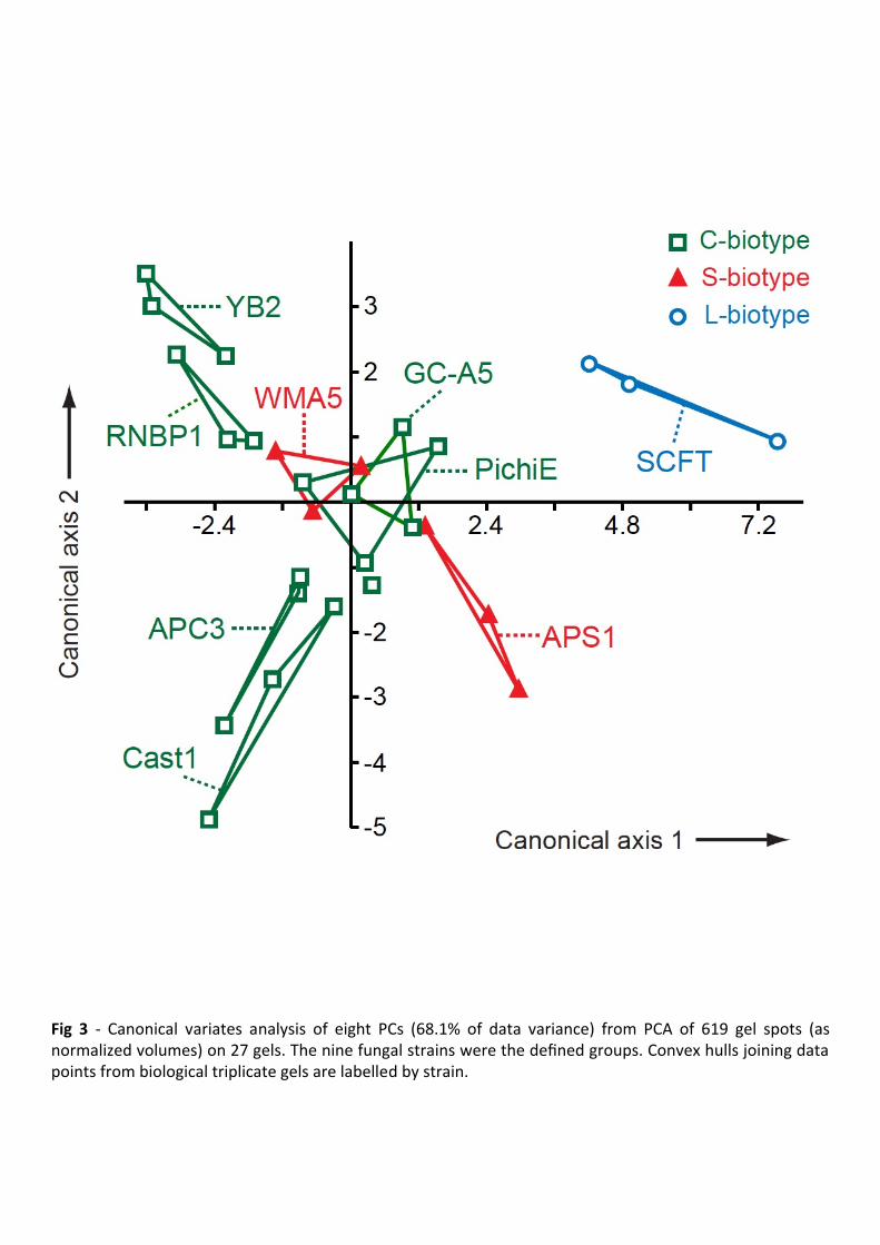
We first applied PCA to the spot data. As the first two PCs accounted for only 32% of 287
overall variance, pairwise PC scores plots offered limited explanatory power. However, we 288
recruited 68.1% of the data variance by using CVA on the scores of the first eight PCs (Fig 3). 289
CVA derives linear combinations of variables (here, PCs of the 2-DE data) to produce 290
maximal, and second-to-maximal, separation between defined groups (here, fungal strains) on 291
the first two canonical axes. 292
On the CVA plot (Fig 3), the slow-growing YB2 culture occupied the most negative region 293
of the (vertical) canonical axis 2, while faster growing cultures occupied the positive region. A 294
possible relation between growth properties and proteomes of the cultures was supported by a 295
correlation coefficient of -0.752 (p < 0.05) between growth rates and mean values on axis 2 for 296
each strain. 297
The L-biotype SCFT appeared separate from all other strains on the CVA plot (Fig 3). For 298
statistical support, we used permutation with 2000 pseudoreplicates on the Mahalanobis 299
squared distance (MD2) between the multivariate data (i.e., scores on eight PCs) for any two 300
defined groups. This non-parametric test confirmed a significant distance between SCFT and 301
the C- and S-biotypes (MD2 = 33.36; p < 0.01). 302
On the other hand, the S-biotypes APS1 and WMA5 did not associate as a distinct group on 303
the CVA plot, being interspersed with the C-biotypes GC-A5 and PichiE (Fig 3). Furthermore, 304
the two-group permutation test did not significantly separate the APS1/WMA5 pair from the 305
other biotypes (MD2 = 5.683; p = 0.06). The proteomic similarity of the S- and C-biotypes 306
could be seen as consistent with the DNA evidence that these biotypes are not phylogenetically 307
distinct (de Arruda et al. 2005; Marelli et al. 2009). 308
Certain C-biotypes occurred in proximity on the CVA plot (Fig 3). One apparent association 309
was Cast1/APC3, and this pair was significantly separated from all other strains in the 310
permutation test (MD2 = 12.61; p < 0.01). Cast1 and APC3 were isolated over a decade apart in 311
different Brazilian states (Table 1), but do share the property of being able to infect the cacao 312
clone Scavina 6, which is resistant to many witches’ broom strains, including GC-A5 (Shaw 313
and Vandenbon 2007). However, the relative virulence of these strains differs on other cacao 314
clones (Shaw and Vandenbon 2007). RNBP1 and YB2 were likewise in proximity on the CVA 315

MANUSCRIP
T
ACCEPTED
ACCEPTED MANUSCRIPT
12
plot, and as a pair were significantly separated from all other strains in the permutation test 316
(MD2 = 12.50; p < 0.01). RNBP1 and YB2 were, again, isolated over a decade apart, but 317
geographically both came from the Western reaches of the Amazon basin (Table 1). 318
Identification of proteins of M. perniciosa cultures 319
Information on sufficiently abundant proteins on the APC3 and Cast1 C-biotype gels was 320
obtained by MASCOT searches of databases using MS/MS sequences of tryptic peptides from 321
2-DE gel spots (Table 2). For most queries, an accession from the genus Moniliophthora was 322
the best match. At the time of this study, the M. perniciosa genome version deposited at 323
DDBJ/EMBL/GenBank was lower quality (accession ABRE, 1.9× coverage) than that of M. 324
roreri (accession LATX, 91× coverage). Consequently, the M. roreri genome provided the 325
MASCOT matches in 16 queries, while M. perniciosa provided only 11, seven of which were 326
partial sequences (Table 2). For one query (spot 12), the MASCOT match was from another 327
genome-sequenced agaric Schizophyllum commune (Ohm et al. 2010), but this sequence 328
showed 94% identity to an M. perniciosa accession, EEB94528. 329
Another new resource is the WBDTA, based on RNA-seq libraries of RNAs from a range of 330
cultures and pathogenic stages of M. perniciosa (Teixeira et al. 2014). NCBI database 331
information (function and InterPro domains) on accessions similar to the 2-DE spots (Table 2) 332
was used further, to identify similar transcripts in the WBDTA. Transcripts (MP identifiers) 333
with high identity to the 2-DE spots peptides are in Table 3. The 14-day-old dikaryotic 334
mycelial cultures library of the WBDTA would be the most comparable to the cultures we 335
analyzed. Table 3 therefore reports expression of the relevant transcripts in this library, as well 336
as the library in which they exhibited maximal expression. Eighteen of the 20 matched genes 337
showed substantial expression in 14-day-old dikaryotic mycelia (over 20% of the maximal 338
expression during the life-cycle), making a relationship to our 2-DE spots plausible (Table 3). 339
An outstanding feature of the M. perniciosa proteomes (Tables 2-3) was the prominence of 340
putative aldo-keto reductases (AKRs). The AKR superfamily has a common structure and 341
reaction mechanism, involving NADPH-dependent oxido-reduction of carbonyl compounds, 342
but it encompasses diverse functional roles across all phyla (Mindnich and Penning 2009). We 343
found an AKR, spot 1, that was among the most abundant proteins in all cultures (Fig 1), 344
irrespective of biotype (see Supplementary Fig 2 for quantitative data). Spot 1 appeared to 345
correlate with culture growth rate (coefficient 0.77, p < 0.05), having lowest abundance in the 346
slow-growing YB2 C-biotype, and highest in the fast-growing APS1 S-biotype. Three other 347

MANUSCRIP
T
ACCEPTED
ACCEPTED MANUSCRIPT
13
spots were assigned as AKRs (Tables 2-3). Two of these (16, 17) were moderately abundant in 348
all strains, but spot 4 showed great variability (Supplementary Fig 2). Spot 4 was present in all 349
replicates of the C-biotype APC3, but in other strains such as the C-biotype Cast1, it was not 350
detected (Fig 4 inset). The peptide sequences of spot 4 were found in spot 1 (Supplementary 351
Table 3), and each could be matched to an M. roreri accession with a theoretical molecular 352
weight (36.4 kDa) and pI (6.2) similar to the gel estimates for spot 1 (35 kDa, pI 6.5). Spot 4 353
was estimated to have a similar molecular weight (37 kDa) but more basic pI (8.5), suggesting 354
a modified isoform of the same protein. 355
Such prominence of AKRs has not been widely reported in fungal cultures on complete 356
media. Leal et al. (2010) found M. perniciosa AKR transcripts were induced in nitrogen-357
limited liquid cultures, and were expressed in infected cacao tissues. They proposed AKRs 358
belong to a suite of ‘virulence life-style genes’ that enable colonization of the host 359
environment (Leal et al. 2010). One virulence-associated AKR is the AFTS1 gene of 360
Alternaria alternata, involved in biosynthesis of a toxin for pathogenicity on strawberry (Ito et 361
al. 2004). In cultures of Ustilago maydis, expression of the AKR YakC increased in the 362
transition to filamentous growth associated with the pathogenic life-style (Böhmer et al. 2007). 363
Other evidence for AKRs in fungal-plant interactions includes the AAD1 aryl-alcohol 364
dehydrogenase of the lignin-degrader Phanerochaete chrysosporium (Yang et al. 2012), and 365
up-regulation of AKRs in the Rhizophagus irregularis-Medicago truncatula symbiosis 366
(Tisserant et al. 2013). In other fungal systems, AKRs detoxify xenobiotics, such as 367
pharmaceuticals in Candida glabrata (Farahyar et al. 2013). Evidence also supports a role for 368
fungal AKRs in stress responses. Yeast mutants defective in AKR genes, for example, 369
exhibited abnormal oxidative and heat stress (Chang and Petrash 2008). The putative AKR 370
gene MP13440, which shared high identity with the spots 1, 4 and 17 peptides, had highest 371
expression in infected fruit in the WBDTA (Table 3), but little (< 1% of maximum) in green 372
brooms. The role of AKRs in witches’ broom disease awaits elucidation. 373
Several other proteins in our cultures would be consistent with a stress response syndrome 374
(Tables 2-3). These included two heat-shock proteins found in all strains (spots 11 and 12). In 375
addition, a 1-cys peroxiredoxin (spot 6) was detected on most gels, generally in high amounts 376
in the faster-growing strains, but showed considerable variability even between replicates 377
(Supplementary Fig 2). Peroxiredoxins are widely distributed peroxide-decomposing enzymes 378
(Monteiro et al. 2007). Cobos et al. (2010) detected peroxiredoxin, and heat-shock protein, in 379

MANUSCRIP
T
ACCEPTED
ACCEPTED MANUSCRIPT
14
liquid cultures of the grapevine pathogen Diplodia seriata, and speculated that peroxiredoxin 380
could contribute to pathogenesis by counteracting host-produced hydrogen peroxide. 381
Spot 18 had a negative correlation with growth rate (coefficient -0.78, p < 0.05), being 382
found in greatest abundance in the slow-growing YB2 cultures (Supplementary Fig 2). It 383
showed homology to mannitol-1-phosphate dehydrogenase (Tables 2-3), which functions in 384
biosynthesis of mannitol, a compatible solute produced by fungi in response to various stresses 385
(Dijksterhuis and De Vries 2006). Interestingly, mannitol-1-phosphate dehydrogenase appears 386
to have been acquired by the Moniliophthora genus via horizontal gene transfer from firmicute 387
bacteria (Tiburcio et al. 2010). These authors point out that Moniliophthora relatives are 388
saprotrophs (Aime and Phillips-Mora 2005), so a pre-pathogenic ancestor might have occupied 389
soil and decomposed organic material, in which firmicutes are common. Acquisition of 390
mannitol-1-phosphate dehydrogenase by a Moniliophthora ancestor might have contributed to 391
the evolution of pathogenicity. Alternaria alternata mutants in this enzyme, for example, were 392
less virulent on tobacco (Vélëz et al. 2008). Ascomycetes such as Alternaria, however, appear 393
to have acquired mannitol-1-phosphate dehydrogenase by a different horizontal gene transfer, 394
from actinobacteria (Tiburcio et al. 2010). In contrast, most basidiomycetes other than 395
Moniliophthora appear to lack mannitol-1-phosphate dehydrogenase, which Tiburcio et al. 396
(2010) speculate is one reason why there are more phytopathogens among ascomycetes than 397
basidiomycetes. This protein is therefore a further possible ‘virulence life-style’ function in the 398
cultured M. perniciosa proteome. 399
About one-third of the gel spots in Tables 2-3 were assignable to central metabolism 400
enzymes generally encountered in fungal proteomic studies. Malate dehydrogenase (spots 2 401
and 14), phosphoglycerate kinase (spot 3), and glyceraldehyde-3-phosphate dehydrogenase 402
(spots 10 and 20) featured in proteomic analyses of cultures of M. perniciosa (Silva et al. 403
2013), other agaricomycetes such as Rhizoctonia solani (Kwon et al. 2014) and Phanerochaete 404
chrysosporium (Yildirim et al. 2014), and ascomycetes including Diplodia seriata (Cobos et 405
al. 2010) and Aspergillus fumigatus (Vödisch et al. 2009). Since M. perniciosa has been found 406
to possess only one glyceraldehyde-3-phosphate dehydrogenase gene (Lima et al., 2009), spot 407
20 was likely a modified version of spot 10, with a similar molecular weight, but slightly less 408
basic pI (Fig 4). Two isoforms with similar molecular weight and pI were also found in the 409
pathogenic ascomycete Paracoccidioides brasiliensis (Barbosa et al., 2006). 410
For each of these ubiquitous enzymes, specialized extra functions have been proposed. 411
Glyceraldehyde-3-phosphate dehydrogenase has been identified as a virulence factor in 412

MANUSCRIP
T
ACCEPTED
ACCEPTED MANUSCRIPT
15
mycoses caused by Candida albicans and Paracoccidioides brasiliensis (Barbosa et al. 2006; 413
Seidler 2013). The other glycolytic enzymes enolase (spot 13) and phosphoglycerate kinase 414
have also been implicated in mycoses (Pancholi and Chhatwal 2003). Malate dehydrogenase 415
may be indirectly involved in virulence in M. perniciosa. Oxaloacetate produced by this 416
enzyme may be converted into oxalate, from which M. perniciosa can make calcium oxalate, 417
which appears to play a role in pathogenesis (Ceita et al. 2007; do Rio et al. 2008). The genes 418
MP02237, MP03476, MP05722, MP05723, MP12535 with similar sequences to these 419
carbohydrate metabolism enzymes (Table 3) all had strong expression in in the WBDTA green 420
broom libraries (252-1955 RPKM). 421
Spot 9 was matched to M. perniciosa accession EEB89936 (hypothetical protein 422
MPER_11918),which had 61% homology with the Pleurotus ostreatus protein pleurotolysin B, 423
of the membrane pore-forming aegerolysin family (Tomita et al. 2004). The discrepancy 424
between theoretical (57.2 kDa) and observed (41 kDa) molecular weights of this protein was 425
consistent with the reported proteolytic cleavage in P. ostreatus extracts of the 59 kDa 426
pleurotolysin B into a 41 kDa fragment (Tomita et al. 2004). Using an M. perniciosa culture 427
system based on Griffith and Hedger (1993), Pires et al. (2009) found differential expression 428
of aegerolysin transcripts during fructification, and have already associated MPER_11918 with 429
the probable pleurotolysin B of M. perniciosa. A role for aegerolysins in the later stages of 430
development would be consistent with the fact that we only observed spot 9 in the fast-growing 431
Cast1 cultures (Fig 4), whose mycelia may have been in a more advanced developmental state 432
(though basidiome formation was observed in these cultures and has not been reported from 433
any Petri dish cultures). The WBDTA gene most similar to spot 9, MP13610, had strongest 434
expression in basidiome primordia and little in mycelial cultures (Table 3). Similar comments 435
apply to the putative NAD-dependent epimerase spot 5 (Fig 4), of unknown function, and its 436
most similar WBDTA gene MP04655 (Table 3). 437
Other library matches of potential relevance to pathogenesis included a putative nitrilase 438
(spot 15), a function highlighted in the M. perniciosa genome by Mondego et al. (2008) as an 439
auxin biosynthesis step, via hydrolysis of indole-3-acetonitrile. Alternatively, nitrilase could 440
detoxify plant-produced cyanides (O’Reilly and Turner 2003). Spot 21 was identified as an 441
NAD-dependent formate dehydrogenase, which in M. perniciosa may have a role in 442
catabolism of methanol released by hydrolysis of methylesterified host pectins (Mondego et al. 443
2008; De Oliveira et al. 2012). 444
It should be stated that the subset of proteins identified by MS/MS were self-selected for 445

MANUSCRIP
T
ACCEPTED
ACCEPTED MANUSCRIPT
16
sufficient abundance on the gels. The sequenced protein spots in Supplementary Fig 2 446
contributed 12.9% of the total variance explored by CVA (Fig 3). Consequently, the proteomic 447
phenotypes indicated by CVA will remain ‘black boxes’ until a more comprehensive 448
characterization of the protein populations. Nonetheless this study supports the potential of 449
multivariate analysis and sequence informatics for understanding fungal proteomes. 450
451

MANUSCRIP
T
ACCEPTED
ACCEPTED MANUSCRIPT
17
452
Conclusions 453
Cultures of geographically diverse M. perniciosa isolates exhibited differences in mycelial and 454
basidiome morphology, and also in rates of saprotrophic growth and fructification, but no 455
biotype-specific characters were observable. In infection experiments, moreover, one of the C-456
biotypes caused symptoms on tomato, a putative S-biotype host. The lack of a clear distinction 457
between C- and S-biotypes also applied at the proteome level, as multivariate analyses of 2-DE 458
spot patterns did not discriminate these two biotypes. These observations accord with genetic 459
studies that failed to separate C- and S-biotypes (De Arruda et al. 2005; Marelli et al. 2009). 460
The single L-biotype, however, was statistically different in our proteomic analyses. 461
Peptide sequencing of 2-DE spots from M. perniciosa cultures confirmed the utility of 462
recent genome sequencing, including M. roreri, which contributed a number of functional 463
annotations. The proteome of in vitro cultured M. perniciosa was suggestive of the ‘virulence 464
life-style’ proposed on the basis of transcript analyses by Leal et al. (2010). Unlike these 465
authors, we did not subject the saprotrophic cultures to any treatment designed to mimic a host 466
environment and yet, interestingly, the sampled proteome presented a high proportion of 467
functions indicative of pathogenicity. 468
469
Acknowledgements 470
We dedicate this paper to the memory of Dr. Sandra Pierre who died in March 2017. She had a 471
huge enthusiasm for cocoa research and will be greatly missed by her former colleagues. 472
This work was funded by Cocoa Research UK Ltd and the Government of the Netherlands. We 473
are grateful for technical assistance and advice from James Heald, Martyna Matuszyk, Sarah 474
Tvedt, Jon Lamb, Martin Swain, Tom Thomas, Pat Causton, Anthony Pugh, Gwen Jenkins and 475
Joanne Hamilton. 476
477
Appendix A. Supplementary data 478
Supplementary data associated with this article can be found, in the online version, at 479
480

MANUSCRIP
T
ACCEPTED
ACCEPTED MANUSCRIPT
18
481
REFERENCES 482
Aime MC, Phillips-Mora W, 2005. The causal agents of witches’ broom and frosty pod rot of 483
cacao (chocolate, Theobroma cacao) form a new lineage of Marasmiaceae. Mycologia 97: 484
1012-1022. 485
Alvim FC, Mattos EM, Pirovani CP, Gramacho K, Pungartnik C, Brendel M, Cascardo JCM, 486
Vincentz M, 2009. Carbon source-induced changes in the physiology of the cacao pathogen 487
Moniliophthora perniciosa (Basidiomycetes) affect mycelial morphology and secretion of 488
necrosis-inducing proteins. Genetics and Molecular Research 8: 1035-1050. 489
Barbosa MS, Báo SN, Andreotti PF, Faria FP, Felipe MSS, Feitosa LS, Mendes-Giannini MJS, 490
Soares CMA, 2006. Glyceraldehyde-3-phosphate dehydrogenase of Paracoccidioides 491
brasiliensis is a cell surface protein involved in fungal adhesion to extracellular matrix 492
proteins and interaction with cells. Infection and Immunity 74: 382-389. 493
Bastos CN, Evans HC, 1985. A new pathotype of Crinipellis perniciosa (witches’ broom 494
disease) on solanaceous hosts. Plant Pathology 34: 306-312. 495
Böhmer M, Colby T, Böhmer C, Bräutigam A, Schmidt J, Bölker M, 2007. Proteomic analysis 496
of dimorphic transition in the phytopathogenic fungus Ustilago maydis. Proteomics 7: 675-497
685. 498
Ceita GDO, Macedo JNA, Santos TB, Alemanno L, Gesteira AD, Micheli F, Mariano AC, 499
Gramacho KP, Silva DDC, Meinhardt L, Mazzafera P, Pereira GAG, Cascardo J, 2007. 500
Involvement of calcium oxalate degradation during programmed cell death in Theobroma 501
cacao tissues triggered by the hemibiotrophic fungus Moniliophthora perniciosa. Plant 502
Science 173: 106-117. 503
Chang Q, Petrash JM, 2008. Disruption of aldo-keto reductase genes leads to elevated markers 504
of oxidative stress and inositol auxotrophy in Saccharomyces cerevisiae. Biochimica et 505
Biophysica Acta 1783: 237–245. 506
Cobos R, Barreiro C, Mateos RM, Coque JJR, 2010. Cytoplasmic- and extracellular-proteome 507
analysis of Diplodia seriata: a phytopathogenic fungus involved in grapevine decline. 508
Proteome Science 8: 46. 509

MANUSCRIP
T
ACCEPTED
ACCEPTED MANUSCRIPT
19
Cohen BL, 1973. Growth of Apergillus nidulans in a thin liquid layer. Microbiology 76: 277-510
282. 511
De Arruda MCC, Sepulveda Ch. GF, Miller RNG, Ferreira MASV, Santiago DVR, Resende 512
MLV, Dianese JC, Felipe MSS, 2005. Crinipellis brasiliensis, a new species based on 513
morphological and molecular data. Mycologia 97: 1348-1361. 514
Deganello J, Leal GA, Rossi ML, Peres LEP, Figueira A, 2014. Interaction of Moniliophthora 515
perniciosa biotypes with Micro-Tom tomato: a model system to investigate the witches’ 516
broom disease of Theobroma cacao. Plant Pathology 63: 1251-1263. 517
De Maesschalck R, Jouan-Rimbaud D, Massart DL, 2000. The Mahalanobis distance. 518
Chemometrics and Intelligent Laboratory Systems 50: 1-18. 519
De Oliveira BV, Teixeira GS, Reis O, Barau JG, Teixeira PJPL, do Rio MCS, Domingues RR, 520
Meinhardt LW, Paes Leme AF, Rincones J, Pereira GAG, 2012. A potential role for an 521
extracellular methanol oxidase secreted by Moniliophthora perniciosa in Witches’ broom 522
disease in cacao. Fungal Genetics and Biology 49: 922-932. 523
Díaz-Valderrama JR, Aime MC, 2016. The cacao pathogen Moniliophthora roreri 524
(Marasmiaceae) possesses biallelic A and B mating loci but reproduces clonally. Heredity 525
116: 491-501. 526
Dijksterhuis J, De Vries RP, 2006. Compatible solutes and fungal development. Biochemical 527
Journal 399: e3-e5. 528
Do Rio MCS, de Oliveira BV, de Tomazella DPT, Silva JAF, Pereira GAG, 2008. Production 529
of calcium oxalate crystals by the Basidiomycete Moniliophthora perniciosa, the causal 530
agent of witches’ broom disease of cacao. Current Microbiology 56: 363-370. 531
Farahyar S, Zaini F, Kordbacheh P, Rezaie S, Safara M, Raoofian R, Heidari M, 2013. 532
Overexpression of aldo-keto-reductase in azole-resistant clinical isolates of Candida 533
glabrata determined by cDNA-AFLP. DARU Journal of Pharmaceutical Sciences 21:1. 534
Fernández-Acero FJ, Jorge I, Calvo E, Vallejo I, Carbú M, Camafeita E, Garrido C, López JA, 535
Jorrín J, Cantoral JM, 2007. Proteomic analysis of phytopathogenic fungus Botrytis cinerea 536
as a potential tool for identifying pathogenicity factors, therapeutic targets and for basic 537
research. Archives of Microbiology 187: 207-215. 538

MANUSCRIP
T
ACCEPTED
ACCEPTED MANUSCRIPT
20
Fernández-Acero FJ, Jorge I, Calvo E, Vallejo I, Carbú M, Camafeita E, López JA, Cantoral 539
JM, Jorrín J, 2006. Two-dimensional electrophoresis protein profile of the phytopathogenic 540
fungus Botrytis cinerea. Proteomics 6: S88-S96. 541
Franco SF, Baroni RM, Carazzolle MF, Teixeira PJPL, Reis O, Pereira GAG, Mondego JMC, 542
2015. Genomic analyses and expression evaluation of thaumatin-like gene family in the 543
cacao fungal pathogen Moniliophthora perniciosa. Biochemical and Biophysical Research 544
Communications 466: 629-636. 545
Gomes DS, Lopes MA, Menezes SP, Ribeiro LF, Dias CV, Andrade BS, de Jesus RM, Pires 546
ABL, Goes-Neto A, Micheli F, 2016. Mycelial development preceding basidioma formation 547
in Moniliophthora perniciosa is associated to chitin, sugar and nutrient metabolism 548
alterations involving autophagy. Fungal Genetics and Biology 86: 33-46. 549
Griffith GW, Hedger JN, 1993. A novel method for producing basidiocarps of the cocoa 550
pathogen Crinipellis perniciosa using a bran-vermiculite medium. Netherlands Journal of 551
Plant Pathology 99: 227-230. 552
Griffith GW, Hedger JN, 1994a. The breeding biology of biotypes of the witches’ broom 553
pathogen of cocoa, Crinipellis perniciosa. Heredity 72: 278-289. 554
Griffith GW, Hedger JN, 1994b. Spatial distribution of mycelia of the liana (L-) biotype of the 555
agaric Crinipellis perniciosa (Stahel) Singer in tropical forest. New Phytologist 127: 243-556
259. 557
Griffith GW, Nicholson JN, Nenninger A, Birch RN, Hedger JN, 2003. Witches’ brooms and 558
frosty pods: two major pathogens of cacao. New Zealand Journal of Botany 41: 423-435. 559
Hammer Ø, Harper DAT, Ryan PD, 2001. PAST: paleontological statistics software package 560
for education and data analysis. Palaeontologia Electronica 4(1): 9pp. 561
Ito K, Tanaka T, Hatta R, Yamamoto M, Akimitsu K, Tsuge T, 2004. Dissection of the host 562
range of the fungal plant pathogen Alternaria alternata by modification of secondary 563
metabolism. Molecular Microbiology 52: 399-411. 564
Kwon YS, Kim SG, Chung WS, Bae H, Jeong SW, Shin SC, Jeong MJ, Park SC, Kwak YS, 565
Bae DW, Lee YB, 2014. Proteomic analysis of Rhizoctonia solani AG-1 sclerotia 566
maturation. Fungal Biology 118: 433-443. 567
Leal GA, Gomes LH, Albuquerque PSB, Tavares FCA, Figueira A, 2010. Searching for 568
Moniliophthora perniciosa pathogenicity genes. Fungal Biology 114: 842-854. 569

MANUSCRIP
T
ACCEPTED
ACCEPTED MANUSCRIPT
21
Lima JO, Pereira JF, Rincones J, Barau JG, Araujo EF, Pereira GAG, Queiroz MV, 2009. The 570
glyceraldehyde-3-phosphate dehydrogenase gene of Moniliophthora perniciosa, the causal 571
agent of witches' broom disease of Theobroma cacao. Genetics and Molecular Biology 32: 572
362-366. 573
Lopes JRM, Luz EDMN, Bezerra JL, 2001. Suscetibilidade do cupuaçuzeiro e outras espécies 574
vegetais a isolados de Crinipellis perniciosa obtidos de quatro hospedeiros diferentes no sul 575
da Bahia. Fitopatologia Brasileira 26: 601-605. 576
Mackey AJ, Haystead TAJ, Pearson WR, 2002. Getting more from less. Algorithms for rapid 577
protein identification with multiple short peptide sequences. Molecular and Cellular 578
Proteomics 1: 139-147. 579
Marelli JP, Maximova SN, Gramacho KP, Kang S, Guiltinan MJ, 2009. Infection biology of 580
Moniliophthora perniciosa on Theobroma cacao and alternate solanaceous hosts. Tropical 581
Plant Biology 2: 149-160. 582
Meinhardt LW, Bellato CD, Rincones J, Azevedo RA, Cascardo JCM, Pereira GAG, 2006. In 583
vitro production of biotrophic-like cultures of Crinipellis perniciosa, the causal agent of 584
witches' broom disease of Theobroma cacao. Current Microbiology 52: 191-196. 585
Meinhardt LW, Costa GGL, Thomazella DPT, Teixeira PJPL, Carazzolle MF, Schuster SC, 586
Carlson JE, Guiltinan MJ, Mieczkowski P, Farmer A, Ramaraj T, Crozier J, Davis RE, Shao 587
J, Melnick RL, Pereira GAG, Bailey BA, 2014. Genome and secretome analysis of the 588
hemibiotrophic fungal pathogen, Moniliophthora roreri, which causes frosty pod rot disease 589
of cacao: mechanisms of the biotrophic and necrotrophic phases. BMC Genomics 15: 164. 590
Meinhardt LW, Rincones J, Bailey BA, Aime MC, Griffith GW, Zhang D, Pereira GAG, 2008. 591
Moniliophthora perniciosa, the causal agent of witches’ broom disease of cacao: what’s 592
new from this old foe? Molecular Plant Pathology 9: 577-588. 593
Mindnich RD, Penning TM, 2009. Aldo-keto reductase (AKR) superfamily: Genomics and 594
annotation. Human Genomics 3: 362-370. 595
Mondego JM, Carazzolle MF, Costa GG, Formighieri EF, Parizzi LP, Rincones J, Cotomacci 596
C, Carraro DM, Cunha AF, Carrer H, Vidal RO, Estrela RC, García O, Thomazella DP, de 597
Oliveira BV, Pires AB, Rio MC, Araújo MR, de Moraes MH, Castro LA, Gramacho 598
KP, Gonçalves MS, Neto JP, Neto AG, Barbosa LV, Guiltinan MJ, Bailey BA, Meinhardt 599
LW, Cascardo JC, Pereira GA, 2008. A genome survey of Moniliophthora perniciosa gives 600
new insights into Witches' Broom Disease of cacao. BMC Genomics 9: 548. 601

MANUSCRIP
T
ACCEPTED
ACCEPTED MANUSCRIPT
22
Monteiro G, Horta BB, Pimenta DC, Augusto O, Netto LES, 2007. Reduction of 1-Cys 602
peroxiredoxins by ascorbate changes the thiol-specific antioxidant paradigm, revealing 603
another function of vitamin C. Proceedings of the National Academy of Sciences USA 104: 604
4886-4891. 605
Morphew RM, Eccleston N, Wilkinson TJ, McGarry J, Perally S, Prescott M, Ward D, 606
Williams D, Paterson S, Raman M, Ravikumar G, Saifullah MK, Abidi SMA, McVeigh P, 607
Maule AG, Brophy PM, LaCourse EJ, 2012. Proteomics and in silico approaches to extend 608
understanding of the glutathione transferase superfamily of the tropical liver fluke Fasciola 609
gigantica. Journal of Proteome Research 11: 5876-5889. 610
Ohm RA, de Jong JF, Lugones LG, Aerts A, Kothe E, Stajich JE, de Vries RP, Record E, 611
Levasseur A, Baker SE, Bartholomew KA, Coutinho PM, Erdmann S, Fowler TJ, Gathman 612
AC, Lombard V, Henrissat B, Knabe N, Kües U, Lilly WW, Lindquist E, Lucas S, 613
Magnuson JK, Piumi F, Raudaskoski M, Salamov A, Schmutz J, Schwarze FWMR, van 614
Kuyk PA, Horton JS, Grigoriev IV, Wösten HAB, 2010. Genome sequence of the model 615
mushroom Schizophyllum commune. Nature Biotechnology 28: 957-965. 616
O’Reilly C, Turner PD, 2003. The nitrilase family of CN hydrolysing enzymes - a comparative 617
study. Journal of Applied Microbiology 95: 1161-1174. 618
Pancholi V, Chhatwal GS, 2003. Housekeeping enzymes as virulence factors for pathogens. 619
International Journal of Medical Microbiology 293: 391-401. 620
Pires ABL, Gramacho KP, Silva DC, Góes-Neto A, Silva MM, Muniz-Sobrinho JS, Porto RF, 621
Villela-Dias C, Brendel M, Cascardo JCM, Pereira GAG, 2009. Early development of 622
Moniliophthora perniciosa basidiomata and developmentally regulated genes. BMC 623
Microbiology 9: 158. 624
Ploetz RC, Schnell RJ, Ying Z, Zheng Q, Olano CT, Motamayor JC, Johnson ES, 2005. 625
Analysis of molecular diversity in Crinipellis perniciosa with AFLP markers. European 626
Journal of Plant Pathology 11: 317-326. 627
Rincones J, Mazotti GD, Griffith GW, Pomela AW, Figueira A, Queiroz MV, Pereira JF, 628
Azevedo RA, Pereira GAG, Meinhardt LW, 2006. Genetic variability and chromosome-629
length polymorphisms of the witches’ broom pathogen Crinipellis perniciosa from various 630
plant hosts in South America. Mycological Research 110: 821-832. 631

MANUSCRIP
T
ACCEPTED
ACCEPTED MANUSCRIPT
23
Schlumberger S, Kristan KC, Ota K, Frangež R, Molgό J, Sepčić K, Benoit E, Maček P, 2014. 632
Permeability characteristics of cell-membrane pores induced by ostreolysin A/pleurotolysin 633
B, binary pore-forming proteins from the oyster mushroom. FEBS Letters 588: 35-40. 634
Seidler NW, 2013. GAPDH, as a virulence factor. Advances in Experimental Medicine and 635
Biology 985: 149-178. 636
Blackwell Publishing Ltd 637
Shaw MW, Vandenbon AE, 2007. A qualitative host-pathogen interaction in the Theobroma 638
cacao-Moniliophthora perniciosa pathosystem. Plant Pathology 56: 277-285. 639
Silva FAC, Pirovani CP, Menezes S, Pungartnik C, Santiago AS, Costa MGC, Micheli F, 640
Gesteira AS, 2013. Proteomic response of Moniliophthora perniciosa exposed to 641
pathogenesis-related protein-10 from Theobroma cacao. Genetics and Molecular Research 642
12: 4855-4868. 643
Teixeira PJPL, Thomazella DPT, Reis O, do Prado PFV, do Rio MCS, Fiorin GL, José J, Costa 644
GGL, Negri VA, Mondego JMC, Mieczkowski P, Pereira GAG, 2014. High-resolution 645
transcript profiling of the atypical biotrophic interaction between Theobroma cacao and the 646
fungal pathogen Moniliophthora perniciosa. The Plant Cell 26: 4245-4269. 647
Thomazella DPT, Teixeira PJPL, Oliveira HC, Saviani EE, Rincones J, Toni IM, Reis O, 648
Garcia O, Meinhardt LW, Salgado I, Pereira GAG, 2012. The hemibiotrophic cacao 649
pathogen Moniliophthora perniciosa depends on a mitochondrial alternative oxidase for 650
biotrophic development. New Phytologist 194: 1025-1034. 651
Tiburcio RA, Costa GGL, Carazzolle MF, Mondego JMC, Schuster SC, Carlson JE, Guiltinan 652
MJ, Bailey BA, Mieczkowski P, Meinhardt LW, Pereira GAG, 2010. Genes acquired by 653
horizontal transfer are potentially involved in the evolution of phytopathogenicity in 654
Moniliophthora perniciosa and Moniliophthora roreri, two of the major pathogens of cacao. 655
Journal of Molecular Evolution 70: 85-97. 656
Tisserant E, Malbreil M, Kuo A, Kohler A, Symeonidi A, Balestrini R, Charron P, Duensing 657
N, Freidit Frey N, Gianinazzi-Pearson V, Gilbert LB, Handa Y, Herr JR, Hijri M, Koul R, 658
Kawaguchi M, Krajinski F, Lammers PJ, Masclaux FG, Murat C, Morin E, Ndikumana S, 659
Pagni M, Petitpierre D, Requena N, Rosikiewicz P, Riley R, Saito K, San Clemente H, 660
Shapiro H, van Tuinen D, Bécard G, Bonfante P, Paszkowski U, Y. Shachar-Hill YY, 661
Tuskan GA, Young JPW, Sanders IR, Henrissat B, Rensing SA, Grigoriev IV, Corradi N, 662
Roux C, Martin F, 2013, Genome of an arbuscular mycorrhizal fungus provides insight into 663

MANUSCRIP
T
ACCEPTED
ACCEPTED MANUSCRIPT
24
the oldest plant symbiosis. Proceedings of the National Academy of Sciences USA 110: 664
20117-20122. 665
Tomita T, Noguchi K, Mimuro H, Ukaji F, Ito K, Sugawara-Tomita N, Hashimoto Y, 2004. 666
Pleurotolysin, a novel sphingomyelin-specific two-component cytolysin from the edible 667
mushroom Pleurotus ostreatus, assembles into a transmembrane pore complex. Journal of 668
Biological Chemistry 279: 26975-26982. 669
Vélëz H, Glassbrook NJ, Daub ME, 2008. Mannitol biosynthesis is required for plant 670
pathogenicity by Alternaria alternata. FEMS Microbiology Letters 285: 122-129. 671
Vödisch M, Albrecht D, Lessing F, Schmidt AD, Winkler R, Guthke R, Brakhage AA, 672
Kniemeyer O, 2009. Two-dimensional proteome reference maps for the human pathogenic 673
filamentous fungus Aspergillus fumigatus. Proteomics 9: 1407-1415. 674
Yang DD, François JM, de Billerbeck GM, 2012. Cloning, expression and characterization of 675
an aryl-alcohol dehydrogenase from the white-rot fungus Phanerochaete chrysosporium 676
strain BKM-F-1767. BMC Microbiology 12: 126. 677
Yildirim V, Özcan S, Becher D, Büttner K, Hecker M, Özcengiz G, 2011. Characterization of 678
proteome alterations in Phanerochaete chrysosporium in response to lead exposure. 679
Proteome Science 9: 12. 680
681

MANUSCRIP
T
ACCEPTED
ACCEPTED MANUSCRIPT
25
682
Table 1 - Provenance of M. perniciosa cultures. 683
Strain Biotype Origin Host if recorded Date Collectora CBS
Accession No.
APC3 C Almirante, Bahia, Brazil Scavina 6 cocoa pod Late 1990’s AP CBS
142684
Cast1 C Castanhal, Rondȏnia, Brazil
Cocoa tree in area of Scavina 6 resistance
breakdown Early 1980’s BJW
CBS 142685
GC-A5 C Gran Couva, Trinidad Dead cocoa broom Early 1980’s BJW CBS
142682
PichiE C Pichilingue, Ecuador Dead cocoa broom Early 1980’s BJW CBS
142683
RNBP1 C Quillabamba, Peru Dead cocoa broom June 1998 JNH CBS
142680
YB2 C Japacani, Bolivia Dead cocoa broom June 1987 EAWB CBS
142679
APS1 S Minas Gerais, Viçosa,
Brazil Living Lobeira branch Late 1990’s AP
CBS 142681
WMA5 S Manaus, Amazonas,
Brazil Solanum rugosum Jan. 1991 FJW
CBS 142677
SCFT L San Carlos, Napo,
Ecuador Liana (Arrabidaea sp.) 1987 GWG
CBS 142678
a A. Pomella (AP); BJ Wheeler (BJW); JN Hedger (JNH); EA Wyrley-Birch (EAWB); 684
FJ Wilson (FJW); GW Griffith (GWG). 685
686

MANUSCRIP
T
ACCEPTED
ACCEPTED MANUSCRIPT
26
687
Table 2 Identifications of M. perniciosa mycelial proteins using MS/MS. 688
Spot MASCOT
score a Unique
peptides b Predicted function Accession (species)
1 319 5/5 Aldo-keto reductase c XP_007856327 (M. roreri)
283 4/4 Aldo-keto reductase d EEB99046 (M. perniciosa)
2 352 5/5 Malate dehydrogenase d EEB94126 (M. perniciosa)
3 321 6/6 Phosphoglycerate kinase d EEB89461 (M. perniciosa)
4 195 3/3 Aldo-keto reductase c ESK84368 (M. roreri)
5 470 7/8 NAD-dependent epimerase/dehydratase c XP_007844083 (M. roreri)
6 422 6/8 1-Cys peroxiredoxin c XP_007845258 (M. roreri)
355 6/7 1-Cys peroxiredoxin d EEB97022 (M. perniciosa)
7 257 4/5 Acetyl-acetyltransferase c XP_007843079 (M. roreri)
8 263 5/5 Aspartate aminotransferase c XP_007843679 (M. roreri)
9 235 3/3 Pleurotolysin B homologue d EEB89936 (M. perniciosa)
53 1/1 Erylysin B c
XP_007849236 (M. roreri)
10 335 4/5 Glyceraldehyde-3-phosphate dehydrogenase d EEB90046 (M. perniciosa)
267 3/4 Glyceraldehyde-3-phosphate dehydrogenase c XP_007846800 (M. roreri)
11 339 5/5 Heat-shock protein hss1 c XP_007847698 (M. roreri)
12 101 2/3 Heat shock protein sks2 d XP_003035960 (S. commune)
13 193 2/2 Enolase c XP_007847255 (M. roreri)
14 484 6/6 Malate dehydrogenase d EEB96289 (M. perniciosa)
475 7/7 Malate dehydrogenase c XP_007846805 (M. roreri)
15 344 5/5 Nitrilase c XP_007842695 (M. roreri)
16 148 2/3 Aldo-keto reductase c XP_007854364 (M. roreri)
134 2/3 Aldo-keto reductase d EEB98988 (M. perniciosa)
17 69 1/2 Aldo-keto reductase d EEB97935 (M. perniciosa)
18 142 3/3 Mannitol-1-phosphate dehydrogenase c XP_007855888 (M. roreri)
19 318 4/4 MPER_00772 (unknown function) EEB99535 (M. perniciosa)
20 144 3/3 Glyceraldehyde-3-phosphate dehydrogenase c XP_007846800 (M. roreri)
21 290 5/5 NAD-dependent formate dehydrogenase c AFO55209 (M. perniciosa)
22 86 2/2 Putative anhydrolase d XP_007855443 (M. roreri) 689
a MASCOT scores over 68 were considered significant (p < 0.05). 690
b Peptide sequences are in Supplementary Table 3. 691

MANUSCRIP
T
ACCEPTED
ACCEPTED MANUSCRIPT
27
c Function assignment as database accession. 692
d Function prediction from BLAST search.693

MANUSCRIP
T
ACCEPTED
ACCEPTED MANUSCRIPT
28
694

MANUSCRIP
T
ACCEPTED
ACCEPTED MANUSCRIPT
29
Table 3 Comparison of M. perniciosa mycelial proteins (Table 2) with transcript sequences in the Witches’ Broom Disease Transcriptome Atlas (WBDTA).
Spot
Query/database sequence overlap
Gene IDa Predicted function
Expression in WBDTA RNA-Seq libraries (Teixeira et al. 2014)
Amino acids Identity (%) 14-day-old dikaryotic
mycelia (RPKM)b Peak expression in life-cycle
(RPKM)b
1 90 100 MP13440 Aldo-keto reductase 668 Infected young fruit shell (2427)
2 61 100 MP12535 Malate dehydrogenase 462 Non-germinating spores (742)
3 69 100 MP02237 Phosphoglycerate kinase 321 Green broom (469)
4 32 100 MP13440 Aldo-keto reductase 668 Infected young fruit shell (2427)
5 99 100 MP04655 NAD-dependent epimerase/dehydratase 10 Basidiocarps (1662)
6 116 100 MP16197 1-Cys peroxiredoxin 736 7-day-old dikaryotic mycelia (3303)
7 48 100 MP01795 Acetyl-acetyltransferase 701 Young dikaryotic mycelia (807)
8 56 100 MP00138 Aspartate aminotransferase 547 Germinating spores (608)
9 46 100 MP13610 Pleurotolysin B homologue 29 Basidiocarp primordia (2578)
10 67 100 MP05723 Glyceraldehyde-3-phosphate dehydrogenase 11600 Senescent dikaryotic mycelium (23464)
11 58 100 MP00096 Heat-shock protein hss1 1290 Infected necrotic fruit shell (5313)
12 24 100 MP00146 Heat shock protein sks2 224 Basidiocarp primordia (980)
13 31 100 MP03476 Enolase 441 Green broom (1005)
14 136 99.3 MP05722 Malate dehydrogenase 869 Green broom (1955)
15 79 100 MP00452 Nitrilase 882 Senescent dikaryotic mycelium (1166)
16 38 100 MP12009 Aldo-keto reductase 200 Young dikaryotic mycelium (473)
17 12 92-100 c MP13440 Aldo-keto reductase 668 Infected young fruit shell (2427)
18 30 100 MP10747 Mannitol-1-phosphate dehydrogenase 6897 14-day-old dikaryotic mycelia (6897)
19 - -
20 42 100 MP05723 Glyceraldehyde-3-phosphate dehydrogenase 11600 Senescent dikaryotic mycelium (23464)
21 55 100 MP03866 NAD-dependent formate dehydrogenase 2872 28-day-old dikaryotic mycelia (4213)
22 - -
t
a Identifiers of the WBDTA (http://bioinfo08.ibi.unicamp.br/wbdatlas). b RPKM, reads per kilobase per million mapped reads.
c Polymorphic peptide: YLQENVGAGSIK (in MP13440) and YLKENVGAGSIK (in EEB97935, Table 2), which MS/MS was not able to distinguish.

MANUSCRIP
T
ACCEPTED
ACCEPTED MANUSCRIPT
30
Figure legends
Fig 1 - Mycelia of M. perniciosa strains cultured on MYEA (12 d, 25°C) in 9 cm Petri dishes.
C-biotypes: Cast1, APC3, RNBP1, YB2, PichiE, GC-A5; S-biotypes: WMA5, APS1;
L-biotype: SCFT. Below: 2-DE gel of each strain (arrow locates spot 1 for orientation).
Proteins were separated by pH 3-10 on 12.5% SDS-PAGE and stained with Coomassie
Phastgel Blue R-250.
Fig 2 - Plants inoculated with M. perniciosa basidiospores (defoliated to show symptoms).
(A) Cacao four weeks after mock-inoculation (control), C-biotype infection (APC3) or S-
biotype infection (WMA5). (B) Tomato three weeks post-inoculation with the C-biotype Cast1,
exhibiting stem fasciation (left), stem swelling (centre) and shoot proliferation (right). Inset
shows uninoculated control stem.
Fig 3 - Canonical variates analysis of eight PCs (68.1% of data variance) from PCA of 619 gel
spots (as normalized volumes) on 27 gels. The nine fungal strains were the defined groups.
Convex hulls joining data points from biological triplicate gels are labeled by strain.
Fig 4 - 2-DE gel of proteins from cultured M. perniciosa strain APC3. Samples (100 ng) were
subjected to isoelectric focusing in a non-linear IPG strip (7 cm, pH 3-10), then 12.5% SDS-
polyacrylamide gel electrophoresis as second dimension. Proteins were stained with Coomassie
Phastgel Blue R-250. Arrows indicate peptide-sequenced spots, numbered as in Tables 2-3.
Inset shows part of a gel of strain Cast1 corresponding to the rectangle in the APC3 main
image.

MANUSCRIP
T
ACCEPTED
ACCEPTED MANUSCRIPT
Fig 1 - Mycelia of M. perniciosa strains cultured on MYEA (12 d, 25°C) in 9 cm Petri dishes. C-biotypes:Cast1, APC3, RNBP1, YB2, PichiE, GC-A5; S-biotypes: WMA5, APS1; L‑biotype: SCFT. Below: 2-DEgel of each strain (arrow locates spot 1 for orientation). Proteins were separated by pH 3-10 on 12.5% SDS-PAGE and stained with Coomassie Phastgel Blue R-250.

MANUSCRIP
T
ACCEPTED
ACCEPTED MANUSCRIPT
Fig 2 - Plants inoculated with M. perniciosa basidiospores (defoliated to show symptoms). (A) Cacao fourweeks after mock-inoculation (control), C-biotype infection (APC3) or S-biotype infection (WMA5). (B)Tomato three weeks post-inoculation with the C-biotype Cast1, exhibiting stem fasciation (left), stemswelling (centre) and shoot proliferation (right).
MANUSCRIP
T
ACCEPTED
ACCEPTED MANUSCRIPT
Fig 3 -‐ Canonical variates analysis of eight PCs (68.1% of data variance) from PCA of 619 gel spots (asnormalized volumes) on 27 gels. The nine fungal strains were the defined groups. Convex hulls joining datapoints from biological triplicate gels are labelled by strain.

MANUSCRIP
T
ACCEPTED
ACCEPTED MANUSCRIPT
Fig 4 - 2-DE gel of proteins from cultured M. perniciosa strain APC3. Samples (100 ng) were subjected toisoelectric focusing in a non-linear IPG strip (7 cm, pH 3-10), then 12.5% SDS-polyacrylamide gelelectrophoresis as second dimension. Proteins were stained with Coomassie Phastgel Blue R-250. Arrowsindicate peptide-sequenced spots, numbered as in Tables 2-3. Inset shows part of a gel of strain Cast1corresponding to the rectangle in the APC3 main image.