Embed Size (px)
Citation preview

An-Najah National University
Faculty of Graduate Studies
Sanitary Assessment of Viral Diseases
Infecting Figs in Northern
West Bank -Palestine
By
Mona Mohammed Yousef Mahmoud
Supervisor
Dr. Raed Alkowni
This Thesis is Submitted in Partial Fulfillment of the requirements for
the Degree of Master in Biological Sciences, Faculty of Graduate
Studies, An-Najah National University, Nablus - Palestine.
2015


III
Dedication
I dedicate my thesis to my lovely parents ( Mohammed and Ferial
), my sister (Manal), my brother (Yousef), my dear fiance
(Khaleel) and for my best friend Maram Saqer with love and
respect.

IV
Acknowledgements
First, I would like to thank my supervisor Dr. Raed Alkowni for
his encouragement, patient and help throughout this study.
My thanks for the faculty members of Graduate Studies and
Biology department at An-Najah National University for their
support during my Master program.
I would like to thank my colleagues Basema Abu-Rumaila, Isra'a
Rabaya'a, Maram Saqer, Mays Sarah and Naela Abu-Hatab for
supporting during work lab.
Above all, special thanks to my lovely parents for every things.


VI
List of Contents
No Subject Page
Committee Decision ii
Dedication iii
Acknowledgement iv
Declaration v
List of contents vi
List of tables ix
List of figures x
List of abbreviations xii
Abstract xiv
Chapter One: Introduction 1
1.1 Introduction 2
1.2 The Fig 3
1.2.1 Botany 3
1.2.2 Botanical Characteristics 3
1.2.3 World Market 4
Chapter Two: Literature Review 7
2.1 Plant Pathology 8
2.2 Fig Viruses 9
2.2.1 Virus with dsDNA Genome 11
2.2.2 Virus with (+) ssRNA Genome 11
2.2.3 Virus with (-) ssRNA Genome 11
2.2.4 Virus with dsRNA Genome 12
2.3 Principles of Molecular Diagnosis 12
2.3.1 Gene Amplification (PCR-Polymerase Chain
Reaction)
14
2.4 Objectives of the Thesis 15
Chapter Three: Materials and Methods 16
3.1 Field Survey and Sample Collection 17
3.2 Total RNA Extraction 19
3.3 Revers transcription of RNA into cDNA 20
3.4 Gene Amplification 20
3.5 Agarose Gel Electrophoresis 22
3.6 Sequencing and data analysis 22
Chapter Four: Results 24
4.1 Field Survey and Biological Identification of Virus-
Associated Systems in Palestinian Fig Growing Area
25
4.2 Total RNA Extraction 31
4.3 Detection of FMV by PCR 31

VII
4.4 Detection of FBV by PCR 32
4.5 Detection of FCV by PCR 33
4.6 Results of Gene Amplification 34
4.7 Prevalence of Viruses in Each Provinces 35
4.8 Sequences 37
Chapter Five: Discussion, Conclusion and
Recommendation 38
5.1 Discussion 39
5.2 Conclusion and Recommendations 42
References 46
Appendixes 54
ب اخض

VIII
List of Tables No. Table Title Page
2-1 List of viruses infecting fig trees and the year of their
identification.
9
2-2 Recently identified fig infecting viruses.(ICTV, 2014) 10
3-1 Identified fig infecting viruses with their accession
numbers and primer sequences used for RT-PCR
analysis.
21
4-1 Analysis of gene amplification (RT-PCR) tested
samples of fig.
35

IX
List of Figures No. Figure Title page
1-1 Worldwide production of figs of fresh figs in period
2006-2012, of four Mediterranean countries producers
in the world (ton/year) (FAO data, 2014).
5
1-2 Production of fig in northern Palestine (ton/ year). 6
3-1 Location and distribution of collected samples in
northern West Bank - Palestine. 18
4-1 Viral disease symptoms were observed in the fig
yards which were grown in northern West Bank-
Palestine.
28
4-2 Yellow-ring spots and necrosis were noticed on the
fruit.
30
4-3 Example of electrophoresis of total RNA extraction. 31
4-4 PCR results of FMV. 32
4-5 PCR results of FBV. 33
4-6 PCR results of FCV. 34
4-7 Incidence of FMV, FBV and FCV infections in five-
growing provinces of northern West Bank- Palestine
as determined by RT-PCR assays.
36
4-8 Virus incidence on northern West Bank- Palestine fig
provinces.
37
5-1 Compared with the previous research on viral diseases
of figs with northern West Bank- Palestine Results.
40

X
List of Abbreviations Bp Base pair
cDNA Complementary DNA
Cm Centimeter
D.D.H2O Double distilled water
DNA Deoxyribonucleic acid
DNTPs Deoxyribonucleotide triphosphates
DTT Dithiothreitol
EDTA Ethelendiaminetetraacetic acid
EtOH Absolute ethanol
FBV-1 Fig badnavirus-1
FCV Fig cryptic virus
FMD Fig mosaic diseases
FMMaV Fig mild mottle-associated virus
FMV Fig mosaic virus
G Gram
Ha Hectare
KOAc Potassium acetate
Ml Mille liter
Mm Mille mole
M-MLV Moloney murine leukemia virus
µl Micro litter
NaI Sodium iodide
NaOAc Sodium acetate
Na2S2O5 Sodium metabisulfite
NLS Sodium lauryl sarcosine
No. Number
PCR Polymerase chain reaction
PVP Polyvinylpyrrolidone
RNA Ribonucleic acid
Rpm Revolution per minute
RT-PCR Reverse transcriptase – PCR
Taq Thermus aquaticus
Tm Temperature
TNA Total nucleic acids
ºC Degree Celsius

XI
Sanitary assessment of viral diseases infecting figs in northern West
Bank- Palestine.
By
Mona Muhammed Yousef Mahmoud
Supervisor
Dr. Raed Alkowni
Abstract
The available data about the sanitary status of crops in Palestine is scarce,
due to the lack of diagnostic facilities and equipment, for diseases, lack of
specialized technicians, scientists and poor organization of nurseries and
farming activities. Fig (Ficus carica) is one of the old and the most
common fruit tree grown in all the Mediterranean countries, which is
among those exposed to diseases and pathogens. Recent investigation
indicated that fig mosaic disease was the main pathogenic agent. In this
study, field surveys were conducted in the areas of northern region of West
Bank- Palestine (Jenin, Tulkarem, Qalqilya, Nablus, Tubas, Salfit and
Ramallah) for symptom observation during the two growing seasons of
2014 and 2015. In addition to that, a total of 50 fig samples were collected
from the surveyed areas: Jenin (12), Nablus (16), Ramallah (5), Qalqilya
(12) and Tulkarem (5 samples), to detect any of Fig mosaic virus (FMV),
Fig badnavirus-1 (FBV-1), Fig cryptic virus (FCV), and Fig mild mottle-
associated virus (FMMaV) by using RT-PCR with sets of specific primers.
During the field surveys almost two third of visually inspected trees
demonstrated one or more of viral symptoms. The symptoms appeared on
leaves as well as fruits. The symptoms on leaves were reported as mosaic,
mottling; yellowing and deformation. Surprisingly the symptoms appeared

XII
on newly growing leaves, and asymmetrically on trees, suggested the
uneven distribution of the virus on the plant. Notably, mite infestations
were found on some of those trees inspected as symptomatic, which may
indicate they play a role as putative transmissible agents to the virus.
Moreover, newly formed figs, were exhibiting yellow ring spots varied in
size and strength, explaining the systematic paths of the viral infections and
leading to believe of the virus existence and transmissibility within the
propagated materials. Necrotic spots were also noticed on infected fig fruit.
Molecularly, by using RT-PCR, all inspected viruses except of FMMaV,
were detected in tested samples, with overall incidence of (78%). The most
spread virus was FMV (60%) followed by FBV-1 (46%). Also were
detected about 30% of mixed infection between FMV and FBV-1. Portions
of the FMV and FBV genomes were sequenced and showed high similarity
with published ones in GenBanks (98-100%). This study is reporting the
deterioration of the sanitary status of figs in northern region of West Bank,
as indicating by the high incidence of virus- infections in fig yards planted
and growing there for many years. This could be considered alarming to fig
production and extension in the country due to significant losses caused by
the virus. As this study offered insight on the sanitary status of fig in the
country, it is also highly recommended to use and produce of healthy fig
seedlings and propagating materials.
Key words: fig viruses, RT-PCR, Ficus carica, FMD.

1
Chapter One
Introduction

2
1.1 Introduction
Agriculture is considered an important department for Palestinians in West
Bank and Gaza Strip (WBGS). Also it plays a crucial role in protecting the
environment and enhancing biodiversity. Palestine is described by a wide
zone of environmental conditions and rich natural biodiversity (FAO,
2009).
Fig (Ficus carica) is an old common fruit grown in all the Mediterranean
countries. It considered as one of the economically important crops (Aljane
et al., 2008; Flaishman et al., 2008). Figs are deciduous flowering plants in
Mulberry family (USDA, 2011; NGA, 2011). Recently, many types of
viruses infect fig trees in different countries were detected (Elci et al.,
2012).
Diseases induced by intracellular infectious agents (viruses, viroids,
phytoplasma) represent a major threat, particularly to fruit trees and can
stunt growth. The wide geographical incidence of these diseases is a result
of the inefficacy of the standard methods used to control plant viruses.
Their spread is also due to vectors (any agent that carries and transmits a
disease) and from the diffusion of vegetative-reproductive plant material
(material that reproduces asexually). It is humans, however, who are
mainly responsible for spreading infectious diseases over long distances,
through the uncontrolled circulation of plant material and trading stock that
may be virus-infected (Alkowni and Srouji, 2009; ARIJ, 1994).
Living organisms are cellular that able to grow and reproduce
independently. The simplest creatures living on earth today that satisfy

3
these criteria are bacteria, but viruses are simpler than that. Viruses are
non– cellular form of life. They are obligate intracellular parasites and they
exist as inert particles (virions) outside the cell. All viruses have the same
basic structure: a core of nucleic acid surrounded by a protein. Viruses
occur in virtually every kind of organism that has been investigated for
their presence. However, each type of virus can replicate in only a very
limited number of cell types, such as enveloped viruses that are usually
infected animal cells. Unlike non-enveloped viruses that infect plant cells
according to the difference in the structures between them. The suitable
cells for a particular virus are collectively referred to as its host range. The
size of the host range reflects the histories of co-evolution of the virus and
its potential hosts (Peter et al., 2001).
1.2 The fig
1.2.1 Botany
The fig tree is a deciduous fruit tree; botanically it is inserted in the order
Urticales, family Moracee, and genus Ficus. This genus includes about
1000 species, most are native to the tropics or sub-tropics. The most
important species for the production of the fruit is Ficus carica, with
chromosome number 2n = 26 (Morton, 2012; Flaishman et al., 2008).
1.2.2 Botanical Characteristics
The average lifespan of fig trees is between 50 and 70 years. The wood is
light and soft, easily subject to decay, with little or no use. The roots have
high penetration strength, speed of growth and ability to fit into the soil,

4
even rocky, leading to absorption up water from wet areas relatively
distant. Fig trees and fruits contain typical latex secreting cells, producing
milky exudates characteristic to all fig cultivars (Crane et al., 1950).
Latex is the cytoplasmic fluid of laticiferous tissues that contain the usual-
organelles of plant cells such as nucleus, mitochondria and Golgi
apparatus. It has been suggested that latex secretion is a defense against
mechanical wounding and/or herbivores such as insects, vertebrates,
microorganisms, and fungi (John, 1993). Finally, figs' leaves are pubescent,
darker at the top, otherwise stinging depending on the cultivar, with a
variable number of lobes more or less pronounced and separated from
breasts of different depths. (Condit, 1955; Janzen, 1979).
1.2.3 World Market
Statistically, figs are harvested from 427000 ha around the world (FAO,
2009). The Mediterranean has been the most important region of fig
production from ancient time, representing more than 82 % of the total
world annual production. According to the latest FAO data in 2014, the
world's largest producer analyzing the last three years is Turkey, Egypt,
Syria and Algeria (FAO, 2014). It can be seen as the world's largest
producers are those of the Mediterranean basin (Figure 1-1 ).

5
Figure 1-1. Worldwide production of fresh figs in the period 2006-2012, of four
Mediterranean countries, which are the main producers in the world (ton/year) (FAO
data, 2014).
Palestine is characterized by a wide range of environmental conditions and
rich natural biodiversity. The fig trees are grown all over the country in
mixture with other fruit trees like olive (Alkowni et al., 2015). According
to the statistics of the Ministry of Agriculture Center in Ramallah –Balou'
(personal communication), it has been monitoring decline in the production
of fig in northern West Bank- Palestine. Figure (1-2).
0 50000 100000 150000 200000 250000 300000 350000
2006
2007
2008
2009
2010
2011
2012
Syria
Algeria
Egypt
Turkey

6
Figure 1-2. Production of figs in northern West Bank- Palestine (ton/ year).
2309
1870 1840
0
500
1000
1500
2000
2500
2010 2012 2013
Northern Palestine

7
Chapter Two
Litrature Review

8
2.1 Plant Pathology
Understanding of plant diseases is important to humans, because they cause
damage to plants and plant products. Diseases can be caused by pathogens
such as fungi, bacteria, and viruses. Fig mosaic disease is transmitted by
asexually vegetative propagation and grafting, but not by seed (Condit and
Horn, 1933; Blodgett and Gomec, 1967). The natural vector of FMD is the
eriophyid mite (Aceria ficus) (Flock and Wallace, 1955), with a high
percentage of the population (70%) able to transmit the disease (Skara et
al., 2006).
Mites of two families, Eriophyidae and Tetranychidae, have been shown to
transmit several plant viruses. Virus transmission by eriophyid mites seems
to be quite specific, since each of these mites has a restricted host range and
it is the only known vector for the virus or viruses that they transmit. Some
of the mite-transmitted viruses are stylet borne, while others are circulatory
and, of the latter, at least one persists through the molts. The transmission
of the pathogen to work of eriophyid mites has been confirmed through the
use of electron microscopy and molecular analysis (Caglayan et al., 2010).
The consistent presence of putative members of the genera Potyvirus,
Carlavirus, Closterovirus and Ampelovirus in fig plants infected from
different countries
(Namba, 1983; Grbelja and Eric, 1983; Elbeaino et al., 2007a, 2007b)
currently suggests that the spread of the virus of the fig tree may be
involved other types of carriers such as aphids and scale insects.

9
2.2 Fig Viruses
Recent studies have reported of FMD observation, in leaf tissues fig
symptomatic of viral particles of different morphology (Grbelja and Eric,
1983; Serrano et al., 2004). Several viruses infecting fig trees have been
reported (Table 2-1). Fig leaf mottle-associated viruses 1 and 2 (Elbeaino
et al., 2006; Elbeaino et al., 2007a, 2007b), Fig mosaic virus (Walia et al.,
2009; Elbeaino et al., 2009a, 2009b), Fig latent virus 1 (Gattoni et al.,
2009), Fig mild mottle-associated virus (Elbeaino et al., 2010), Arkansas
fig closteroviruses 1 and 2 (Elbeaino et al., 2011a), badnavirus-1
(Tzanetakis and Martin, 2010) and Fig cryptic virus (Elbeaino et al.,
2011b).
Table 2-1. List of viruses infecting Fig trees and the year of their
identification
These viruses which infect fig plants can cause problems such as the
impact on the productivity of the crop on the wide agricultural areas and
viruses may effect on fruiting period of the plant.
Virus Year Reference
Fig leaf mottle-associated virus 1
(FLMaV-1)
2006 Elbeanion et al. 2006
Fig leaf mottle-associated virus 2
(FLMaV-2)
2007 Elbeanion et al.
2007a,b
Fig mosaic virus (FMV) 2009 Walia et al. 2009
Fig latent virus 1 (FLV-1) 2009 Gattoni et al. 2009
Arkansas fig closterovirus-1 (AFCV-1 ) 2010 Tzanetakis and Marten
2010
Fig mild mottle-associated virus (FMMaV) 2010 Elbeanion et al. 2010
Fig cryptic virus (FCV) 2012 Elbeanion et al. 2011
Fig badnavirus-1 (FBV-1) 2012 Laney et al. 2012
10
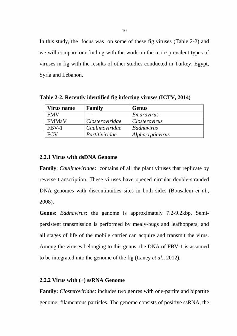
In this study, the focus was on some of these fig viruses (Table 2-2) and
we will compare our finding with the work on the more prevalent types of
viruses in fig with the results of other studies conducted in Turkey, Egypt,
Syria and Lebanon.
Table 2-2. Recently identified fig infecting viruses (ICTV, 2014)
2.2.1 Virus with dsDNA Genome
Family: Caulimoviridae: contains of all the plant viruses that replicate by
reverse transcription. These viruses have opened circular double-stranded
DNA genomes with discontinuities sites in both sides (Bousalem et al.,
2008).
Genus: Badnavirus: the genome is approximately 7.2-9.2kbp. Semi-
persistent transmission is performed by mealy-bugs and leafhoppers, and
all stages of life of the mobile carrier can acquire and transmit the virus.
Among the viruses belonging to this genus, the DNA of FBV-1 is assumed
to be integrated into the genome of the fig (Laney et al., 2012).
2.2.2 Virus with (+) ssRNA Genome
Family: Closteroviridae: includes two genres with one-partite and bipartite
genome; filamentous particles. The genome consists of positive ssRNA, the
Virus name Family Genus
FMV --- Emaravirus
FMMaV Closteroviridae Closterovirus
FBV-1 Caulimoviridae Badnavirus
FCV Partitiviridae Alphacrpticvirus

11
transmission is semi-persistent by aphids and other carriers such as
whiteflies, coccidian and pseudocode.
Genus: Closterovirus: virion contain a single strand of RNA from 14.5 to
19.3 kb in size. The genome is mono-partite, carriers are natural especially
aphids. FLMaV and FMMaV are examples on fig (Martelli et al., 2002).
2.2.3 Virus with (-) ssRNA Genome
Family: ---
Genus: Emaravirus: It have four segments of negative ssRNA of about 7.0,
2.3, 1.6 and 1.4 kb ( NCBI, 2012).The newly-detected RNA-5 and RNA-6
contain two further proteins of unknown function (Ishikawa et al., 2012).
2.2.4 Virus with dsRNA Genome
Family: Partitiviridae: Includes fungi and plants host. The plant virus
particles are isometric, bipartite genome consisting of two molecules of
linear dsRNA. Infection usually latent and the transmission is by seed and
pollen, not for vectors or graft.
Genus: Alphcryptovirus: typically contain two dsRNA segments of about
1.5 and 2.0 kbp in length (Nibert et al., 2013).
2.3 Principles of Molecular Diagnosis
The tools of diagnosis of viruses that infect fruit tree crops must possess the
qualities of versatility and ease of application and with a high sensitivity
and specificity, which allow identifying viruses already present in low

12
concentrations in the tissues of woody plants, especially in the dormant
material outside of the growing season.
Molecular techniques are rapidly developing in plant virology, where
versatility of use and the reliability of results are mprover. It can be used
as a tool for basic research and diagnostic tools. The possibility of in vitro
enzymatic synthesis of DNA sequences complementary, corresponding to
large portions or the entire viral RNA genome, has allowed in recent years
to develop methods of molecular investigation which aim to search for
specific regions of the viral genome. The ability to explore, for diagnostic
and / or identification, the entire viral genome and not just the portion
encoding the capsid protein (which represents approximately 10% of the
entire genome) is the strength of these techniques against those serological.
Other portions of the same genome may, in some moments of the
replication cycle of the virus in infected cells, accumulate in moderate
amounts in the form of double-stranded RNA encapsidation or RNA
replication (Naidu and Hughes, 2001).
The molecular diagnosis has found great possibilities of use especially in
the case of a plant virus with a single stranded RNA which represents 75%
of plant viruses. It is particularly useful where there are limitations in the
use of serological techniques, for example in the diagnosis of:
unstable viruses.
poorly immunogenic virus.
Viruses which it is difficult to obtain purified preparations.
13
Viruses with low titer in the natural and not transmissible
mechanically.
RNA satellites.
Virus strains serologically very close to or indistinguishable.
The techniques of molecular diagnosis are much more sensitive than
serological (sensitivity of the order of picograms, 10-12, that is 1000 times
more sensitive than serological techniques) for which they are able to
detect the presence of the virus even in asymptomatic plants although
infected. The nucleotide sequence makes every single nucleic acid strand
that can base-pair (hybridize) with another filament only and exclusively in
a complementary way. The uniqueness of these relationships (sequence,
complementarities, hybridization) enables molecular diagnosis (Codo,
2012).
2.3.1 Gene Amplification (PCR-polymerase chain reaction)
The PCR (Polymerase chain reaction) molecular tool, developed in the
1980s by Kary Mullis, that allows to selectively amplify the DNA
sequences related to the virus to be identified (sequence "target"), from a
complex mixture of nucleic acids present in the plant sample to be
analyzed, by the use of a DNA thermo-stable polymerase (Taq-
polymerase) and a pair of specific primers, of two short sequences
(primers) that serve as trigger for the reaction. These primers are
complementary to the sequences which delimit the DNA fragment that you
want to amplify, in our case the virus (which, for RNA viruses, has been

14
previously reverse transcribed into cDNA). Standard RT-PCR (Chiumenti
et al., 2013) was used for FMV amplification, adding specific reagents and
enzymes, and subjecting the samples to temperature cycles and appropriate
variables, one can obtain an exponential increase of the DNA fragment of
interest (amplified product) which reaches such concentrations to be
detected with a common electrophoresis (visible on a support solid - gel) in
the form of colored band with an intercalating agent fluorescent UV is Red
gel (Biotium, USA).
2.4 Objectives of the Thesis
Within the frame work of international concern in production of healthy
propagated materials as the first step toward food security, viruses and
virus-like pathogens were the main problematic pathogens to be considered
in any Phytosanitary program (certified healthy plants). For that, sanitary
status of crops must be assessed first, with particular emphasis on viral
diseases infections. Thus, this research study aimed to assess and report the
sanitary status of Fig trees in Northern West Bank; the most fig growing
areas of Palestine. For that, the main objectives of this research study were
to do:
Field surveys, to monitor the presence of virus diseases on figs, and
the symptoms they cause.
Molecular detection of some widely spread viruses using RT-PCR
techniques.
Studying portion of genome sequence variations for some of these
detected viruses

15
Chapter Three
Materials and Methods

16
3.1. Field Surveys and Samples Collection
Several fields which are grown with fig trees in northern state of West
Bank- Palestine were subjected to the inspections to detect any viral
symptoms. These fig yards were visited during the main growing seasons
of the year 2014 and 2015.
Viral symptoms were investigated for several trees randomly selected for
each fields and the incidence of infections was reported. In addition to that,
the severity of the symptoms had been reported. Some of the fields were
visited twice in the two successive growing season and symptoms were
inspected and compared.
For molecular assessments, almost 50 samples were collected from main
areas know to be planted with figs (Figure 3-1). 3-5 samples were collected
randomly from each visited field for later lab tests for the presence of four
fig viruses. This number was limited to ensure the best coverage of
geographical distribution; origin of material; age of the plants; and cultural
methods. The collections were made from both symptomless and
symptomatic trees.

17
Figure 3-1. Location and distribution of collected samples in northern West Bank-
Palestine.
Each sample was composed from several petioles collected from different
sites of the tree; grouped and labeled accordingly. These samples were
placed in plastic bags and preserved in ice-bag container and transported at
the same day to the Research Laboratory of Dr. Raed Alkowni, at the
Department of Biology, at Al- Najah National University- Nablus,
Palestine, and stored in the refrigerator at 4°C.
3.2 Total RNA Extraction
The total RNA extracted were extracted from approximately 0.1-0.4 g of
the petioles, grinded in 1 ml of grinding buffer (4 M guanidine thiocyanate;

18
2 M of NaOAc, pH 5.2; 25 mM EDTA; 1 M of KOAc; 2.5% PVP-40 and
1% sodium metabisulfite) and were silica-purified (Foissac et al., 2001).
The mixture was then placed in 1.5 ml micro-tube, vortex and spin
centrifuge then left for few minutes on ice. 500 µl of the upper aqueous
phase were collected into another micro-tube and 150 µl of NLS to 10%.
The mixture was stirred, incubated at room temperature for a few minutes;
then samples were placed in heating block at 70° C for 10 minutes with
occasional shaking. After that, the samples were cooled for at least 5
minutes on ice and centrifuged at 13,000 rpm for 3 minutes. The extract
was transferred into clean tubes joining 500 µl of NaI (6 M), 250 µl of
EtOH at 99.97% and 35 µl of silica resuspended well and left at room
temperature. After at least 10 minutes, stirring occasionally, 60 seconds
centrifugation at 6000 rpm was applied. The supernatant was removed and
the tube contains the pellet was dried. Proceed with two successive washes,
using the appropriate washing buffer. Finally the pellets were added 150 µl
of d.d.H2O, RNase-free, processing samples for 4 minutes at 70 ° C and
then in ice. The mixture was centrifuged for three minutes at full speed
(13000 rpm) and the supernatant was taken and stored at -20 ° C.
3.3 Reverse Transcription of RNA into cDNA
10 µL of extracted RNA with 1 µl of Random Primers (short randomized
hexamers were used at a concentration of 1μg / μl). It was placed for 5
minutes at 95 ° C (denaturation of nucleic acid). The mixture for the
synthesis of cDNA consists of:

19
2 µL of M-MLV buffer 10 X,
0.5 µL of 10 mM dNTPs,
2 µL of 10 mM DTT,
1 µL of reverse transcriptase 200 units / µl (enzyme M-MLV),
3.5 µL of ddH2O ( RNase free water)
The reaction took place in a final volume of 20 µl. The tubes were shaken,
followed by brief centrifugation and incubated in a thermostat at 37° C for
2 h. At the end of the reaction, the cDNA was synthesized, cooled in ice
and stored at - 20 ° C for later use.
3.4 Gene Amplification
The viruses that were examined are : FMV, FBV-1, FCV and FMMaV.
Table 3-1, is listing the names of the viruses associated with their primers
used, with relative nucleotide sequence and size of amplicon (the size of
the fragment of the target molecule to be amplified).
Table 3-1: identified fig infecting viruses with their accession numbers
and primes sequences used for RT-PCR analysis
Virus Primer sequence
5'-3'
Amplicon
size (bp) References
FMV
F
R CGGTAGCAAATGGAATGAAA
AACACTGTTTTTGCGATTGG 302
Elbeaino et
al. 2009a;&
b
FMMaV F
R
AAGGGGAATCTACAAGGGTCG
TATTACGCGCTTGAGGATTGC 311
Elbeaino et
al. 2010
FBV-1
F
R ACCAGACGGAGGGAAGAAAT
TCCTTGCCATCGGTTATCTC 474
Tzanetakis
and Martin
2010
FCV F
R
TCGGATTGTCTTTGGAGAGG
CGCATCCACAGTATCCCATT 353
Elbeaino et
al. 2011b

20
The reaction of gene amplification was performed on an aliquot of the
cDNA which it had been added to the amplification mixture that containing
the nucleotides and the sense and antisense primers for the detection of the
segment of the cDNA to be amplified.
Then, 2.5 µl of cDNA were added to the mixture of amplification which is
consisting of 5 µl of 5x reaction buffer, 0.5 μl dNTP (10 mM), 0.5 μl sense
primer , 0,5 µl antisense primer, 0.2 μl Taq polymerase (5U/µl)
(Fermentas®) and sterile water (15.8 µl) up to 25 µl of total volume. The
reactions were carried out using 35 cycles of amplification in a thermo-
cycler with the following parameters set:
Denaturation of cDNA molecules: 94 ° C for 2 minutes;
30 cycles of amplification with the following segments: 94 ° 30 sec,
55 ° C 30 sec, 72 ° C 45;
Extension of the final nucleotide chain, through the action of Taq
polymerase: 72 ° C for 7 min.
The amplified products were subsequently stored at 4 ° C.
3.5 Agarose Gel Electrophoresis
For the visualization of the amplified products from the enzymatic reaction,
1.2% agarose gel was prepared, depending on the size of the fragments, in
1x TAE for PCR product. The agarose solubility in buffer was added to 1µl
GelRed (Biotium, USA) which intercalates into DNA and become
fluorescent when excited by ultraviolet rays. That’s why it is used to detect
the DNA fragments. The size of the amplified bands was determined by

21
comparison with a commercial marker of DNA, the 1 kb Ladder
Invitrogen®, extent of 5 µl. The run was made at first with a potential
difference of 80 V, to facilitate the escape of the samples from the wells in
the gel matrix, and continued at 100 V. After the electrophoretic run, the
gel was observed and photographed on a trans-illuminator light emitting
ultraviolet GelDoc-It (UVP, USA).
3.6 Sequencing and data analysis
The PCR products were cleaned with ChargeSwitch®-Pro PCR Clean-Up
Kit (Invitrogen, USA), following the manufacturer's protocol. DNA PCR
products were sequenced by dideoxynucleotide chain termination method
using 3130 Genetic Analyzer (Applied Biosystem®, Bethlehem University,
Palestine). The sequencing PCR reaction was performed with primers used
singly in forward and reverse reaction and BigDye® Terminator v 1.1
Cycle Sequencing Kit (Applied Biosystem®, USA).
The DNA sequences of 5 samples of FBV, 3 samples of FMV and one
sample of FCV were compared with available sequences in NCBI
(National Center for Biotechnology Information) using BLAST and
multiple alignments were done by using ClustalW.

22
CHAPTER FOUR
RESULTS

23
4.1 Field surveys and biological identification of virus-associated
symptoms in Palestinian fig growing areas
Field surveys inspections to check for any visual viral disease symptoms
had been carried out in the fields of figs during the growing seasons of
2014 and 2015, to reveal that viral disease symptoms were rather broad and
diverse. Almost two third of visually inspected fig trees were found with
one or more viral symptoms. These symptoms were exhibited on leaves
and/or fruits. The symptoms on leaves were reported as mosaic; mottling;
vein clearing; yellowing and deformation (Figure 4-1).
A B

24
C D
F E
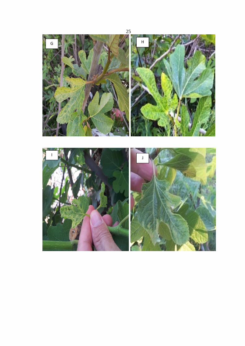
25
G H
J I

26
Figure 4-1. Viral disease symptoms were observed in the fig yards which were grown
in northern West-Bank of Palestine. Several types of symptoms were noticed on fig
leaves, such as mottling (A-B); vein clearing (C-D-E); yellowing (F-G-H); deformation
(I-J-K) and mosaic (L-M-N).
N M
K L

27
Meanwhile the fruits were examined to reveal local lesions, chlorosis, ring
spots, and mosaic. It was noticed that the newly formed figs were
exhibiting yellow ring spots (Figure 4-2) which were varied in their sizes.
The developments of infected fruits were monitored and the necrotic spots
were firstly noticed and heavily fruit drops were found later on.
A B
C D

28
Figure 4-2. Yellow ring spots and necrosis were noticed on the fruit (A-G). These fruits
were dropped down later.
E
G
F

29
4.2 Total RNA Extraction
The extracted total RNA were analyzed in 1.2% agarose gel (Figure 4-3).
This was done for all extracted RNAs to ensure their quality extractions.
Figure 4-3. Example of electrophoresis of total RNA extraction. 1.2% agarose gel in
TBE, colored with GelRed (Biotium). Lane 1-6 represents TNA from different fig leaf
samples.
4.3 Detection of FMV by PCR
DNA samples which tested positively for FMV were able to amplify the
expected PCR products of a size equal to 302 bp; using specific primer
(Elbeaino et al, 2009a, b). Results about detection of FMV using PCR in
fig samples as presented in Figure 4-4. It showed that 60% of samples were
infected with FMV.

30
Figure 4-4. PCR results of FMV: Lane M contained ladder (1Kb DNA ladder RTU,
GeneDireX)), lane 4 represents negative sample, lane 1, 2 and 3 represents FMV
positive sample on 302 bp.
4.4 Detection of FBV by PCR
Positive samples were those generated the expected PCR products with size
equals to 474 bp. The detection of FBV was done by using PCR with
specific primers (Tzanetakis and Martin, 2010) as presented in Figure 4-3.
Results of this research showed that 46% of samples were infected with
FBV.

31
Figure 4-5. PCR results of FBV: Lane M contained ladder (1Kb DNA ladder RTU,
GeneDireX)), lane 1 and 2 represents negative samples, lane 3 represents FBV positive
sample on 474 bp.
4.5 Detection of FCV by PCR
Results of this research showed that only (2%) of fig samples were infected
with FCV. Only one sample of fig was detected using specific primers
(Elbeaino et al., 2011b). The size was equal to 353 bp as presented in
(Figure 4-4).

32
Figure 4-6. PCR results of FCV: Lane M contained ladder (1Kb DNA ladder RTU,
GeneDireX), lane 1 represents negative sample, lane 2 represents FCV positive sample
on 353 bp.
4.6 Results of Gene Amplification
From the analysis of results from assays of molecular diagnosis RT-PCR,
relating to each virus, there has been a presence in percentage of 60% of
FMV, 46% of FBV and 2% of FCV (Table 4-1)

33
Table 4.1 Analysis of gene amplification (RT-PCR) tested samples of
fig.
Region Tested Positive % FMV % FMMaV % FCV % FBV %
Jenin 12 10 83 8 67 0 0 0 0 6 50
Nablus 16 14 87.5 11 68.8 0 0 0 0 9 56.3
Ramallah 5 4 80 3 60 0 0 0 2 40
Qalqilyah 12 8 67 5 42 0 0 1 8 4 33
Tulkarem 5 3 60 3 60 0 0 0 0 2 40
Total 50 39 78 30 60 0 0 1 2 23 46
4.7 Prevalence of Viruses in Each Province
As regards instead FMMaV, it was not found to be present in any of the
samples analyzed. The results showed that the high percentage of FMV
was recorded from Nablus 68.8% and Jenin 66.7%, as well as FBV from
Nablus 56.3% and Jenin 50% (Figure 4-7).
Figure 4-7. Incidence of FMV, FBV and FCV infections in five-growing provinces of
northern West Bank- Palestine as determined by RT-PCR assays.
0%
10%
20%
30%
40%
50%
60%
70%
Jenin Nablus Tulkarem Qalqiya Ramalla
50%
56.30%
40%
33.30%
40%
0 0 0
8%
0
66.70% 68.80%
60%
42%
60%
FBV
FCV
FMV

34
The results show that Nablus province have the most total infected rate
with 88% in Nablus (14/16), 83% in Jenin (10/12), 80% in Ramallah (4/5),
67% in Qalqilya (8/12) and 60% Tulkarem (3/5). (Figure 4-8).
Figure 4-8. Virus incidence on northern West Bank- Palestine fig provinces.
4.8 Sequencing
Depending on the results observed and because of the high percentage of
both FMV and FBV, five samples of FBV, four samples of FMV and the
only sample of FCV were selected from each province ( Appendix A).
Results showed that high similarity with published ones in GenBank 98-
100% (Appendix B).
Nablus 88%
Qalqilia 67%
Jenin 83%
Ramalla 80%
Tulkarem 60%

35
Chapter Five
Disscusion, Conclusion and
Recommendation
36
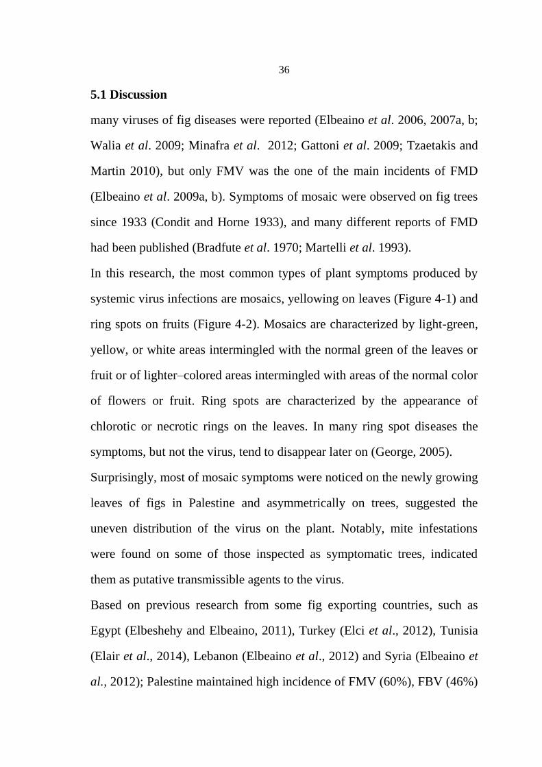
5.1 Discussion
many viruses of fig diseases were reported (Elbeaino et al. 2006, 2007a, b;
Walia et al. 2009; Minafra et al. 2012; Gattoni et al. 2009; Tzaetakis and
Martin 2010), but only FMV was the one of the main incidents of FMD
(Elbeaino et al. 2009a, b). Symptoms of mosaic were observed on fig trees
since 1933 (Condit and Horne 1933), and many different reports of FMD
had been published (Bradfute et al. 1970; Martelli et al. 1993).
In this research, the most common types of plant symptoms produced by
systemic virus infections are mosaics, yellowing on leaves (Figure 4-1) and
ring spots on fruits (Figure 4-2). Mosaics are characterized by light-green,
yellow, or white areas intermingled with the normal green of the leaves or
fruit or of lighter–colored areas intermingled with areas of the normal color
of flowers or fruit. Ring spots are characterized by the appearance of
chlorotic or necrotic rings on the leaves. In many ring spot diseases the
symptoms, but not the virus, tend to disappear later on (George, 2005).
Surprisingly, most of mosaic symptoms were noticed on the newly growing
leaves of figs in Palestine and asymmetrically on trees, suggested the
uneven distribution of the virus on the plant. Notably, mite infestations
were found on some of those inspected as symptomatic trees, indicated
them as putative transmissible agents to the virus.
Based on previous research from some fig exporting countries, such as
Egypt (Elbeshehy and Elbeaino, 2011), Turkey (Elci et al., 2012), Tunisia
(Elair et al., 2014), Lebanon (Elbeaino et al., 2012) and Syria (Elbeaino et
al., 2012); Palestine maintained high incidence of FMV (60%), FBV (46%)

37
(Figure 5-1). Meanwhile FCV was detected in few samples, showing less
incidences compared with other countries.
Figure 5-1. Compared with the previous research on viral diseases of figs with northern
West Bank- Palestine Results.
Clearly, this study was revealed that the most infected viruses were FMV
and FBV are in agreement with a previous survey study of Alkowni
research (Alkowni et al., 2015). In addition, the fact that FM was recorded
in all provinces and field in northern West Bank indicates its seriousness,
similarity with a study in Jordan (Al Mughrabi and Anfoka, 2000).
Virus–virus interactions in plants may be of crucial significance for the
understanding of viral pathogenesis and evolution and consequently for the
development of efficient and stable control strategies (Syller, 2011). In
Turkey, complex infection were detected in the most samples and the most
0%
10%
20%
30%
40%
50%
60%
70%
80%
90%
30%
8.90% 2%
4%
28.30%
12.20% 8.40%
26.5%
79%
46.70%
56.70%
37.12% 60%
60%
82%
46%
FCV
FMMaV
FMV
FBV

38
common viruses were FMV+FBV-1 with 90% (Elci et al., 2012) and 52%
in Syria (Elbeaino et al., 2012). In this research, mixed infections of fig
viruses (FMV+FBV-1) were found in the tested cultivars from different
provinces with 30%. Hence, most attentions have been paid to virus
interactions in multiple infections in infected fig trees.
The results show that Nablus province have the most total infected rate
(88%) when it was compared with others (Figure 4-8). This could be
referred to most fig nurseries which are located in this province produced
by cutting from infected mother trees. Also, pruning tools are used without
disinfection between cuttings and may even be shared by different
nurseries. The weather conditions in Nablus, with moderate humidity and
temperatures, are very favorable for plant propagation, but also favor for
the vectors that spread viral diseases. Direct correlation between the
geographic distribution and incidence of viral pathogen affecting of
agricultural were noticed in this research study. Moreover, due to the
different geographic distribution of the fig tree cultivation in the provinces
that have been previewed in the research. Depending on the Ministry of
Agriculture data in Ramallah- Balou, Till area in Nablus, is the most
widely distributed and growing figs (29%). Jaba' - Jenin (10%) , Senjil -
Ramallah (6%), Jauos - Qalqilya (5%) and Attel - Tilkarem (2%). So, It is a
direct correlation between the geographic distribution and viral pathogen
affecting of agricultural crop.
Portions of the FMV and FBV genomes were sequenced to reveal high
similarity with published ones in Gen bank (98-100%) (Appendix A and B)

39
The picture that emerges from the molecular diagnosis of the virus of the
fig tree in the numerous accessions of native varieties sampled in this study
is quite worrying implications for the quality of fruit production. The
almost ubiquitous presence of several viruses, among which the FMV,
mirrors the presence of symptoms as extensive mosaic were found. FMV,
as known to be transmitted quickly and with high efficiency by means of
the mite, it was therefore obviously noticed over large areas of fig plants.
However, the majority of the symptoms observed, can't be attributed to a
given virus, among those least sought and found, given the abundance of
mixed infections of more viruses. The wide range of symptoms described
could then be determined by phenotypic synergy due to the association of
different viruses, some of which certainly still unknown.
The limited presence of FCV is equally understandable as given to their
transmission difficulties. The cryptic virus did not have any known carriers
and could be transmitted mostly by seeds or asexual propagation. The
constant infection of FBV-1 instead is certainly due to its ancestral co-
integration into the genome of fig, and the most likely transmission
efficiency through scale insects or other potential insect vectors.
5.2 Conclusion and Recommendations
The outcome of this preliminary work extends the knowledge on the spread
of fig viruses in the Mediterranean region, particularly in northern West
Bank- Palestine. This is the first report in northern West- Bank fig orchards
of FBV, FMV, FCV and FMMaV, talking about the sanitary assessment of

40
fig viruses and the percentage of appearance on fig trees. Although this
assessment was limited to 50 trees, the results obtained clearly indicate
how the sanitary status of fig crop has deteriorated in northern West Bank
(78% of viral infection). Special worrying is for the incidence of FMV,
since this has proved to be the unique virus closely correlated with the
FMD (Elbeaino et al., 2009a). High incidence of FMV is not surprising
considering the way this virus spreads in figs through infected propagating
material (cuttings and grafting), and natural vectors (eriophyid mites)
(Alkowni et al., 2015). In Palestine there is no information on the presence
of Aceria ficus (Eriophyidae). However, such presence in the Palestinian
orchards would likely aggravate the sanitary status and the level of
infections in the surveyed areas. The several FMV-infected samples found
in association with most of the mosaic symptoms in the field further
confirms what was previously reported regarding the etiology of FMV. In
Palestine, fig production is in developing emphasis for more intensive fig
cultivation , three thousand tons are produced in almost three thousand and
five hundred acres in West Bank in 2010 (PCBS, 2010). Figs like other
agriculture crops are infected with viruses. The widespread and severe
symptoms were presented on young fig leaves can cause problems such as
the impact on the productivity of the crop. It may become more of an
agricultural problem with the introduction of intensive cultivation and
viruses may effect on fruiting period of the plant. Also, as the results show
and based on the geographical distribution, the probability of infection
increases with the agricultural area of the crop.

41
The knowledge we have gained on the incidence of virus diseases of fig in
northern West Bank provides information on which a sanitary selection,
sanitation and certification programs can be initiated for the production of
healthy propagating plant material of fig in this country. In recent years
they have added new consisting in particular applications of in-vitro culture
of plant tissues: somatic embryogenesis and cryotherapy.
This study is come out with many recommendations regarding to the fig
industry. The setting of a regional plan of defense against virus diseases of
a particular crop must be based on knowledge of essentially harmful
viruses in a given area; the modes of transmission and the epidemiological
and ecological conditions favorable to infection. The prevention of viral
infections is mainly based on the adoption of the following measures:
Use of seed and planting material free of viruses.
Elimination of the possibility of infection.
Growing species and / or varieties tolerant or resistant to the virus
or to the carrier.
Integrating conventional technologies with molecular biology and genetic
engineering could enhance desirable characteristics of agricultural crops
while reducing the expression of undesirable ones. Using improved
conventional breeding in fig, by molecular markers or by the newly
introduced genetic engineering technology, could enhance new properties,
such as health-promoting compounds.

42
References
Aljane F., Ferchichi A., and Boukhris M. Pomological charachteristics
of local fig (Ficuscarica) cultivars in southern Tunisia. Acta
Horticulturae. 2008; 798: 123-128.
Alkowni R., Chiumenti M Minafra A., and Martelli. GP. A survey for
fig infecting viruses in Palestine. Journal of Plant Pathology. 2015; 97:
383-386.
Alkowni R., and Srouji F. Food Security and Viral Diseases (in Fruit
Trees). Palestine Economic Policy Research Institute (MAS). Ramallah-
Palestine. [In Arabic]. 2009.
Al-Mughrabi K.I., and Anfoka G.H. Distribution of fig mosaic in
Jordan. Phytopathol. Mediterr. 2000, 39:263-270
ARIJ. Dry land Farming in Palestine. Applied Research Institute of
Jerusalem, Bethlehem, Palestine. 1994.
Blodgett E.C., Gomec C. Fig mosaic. Plant Disease Reports. 1967; 51:
893-896.
Bousalem M., Douzery F.J.P., Seal S.E. Taxonomy, molecular
phylogeny and evolution of plant reverse transcripting viruses
(family Caulimoviridae) inferred from full-length genome and
reverse transcriptase sequences. Arch. Virol. 2008; 153: 1085-1102.
Bradfute O.E., Whitmoyer R.E., Nault R.L. Ultrastructure of plant
leaf tissue infected with mite-born viral-like particles. Proc. Elect.
Micros. Soc. America. 1970; 28: 178–179.
Caglayan K., Serce C.U., Barutcu E., Kaya K., Medina V., Gazel M.,
Sovlu S., and Caliskan O. Comparison by sequence-based and
electron microscopic analyses of Fig mosaic virus isolates obtained

43
from field and experimentally inoculated fig plants. Plant Diseases.
2010; 94: 1448-1452.
Chiumenti M., Campanale A., Bottalico G., Minafra A., De Stradis A.,
Savino A., Martelli G. P. Sanitation Trials for the production of
virus-free fig stocks. Journal of Plant Pathology. 2013; 95: 655-658.
Codo F. Application of molecular diagnostic techniques for viral testing. NCBI. Open Virol.
J. 2012; 6: 104-114.
Condit I.J. Fig Verities. Hilgardia: A Journal of Agricultural Science
Published by the California Agricultural Experiment Station; February.
1955; 11: 323-326.
Condit I.J., and Horne W.T. A mosaic of the fig in California.
Phytopathology. 1933; 23: 887-896.
Crane J.C., and Brown J.B. Growth of the fig fruit, Ficus carica var.
Mission. Proc. Am. Soc. Hort. Sci. 1950; 56: 93-97.
Elair M., Mahfoudhi N., Bayoudh C., Selmi I., Mars M., and Dhouibi
M.H. Sanitary Selection of Virus-Tested Fig (Ficus carica) Cultivars
in Tunisia. Journal of Plant Protection. 2014; 9: 100-109.
Elbeaino T., Abou Kubaa R., Ismaeil F., Mando J., and Digiaro M.
Viruses and hop stunt viroid of fig trees Syria. Journal of Plant
Pathology. 2012; 94: 687-691.
Elbeaino T., Choueiri E., Hobeika and Digiaro, M. Presence of Fig
mottle-associated virus 1 and 2 in Lebanese fig ochards. J. Plant
Pathol. 2007b; 89: 409-411.
Elbeaino T., Digiaro M., De Stradis A., Martelli G.P. Partial
characterization of a closterovirus associated with a chlorotic
mottling of fig. Journal of Plant Pathology. 2006; 88: 187-192.

44
Elbeaino T., Digiaro M., De Stradis A., Martelli G.P. Identification of a
second member of the family Closteroviridae in mosaic-diseased figs.
Journal of Plant Patholog. 2007a; 89: 119-124.
Elbeaino T., Digiaro M., Alabdullah A., De Stradis A., Minafra A.,
Mielke N., Castellano M.A., Martelli G.P. A multipartite single-
stranded negative-sense RNA virus is the putative agent of fig
mosaic disease. Journal of General Virolog. 2009a; 90: 1281-1288.
Elbeaino T., Digiaro M., De Stradis A., Martelli G.P. Complete
nucleotide sequence of four RNA segments of Fig mosaic virus.
Archives of Virolog. 2009b; 154: 1719-1727.
Elbeaino T., Digiaro M., Martelli G.P. Complete nucleotide sequence
of Fig fleck-associated virus, a novel member of the the family
Tymoviridae. Virus Research. 2011a; 161: 198-202.
Elbeaino T., Heinoun K., Digiaro M., Martelli G.P. Fig mild mottle-
associated virus, a novel closterovirus infecting fig. Journal of Plant
Pathology. 2010; 92: 165-172.
Elbeaino T., Mortada C., Digaro M., Choueiri E. Suurvey on fig
viruses in Lebanon. Acta horticulture. 2012; 940: 665-668
Elbeaino T., Kubaa R.A., Digiaro M., Minafra A., Martelli G.P. The
complete nucleotide sequence and genome organization of Fig
cryptic virus, a novel bipartite dsRNA virus infecting fig, widely
distributed in the Mediterranean basin. Virus Gene. 2011b; 42: 415-
421.
Elbeshehy E.K.F., and Elbeaino T. Viruses infecting figs in Egypt.
Phytopathol. Mediterr. 2011; 50: 327−332.
Elci E., Serce C., Gazel M., and Caglayan K. Molecular Detection and
Comparative Sequence Analysis of Viruses Infecting Fig Trees in

45
Turkey. J Phytopathology. Mustafa Kemal University, Antakya-Hatay,
Turkey. 2012; 160: 418–423.
FAO. Food and Agriculture Organization of the United Nations.
2009; available from: http://faostat.fao.org/default.aspx
FAO. The FAO statistical database-Agriculture. 2014 January 3;
available from:
http://faostat.fao.org/DesktopDefault.aspx?PageID=339&lang=en&coun
try=4
Flaishman M., Rodov V., and Stover E. The Fig. Botany, Horticulture,
and Breeding. Horticultural Reviews, John Wiley & Sons, Inc. 2008;
34: 113-197.
Flock R.A., and Wallace J.M. Transmission of fig mosaic by the
eriophyd mite Aceria ficus. Phytothology. 1955; 45: 52-54.
Foissac X., Svanella-Dumas L., Gentit P., Dulucq M.J., Marais A.,
Candresse T. Polyvalent degenerate oligonucleotides reverse
transcription-polymerase chain reaction: a polyvalent detection and
characterization tool for Trichoviruses, Capilloviruses, and
Foveaviruses. Phythopathology. 2005; 95: 617-625.
Gattoni G., Minafra A., Castellano M.A., De Stradis A., Boscia D.,
Elbeaino T., Digiaro M., Martelli G.P. Some properties of Fig latent
virus 1, a new member of the family Flexiviridae. J Plant Pathol.
2009; 91: 543–552.
George N. Agrios. Plant Pathology. Fifth edition. 2005; 737.
Grbelja J., and Eric Z. Isolation of a potyvirus from Ficus carica. L.
Acta Bot. Croat. 1983; 42:11-14.
ICTV. International Committee of Taxonomy of Viruses [Internet].
Virology Division-IUMS. 2014; available from:
http://www.ictvonline.org/virusTaxonomy.asp

46
Ishikawa K., Maejima K., Komatsu K., Kitazawa Y., Hashimoto M.,
Takata D. Yamaji Y., Namba S. Identification and characterization of
two novel genomic RNA segments of fig mosaic virus, RNA5 and
RNA6. J. Gen. Virol. 2012; 93: 1612–1619. Janzen H. How to be a Fig.
Department of Biology, University of Pennsylvania, Philadelphia. 1979;
10: 13-51.
John P. In Biosynthesis of the Major Crop Products, Wiley, New
York. 1993; 114-126.
Laney A.G., Hassan M., Tzanetakis I.E. An integrated badnavirus is
prevalent in fig germplasm. Phytopathology. 2012; 102: 1182-1189
Martelli G.P., Agranovsky A.A., Bar-Joseph M., Boscia D., Candresse
T., Coutts R.H.A., Dolja V.V.,Falk B.W., Gonsalves D., Jelkmann W.,
Karasev A.V., Minafra A., Namba S., Vetten H.J., Wisler G.C.,
Yoshikawa N. The family Closteroviridae revised. Virology division
news. Arch. Viro. 2002; 2039-2044.
Martelli G.P., Castellano M.A., Lafortezza R. An ultrastructural study
of fig mosaic. Phytopathologia Mediterranea. 1993; 32: 33-43.
Minafra A., Chiumenti M., Elbeaino T., Digiaro M., Bottalico G.,
Pantaleo V., Martelli G.P. Occurrence of Fig badnavirus- 1 in fig
trees from different countries and in symptomless seedlings. Journal
of Plant Pathology. 2012; 94: S4.105.
Morton J. Fruits of warm climates [Internet]. Horticulture &
Landscape Architecture. 2012 May15; available from:
http://www.hort.purdue.edu/newcrop/morton/fig.html#Toxicity
Naidu R.A. and Hughes J.d.A. Methods for the detection of plant
virus diseases. Proceedings of a Conference Organized by IITA,
International Institute of Tropical Agriculture, Nigeria. 2001; 233–260.

47
NCBI. National Center of Biotechnology Information [Internet]. 2012
Sep 12; available from:
http://www.ncbi.nlm.nih.gov/pmc/articles/PMC3499817/
Nibert M.L., Said A. Ghabrial S.A., Edgar Maiss E., Lesker T.
Reorganization of family Partitiviridae to contain 4 new genera of
plant and/or fungal viruses, with elimination of 3 current genera
and creation of 16 new species. ICTV, 2013; 1-50.
Namba, S. Fig virus in Handbook of Plant Viruses. K. Yora, Y. Saito,
Y. Doi, T. Inoue and K. Tomaru ads. Asakura Shoten, Tokyo, Japan.
1983; 326-327.NGA (National Gardening Association) [Internet]. 2011
Available from: http://www.garden.org/plantguide/?q=show&id=3328
PCBS. (Palestine Central Bureau of Statistics). Agro. Census-
Palestinian territories. Ramalla – Palestine. 2010.
Peter H., Raven, Georgo B., Johnson. Biology. The McGraw-Hill
Companies. 2001; 666-667.
Serrano L., Raman J., Segarra J., Medina V., Achon M.A., Lopez M.,
and Juarez M. New approch in the identification of the causal agent
of fig disease. Acta Horticulturae. 2004; 657: 559.
Skare J.M., Wijkamp I., Denham I., Rezende J.A.M., Kitajima E.W.,
Park J.W., Desvoyes B., Rush C.M., Michels G., Scholtof K.B., and
Scholtof H.B. A new eriophyid mite-borne membrane-envelope
virus-like complex isolated from plants. Virology. 2006; 347: 343-353
Syller J. Facilitative and antagonistic interactions between plant
viruses in mixed infections. Molecular Plant Pathology. 2012 February;
available at: http://onlinelibrary.wiley.com/doi/10.1111/j.1364-
3703.2011.00734.x/abstract
Tzanetakis I. and Martin R. New viruses found in fig exhibiting
mosaic symptoms. 21st International Conference on Virus and other

48
Graft Transmissible Diseases of Fruit Crops. Julius-Kuhn-Archiv. 2010;
427.
USDA (United State Department of Agriculture) [Internet]. 2011
May11; available
from;https://plants.usda.gov/java/ClassificationServlet?source=display&
classid=FICUS
Walia J., Salem N., and Falk B. Partial sequence and survey analysis
identify a multipartite, negative-sense RNA virus associated with fig
mosaic. Plant Diseases. 2009; 93: 4.

49
Appendixes
Appendex A
Sequences
>FBV-1/J (98%)
TCAATGTTGGTTTGCTTACGAATAAGCCTGTGACGGATTACCTA
GCAAGCAGGGGAGTCCAAGCTTTGCCGGGAAGAAGATACAGAT
CGGAGATGCTACGAGGAAGAAACTGGATCATAAGGCAGCCACA
GATCCAGGCGGCAATGATGCCAAGGAACGTGGAAACAAGGA
>FBV-1/ Q (99%)
TCAGGACTCAATGTTGGATTTGCTTACAGAATAAGCCTGTGACG
GATTACCTAGCAAGCAGGGGAGTCCAAGCTTTGCCGGGAAGAA
GATACAGATCGGAGATGCTACGAGGAAGAAACTGGATCATAAG
GCAGCCACAGATCCAGGCGGCAATGATGCCAAGGAACGTGGAA
ACAAGGA
>FBV-1/T (99%)
ACATGCAATGTTGGATTTGCTTACAGAATAAGCCTGTGACGGAT
TACCTAGCAAGCAGGGGAGTCCAAGCTTTGCCGGGAAGAAGAT
ACAGATCGGAGATGCTACGAGGAAGAAACTGGATCATAAGGCA
GCCACAGATCCAGGCGGCAATGATGCCAAGGAACGTGGAAACA
AGG

50
>FBV-1/R (98%)
GTTGGTTTGCTTACGAATAAGCTGTGACGGATTACCTAGCAAGC
AGGGGAGTCCAAGCTTTGCCGGGAAGAAGATACAGATCGGAGA
TGCTACGAGGAAGAAACTGGATCATAAGGCAGCCACAGATCCA
GGCGGCAATGATGCCAAGGAACGTGGAAACAAGGAGATTTTTTT
T
>FBV-1/N (99%)
TGTGACGGATTACCTAGCAAGCAGGGGAGTCCAAGCTTTGCCGG
GAAGAAGATACAGATCGGAGATGCTACGAGGAAAAAACTGGAT
CATAAGGCAGCCACAGATCCAGGCGGCAATGATGCCAAGGAAC
GTGGAAACAAGGGTTGGATTTGCTTACAGAATAAGCGGGCAAG
G
>FMV/J (98%)
ATTGCGGTTACGGCGCTGTTTTATTTTATAAAGCGTTAAAAGTTC
CAAGGATACCACCCCTTTGAGAATTCGCCGCTTCGGGATACCAT
TTGTGTTTCCAACAAGATCAACATTAATCTTGCCAGTCTTTGCTC
AACAAGATCAACATTAATCTTGCCAGTCTTTGGTTTCCAACAAG
ATCAACATTAATCTTGCCAGTCTTTAAAAGTTACCAGCCTGCCA
GTTATAAATTTTTGGATCA

51
>FMV/Q (100%)
TTCGGTATGTGTTTCCAACAAGATTTCCACGCTCAACTTATCCTT
GAATCTGCCAGGGAACCAATTAATTTGGTTTCCAACAAGATCAA
CAAATGGTACCCATTAATCTTGCCAGTCTTTAAAAGTTCAACAA
GATTTGCCAGTTGCCAGTCTTT
>FMV/R (99%)
CTGTTATTGTGTTTCCAACAAGATCAACATTAATCTTGCCAGTCT
TTAAAAGTTCTTTCTTACCAGCTTTGATACCATTATCTTGAATCT
GCCAAGGGAACCAGTTATAAATTGCTGGAAATTTGGATCAAATG
GTACCAAATCTATATTG
>FCV/Q (98%)
ATGGAAGCAGGTCTTATAGAGATTGGAAACGACACTTATTGAAG
GAGGTTTTGACCACGTCCCATTGAATCGAGCTCATCAGCCGGCT
ACGGCTATGTTAAAGTTCGGATTGTCTTTCGTTCAAAGGAAATG
CTTAGGTTAACAAATGTGCAGTCTTACTCAGAGAC

52
Appendex B
Cladogram Tree
Phylogenetic relationship of FMV, FBV and FCV, computed by Clustal W

انوطية انجاح جايعة
كهية انذراسات انعهيا
ؤثز عهى ج نأليزاض انفيزوسية انحينحقييى انصحي ا
فهسطي -انضفة انغزتية ثاجات انحي في شال
إعذاد
يوسف يحوديى يحذ
إشزاف
د. رائذ انكوي
ووهانع تزايج في اناجسحيز درجة ىهع انحصول ثاتهنحط اسحكاال األطزوحة هذ قذيث
سطيهف -سهات في انوطية، انجاح جايعة في يا،هانع انذراسات يةهتك انحياجية،
5102

ة
-انضفة انغزتية في شالؤثز عهى ثاجات انحي ج نأليزاض انفيزوسية انحي انحقييى انصحي
فهسطي
إعذاد
يى يحذ يوسف يحود
إشزاف
انكوي د. رائذ
انهخص
ره ؼذح ،ػغ اظح حبط١ اضساػ١خ ف فغط١ ذسح ف اج١ببد اخبطخ ػ ا بن
عء ،أعجبة ب9 ػذ رفش ؼذاد زشخ١ض األشاع, مض اف١١ ازخظظ١ اؼبء
ألذ أاع افاو األوثش ( Ficus caricaاألشطخ اضساػ١خ. شدشح از١ ) اشبررظ١
ح١ث رؼزجش ث١ أئه افاو از ثذا حع اجحش األث١غ ازعط، ش١ػب ف و
ح أ شع أشبسد اذساعبد األخ١شرزؼشع إلطبثخ ثبألشاع ادشاث١ اغججخ شع.
. ف زا اذساعخ أخش٠ذ ػ١خ غح ١ذا١خ ف اشع اشئ١غ ازشش فغ١فغبء از١
عف١ذ ،طثبط ،بثظ ،لم١١خ ،طىش ،فغط١ )خ١-ابطك اشب١خ اؼفخ اغشث١خ
،. ثبإلػبفخ إ ره4102-4102ي ع١ ػ ازا اهلل ( شالجخ األػشاع ره خال سا
بثظ ،(04خ١ ) از١ ره ابطك از ر غحب ػ اح ازب9 ػ١خ 21ر خغ
ىشف ػ أاع ف١شعبد از١ ث ،(2( أخ١شا طىش ) 04لم١١خ ) ،(2سا اهلل ) ،(01)
Fig mosaic virus (FMV), figbadnavirus-1 (FBV-1), Fig cryptic virus
(FCV) (FMMaV) Fig mild-associated virus رم١خ ابعخ اؼىغره ثبعزخذا-
ثبالػزبدثبدئبد زخظظخ ف ازفبػ. خالي اغح ا١ذا ثخد ،رفبػ اجشح ازغغ
فمذ ظشد ،أػشاع ف١شع١خس از١ ػ١ بشدأ ثث١ب ٠مشة خذ ،ػ اىشف اجظش
،ازجم١غ ،أػشاع اسق9 افغ١فغبء أثخ .اثشحاألػشاع ػ األساق وزه ػ
ظس األػشاع اف١شع١خ ػ األساق حذ٠ثخ ا ثشى ،طفشاس ازش. ب أثبس اذشخاإل
ر اؼثس ػ رفش ادذ٠ش ثبزوش أ زضا٠ذ ازص٠غ اغ١ش زبث ألػشاع ػ اشدشح.
. ػ احزب١خ و ابل شع داخ ثزه ،ثبألػشاع ػ ره األشدبس اظبثخ اؼبوت
حذ٠ثخ ا ظبثخ ثجمغ طفشاء حم١خ اشى رخزف ف احد أساقر الحظخ ،إػبفخ إ ره

ج
ؤد٠خ إ االػزمبد ثخد ،دالخ ػ أ اإلطبثخ اف١شع١خ أخزد غىب د١ب ،اشذح
مبي داخ ابدح اح١خ. لذ ر الحظخ ثمغ خش٠خ ػ أشدبس اف١شط لذسر ابئخ ػ االز
ر اىشف ،رفبػ اجشح ازغغ-ثبعزخذا رم١خ ابعخ اؼىغ ،خض٠ئ١ب اظبثخ. ثبس از١
%(. وب أوثش أاع اف١شعبد 87ف اؼ١بد از ر فحظب ػ اؼذي اؼب إلطبثخ )
FMV%(. خذ ض٠ح ػذ ف١شط 21) FBV-1 ١٠ %FMV (11) ازشبسا
FBV-1 ( 01ب ٠مبسة .)% أظش رغغ اد١ ػ١بد ف١شط لذFMV FBV-1
ف ز .%(011-%87خد رشبث ػب غ ره از ششد ف ثه اؼبد اد١ )
ازذس اظح ز١ ف اطمخ اشب١خ اؼفخ اغشث١خ خظطب مذ ثبد خ١ب ،اذساعخ
عاد ػذ٠ذح. ٠ؼزجش زا عبحبد از١ اضسػخ ز ثبسرفبع ػذد حبالد اؼذ اف١شع١خ ف
ػ احظي وج١شحب زه اف١شعبد خغبئش زخ إ اإلسشبد ازظ١ امك بدافؼ
إ ازخػ اػغ اظح ز١ ف اجالد غ اؼءاذساعخ ز مذأ. وب اإلزبج
أشزبي ر١ طح١خ خب١خ اف١شعبد.اعزخذا
أشاع ،از١ ،رفبػ اجشح ازغغ -رم١خ ابعخ اؼىغ از١، ف١شعبد9 انزئيسية اتانكه
از١.رجشلش