Embed Size (px)
Citation preview
DIFFERENTIATION OF HUMAN INDUCED PLURIPOTENT STEM CELLS
TO OLIGODENDROCYTE PROGENITOR CELLS
A THESIS
SUBMITTED TO THE FACULTY OF THE GRADUATE SCHOOL OF THE UNIVERSITY OF MINNESOTA
BY
Sandhya Subramaniam
IN PARTIAL FULFILLMENT OF THE REQUIREMENTS
FOR THE DEGREE OF
MASTER OF SCIENCE
Ann Parr
James Dutton
December 2012

© Sandhya Subramaniam, 2012

Acknowledgements
I would like to thank my advisor Dr. Ann Parr for giving me this opportunity to work in
her laboratory. Also I would like to thank Dr. James Dutton for his support and
guidance throughout my project. I would like to thank Dino Terzic, Mike Ritchie,
Christina DiBartolomeo, Tarini Goyal and other members of the Parr lab for their
support. I would like to acknowledge Lucas Greder, Joseph Dalton and other members
of the slack lab for their timely counsel and assistance. I would also like to thank Dr.
Susan Keirstead for her guidance and counsel during the past year. Finally I would like
to thank my family and friends for their patience and support.
i
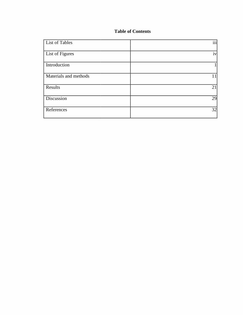
Table of Contents
List of Tables iii
List of Figures iv
Introduction 1
Materials and methods 11
Results 21
Discussion 29
References 32

ii
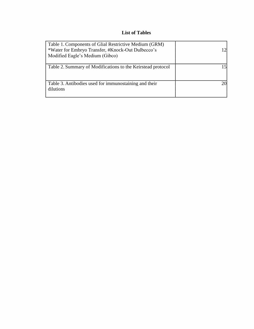
List of Tables
Table 1. Components of Glial Restrictive Medium (GRM)
*Water for Embryo Transfer, #Knock-Out Dulbecco’s 12
Modified Eagle’s Medium (Gibco)
Table 2. Summary of Modifications to the Keirstead protocol 15
Table 3. Antibodies used for immunostaining and their 20 dilutions

iii
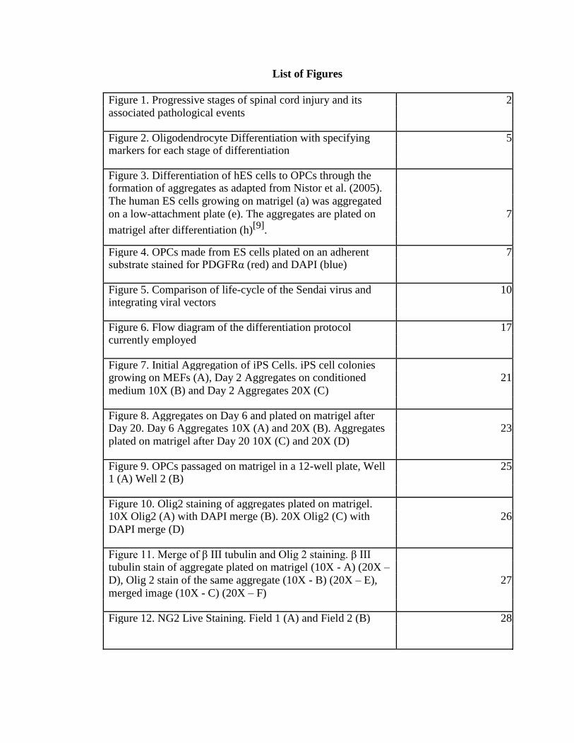
List of Figures
Figure 1. Progressive stages of spinal cord injury and its 2
associated pathological events
Figure 2. Oligodendrocyte Differentiation with specifying 5 markers for each stage of differentiation
Figure 3. Differentiation of hES cells to OPCs through the
formation of aggregates as adapted from Nistor et al. (2005).
The human ES cells growing on matrigel (a) was aggregated
on a low-attachment plate (e). The aggregates are plated on 7
matrigel after differentiation (h)[9]
.
Figure 4. OPCs made from ES cells plated on an adherent 7 substrate stained for PDGFRα (red) and DAPI (blue)
Figure 5. Comparison of life-cycle of the Sendai virus and 10 integrating viral vectors
Figure 6. Flow diagram of the differentiation protocol 17 currently employed
Figure 7. Initial Aggregation of iPS Cells. iPS cell colonies
growing on MEFs (A), Day 2 Aggregates on conditioned 21
medium 10X (B) and Day 2 Aggregates 20X (C)
Figure 8. Aggregates on Day 6 and plated on matrigel after
Day 20. Day 6 Aggregates 10X (A) and 20X (B). Aggregates 23
plated on matrigel after Day 20 10X (C) and 20X (D)
Figure 9. OPCs passaged on matrigel in a 12-well plate, Well 25 1 (A) Well 2 (B)
Figure 10. Olig2 staining of aggregates plated on matrigel.
10X Olig2 (A) with DAPI merge (B). 20X Olig2 (C) with 26
DAPI merge (D)
Figure 11. Merge of β III tubulin and Olig 2 staining. β III
tubulin stain of aggregate plated on matrigel (10X - A) (20X –
D), Olig 2 stain of the same aggregate (10X - B) (20X – E), 27
merged image (10X - C) (20X – F)
Figure 12. NG2 Live Staining. Field 1 (A) and Field 2 (B) 28

iv
INTRODUCTION
According to the 2011 annual statistical report obtained by the National Spinal
Cord Injury Statistical Center (NSCISC) at the University of Alabama at Birmingham,
approximately 12,000 new cases of Spinal Cord Injury (SCI) are reported each year
with the average life expectancy decreasing by at least 10 years depending on the
severity of the injury[1]
. The 2011 annual statistical report by the NSCISC also reports
that the yearly expenses of a paraplegic patient post injury is approximated to $66,106
and increases with the severity of injury[1]
. It has been identified that 67% of spinal
cord injuries occur due to vehicular accidents and falls.
SCI is a physically debilitating and emotionally taxing problem for patients.
They can incur a permanent loss of sensory and motor function below the level of
injury. Several functions of the autonomous nervous system such as bladder control,
urination and sexual arousal can also be affected after a spinal cord injury. Depending
on the level of injury and symptoms displayed, spinal cord injuries have different
degrees of severity. The most common grading system is the ASIA scoring system
described by the American Spinal Injury Association[2]
. This grades injuries from A to
E, where “A” is a clinically complete injury with no motor or sensory function below
the level of injury. “E” is a normal individual with no motor or sensory dysfunction, and
the grades in between describe varying degrees of clinically incomplete injury. An
injury that lies in the cervical region of the spinal cord ie. C1-C7 can cause weakness of
all four limbs known as quadriparesis, or paralysis known as quadriplegia. Depending
on the location of injury in cervical spine, the severity of symptoms increase. For
1
example, a high cervical injury may cause death or ventilator dependency for survival
with an increasing gain of function with respect to respiratory control and digestion as
the level of injury progresses caudally through C2 – C7. An injury to the thoracic or
upper lumbar levels of the spinal cord can cause loss of function to the lower
extremities, referred to as paraparesis or paraplegia.
The majority of cases of SCI in humans occur in the form of a contusion. A
contusion injury is an impact to the spinal cord that damages the capillary blood vessels
causing leakage of blood into the spinal cord. This leakage of blood and blood products
into the spinal cord cause a series of prolonged events referred to as “secondary injury”.
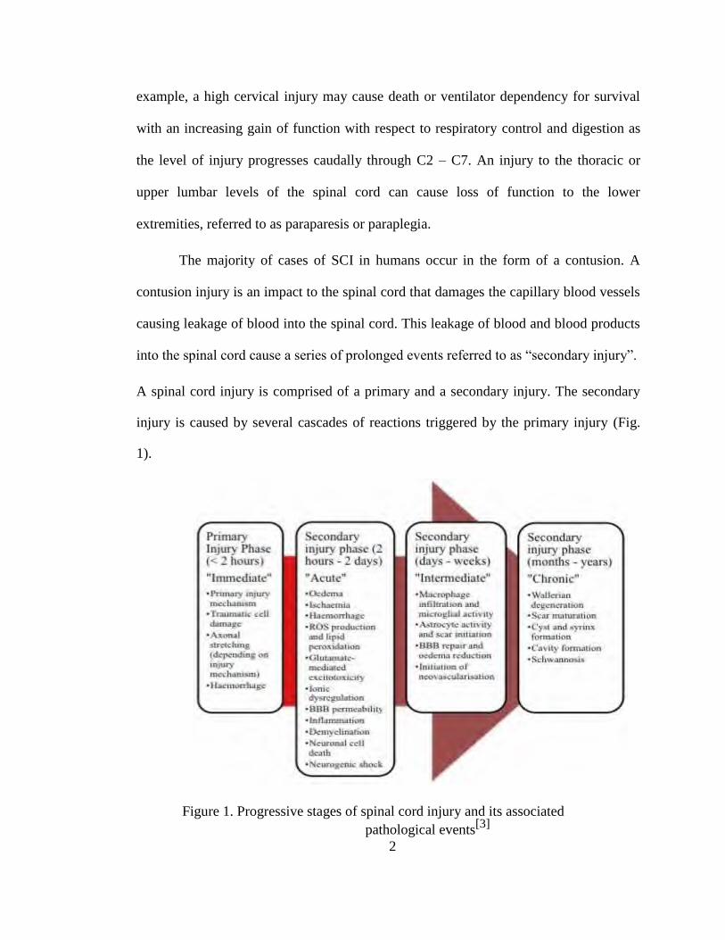
A spinal cord injury is comprised of a primary and a secondary injury. The secondary
injury is caused by several cascades of reactions triggered by the primary injury (Fig.
1).
Figure 1. Progressive stages of spinal cord injury and its associated
pathological events[3]
2
Apart from the structural damage to the column, cellular, biochemical and
immunological reaction within the injury site can lead to wide-spread loss of neuronal
function around the primary injury. Several of these reactions are triggered by the
damage to the blood vessels and capillaries within the spinal cord[13]
.
Damage to the blood vessels triggers several cascades such as the clotting
cascade and the complement cascade. Activation of complements and other such
cytokines in the primary injury area, invites immune cells such as macrophages and
other antigen presenting cells. Hemorrhaging in the gray matter upon contusion injury
activates the microglia and astrocytes in the surrounding region to form the glial scar in
that location. Invasion of immune cells generates a cyst in the region of primary
injury[11,12,13]
. This leads to further necrosis of neurons surrounding the glial scar.
Permeability of the Blood Brain Barrier (BBB) is also affected thereby, losing its
filtration specificity[11,12,13]
. Change in permeability of the BBB leads to increased
diffusion of neuro-degenerative molecules into the spinal cord. Free radicals are also
produced as a reaction to the primary injury.
The free radicals and other ions produced in the injury site induce excitotoxicity.
Excitotoxicity forms a major reason for neuronal apoptosis as well as oligodendrocyte
apoptosis in the surrounding regions[11,12,13]
. Oligodendrocyte apoptosis causes increased
demyelination of axons in the white matter. Death of myelinated axons and demyelination
events lead to the production of myelin debris in and around the injury location. Myelin
debris is also toxic to the oligodendrocytes leading to wide-spread death of
oligodendrocytes in the periphery of the injury location[11,12,13]
. Demyelination of axons in
the white matter leave the longitudinally traversing axons intact but they
3
lose their function due to loss of the insulating myelin sheath around their axons. With
the progressing secondary injury, the axons in the white matter may remain intact but
become non-functional due to demyelination. Although the extent to which this occurs
in humans is controversial, remyelination of these demyelinated axons suggests a
potential strategy for improving neurological function after SCI.
OLIGODENDROCYTES AND THEIR SIGNIFICANCE IN SCI:
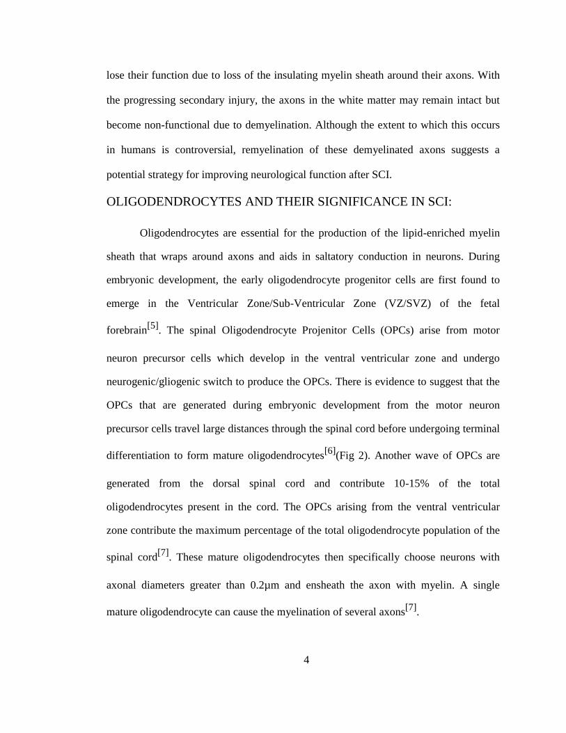
Oligodendrocytes are essential for the production of the lipid-enriched myelin
sheath that wraps around axons and aids in saltatory conduction in neurons. During
embryonic development, the early oligodendrocyte progenitor cells are first found to
emerge in the Ventricular Zone/Sub-Ventricular Zone (VZ/SVZ) of the fetal
forebrain[5]
. The spinal Oligodendrocyte Projenitor Cells (OPCs) arise from motor
neuron precursor cells which develop in the ventral ventricular zone and undergo
neurogenic/gliogenic switch to produce the OPCs. There is evidence to suggest that the
OPCs that are generated during embryonic development from the motor neuron
precursor cells travel large distances through the spinal cord before undergoing terminal
differentiation to form mature oligodendrocytes[6]
(Fig 2). Another wave of OPCs are
generated from the dorsal spinal cord and contribute 10-15% of the total
oligodendrocytes present in the cord. The OPCs arising from the ventral ventricular
zone contribute the maximum percentage of the total oligodendrocyte population of the
spinal cord[7]
. These mature oligodendrocytes then specifically choose neurons with
axonal diameters greater than 0.2µm and ensheath the axon with myelin. A single
mature oligodendrocyte can cause the myelination of several axons[7]
.
4
Figure 2. Oligodendrocyte Differentiation with specifying markers for each stage
of differentiation. [4]
Some OPCs that migrate from the embryonic point of origin are believed to
reside in the brain and spinal cord without undergoing terminal differentiation. It can be
hypothesized that these resident OPCs are involved in the oligodendrocyte turn-over in
the central nervous system and also in minor injury repair. OPCs retain their
proliferative capacity and migrate to regions requiring myelination before undergoing
terminal differentiation. These characteristics of OPCs make them an attractive target
for cellular therapy in demyelinating diseases and SCI. In SCI, there is progressive
demyelination of axons in the white matter during secondary injury. It appears that the
resident OPCs are unable to mount a sufficient response to repair this damage.
Therefore transplantation of OPCs could potentially repair this damage and restore
some functionality previously lost.
There is evidence that transplantation of OPCs does result in functional
improvement in a rodent model of SCI[9]
. While remyelination may contribute to this
improvement, there is also evidence that there are other mechanisms involved[9,27]
,
5
mainly because of the early improvement identified. The transplantation of OPCs in the
rat optic chiasm model displayed an improvement in the P1 wave-latency, which is a
determinant of the functionality of the neurons, within the first week of
transplantation[27]
. OPCs produced from human Embryonic Stem cells also express
neurotrophic factors which may be involved in inhibiting axonal dieback and assist in
regeneration of transected neurons[28]
. Transplanted OPCs are also found to interact
with and modulate the gene expression pattern of resident phagocytic cells and decrease
the inflammatory response in the injury site[29]
. OPCs are believed to have other
paracrine effects around the injury site which helps decrease the pathology of the injury
and provide a favorable environment for regeneration.
HUMAN EMBRYONIC STEM CELL DERIVED OPCs
Human Embryonic Stem (ES) cells are derived from the inner cell mass in the
blastocyst stage of a developing embryo. These cells are pluripotent in nature and can
be maintained in culture indefinitely. Under defined growth conditions, these cells can
be differentiated into the cells of ectodermal, mesodermal and endodermal lineages. In
studies by other groups, these cells were subjected to specific growth conditions in
order to direct them towards the oligodendrocyte lineage (Fig 3). This protocol involved
the use of a Glial Restrictive Medium (GRM) and Retinoic Acid[8]
. During embryonic
development, Retinoic Acid is responsible for triggering human ES cells towards
forming spinal progenitors[6]
. The protocol used to make OPCs from human ES cells
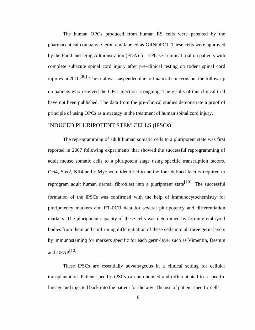
was refined and optimized and a high yield of OPCs was obtained and characterized by
immunostaining and quantification of immune stained cells (Fig 4)[9]
.
6
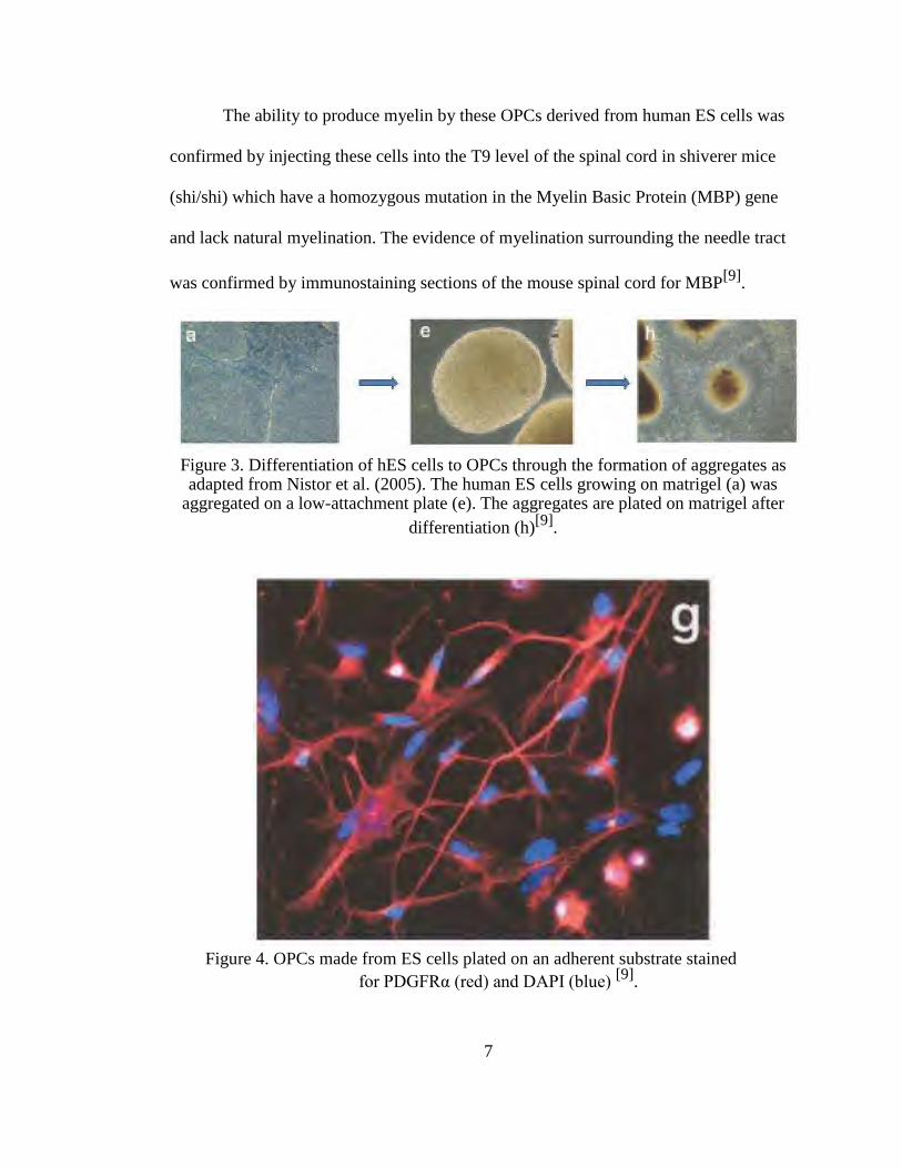
The ability to produce myelin by these OPCs derived from human ES cells was
confirmed by injecting these cells into the T9 level of the spinal cord in shiverer mice
(shi/shi) which have a homozygous mutation in the Myelin Basic Protein (MBP) gene
and lack natural myelination. The evidence of myelination surrounding the needle tract
was confirmed by immunostaining sections of the mouse spinal cord for MBP[9]
.
Figure 3. Differentiation of hES cells to OPCs through the formation of aggregates as adapted from Nistor et al. (2005). The human ES cells growing on matrigel (a) was
aggregated on a low-attachment plate (e). The aggregates are plated on matrigel after
differentiation (h)[9]
.
Figure 4. OPCs made from ES cells plated on an adherent substrate stained
for PDGFRα (red) and DAPI (blue) [9]
.
7
The human OPCs produced from human ES cells were patented by the
pharmaceutical company, Geron and labeled as GRNOPC1. These cells were approved
by the Food and Drug Administration (FDA) for a Phase I clinical trial on patients with
complete subacute spinal cord injury after pre-clinical testing on rodent spinal cord
injuries in 2010[30]
. The trial was suspended due to financial concerns but the follow-up
on patients who received the OPC injection is ongoing. The results of this clinical trial
have not been published. The data from the pre-clinical studies demonstrate a proof of
principle of using OPCs as a strategy in the treatment of human spinal cord injury.
INDUCED PLURIPOTENT STEM CELLS (iPSCs)
The reprogramming of adult human somatic cells to a pluripotent state was first
reported in 2007 following experiments that showed the successful reprogramming of
adult mouse somatic cells to a pluripotent stage using specific transcription factors.
Oct4, Sox2, Klf4 and c-Myc were identified to be the four defined factors required to
reprogram adult human dermal fibroblast into a pluripotent state[10]
. The successful
formation of the iPSCs was confirmed with the help of immunocytochemistry for
pluripotency markers and RT-PCR data for several pluripotency and differentiation
markers. The pluripotent capacity of these cells was determined by forming embryoid
bodies from them and confirming differentiation of these cells into all three germ layers
by immunostaining for markers specific for each germ-layer such as Vimentin, Desmin
and GFAP[10]
.
These iPSCs are essentially advantageous in a clinical setting for cellular
transplantation. Patient specific iPSCs can be obtained and differentiated to a specific
lineage and injected back into the patient for therapy. The use of patient-specific cells
8
theoretically decreases the risk of immune rejection of the transplanted cells
considerably and the antigen compatibility may help in increasing the integrative
capacity of the transplanted cells into the host system. However certain disadvantages
are the potential for teratogenicity, and the time taken after SCI to both culture the
iPSCs and to subsequently differentiate them into OPCs. To date, there are no clinical
trials approved by the FDA for the use of iPSCs in humans.
iPSCs are generated by the insertion of reprogramming factors within a somatic
cell with the help of integrating viral vectors. The disadvantage of using such vectors is
the integration of the viral genome with the host cell genome. This may lead to the
expression of viral genes and proteins in the pluripotent cells which may elicit an
adverse reaction in a human host during transplantation. The need for alternative
delivery methods for the reprogramming factors into the cell is necessary to reduce the
risk of adverse side-effects during a clinical trial. Human iPSCs have been generated
with the help of Sendai Virus as a vehicle[31]
. The sendai virus is a non-integrating
RNA virus whose genome gets translated into protein in the cytoplasm (Fig. 5). The
viral proteins are cleared from the cells in subsequent cycles of host cell replication thus
eliminating the risk of an immune reaction. Sendai virus is non-tumorigenic and non-
pathogenic to humans. Sendai virus membrane fusion with host cells occurs efficiently
with low species and cell specificity. These properties of the sendai virus make them an
advantageous vehicle for delivering foreign DNA into human cells. These iPSCs
theoretically present a lesser risk during clinical transplantation than other iPSCs
produced using integrating viruses.
9
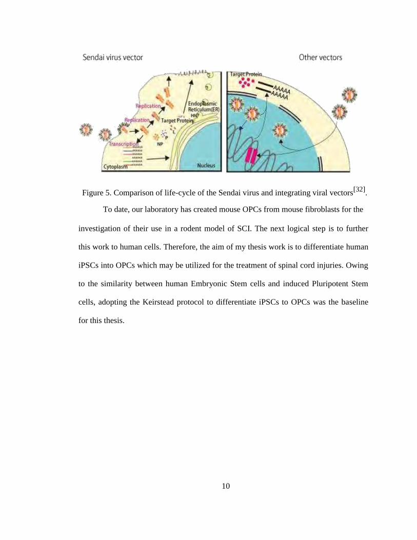
Figure 5. Comparison of life-cycle of the Sendai virus and integrating viral vectors[32]
.
To date, our laboratory has created mouse OPCs from mouse fibroblasts for the
investigation of their use in a rodent model of SCI. The next logical step is to further
this work to human cells. Therefore, the aim of my thesis work is to differentiate human
iPSCs into OPCs which may be utilized for the treatment of spinal cord injuries. Owing
to the similarity between human Embryonic Stem cells and induced Pluripotent Stem
cells, adopting the Keirstead protocol to differentiate iPSCs to OPCs was the baseline
for this thesis.
10
MATERIALS AND METHODS
MAINTAINING INDUCED PLURIPOTENT STEM CELL LINE
Human iPSCs (made using sendai virus) were kindly provided by Lucas Greder
on irradiated Mouse Embryonic Fibroblasts (MEFs) (provided by Joseph Dalton). The
iPSCs produced using sendai virus displayed a lack of transgene expression by passage
8. Absence of viral protein in the iPSC colonies was confirmed using western blots. The
iPSCs generated using the sendai virus showed a similar global gene expression profile
to iPSCs genererated using a lentiviral vector.[33]
These iPSCs were supplemented with
pluripotent stem cell specific medium consisting of Dulbecco’s Modified Eagle’s
Medium:F12 (DMEM : F12) (Gibco), 20% Knock-out Serum Replacement (KSR)
(Gibco), 1% Non-Essential Amino Acids (NEAA) (Gibco), 2mM L-Glutamine (Gibco),
0.1mM β-Mercaptoethanol (Gibco), 1X Penicillin-Streptomycin (Pen-Strp) (Gibco) and
10ng/ml basal Fibroblast Growth Factor (bFGF) (R&D Systems). Mouse Embryonic
Fibroblasts (MEFs) were plated on gelatin coated plastic 6-well plate (BD Falcon) at a
density of 200,000 cells per well such that they form a uniform monolayer. The iPSCs
were passaged using 1ml of Collagenase IV (1mg/ml) (Gibco) per well and scraped
with a cell scraper (BD Falcon) after 10 minutes incubation at 37˚C. The collagenase
was diluted with medium and centrifuged at 750 rpm for 5 minutes. The pellet was re-
suspended in medium and centrifuged to remove all traces of collagenase. The pellet
was re-suspended once again in medium and placed in a new plate of feeders. The cells
were regularly passaged within 6-8 days when the iPSC colonies were large enough but
11
did not make contact with one another. The cells were split in a 1:2 to 1:3 ration to
maintain confluency.
PREPARATION OF GLIAL RESTRICTIVE MEDIUM (GRM)
The supplements utilized in preparing the Glial Restrictive medium were
reconstituted and aliquoted as follows
S.No.
SUPPLEMENT
COMPANY
RECONSTITUTION
STOCK
CONCENTRATION ALIQUOT
1. B27 Gibco - 50X 1ml
R&D
100 m l glacial
acetic acid,
2. Insulin Systems
10mg/ml 1ml
rest with WET*
1 ml of Ethanol
R&D then
3. Progesterone Systems rest with KO- 63µg/ml 1ml
DMEM#
4.
Putrescine
R&D # 10mg/ml
1ml
KO-DMEM
Systems
5. Sodium R&D WET* 50µg/ml 1ml
Selenite Systems
6. Transferrin Sigma KO-DMEM# 50mg/ml 20µl
Add 1 ml of 1 N
R&D Sodium
7. T3 Systems Hydroxide 40µg/ml 1ml
then KO-DMEM#
Table 1. Components of Glial Restrictive Medium (GRM)
*Water for Embryo Transfer, #Knock-Out Dulbecco’s Modified Eagle’s Medium
(Gibco)
GRM was prepared with Dulbecco’s Modified Eagle’s Medium:F12
(DMEM:F12) supplemented with B-27, 10µg/ml Insulin, 63ng/ml Progesterone, 12
10µg/ml Putrescine, 50ng/ml Sodium Selenite, 50µg/ml transferrin, and 40ng/ml T3.
The medium was then filtered and the tube was covered with aluminium foil to avoid
the break-down of B27.
The GRM consists of several components that are involved in aiding glial
differentiation of the pluripotent cells such as insulin, T3 (triiodo thyronine, a thyroid
hormone), transferrin and selenium ions. Insulin and Insulin-like Growth Factor (IGF)
are known to be involved in oligodendrocyte development and proliferation[20]
.
Thyroid hormone is known to aid in the differentiation of mature oligodendrocytes from
precursor cells. Thyroid hormone also promotes proliferation of cells in a pathway
involving the mitogen Platelet-Derived Growth Factor (PDGF)[20]
. Insulin, Transferrin
and Selenium ions are part of a specialized medium known as ITSF medium which is
known to promote migration of cells during neural crest development[20]
. Epidermal
Growth Factor (EGF) receptor and its signaling is identified to promote proliferation of
uncommitted glial cells that are Olig2+, NG2
+, PDGFRα
+ and Ki67
+ [21]. Retinoic
Acid (RA) is a caudalizing factor commonly utilized in several differentiation protocols
to generate ventral spinal progenitors in glial restricted cells[14,18]
. Therefore, GRM
was supplemented with EGF and RA to promote OPC differentiation.
KEIRSTEAD DIFFERENTIATION PROTOCOL[14]
The iPSCs were treated with 1ml of Collagenase IV (1mg/ml) per well for 10
minutes. The collagenase was then removed and the cells were washed with PBS. 2ml
of Transition Medium which was made of a 1:1 ratio of GRM and Mouse Embryonic
Feeder (MEF) Conditioned iPSC medium supplemented with 4ng/ml of bFGF was then
added to the cells. The cells were dissociated with a cell scraper and the large clumps
13
were broken up by pipetting. They were then placed in an ultra low-attachment 6-well
plate (Costar). The next day (Day 2) the medium was changed to transition medium
supplemented with 4ng/ml bFGF, 20ng/ml EGF (R&D Systems) and 10µM RA (R&D
Systems). From Day 3 to 10 the aggregates were supplemented with GRM containing
20ng/ml EGF and 10µM RA with a change of medium everyday. From Day 11 until
Day 28 the culture was fed with GRM supplemented with 20ng/ml EGF with a change
of medium every other day. Matrigel (BD Biosciences) was diluted 1:10 in DMEM:F12
using chilled pipettes and centrifuge tubes and 1ml was placed in each well of a 6-well
plate and left at room temperature for 24 hours. After Day 28, the aggregates were
placed in the matrigel coated plate.
MANIPULATIONS OF THE KEIRSTEAD PROTOCOL
To improve viability of iPSC aggregates, the Keirstead protocol was modified in
several ways. The aggregation step utilized in the Keirstead protocol was changed to the
protocol utilized to make Embryoid Bodies (EBs) from iPSCs[14]
which involves
treating the iPSCs with Collagenase IV (1mg/ml) and washing twice with ES medium
and centrifuging at 750rpm for 5 minutes. This protocol is similar to the one utilized to
passage iPSCs with the difference being that in the final step, the pellet is reconstituted
in transition medium supplemented with 4ng/ml bFGF and the cells are placed in a low-
attachment plate. The aggregation step was further changed according to STEMDiffTM
Neural Induction Kit from human Pluripotent Stem Cells (hPSCs) where the iPSCs are
treated with Collagenase IV (1mg/ml) and the cells are diluted 1:10 in pluripotent cell
specific medium and centrifuged at 750 rpm for 5 minutes. The transition of the iPSC
aggregates to GRM utilized in the Keirstead Protocol was manipulated such that
14
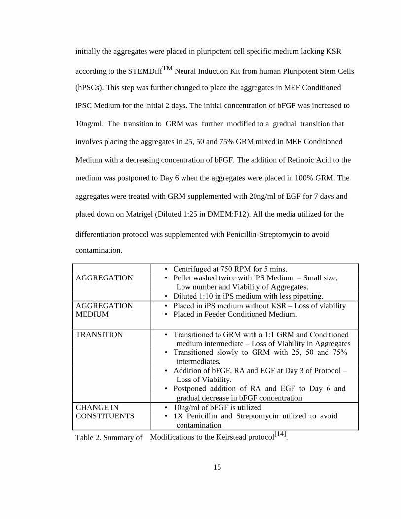
initially the aggregates were placed in pluripotent cell specific medium lacking KSR
according to the STEMDiffTM
Neural Induction Kit from human Pluripotent Stem Cells (hPSCs). This step was further changed to place the aggregates in MEF Conditioned
iPSC Medium for the initial 2 days. The initial concentration of bFGF was increased to
10ng/ml. The transition to GRM was further modified to a gradual transition that
involves placing the aggregates in 25, 50 and 75% GRM mixed in MEF Conditioned
Medium with a decreasing concentration of bFGF. The addition of Retinoic Acid to the
medium was postponed to Day 6 when the aggregates were placed in 100% GRM. The
aggregates were treated with GRM supplemented with 20ng/ml of EGF for 7 days and
plated down on Matrigel (Diluted 1:25 in DMEM:F12). All the media utilized for the
differentiation protocol was supplemented with Penicillin-Streptomycin to avoid
contamination.
• Centrifuged at 750 RPM for 5 mins.
AGGREGATION • Pellet washed twice with iPS Medium – Small size,
Low number and Viability of Aggregates.
• Diluted 1:10 in iPS medium with less pipetting.
AGGREGATION • Placed in iPS medium without KSR – Loss of viability MEDIUM • Placed in Feeder Conditioned Medium.
TRANSITION • Transitioned to GRM with a 1:1 GRM and Conditioned medium intermediate – Loss of Viability in Aggregates
• Transitioned slowly to GRM with 25, 50 and 75%
intermediates.
• Addition of bFGF, RA and EGF at Day 3 of Protocol –
Loss of Viability.
• Postponed addition of RA and EGF to Day 6 and
gradual decrease in bFGF concentration
CHANGE IN • 10ng/ml of bFGF is utilized CONSTITUENTS • 1X Penicillin and Streptomycin utilized to avoid
contamination
Table 2. Summary of Modifications to the Keirstead protocol[14]
.
15
FINAL DIFFERENTIATION PROTOCOL
A schematic of the final Differentiation protocol is represented in Figure 6. The
iPSCs growing on irradiated MEFs were washed with PBS (CellGro) and treated with
collagenase IV (1mg/ml) (1ml/Well) for 10 minutes at 37˚C. The cells were then
scraped using a cell scraper and diluted 1:10 in iPSC medium. The cells were
centrifuged at 750 rpm for 5 minutes. The pellet was reconstituted in MEF Conditioned
iPSC medium supplemented with antibiotics and placed in an ultra low-attachment
plate. The medium was changed to fresh conditioned medium on Day 2. The cells were
then treated to 25%, 50% and 75% Glial Restrictive Medium made with conditioned
medium and supplemented with antibiotics with a decreasing concentration of bFGF
(7.5ng/ml, 5ng/ml and 2.5ng/ml) on consecutive days till Day 5. On Day 6, the cells
were provided with GRM supplemented with 20ng/ml EGF and 10µM RA and the
medium is changed everyday for the next 7 days. On Day 13, the aggregates were given
GRM supplemented with 20ng/ml of EGF with a change of medium every alternate
day. The aggregates were broken up on Day 1 and Day 2. On Day 20, the aggregates
were plated onto matrigel (diluted 1:25 in DMEM:F12).
PASSAGING OPCs PLATED ON MATRIGEL
The OPCs growing out of the aggregates on matrigel were supplemented with
GRM containing 20ng/ml of EGF. The medium was changed every other day. The well
is washed with PBS and treated with warm 0.05% Trypsin-EDTA (HyClone) (500µl per
well of a 6-well plate and 300µl per well of a 12-well plate) for 7 minutes at 37˚C. The
cells were then observed under the microscope for detachment of the cells from the
substrate and the plate was tapped from the bottom and sides to dislodge all the cells 16
from the matrigel. The trypsin-EDTA cell suspension was then diluted 1:10 with
DMEM:F12 medium and centrifuged at 750 rpm for 5 minutes. The pellet was
reconstituted with fresh GRM containing 20ng/ml of EGF and distributed in a new 12-
well plate at a ratio of 1:2 or 1:3 wells to maintain the density required by OPCs for
proliferation. The medium was changed every other day to fresh GRM with 20ng/ml of
EGF to aid in the proliferation of OPCs. The OPCs were passaged at regular intervals
when the cell density reaches the desired confluence. The OPCs were passaged onto
matrigel diluted at a ratio of 1:25 in DMEM:F12.
17
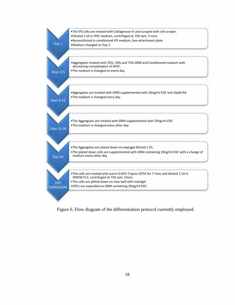
Day 1
Days 3-5 Days 6-12
Days 13-20
Day 20
OPC
EXPANSION
•The iPS cells are treated with Collagenase IV and scraped with cell scraper. •Diluted 1:10 in iPSC medium, centrifuged at 750 rpm, 5 mins •Reconstituted in conditioned iPS medium, low-attachment plate •Medium changed on Day 2
•Aggregates treated with 25%, 50% and 75% GRM and Conditioned medium with
decreasing concentration of bFGF. •The medium is changed on every day.
•Aggregates are treated with GRM supplemented with 20ng/ml EGF and 10µM RA •The medium is changed every day. •The Aggregates are treated with GRM supplemented with 20ng/ml EGF. •The medium is changed every other day
•The Aggregates are plated down on matrigel diluted 1:25. •The plated down cells are supplemented with GRM containing 20ng/ml EGF with a change of
medium every other day
•The cells are treated with warm 0.05% Trypsin-EDTA for 7 mins and diluted 1:10 in
DMEM:F12, centrifuged at 750 rpm, 5mins •The cells are plated down on new well with matrigel. •OPCs are expanded on GRM containing 20ng/ml EGF.
Figure 6. Flow diagram of the differentiation protocol currently employed.
18
IMMUNOHISTOCHEMISTRY
Initially aggregates plated on matrigel were fixed by removing the medium from
the well and adding 1ml of formalin (Protocol) per well of a 6-well plate followed by 20
minutes incubation at room temperature. After 20 minutes, the formalin was removed
and the well was washed twice with PBS. Blocking buffer was prepared by adding 1%
Bovine Serum Albumin (Sigma) and 0.1% Tween-20 (Biorad) to PBS and filtering.
Permeabilizing buffer was prepared by adding Tween-20 to the blocking buffer such
that it has a 1% Tween-20 concentration. The PBS was removed from the cells and
500µl of permeabilizing buffer was added to the well and incubated for 10 minutes at
room temperature. After incubation period, the permeabilizing buffer was removed and
washed 3 times with wash buffer (PBS with 0.1% Tween-20) for 2 minutes each. The
cells were then treated with 1ml of blocking buffer per well and incubated for 30
minutes at room temperature and after incubation they were washed 3 times with wash
buffer (PBS with 0.1% Tween-20) for 2 minutes each. The primary and secondary
antibody treatments were performed according to Table 3. The cells were also stained
with DAPI by diluting DAPI 1:500 in PBS. The cells will be treated with the secondary
antibody alone as a form of negative control.
19

S.No. Primary Secondary
Antibody Company Dilution Antibody Company Dilution
Alexa
Mouse
Fluoro
488
1. Anti β-
Sigma 1:1000 Rabbit Invitrogen 1:500
Tubulin
Anti-
Isotype III
Mouse
IgG
Alexa
Rabbit X
Fluoro
555
Olig 2
2.
Millipore 1:250
Donkey
Invitrogen 1:500
Polyclonal
Anti-
Antibody
Rabbit
IgG
Alexa
Fluoro
3.
Goat Anti R&D 1:100
555 Invitrogen 1:500
hOlig 2 Systems Donkey
Anti-Goat
IgG
Alexa
Fluoro
4.
Rabbit Millipore 1:200
488 Goat Invitrogen 1:500
Anti NG 2 Anti
Rabbit
IgG
Table 3. Antibodies used for immunostaining and their dilutions.
20
RESULTS
AGGREGATION OF iPSCs
The aggregation step employed in the Keirstead protocol[14]
involved treating
the pluripotent stem cells with collagenase and placing them in Transition Medium
followed by dissociating them with a cell scraper. In the Keirstead paper[14]
, since the
ES cells were maintained and passaged on matrigel, the aggregation method utilized
would provide uniform aggregates. In contrast, the iPSCs utilized for this project were
maintained and passaged on MEF feeder cell layers (Fig 7A) which may inhibit uniform
aggregate formation due to the presence of feeder debris. This was overcome by
diluting the cells with iPSC medium and centrifuging at 750 RPM for 5 minutes. This
resulted in depletion of feeder cell debris and formation of uniform aggregates. The
aggregating cells are placed on a plate rocker overnight inside the incubator such that
the cells collect together and ensure the formation of uniform aggregates.
TRANSITION TO GLIAL RESTRICTIVE MEDIUM
A difference in the differentiation capability of human iPSCs and ES cells has
been observed by several groups[15,16]
. The neural differentiation capacity of different
pluripotent lines shows significant variation indicating a possible difference in
pluripotency and epigenetic signals between ES cells and iPSCs[15,16]
. In these studies
the differentiation of each pluripotent cells line was evaluated by their ability to form
PAX 6+ adherent neural rosettes. Differences such as those reported in these papers
may provide an explanation for the difference in the ability of aggregates formed from
ES cells and iPSCs to survive in 1:1 Glial Restrictive Medium (GRM) and MEF
conditioned medium. The iPSCs used in this study exhibited a low survival capacity 21
when subjected to the transition medium (1:1 GRM and conditioned medium)
immediately upon aggregation. The survival of iPSC aggregates was initially
determined by studying their morphology. In these experiments, aggregates of dead
cells appeared very dark with irregular shapes and did not display any growth.
Therefore to increase the survival rate of our iPSCs following aggregation, they were
placed in MEF conditioned medium for the first 2 days with a change of medium on
both days. The aggregates appeared yellow and had a more regular spherical
morphology (Fig 7B, 7C).
Figure 7. Initial Aggregation of iPS Cells. iPS cell colonies growing on MEFs (A), Day 2
Aggregates on conditioned medium 10X (B) and Day 2 Aggregates 20X (C)
22
Subjecting the aggregates to GRM on Day 3, according to the Keirstead
protocol[14]
, rapidly decreased their viability. A concentration of 10µM Retinoic Acid
was utilized in this project at early stages (Day 2) of transition into GRM according to
the Keirstead protocol[14]
which is 100 fold greater than the concentration normally
utilized for neural differentiation[19,25]
and this may cause loss of viability in the
aggregates. Introducing RA and EGF on Day 6 lead to glial differentiation of aggregates
that were better adapted to the GRM.
Figure 8. Aggregates on Day 6 and plated on matrigel after Day 20. Day 6 Aggregates
10X (A) and 20X (B). Aggregates plated on matrigel after Day 20 10X (C) and 20X (D)
DIFFERENTIATION AND PLATING OF AGGREGATES
The aggregates were placed in GRM supplemented with EGF and RA from day
6 to day 12 after transition and expanded further in GRM with EGF alone from day 13
23

to day 20 (Fig 8A, 8B). After expansion, the aggregates were plated onto matrigel at
day 20. Initially, the aggregates were plated and expanded in matrigel diltuted 1:10 in
DMEM:F12 according to the Keirstead protocol[14]
. However the dilution was changed
to 1:25 in DMEM:F12 due to the common usage of this dilution in other projects in our
laboratory and used to coat 6-well and 12-well plates resulting in a uniform matrigel
coating. Aggregates plated on this diluted matrigel adhered well to the plate and
proliferated (Fig 8C, 8D).
In the experiments conducted in our laboratory on mouse iPSC derived OPCs,
the cells were treated with TrpLE, plated and expanded on poly-ornithine[22]
. Poly-
ornithine is routinely utilized for the selection and expansion of neural cell types. When
aggregates formed from human iPSCs were placed in poly-ornithine coated plates for
long periods of time (4 weeks), they did not display any adherence. Treating the human
iPSC generated aggregates with TrpLE before plating onto poly-ornithine, following
more closely the mouse iPSC to OPC protocol, decreased the viability of the aggregates.
Therefore the aggregates were plated on matrigel, and the cells from within migrated
onto the matrigel and exhibited typical OPC morphology (Fig 8C, 8D).
The OPCs growing out of the aggregates on matrigel were passaged using
0.05% Trypsin-EDTA as described in the Keirstead Protocol[14]
. The trypsin was
neutralized using an anti-trypsin solution in the previously established Keirstead’s
protocol. In this project, the cells were treated with 0.05% trypsin-EDTA at 37˚C for 7
mins following which the plate was tapped well to dislodge the cells. The cell
suspension in trypsin was then diluted 1:10 in DMEM:F12. The cells were collected by
centrifugation and redistributed on matrigel coated wells in GRM containing 20ng/ml
24
EGF. The medium was changed every other day to expand the OPCs. These cells were
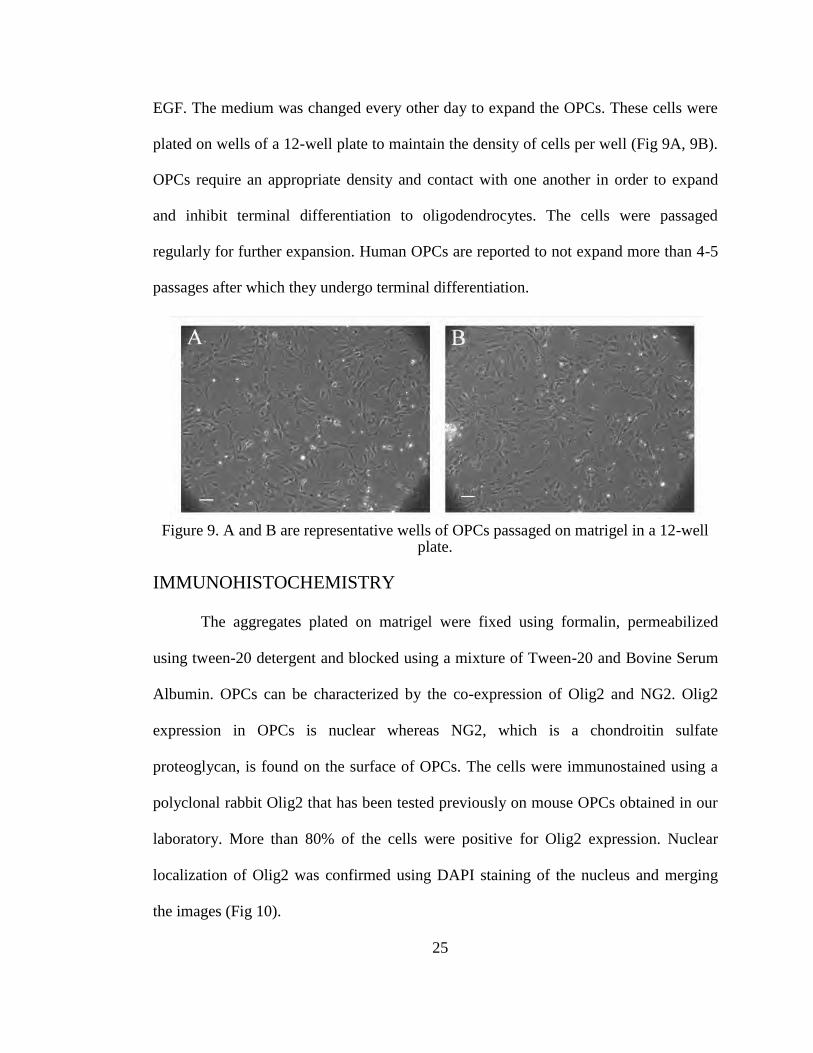
plated on wells of a 12-well plate to maintain the density of cells per well (Fig 9A, 9B).
OPCs require an appropriate density and contact with one another in order to expand
and inhibit terminal differentiation to oligodendrocytes. The cells were passaged
regularly for further expansion. Human OPCs are reported to not expand more than 4-5
passages after which they undergo terminal differentiation.
Figure 9. A and B are representative wells of OPCs passaged on matrigel in a 12-well
plate.
IMMUNOHISTOCHEMISTRY
The aggregates plated on matrigel were fixed using formalin, permeabilized
using tween-20 detergent and blocked using a mixture of Tween-20 and Bovine Serum
Albumin. OPCs can be characterized by the co-expression of Olig2 and NG2. Olig2
expression in OPCs is nuclear whereas NG2, which is a chondroitin sulfate
proteoglycan, is found on the surface of OPCs. The cells were immunostained using a
polyclonal rabbit Olig2 that has been tested previously on mouse OPCs obtained in our
laboratory. More than 80% of the cells were positive for Olig2 expression. Nuclear
localization of Olig2 was confirmed using DAPI staining of the nucleus and merging
the images (Fig 10).
25
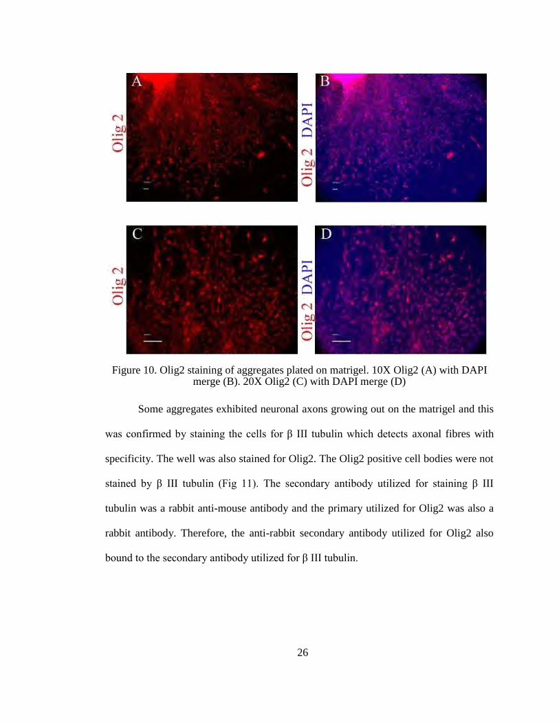
Figure 10. Olig2 staining of aggregates plated on matrigel. 10X Olig2 (A) with DAPI
merge (B). 20X Olig2 (C) with DAPI merge (D)
Some aggregates exhibited neuronal axons growing out on the matrigel and this
was confirmed by staining the cells for β III tubulin which detects axonal fibres with
specificity. The well was also stained for Olig2. The Olig2 positive cell bodies were not
stained by β III tubulin (Fig 11). The secondary antibody utilized for staining β III
tubulin was a rabbit anti-mouse antibody and the primary utilized for Olig2 was also a
rabbit antibody. Therefore, the anti-rabbit secondary antibody utilized for Olig2 also
bound to the secondary antibody utilized for β III tubulin.
26
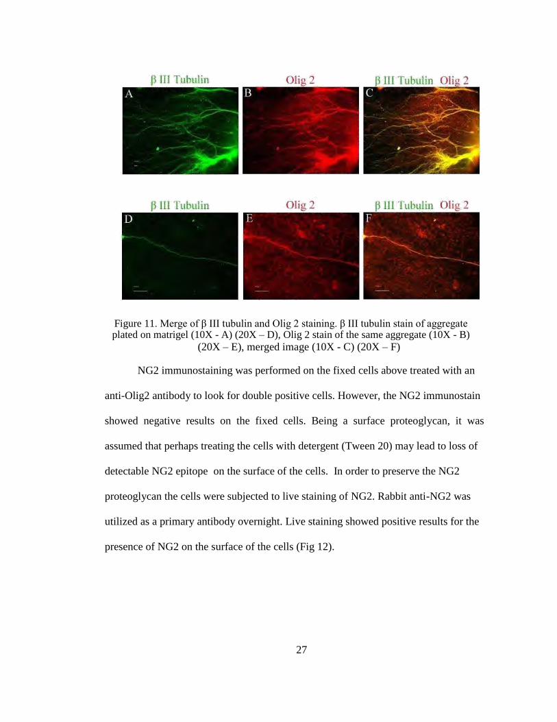
Figure 11. Merge of β III tubulin and Olig 2 staining. β III tubulin stain of aggregate plated on matrigel (10X - A) (20X – D), Olig 2 stain of the same aggregate (10X - B)
(20X – E), merged image (10X - C) (20X – F)
NG2 immunostaining was performed on the fixed cells above treated with an
anti-Olig2 antibody to look for double positive cells. However, the NG2 immunostain
showed negative results on the fixed cells. Being a surface proteoglycan, it was
assumed that perhaps treating the cells with detergent (Tween 20) may lead to loss of
detectable NG2 epitope on the surface of the cells. In order to preserve the NG2
proteoglycan the cells were subjected to live staining of NG2. Rabbit anti-NG2 was
utilized as a primary antibody overnight. Live staining showed positive results for the
presence of NG2 on the surface of the cells (Fig 12).
27
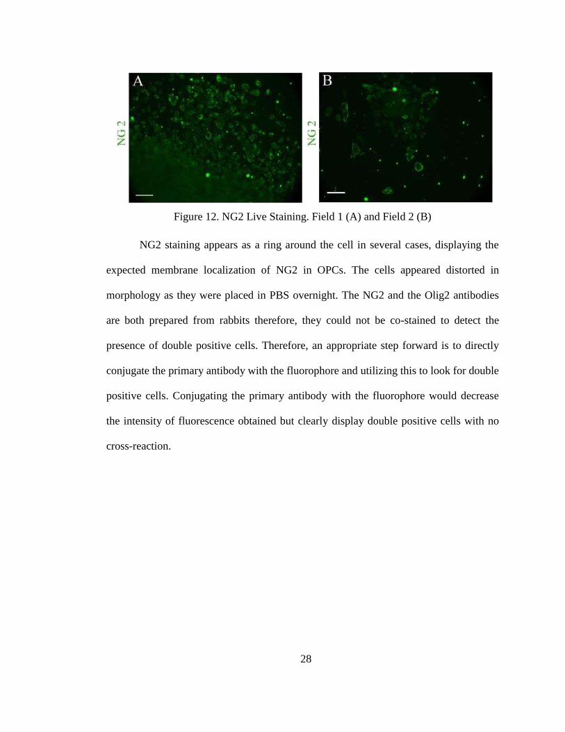
Figure 12. NG2 Live Staining. Field 1 (A) and Field 2 (B)
NG2 staining appears as a ring around the cell in several cases, displaying the
expected membrane localization of NG2 in OPCs. The cells appeared distorted in
morphology as they were placed in PBS overnight. The NG2 and the Olig2 antibodies
are both prepared from rabbits therefore, they could not be co-stained to detect the
presence of double positive cells. Therefore, an appropriate step forward is to directly
conjugate the primary antibody with the fluorophore and utilizing this to look for double
positive cells. Conjugating the primary antibody with the fluorophore would decrease
the intensity of fluorescence obtained but clearly display double positive cells with no
cross-reaction.
28
DISCUSSION
The similarity between iPSCs and ES cells motivated the use of the Keirstead
protocol[14]
in the differentiation of iPSCs to OPCs. The overall concept of the protocol
was successful in differentiating the iPSCs to OPCs with modification at each stage to
better suit the survival of the aggregates made from iPSCs. The OPCs generated from
the iPSCs were primarily confirmed using immunostaining for Olig2 and NG2. The
OPCs produced using this protocol, were expanded on matrigel in GRM supplemented
with EGF and passaged for further expansion.
The modifications to the Keirstead protocol were aimed at producing viable and
uniform aggregates from the iPSCs. Washing the iPSCs with pluripotency specific
medium during the aggregation stage of the protocol decreased the feeder debris and
lead to the production of more uniform aggregates. Placing the aggregates in MEF
feeder conditioned medium for the initial 2 days of the protocol aids in forming stable
aggregates that may be treated to GRM. A gradual transition to GRM with 25%, 50%
and 75% intermediates helps in acclimatizing the aggregates to the drastic change in
medium composition.
The presence of Retinoic acid in this differentiation protocol plays a vital role in
producing spinal progenitors but retinoic acid proved to be toxic in the early stages of
transition to GRM. It was hypothesized that the high concentration of retinoic acid
(10µM) at the early stages of aggregation and differentiation lead to increased cell
death. Postponing the use of retinoic acid to day 6 of the differentiation protocol aided
in increasing the viability of the aggregates.
29
Immunostaining performed for the detection of NG2 displayed a distorted
morphology of the cells as they were placed in PBS overnight. Lack of medium
supplementation lead to loss of morphology of the cells. The primary antibodies of
Olig2 and NG2 that were previously tested on mouse cells were made from rabbit.
Therefore double staining for Olig2 and NG2 was made impossible. These are certain
significant aspects of this project that need attention in order to obtain better results.
The OPCs generated with this method should be further characterized using
immunostaining for other OPC specific markers such as PDGFRα, A2B5[24]
and mRNA
expression analysis must be performed using RT-PCR to confirm the expression of
Olig2, NG2, PDGFRα and A2B5. RT-PCR data for the decrease in pluripotency marker
expression such as Oct4, Nanog and SSEA-4 and increase in glial specific markers such
as Pax6, Sox10, Olig2 and PDGFRα at each stage of differentiation can also provide
further confirmation of OPC generation from iPSCs. Immunostaining for Olig2, NG2 or
Olig2, PDGFRα double positive cells would give positive identification of OPCs
present in the dish[23]
. The cells can be analyzed using flow cytometry to identify the
ratio of Olig2 positive cells to overall number of cells in order to determine the
efficiency of the differentiation protocol in use. The initial number of iPSCs utilized to
make the aggregates would give an overall efficiency of OPC production.
Functional testing of the OPCs produced can be performed by injecting a
purified quantity of these cells into the brain/spinal cord of shiverer mice and staining
the sections for MBP[26]
. These cells can be further characterized by plating on laminin
coated wells which would induce terminal differentiation of OPCs to oligodendrocytes.
The oligodendrocytes produced should exhibit the typical multi-polar morphology and
30
can be functionally tested in vitro by co-culturing them with human motor neurons and
identifying their myelination capacity. These mature oligodendrocytes can also be
immunostained for GalC, MBP and PLP[24]
.
The use of these OPCs in cellular therapy for spinal cord injury holds great
promise as patient specific iPSCs can be used as a source of OPCs and this reduces the
risk of immune-rejection by the patient. As a pre-clinical testing process, spinal cord
injury can be induced in a rodent model using an impactor which would mimic the
contusion injury observed in humans. These OPCs can then be injected in the area
surrounding the injury and the change in functionality of the paralysed portion of these
animals can be recorded with the help of BBB scores[25]
. The animals should also be
sacrificed and sections of the spinal cord immunostained to reveal the changes induced
by these OPCs and identify the target locations towards which they migrate.
It is clear that differentiated cell lines can be generated from iPSCs using
differentiation protocols utilized for ES cells, however these protocols are likely to
required certain modifications to better fit the project goals.
31
REFERENCES 1. Complete public version of the “2011 annual statistical report for the spinal cord
injury model systems” National Spinal Cord Injury Statistical Center,
Birmingham, Alabama.
2. Kirshblum, Steven C, et al. "International standards for neurological
classification of spinal cord injury (revised 2011)." The journal of spinal cord
medicine 34.6 (2011):535-546.
3. Watson, Robert A, and Trevor M Yeung. "What is the potential of
oligodendrocyte progenitor cells to successfully treat human spinal cord
injury?." BMC neurology 11(2011):113-113
4. Back, Stephen A, and Scott A Rivkees. "Emerging concepts in periventricular
white matter injury." Seminars in perinatology 28.6 (2004):405-414.
5. Jakovcevski, Igor, et al. "Oligodendrocyte development and the onset of
myelination in the human fetal brain." Frontiers in neuroanatomy 3(2009):5-5.
6. Bradl, Monika, and Hans Lassmann. "Oligodendrocytes: biology and
pathology." Acta neuropathologica 119.1 (2010):37-53.
7. Olga Momcilovic and Xianmin Zeng. “Neural and Dopaminergic Differentiation
of Human Pluripotent Stem Cells.” Stem Cell Biology and Regenerative
Medicine (2012): 265-287.
8. Keirstead, Hans S, et al. "Human embryonic stem cell-derived oligodendrocyte
progenitor cell transplants remyelinate and restore locomotion after spinal cord
injury." The Journal of neuroscience 25.19 (2005):4694-4705.
32
9. Nistor, Gabriel I, et al. "Human embryonic stem cells differentiate into
oligodendrocytes in high purity and myelinate after spinal cord transplantation."
GLIA 49.3 (2005):385-396.
10. Takahashi, Kazutoshi, et al. "Induction of pluripotent stem cells from adult
human fibroblasts by defined factors." Cell 131.5 (2007):861-872.
11. Ek, C J, et al. "Spatio-temporal progression of grey and white matter damage
following contusion injury in rat spinal cord." PLoS ONE 5.8 (2010):e12021-
e12021.
12. Ek, C J, et al. "Pathological changes in the white matter after spinal contusion
injury in the rat." PLoS ONE 7.8 (2012):e43484-e43484.
13. Rowland, James W, et al. "Current status of acute spinal cord injury
pathophysiology and emerging therapies: promise on the horizon."
Neurosurgical focus 25.5 (2008):E2-E2.
14. Hatch, Maya N, Gabriel Nistor, and Hans S Keirstead. "Derivation of high-
purity oligodendroglial progenitors." Methods in molecular biology
549(2009):59-75.
15. Gai, Hui, et al. "Generation and characterization of functional cardiomyocytes
using induced pluripotent stem cells derived from human fibroblasts." Cell
biology international 33.11 (2009):1184-1193.
16. Hu, Bao-Yang, et al. "Neural differentiation of human induced pluripotent stem
cells follows developmental principles but with variable potency." Proceedings
of the National Academy of Sciences of the United States of America 107.9
(2010):4335-4340.
33
17. Yin, Dezhong, et al. "Comparison of neural differentiation potential of human
pluripotent stem cell lines using a quantitative neural differentiation protocol."
Methods in molecular biology 873(2012):247-259.
18. Hu, Bao-Yang, Zhong-Wei Du, and Su-Chun Zhang. "Differentiation of human
oligodendrocytes from pluripotent stem cells." Nature protocols 4.11
(2009):1614-1622.
19. Hu, Bao-Yang, et al. "Neural differentiation of human induced pluripotent stem
cells follows developmental principles but with variable potency." Proceedings
of the National Academy of Sciences of the United States of America 107.9
(2010):4335-4340.
20. Erceg, Slaven, Mohammad Ronaghi, and Miodrag Stojkovi ć. "Human
embryonic stem cell differentiation toward regional specific neural precursors."
Stem cells 27.1 (2009):78-87.
21. Ivkovic, Sanja, Peter Canoll, and James E Goldman. "Constitutive EGFR
signaling in oligodendrocyte progenitors leads to diffuse hyperplasia in postnatal
white matter." The Journal of neuroscience 28.4 (2008):914-922.
22. O'Shea, K S. "Neural differentiation of embryonic stem cells." Methods in
molecular biology 198(2002):3-14.
23. Baumann, N, et al. "Glial biology and disorders." Current opinion in neurology
and neurosurgery 6.1 (1993):27-33.
24. Cui, Qiao-Ling, et al. "Human fetal oligodendrocyte progenitor cells from
different gestational stages exhibit substantially different potential to myelinate."
Stem cells and development 21.11 (2012):1831-1837.
34
25. Erceg, Slaven, et al. "Transplanted oligodendrocytes and motor neuron
progenitors generated from human embryonic stem cells promote locomotor
recovery after spinal cord transection." Stem cells 28.9 (2010):1541-1549.
26. Izrael, Michal, et al. "Human oligodendrocytes derived from embryonic stem
cells: Effect of noggin on phenotypic differentiation in vitro and on myelination
in vivo." Molecular and cellular neurosciences 34.3 (2007):310-323.
27. Pouya, Alireza, et al. "Human induced pluripotent stem cells differentiation into
oligodendrocyte progenitors and transplantation in a rat model of optic chiasm
demyelination." PLoS ONE 6.11 (2011):e27925-e27925.
28. Zhang, Yi W, JerrodDenham, and R S SThies. "Oligodendrocyte progenitor
cells derived from human embryonic stem cells express neurotrophic factors."
Stem cells and development 15.6 (2006):943-952.
29. Cusimano, Melania, et al. "Transplanted neural stem/precursor cells instruct
phagocytes and reduce secondary tissue damage in the injured spinal cord."
Brain 135.2 (2012):447-460.
30. Geron corporation, Clinical trials Gov Identifier-NCT01217008, Study first
received-October 6th
, 2010
“http://clinicaltrials.gov/ct2/show/NCT01217008?term=geron&recr=Open&ran
k=5”
31. Nakanishi, Mahito, and MakotoOtsu. "Development of Sendai Virus Vectors
and their Potential Applications in Gene Therapy and Regenerative Medicine."
Current gene therapy 12.5 (2012):410-416.
35
32. Adapted from the official website of DNAVEC Corporation,
http://www.dnavec.co.jp/en/technology/technology1.html.
33. Kudva, Yogish C, et al. "Transgene-free disease-specific induced pluripotent
stem cells from patients with type 1 and type 2 diabetes." Stem cells and
translational medicine 1.6 (2012):451-461.
36