Embed Size (px)
Citation preview
Sampling Criteria
• Sampling plans will depend on the question that they are designed to answer
• Basic criteria– Replication– Representative– Random– Controls– Method Validation
Detection of Pathogens in the Environment
Detection of Pathogenic Microbes in Environmental Media
• Three main steps: • (1) recovery, extraction and concentration, • (2) purification and separation, and • (3) assay and characterization.
Assay Methods for Pathogens
• culture or infectivity• viability or activity measurements• immunoassays • nucleic acid assays• Protein or other macromolecular/biochemical
assays• microscopic examinations
Microscopic Methods
History of Microscopy
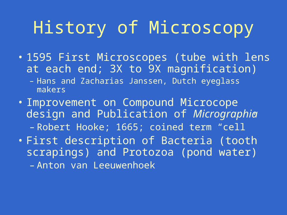
• 1595 First Microscopes (tube with lens at each end; 3X to 9X magnification)– Hans and Zacharias Janssen, Dutch eyeglass makers
• Improvement on Compound Microcope design and Publication of Micrographia– Robert Hooke; 1665; coined term “cell”
• First description of Bacteria (tooth scrapings) and Protozoa (pond water)– Anton van Leeuwenhoek
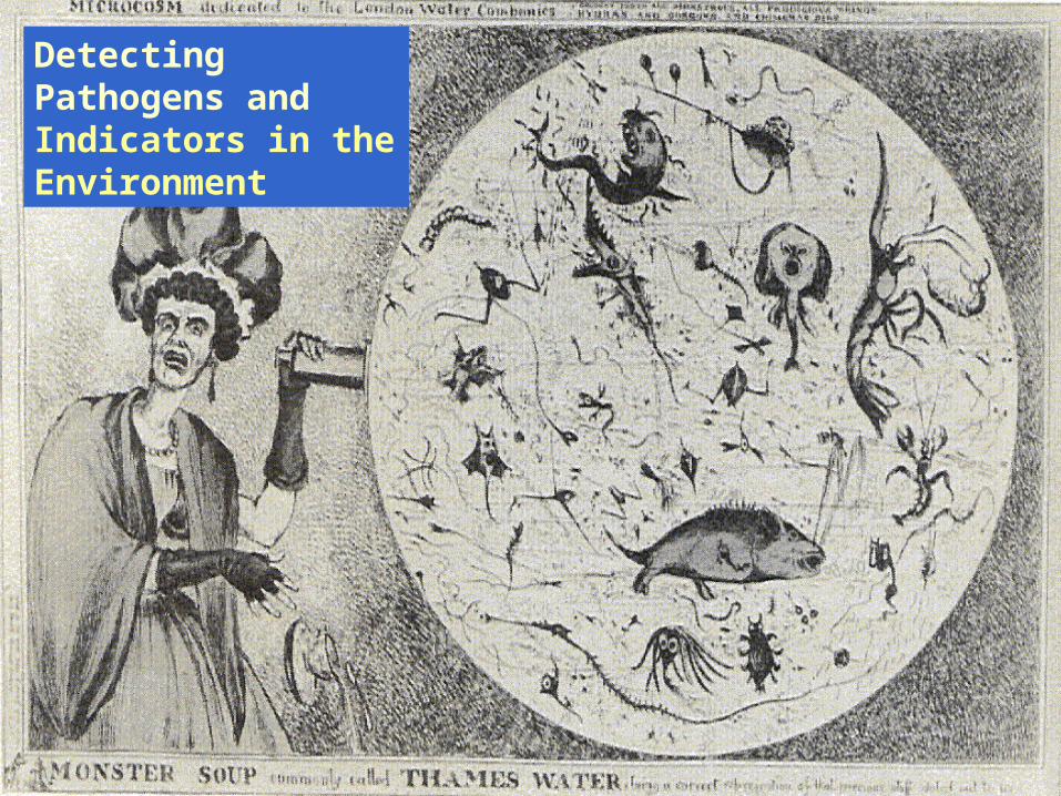
Detecting Pathogens andIndicators in the Environment
Microscopy
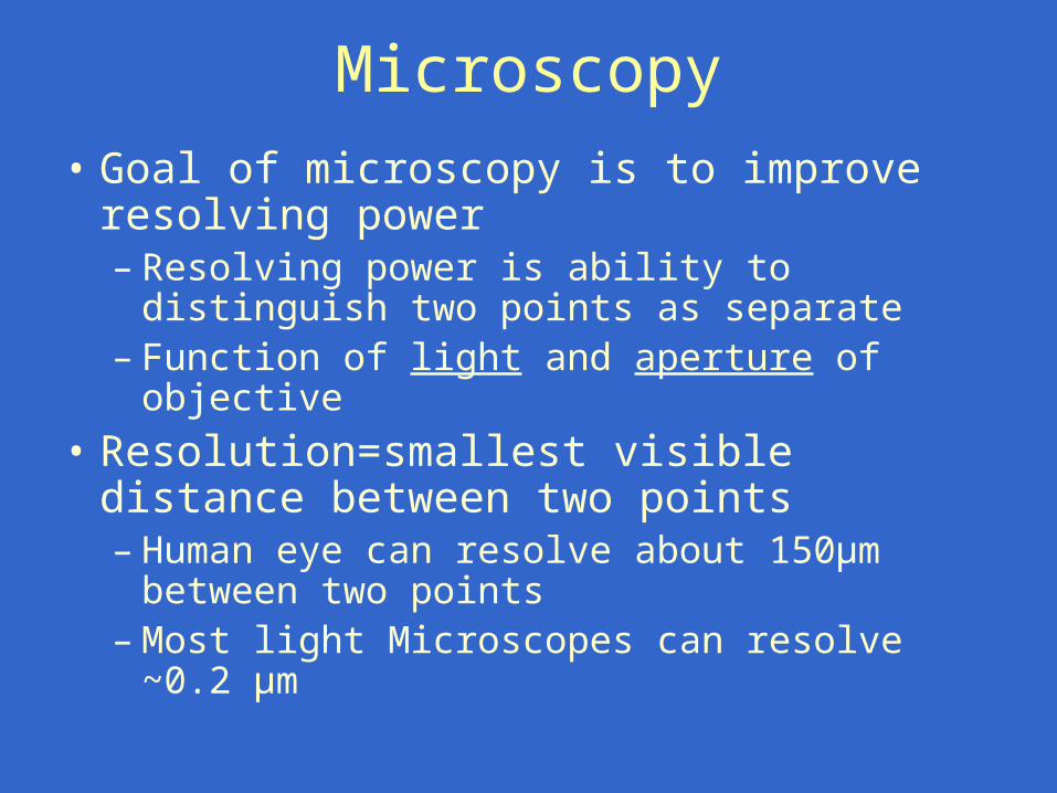
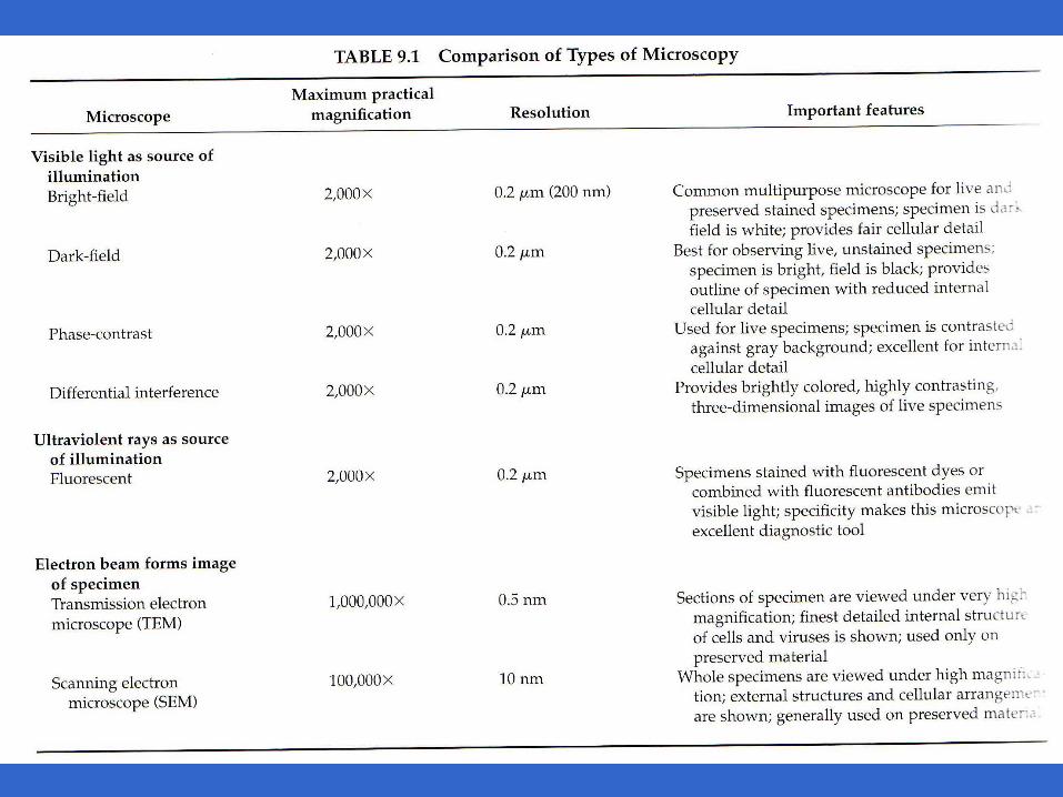
• Goal of microscopy is to improve resolving power– Resolving power is ability to distinguish two
points as separate– Function of light and aperture of objective
• Resolution=smallest visible distance between two points – Human eye can resolve about 150μm
between two points– Most light Microscopes can resolve ~0.2 μm
Magnification
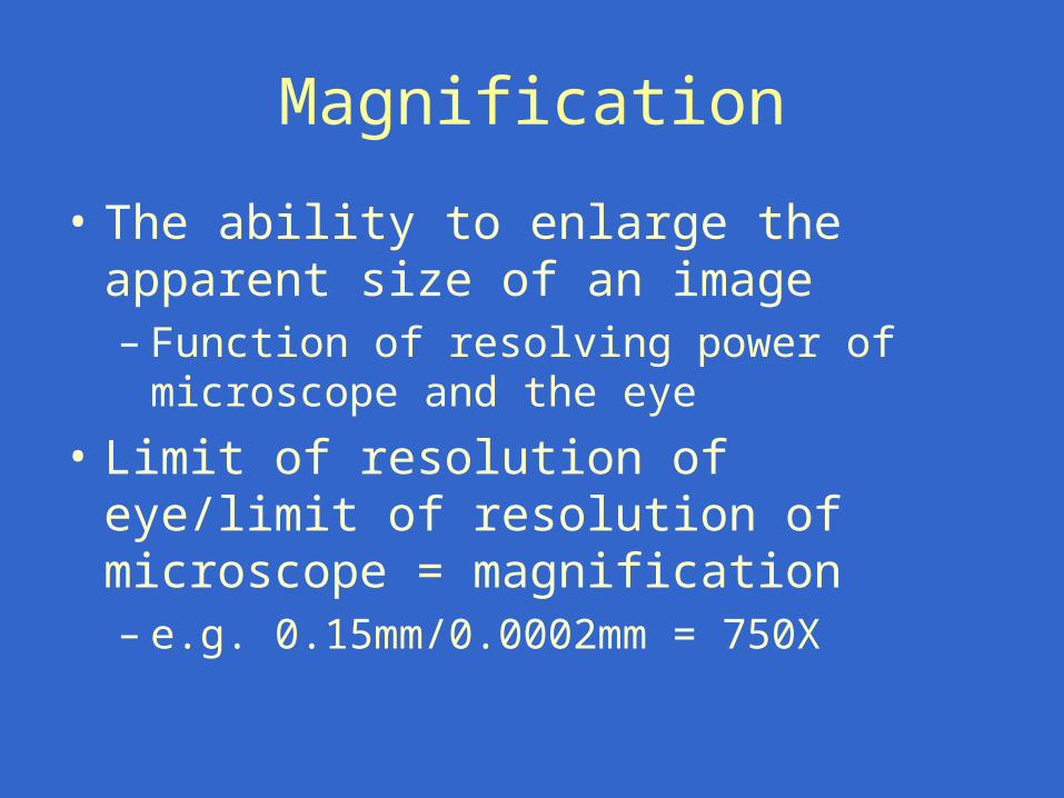
• The ability to enlarge the apparent size of an image– Function of resolving power of microscope
and the eye
• Limit of resolution of eye/limit of resolution of microscope = magnification– e.g. 0.15mm/0.0002mm = 750X
Microscopic and Imaging Detection of Pathogens
• Still widely used for parasites and bacteria• Specific staining and advanced imaging to distinguish target
from non-target organisms– Differential interference contrast microscopy– Confocal laser microscopy
• Distinguish infectious from non-infectious organisms– Combine with infectivity, viability or activity assays
• Overcome sample size limitation due to presence of non-target particles– Flow cytometry and other advanced imaging techniques– Advanced imaging methods require expensive hardware
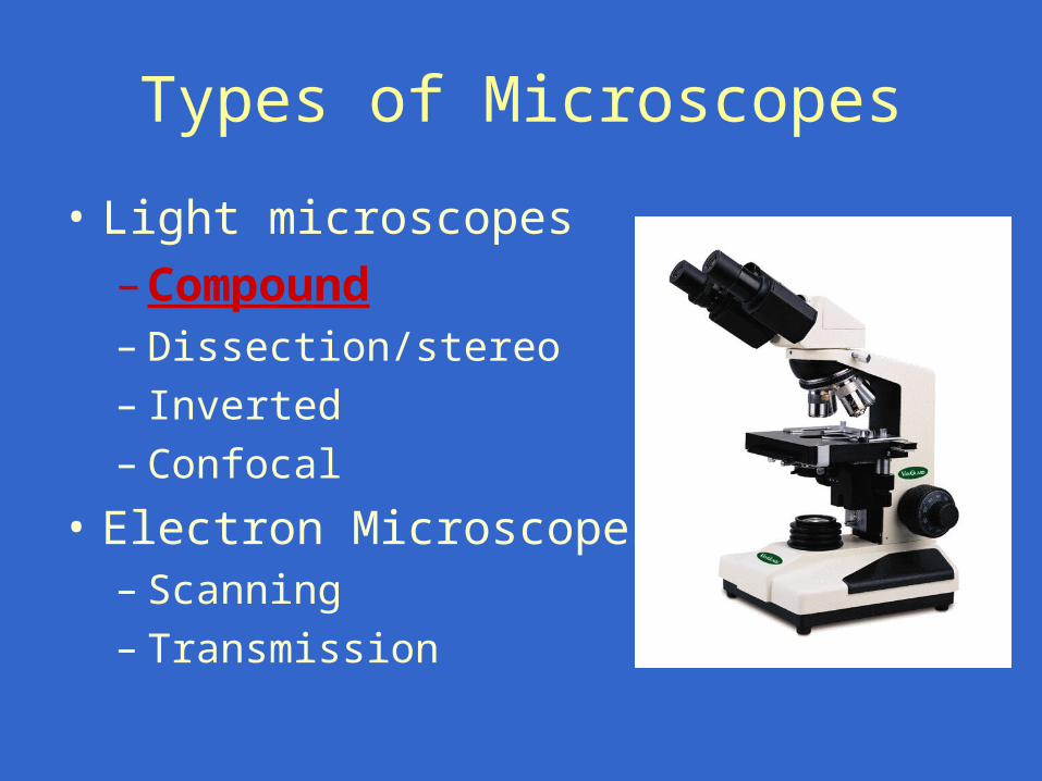
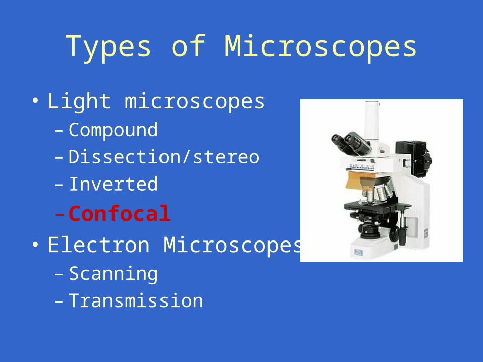
Types of Microscopes
• Light microscopes
–Compound– Dissection/stereo– Inverted– Confocal
• Electron Microscopes– Scanning– Transmission
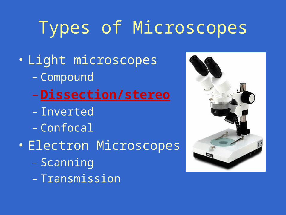
Types of Microscopes
• Light microscopes– Compound
–Dissection/stereo– Inverted– Confocal
• Electron Microscopes– Scanning– Transmission
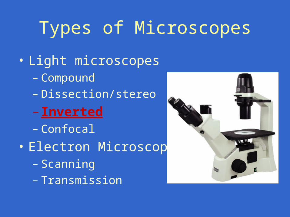
Types of Microscopes
• Light microscopes– Compound– Dissection/stereo
– Inverted– Confocal
• Electron Microscopes– Scanning– Transmission
Types of Microscopes
• Light microscopes– Compound– Dissection/stereo– Inverted
–Confocal
• Electron Microscopes– Scanning– Transmission
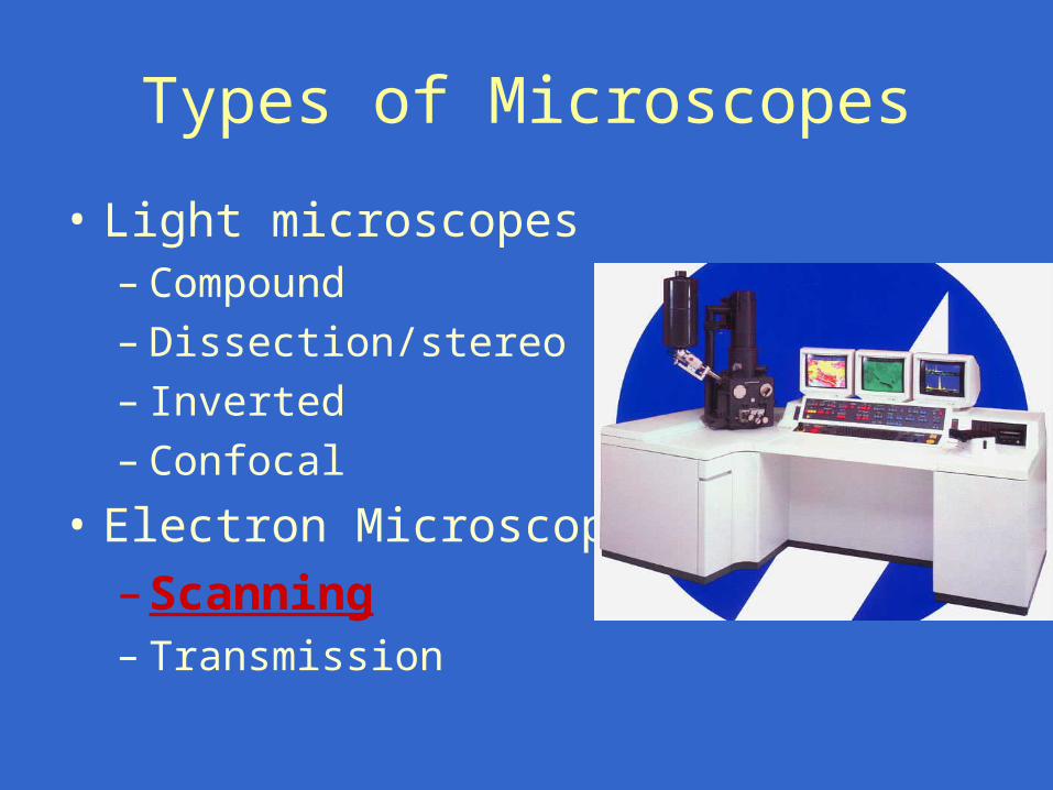
Types of Microscopes
• Light microscopes– Compound– Dissection/stereo– Inverted– Confocal
• Electron Microscopes
–Scanning– Transmission
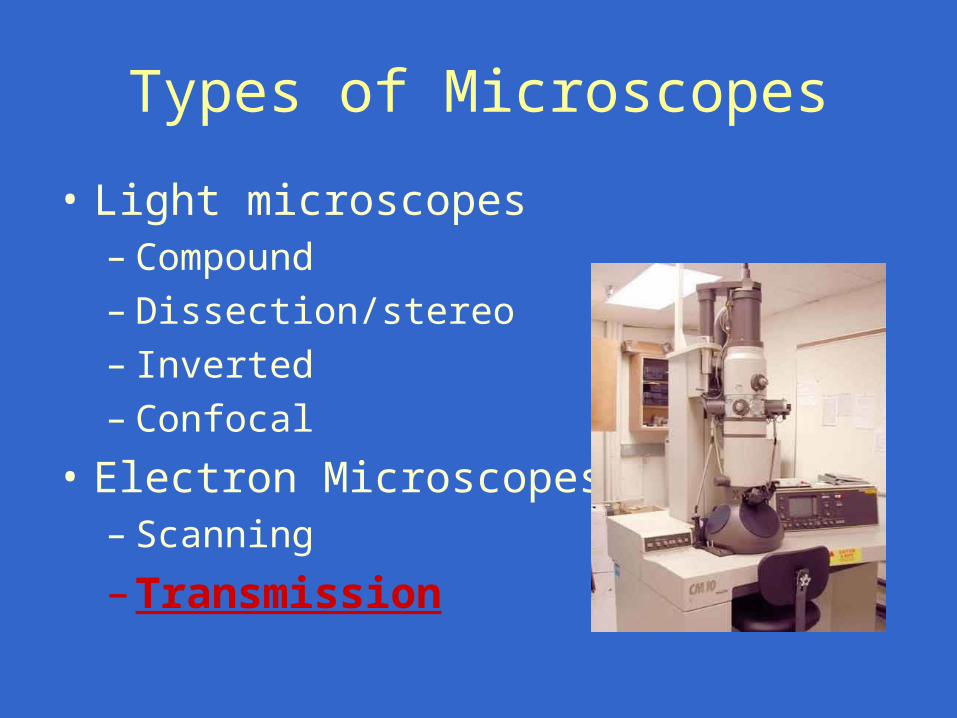
Types of Microscopes
• Light microscopes– Compound– Dissection/stereo– Inverted– Confocal
• Electron Microscopes– Scanning
–Transmission
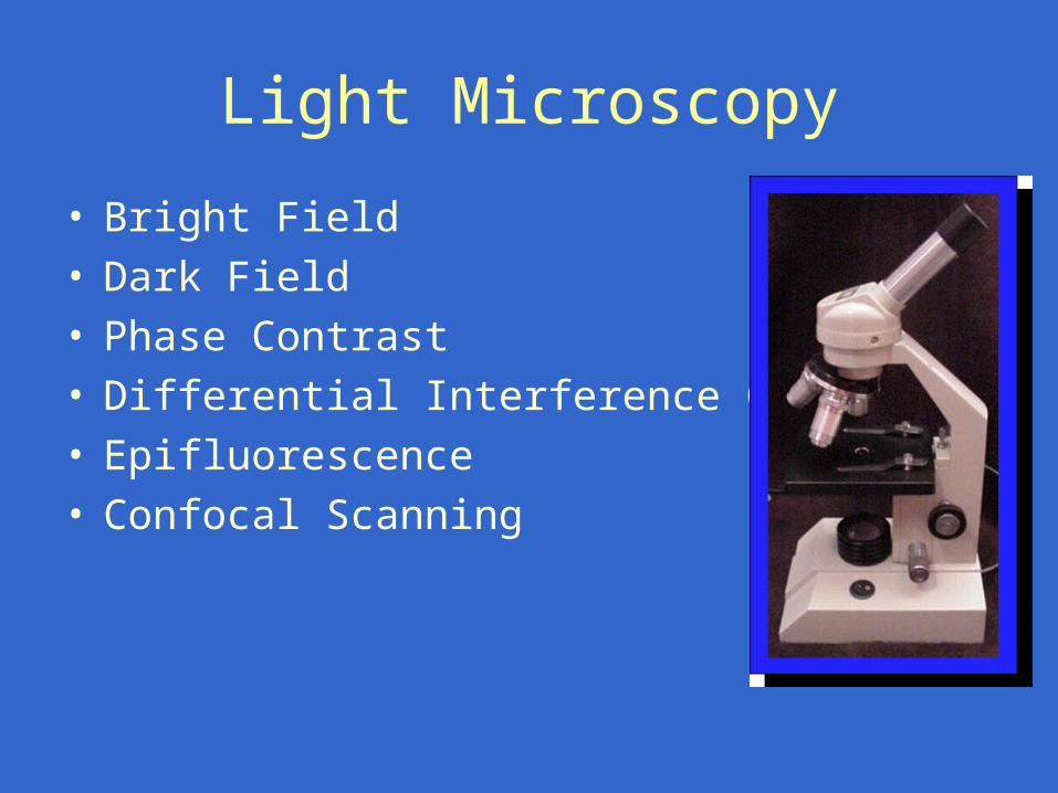
Light Microscopy
• Bright Field• Dark Field• Phase Contrast• Differential Interference Contrast• Epifluorescence• Confocal Scanning
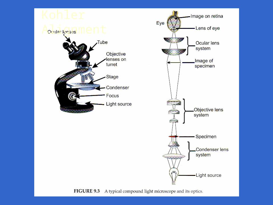
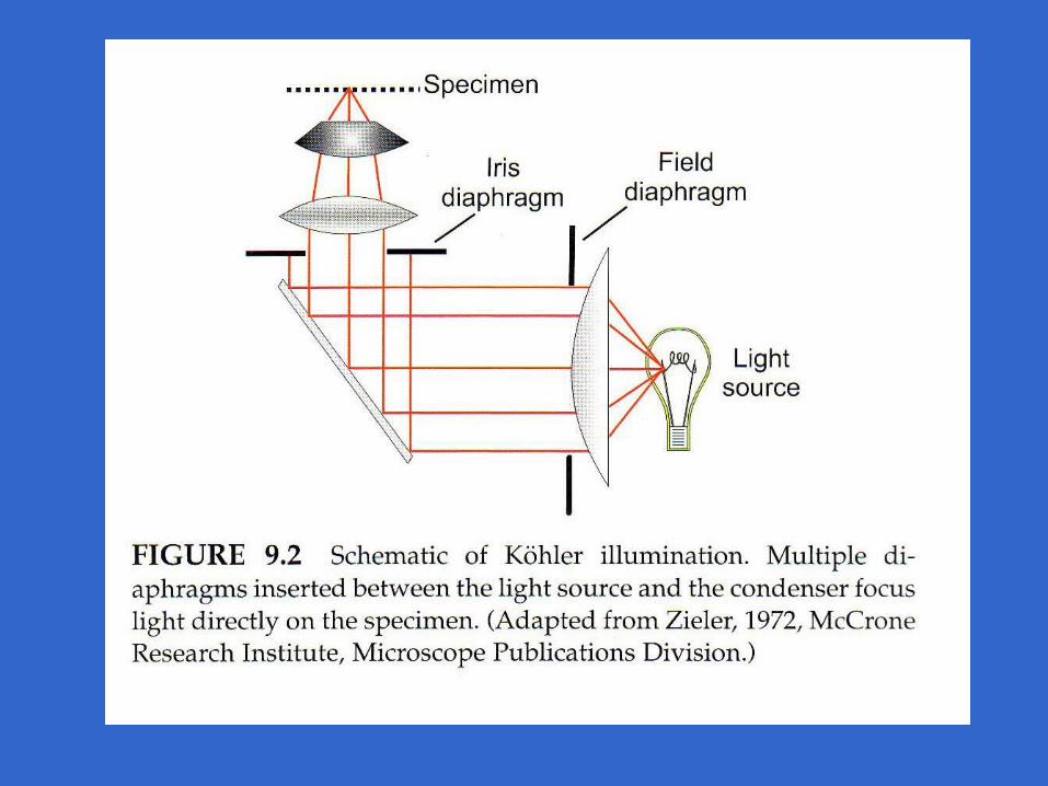
Kohler Alignment
Bright field
• Most common of all light scopes
• Light is transmitted through specimen
• Specimen appears darker than surrounding field
• Typical use: Gram Stains
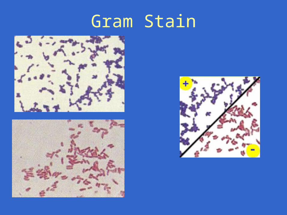
Gram Stain
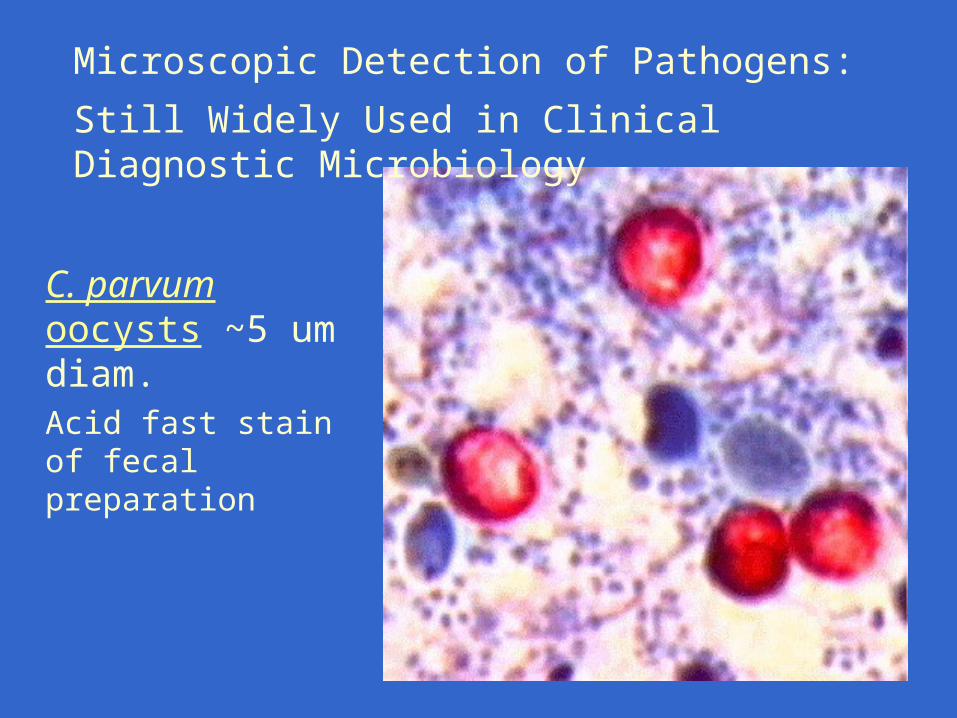
C. parvum oocysts ~5 um diam.Acid fast stain of fecal preparation
Microscopic Detection of Pathogens:
Still Widely Used in Clinical Diagnostic Microbiology
Dark Field
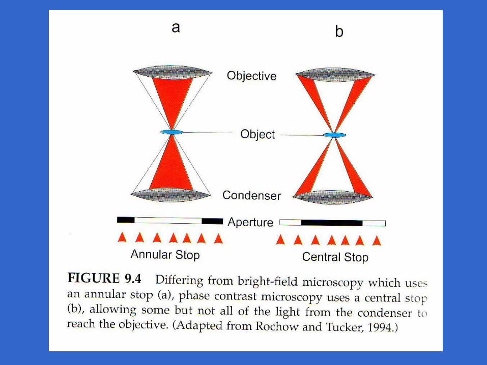
• Used to increase the contrast of a transparent specimen– Contrast = ability to distinguish an object from
surrounding medium
• Specimen appears as a bright image against dark background
• Often used to observe live non-fixed/stained samples, e.g. observe motility and growth
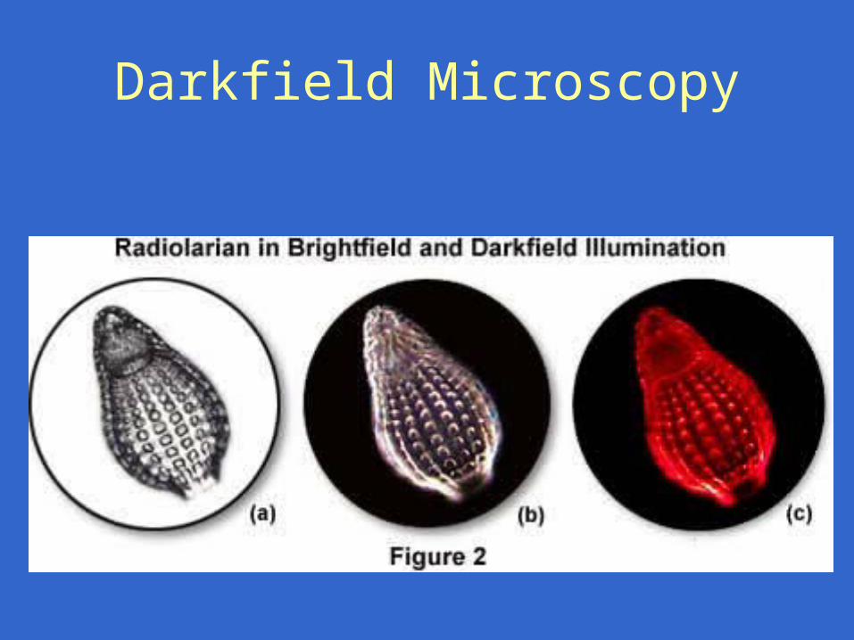
Darkfield Microscopy
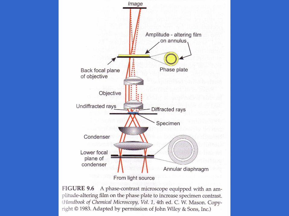
Phase Contrast
• Used to observe fine internal detail
• Takes advantage of differences in density of transparent internal cell components
• Uses a series of diaphragms to separate and recombine direct versus diffracted light rays
DIC• Illuminating light beam is split such that
one beam passes through the specimen creating a phase difference with the second reference beam
• Beams are then combined so that they interfere with eachother
• Allows detection of small changes in in depth or elevation of the surface of the specimen– Thus gives 3d appearance
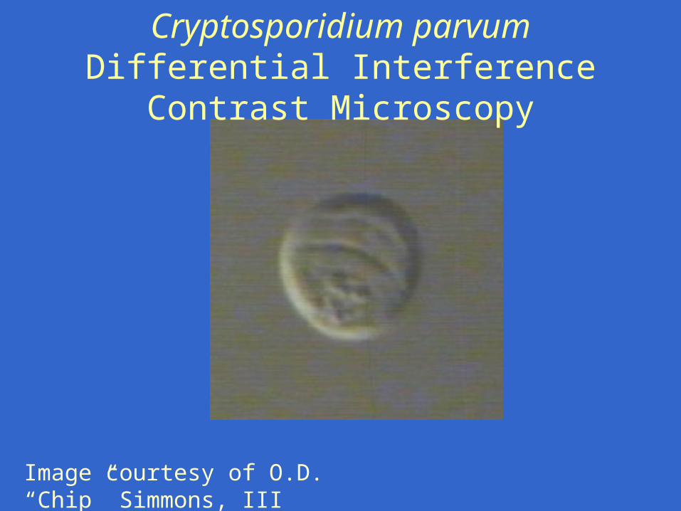
Cryptosporidium parvumDifferential Interference Contrast Microscopy
Image courtesy of O.D. “Chip” Simmons, III
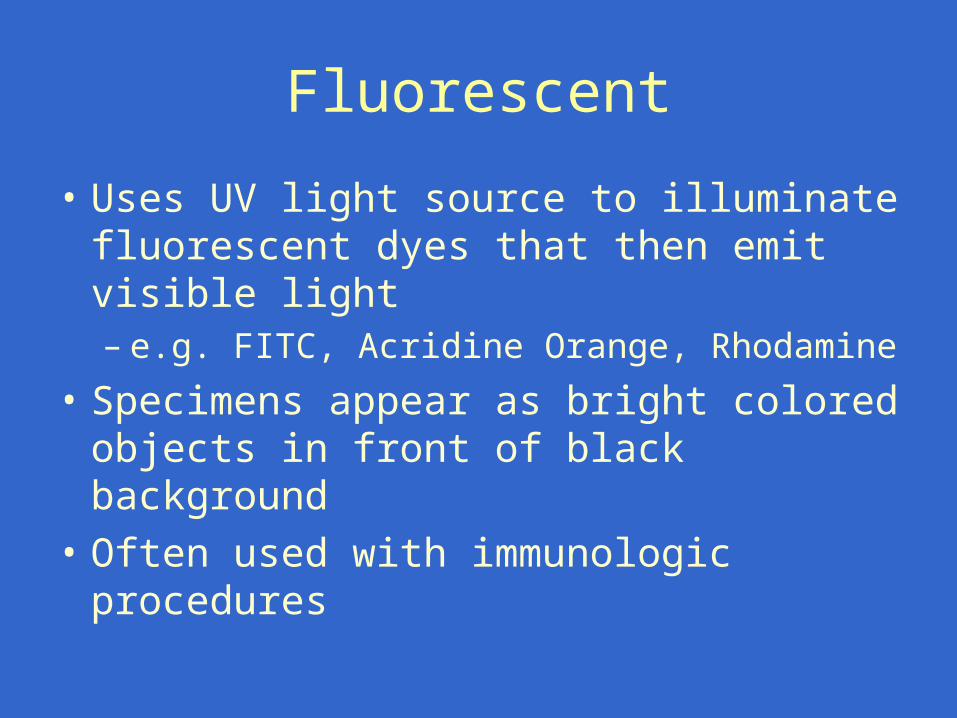
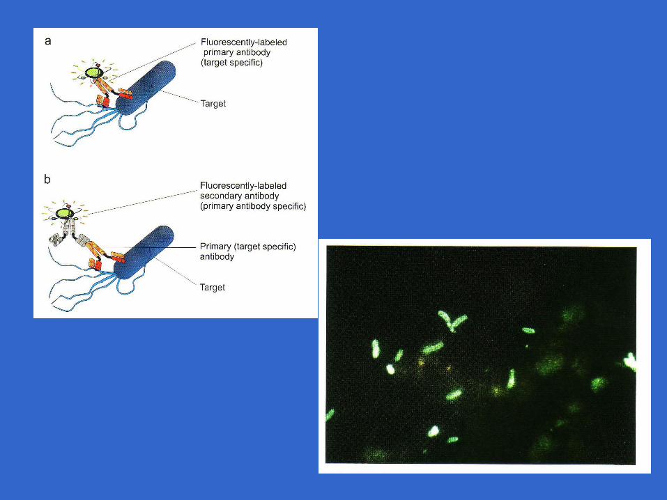
Fluorescent
• Uses UV light source to illuminate fluorescent dyes that then emit visible light– e.g. FITC, Acridine Orange, Rhodamine
• Specimens appear as bright colored objects in front of black background
• Often used with immunologic procedures
Cryptosporidium parvum: Microscopic Analysis of NC field isolate
Differential Interference Contrast
DAPI stain
Immunofluorescence
Images courtesy of O.D. “Chip” Simmons, III
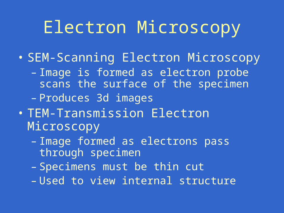
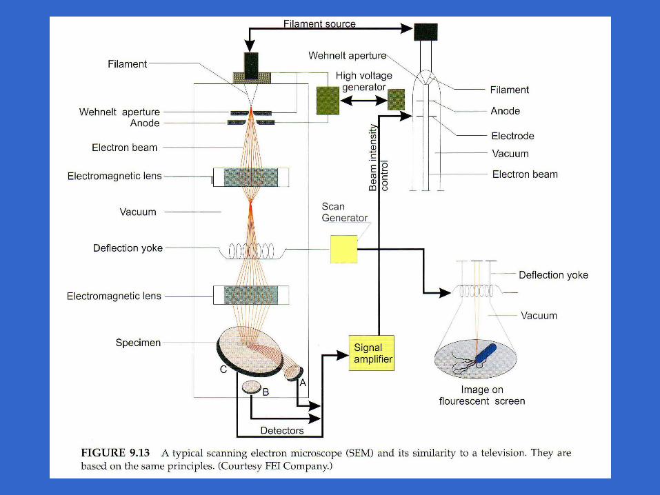
Electron Microscopy
• SEM-Scanning Electron Microscopy– Image is formed as electron probe scans the
surface of the specimen– Produces 3d images
• TEM-Transmission Electron Microscopy – Image formed as electrons pass through
specimen– Specimens must be thin cut– Used to view internal structure
Activity Assays/Vital Dyes
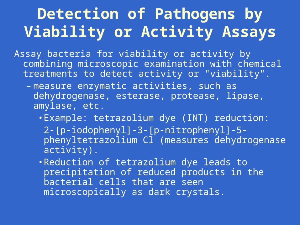
Detection of Pathogens by Viability or Activity Assays
Assay bacteria for viability or activity by combining microscopic examination with chemical treatments to detect activity or "viability". – measure enzymatic activities, such as dehydrogenase,
esterase, protease, lipase, amylase, etc. • Example: tetrazolium dye (INT) reduction:
2-[p-iodophenyl]-3-[p-nitrophenyl]-5-phenyltetrazolium Cl (measures dehydrogenase activity).
• Reduction of tetrazolium dye leads to precipitation of reduced products in the bacterial cells that are seen microscopically as dark crystals.
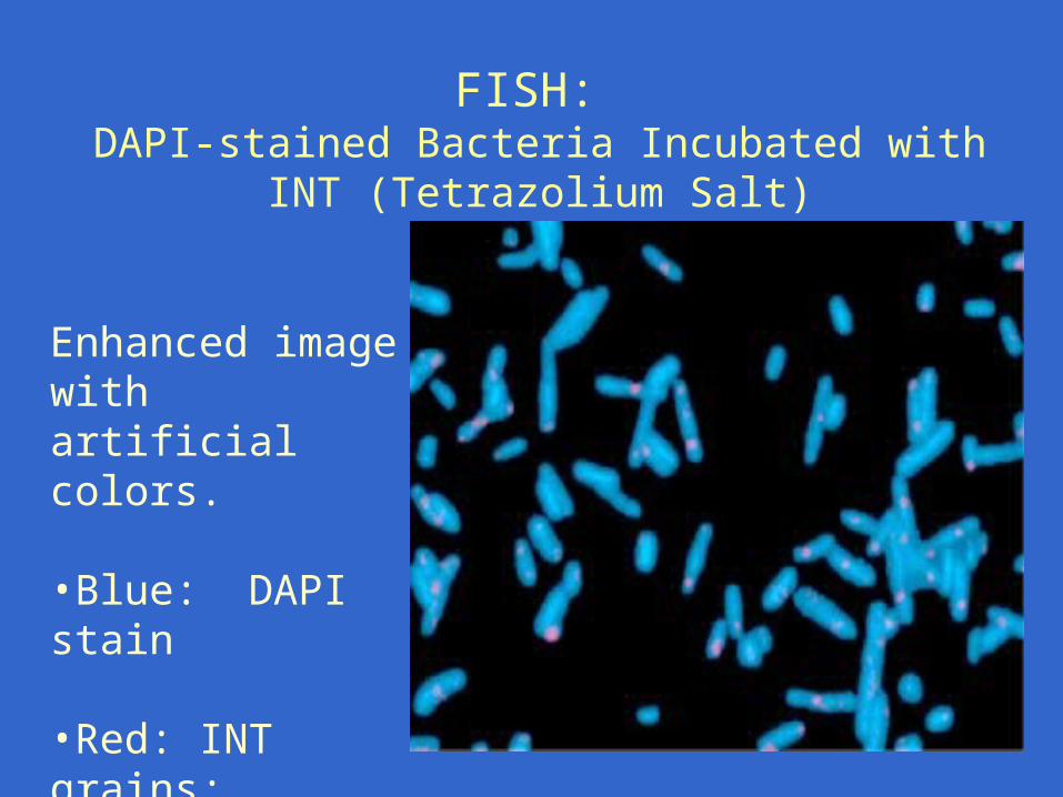
FISH: DAPI-stained Bacteria Incubated with INT (Tetrazolium Salt)
Enhanced image with artificial colors. •Blue: DAPI stain •Red: INT grains; indicate respiratory active bacteria.
Progress in Detection of Bacteria by Viability or Activity Assays
• Combine activity measurement and immunochemical assay (for specific bacteria). – Combine fluorescent antibody (FA) (for detection of specific
bacterium or group) with enzymatic or other activity measurement
• Use image analysis tools to improve detection and quantitation– Flow cytometry– Computer-aided laser scanning of cells or colonies on filters
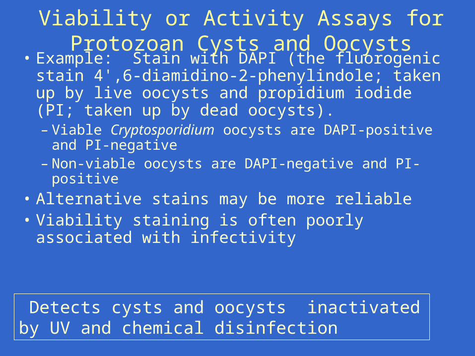
Viability or Activity Assays for Protozoan Cysts and Oocysts
• Example: Stain with DAPI (the fluorogenic stain 4',6‑diamidino‑2‑phenylindole; taken up by live oocysts and propidium iodide (PI; taken up by dead oocysts). – Viable Cryptosporidium oocysts are DAPI-positive and PI-
negative – Non-viable oocysts are DAPI-negative and PI-positive
• Alternative stains may be more reliable• Viability staining is often poorly associated with
infectivity
Detects cysts and oocysts inactivated by UV and chemical disinfection
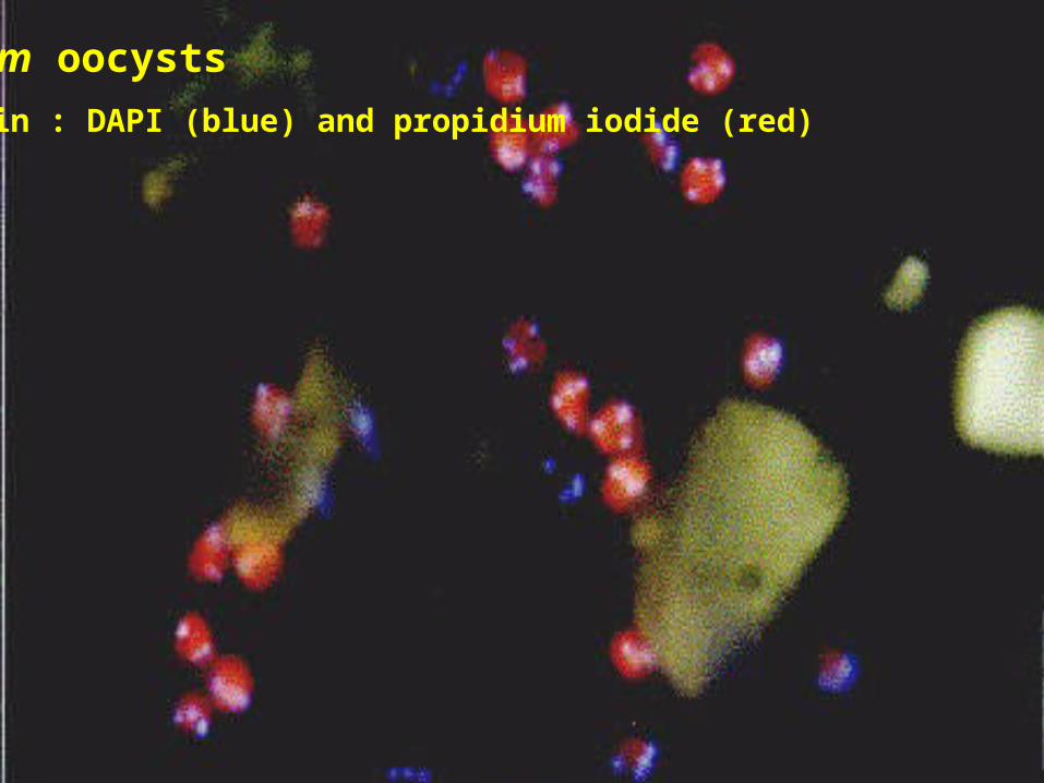
C. parvum oocysts
Dual stain : DAPI (blue) and propidium iodide (red)

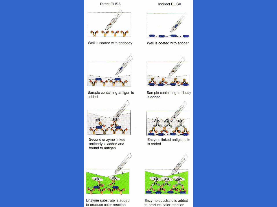
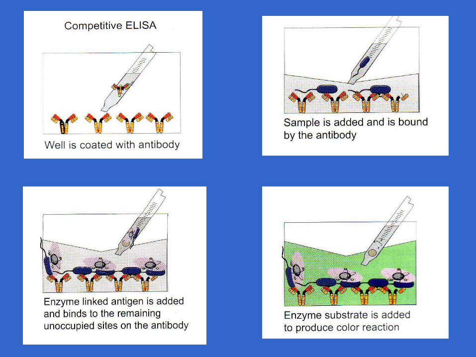
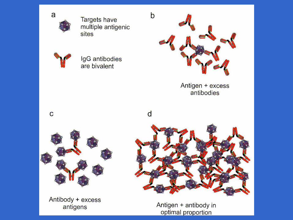
Immunological Methods
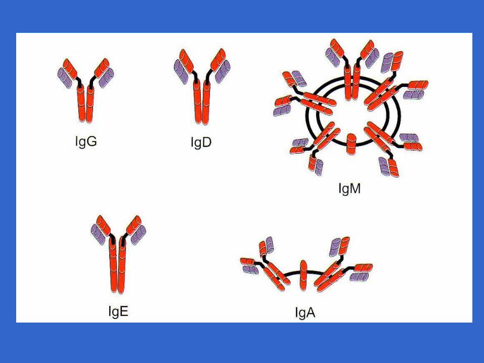
Immunoglbulins
• 5 Classes– IgA secretory– IgD found in plasma but not serum– IgE involved in allergic reactions– IgG humeral response– IgM humeral response
• IgG and IgM most commonly used in immunoassays