Embed Size (px)
Citation preview

University of Groningen
Clinical assessments in Sjögren's syndromeKalk, Wouter Warner Iwe
IMPORTANT NOTE: You are advised to consult the publisher's version (publisher's PDF) if you wish to cite fromit. Please check the document version below.
Document VersionPublisher's PDF, also known as Version of record
Publication date:2001
Link to publication in University of Groningen/UMCG research database
Citation for published version (APA):Kalk, W. W. I. (2001). Clinical assessments in Sjögren's syndrome: the oral component Groningen: s.n.
CopyrightOther than for strictly personal use, it is not permitted to download or to forward/distribute the text or part of it without the consent of theauthor(s) and/or copyright holder(s), unless the work is under an open content license (like Creative Commons).
Take-down policyIf you believe that this document breaches copyright please contact us providing details, and we will remove access to the work immediatelyand investigate your claim.
Downloaded from the University of Groningen/UMCG research database (Pure): http://www.rug.nl/research/portal. For technical reasons thenumber of authors shown on this cover page is limited to 10 maximum.
Download date: 19-05-2018

Salivary imaging
CHAPTER

Chapter 5.1
74
SSUMMARY
Aim - Sialography is commonly used for the purpose of diagnosing Sjögren's syndrome, though itsinvasive nature is often regarded as a serious drawback for routine usage. The aim of this study was toevaluate the morbidity and acceptability of parotid sialography using oil-based contrast fluid.Methods - Twenty-four consecutive sialographic procedures were evaluated by assessing themorbidity and patient's acceptance of the procedure with a standardised questionnaire, and byrecording relevant physical parameters during the procedure.Results - There was good acceptance of the sialographical procedure, and the morbidity was low. Nosigns of overfilling or fausse route were observed in any of the sialograms. On average, 0.74 mlcontrast fluid was infused at a velocity of 0.01 ml/s. The whole procedure was completed within 12minutes.Conclusions - Parotid sialography appears less invasive than is often thought. It has a low morbidityand is well accepted by the patients.

SALIVARY IMAGING
75
M
I
P
MORBIDITY FROM PAROTID SIALOGRAPHY
W.W.I. Kalk, A. Vissink, F.K.L. Spijkervet, J.M. Möller, J.L.N. Roodenburg
Department of Oral and Maxillofacial Surgery, University Hospital Groningen
Oral Surg Oral Med Oral Pathol Oral Radiol Endod, in press
INTRODUCTION
Introduced early in the previous century, sialography has proven a useful techniquethroughout the years. Through retrograde infusion of oil- or water-based iodinecontrast, the architecture of the salivary duct system is visualised radiographically.There is debate ongoing in the literature regarding the current value of contrastsialography for the clinical differentiation of salivary gland disorders. Due to theavailability of new imaging procedures, such as MRI, CT scanning, scintigraphy andultrasonography, its diagnostic indication has significantly been narrowed andlimited to imaging of the ductal system.1-6 For the purpose of diagnosing Sjögren'ssyndrome, sialography is still commonly used, as it has been proven to revealcharacteristic changes of the salivary gland ductal system with rather high sensitivityand specificity.7-10
Following the introduction of water-based contrast fluids in the 1950's11, these fluidshave been advocated in sialography for their better tolerance in the human bodyhaving a physicochemical composition closer to body fluids.12 A comparative studybetween oil-based and water-based contrast revealed no adverse reactions withboth contrast media.13 Nevertheless, invasiveness and poor tolerance is by someclinicians considered a drawback for routine usage of oil-based sialography,especially in SS patients.14 To date, however, the subjective experiences of thepatients have not been described. The aim of this study was to evaluate themorbidity and acceptability of oil-based contrast sialography.
PATIENTS AND METHODS
PatientsTwenty-four consecutive patients who had undergone parotid contrast sialographyfor the diagnosing of Sjögren's syndrome in the period from October 1999 untilApril 2000 at the Department of Oral and Maxillofacial Surgery of the University

Chapter 5.1
76
Hospital Groningen participated in this study. The studied group comprised 22women and 2 men (mean age of 57 years; SD 9; range 41 to 75). The right parotidgland was used 21 times and the left parotid gland three times. Iodine allergy wasapplied as an exclusion criterion for the study, since iodine is present in the contrastfluid used. No patients had to be excluded from the study.
Technical procedure for sialographyAll sialograms were obtained in absence of acute sialadenitis. If clinical signs ofacute inflammation were present, sialography was postponed until clinical signs hadsubsided for at least six weeks. Parotid sialograms were made unilaterally (preferablyof the right gland) in a standardised manner. Reasons for using the left parotid glandincluded probing difficulties and asymmetrical parotid gland swelling. All sialogramswere made by the same clinician. After cannulation of the parotid main duct, thegland was filled through retrograde infusion of an oil-based contrast fluid (LipiodolU.F.®), under low pressure15 using a 2-ml Cornwall® syringe (Becton Dickinson).The patient's sensation of a sudden increase of pre-auricular pressure was used toestimate the proper filling level of the gland, after which two pictures(posteroanterior and lateral) were made. Premature leakage of contrast fluid wasprevented through main duct ligation under local anaesthesia. A General ElectricG1000 was used as X-ray apparatus; pictures were made with an additional filterwith 58 kV during 0.18 s. After removal of the ligature and massaging the parotidgland patients were advised to stimulate salivary gland secretion with citric flavouredgum or candy during the first hours, in order to enhance wash out of remainingcontrast fluid.
Evaluation of the procedureThe sialographical procedure was evaluated by an independent investigator usingthe following two methods: assessment of relevant physical parameters during theprocedure and a standardised patient questionnaire. There was no time intervalbetween the making of the sialogram and the subsequent evaluation, with theexception of the duration of pain sensation after the procedure and the overalljudgement, which were recorded at a recall visit after three weeks. As physicalparameters, the duration of the whole procedure, the duration of contrast infusionand the total amount of infused contrast fluid were recorded. The velocity ofcontrast infusion was in addition calculated. The questionnaire contained multiplechoice questions about presence and severity of pain during the sialographicalprocedure, duration of pain following sialography, and the patients' acceptance of

SALIVARY IMAGING
77
Rthe procedure. Pain severity was graded on a visual analogue scale (0 representingno pain, 10 representing severe pain).
RESULTS
Physical parameters of sialographyThe duration of the whole procedure averaged 12 minutes (mean 716 s, SD 186 s,range 552 – 955 s). The volume of infused contrast fluid averaged 0.74 ml (SD 0.08ml, range 0.50 - 0.90 ml), whereas the mean velocity of infusion was calculated at0.01 ml/s (SD 0.001ml/s, range 0.008 - 0.013 ml/s). There was no evidence ofleakage of contrast medium into the oral cavity until the ligature was removed.Radiographically, all sialograms showed good filling of the gland; no fausse routewas observed.
Morbidity and acceptabilityTwenty-four sialographical procedures were evaluated in twenty-four patients.Nineteen of 24 patients experienced no pain during infusion of the contrast fluid,whereas 5 patients experienced little pain. The pain severity during the procedureaveraged 4.0 in latter patients.Sixteen of 24 patients experienced 'no discomfort' during the sialographicalprocedure, 7 patients found it 'a little unpleasant' and one patient found it 'veryunpleasant'. To the question, which part of the procedure was experienced as themost unpleasant sensation, surprisingly, patients stated most often it was theopening of the mouth (n=7), instead of the placement of the ligature (n=2) or theinfusion of contrast fluid (n=2).Fifteen patients had no pain at all after the procedure and 5 felt soreness for oneday. Two patients stated that a sore feeling had lasted about a week whereas twohad a sore feeling nearby the parotid region lasting about two weeks.To estimate the subjective acceptability of the sialographical procedure, the patientswere requested to judge the procedure using a number between 0 and 10, with 0indication 'very bad experience' and 10 'no problems at all'. The judgementaveraged 8.7.

Chapter 5.1
78
DDISCUSSION
Most of the patients experienced no pain or discomfort during sialography (79%and 67%, respectively). The average patient judgement of the procedure was veryhigh.The mild pain that was felt by 5 of the patients during the contrast infusion was mostlikely related to the perception of raised intraluminal pressure or distension of theparotid capsule from minor glandular enlargement.Soreness in the parotid region for a short period after parotid sialography, asoccurred in a few patients, may relate to presence of some residual contrast fluid inthe gland, especially in cases of sialectasia. Another explanation for the temporarysoreness might be the presence of a subclinical sialadenitis prior to sialography,rendering the gland more sensitive to contrast fluid. The two patients who felt painfor longer than a week both have been examined by their general practitioner, whoconcluded that other reasons than sialography had been responsible for theircomplaints (sinusitis and headache, respectively).Though sialography is considered the imaging procedure of choice for diagnosingSjögren’s syndrome16, it should not be performed in case of iodine allergy toprevent local and systemic allergic reactions. Alternative positive contrast materialsfor iodine, such as barium-sulphate suspensions, are not suitable for sialography dueto a large particle size. Therefore, if confronted with patients with a history of iodineallergy, other imaging techniques such as scintigraphy or ultrasonography should beused instead to visualise salivary gland pathosis. Regarding the use of CT and MRItechniques in diagnosing SS, conflicting results have been reported in literature.2,3,5
Though the use of oil-based contrast fluid has often been associated in literaturewith non-allergic adverse tissue responses or even with damage to the gland14, wehave experienced no complications whatsoever during or after sialography. We feelthat, if lipiodol is restrained to the ductal lumen during the procedure, no adversetissue effects can be expected. Lipiodol remains, due to its hydrophobic nature,much better within the salivary ducts, than water-based contrast fluids that passmore easily through the ductal epithelium.17 As a result, the clearance of contrastfluid differs substantially between oil- and water based contrast. Oil-based fluidleaves the gland with saliva secretion via the main salivary duct, whereas water-based fluid diffuses across the ductal epithelium and is cleared subsequently fromthe circulation by kidneys and liver. Therefore, one might actually expect lessadverse reactions from oil-based contrast than from water-based contrast in thenormal situation.18-20 Only if a iatrogenic fausse route is induced during contrastinfusion, is a less favourable tissue response to be expected from oil-based contrast,

SALIVARY IMAGING
79
R
since in such situation it remains in the gland parenchyma for a long time inducing achronic granulomatous inflammation, as opposed to quick clearance of water-basedcontrast in the same situation. If uncertain about the ductal probing or ifinexperienced with contrast sialography, it seems wise therefore to use water-basedcontrast fluids. This way, adverse tissue reaction from a possible fausse route oroverfilling is minimised.The use of oil-based contrast, whose hydrophobic nature impairs the fluid’s ability tomix with saliva or to pass through epithelial membranes of the salivary ducts,renders much sharper X-ray images than water-based alternatives.13,17,21 Therefore,we prefer the use of lipiodol or other oil-based contrast fluids, yielding optimumquality of sialographical images with, in our hands, no adverse side effects on thesalivary glands of any kind.
Given its low morbidity and its subjective acceptability, sialography of the parotidgland appears, if performed properly, to be less invasive than is often thought. Withsome practice, this diagnostic imaging technique can be applied in ten to fifteenminutes, differentiating between a variety of salivary gland disorders includingSjögren's syndrome.
REFERENCES
1. Napoli V, Tozzini A, Neri E, Calderazzi A, Gabriele M, Bonaretti S, et al. The imaging diagnosis ofSjögren's syndrome: echography, sialography and scintigraphy compared in the study of thesalivary glands. Minerva Stomatol 1996;45:141-148.
2. Izumi M, Eguchi K, Ohki M, Uetani M, Hayashi K, Kita M, et al. MR imaging of the parotid gland inSjögren's syndrome: a proposal for new diagnostic criteria. AJR Am J Roentgenol 1996;166:1483-1487.
3. Tonami H, Ogawa Y, Matoba M, Kuginuki Y, Yokota H, Higashi K, et al. MR sialography inpatients with Sjögren's syndrome. AJNR Am J Neuroradiol 1998;19:1199-1203.
4. Takashima S, Morimoto S, Tomiyama N, Takeuchi N, Ikezoe J, Kozuka T. Sjögren syndrome:comparison of sialography and ultrasonography. J Clin Ultrasound 1992;20:99-109.
5. de Clerck LS, Corthouts R, Francx L, Brussaard C, de Schepper A, Vercruysse HA, et al.Ultrasonography and computer tomography of the salivary glands in the evaluation of Sjögren'ssyndrome. Comparison with parotid sialography. J Rheumatol 1988;15:1777-1781.
6. Saito T, Fukuda H, Horikawa M, Ohmori K, Shindoh M, Amemiya A. Salivary gland scintigraphywith 99mTc-pertechnetate in Sjögren's syndrome: relationship to clinicopathologic features ofsalivary and lacrimal glands. J Oral Pathol Med 1997;26:46-50.
7. Vitali C, Tavoni A, Simi U, Marchetti G, Vigorito P, d'Ascanio A, et al. Parotid sialography andminor salivary gland biopsy in the diagnosis of Sjögren's syndrome. A comparative study of 84patients. J Rheumatol 1988;15:262-267.
8. Pennec YL, Letoux G, Leroy JP, Youinou P. Reappraisal of tests for xerostomia. Clin ExpRheumatol 1993;11:523-528.
Chapter 5.1
80
9. Markusse HM, van Putten WI, Breedveld FC, Oudkerk M. Digital substraction sialography of theparotid glands in primary Sjögren's syndrome. J Rheumatol 1993;20:279-283.
10. Vitali C, Moutsopoulos HM, Bombardieri S. The European Community Study Group on diagnosticcriteria for Sjögren's syndrome. Sensitivity and specificity of tests for ocular and oral involvementin Sjögren's syndrome. Ann Rheum Dis 1994;53:637-647.
11. Katzberg RW. Urography into the 21st century: New contrast media, renal handling, imagingcharacteristics and nephrotoxicity. Radiology 1997;204:297-312.
12. Daniels TE, Benn DK. Is sialography effective in diagnosing the salivary component of Sjögren'ssyndrome? Adv Dent Res 1996;10:25-28.
13. Holtgrave E, Elke M, Lüthy H. Sialography with TELEBRIX 38®. A comparative study with lipiodol-UF in the diagnosis of salivary gland diseases. Dentomaxillofac Radiol 1973;2:68-72.
14. Daniels TE, Fox PC. Salivary and oral components of Sjögren’s syndrome. Rheum Dis Clin NorthAm 1992;18:571-589.
15. Zijlstra G, ten Bosch JJ. Sialography with continuous measurement of pressure outside and insidethe gland. Int J Oral Surg 1975;4:160-167.
16. Stiller M, Golder W, Döring E, Kliem K. Diagnostic value of sialography with both the conventionaland digital subtraction techniques in children with primary and secondary Sjögren’s syndrome.Oral Surg Oral Med Oral Pathol Oral Radiol Endod 1999;88:620-627.
17. Quarnström E. Expirimental sialography: The effects of retrograde infusion of radiographiccontrast media on salivary gland morphology and function. Oral Surg Oral Med Oral Pathol1986;62:668-682.
18. Suzuki S, Kawashima K. Sialographic study of diseases of the major salivary glands. ActaRadiologica Diagnosis 1969;8:465-478.
19. Berman HL, Delaney V. Iodine mumps due to Low-Osmolality Contrast Medium. Am JRöntgenology 1992;159:1099-1100.
20. Christensen J. Iodide mumps after intravascular administration of a nonionic contrast medium.Acta Radiologica 1995;36:82-84.
21. Luyk NH, Doyle T, Ferguson MM. Recent trends in imaging the salivary glands. DentomaxillofacRadiol 1991;20:3-10.


Chapter 5.2
82
SSUMMARY
Aim - Despite the availability of many new imaging procedures, sialography has, after decades of use,maintained its status as the imaging procedure of choice for evaluating the oral component ofSjögren’s syndrome (SS). In this study, the clinical value of sialography as a diagnostic tool in SS wasexplored by assessing its diagnostic accuracy, observer bias and staging potential.Methods - One hundred parotid sialograms were interpreted independently in a blind fashion by twotrained- and two expert-observers. Sialograms were derived from a group of consecutive patients,referred for diagnostics of SS. Patients were categorised as SS and non-SS by the revised Europeanclassification criteria.Results - Trained observers reached a sensitivity of 95 and a specificity of 33 percent for SS bysialogram, whereas expert-observers reached a sensitivity of 87 and a specificity of 84 percent. Therewas only ‘fair’ inter-observer agreement between trained- and expert-observers, whereas both expert-observers showed ‘good’ agreement with one another, according to Cohen’s kappa. Intra-observeragreement was ‘good’ to ‘very good’ for all observers. The four different gradations of sialectasia, i.e.punctate, globular, cavitary and destructive, showed a weak but significant correlation with theduration of oral symptoms.Conclusions - This study markedly shows that the diagnostic value of parotid sialography fordiagnosing SS greatly depends upon the skills of the observer, implying that sialography lacks generalapplicability as a diagnostic tool in SS and requires specific expertise. Nevertheless, given its potentiallyhigh sensitivity and specificity in diagnosing SS as well as its useful staging potential, sialography stillhas its use in the evaluation of the oral component of SS.

SALIVARY IMAGING
83
S
I
PAROTID SIALOGRAPHY FOR DIAGNOSING SJÖGREN’SSYNDROME
W.W.I. Kalk1, A. Vissink1, F.K.L. Spijkervet1, H. Bootsma2, C.G.M. Kallenberg3,J.L.N. Roodenburg1
Departments of 1Oral and Maxillofacial Surgery, and Internal Medicine, divisions of2Rheumatology, and 3Clinical Immunology, University Hospital Groningen
Submitted for publication
INTRODUCTION
Sjögren’s syndrome (SS) is considered a systemic autoimmune disease with theexocrine glands as main target-organs. As a result, the presence of this disease maycause structural damage and secretory dysfunction of the tear- and salivary glands.The tear- and salivary gland involvement with its inherent morbidity is oftenaddressed as the ocular- and the oral component of SS, respectively.The oral component of SS can be evaluated in many ways. Generally, two differentprocedures are practised, i.e. assessment of salivary gland function and salivarygland imaging. Salivary gland function is assessed through measurement of salivarysecretion rate (sialometry) and analysis of salivary composition (sialochemistry).1-3
Salivary gland imaging is currently performed by several procedures includingmagnetic resonance imaging (MRI), computer tomography (CT) scanning,ultrasonography, scintigraphy and sialography.4-9 Despite the availability of newimaging procedures, the oldest procedure of all, sialography has maintained itsstatus as the method of choice for exploring the ductal system of the salivary glandto diagnose SS.10
Sialography reveals the architecture of the salivary duct system radiographically byinsertion of a contrast fluid. This radiographic demonstration of salivary glands invivo was first performed in 1913.11 Four decades ago the sialographic changes seenon sialograms were accurately described and, with regard to chronic sialadenitis,classified into punctate, globular, cavitary and destructive sialectasia (dilatation) ofthe acinar and ductal system.12-14 These four sialectatic changes are thought torepresent increasing glandular damage, respectively, caused by chronic salivarygland inflammation.14 SS is by far the most frequent cause of such chronic salivarygland inflammation. Therefore, by observing sialectasia on a sialogram, the presence(and progression) of SS with regard to its oral component can be determined.

Chapter 5.2
84
P
It has been demonstrated that SS-related sialographic findings such as sialectasia aremore closely related to SS-related clinical symptoms (stimulated parotid salivaryflow, incidence of keratoconjunctivitis sicca) than is the periductal lymphocyticinfiltration of the labial glands.15 In addition, superior sensitivity16-18 and/or specificityfor SS have been frequently ascribed to sialography as compared to labial glandbiopsy.19-23 However, the subjective nature of reading and interpreting a sialogramcauses a certain observer bias, as is the case with diagnostic-imaging tests in general.The amount of observer bias may have a substantial impact on the clinical value of aparticular diagnostic test.In this study the clinical value of sialography as a diagnostic tool in SS was exploredby assessing its diagnostic accuracy, observer bias and staging potential in 100sialograms.
PATIENTS AND METHODS
PatientsIn order to study the clinical value of sialography for diagnosing Sjögren’s syndrome(SS) 100 parotid sialograms were interpreted independently by four observers. Twoobservers had large general experience in judging sialograms, whereas twoobservers were in addition especially experienced in the judging of sialograms withrespect to the diagnosis SS. The observers with general experience are termed‘trained observers’ and the observers with specific SS expertise are termed ‘expert-observers’. Sialograms were derived from a non-selected group of 100 consecutivepatients referred to the outpatient clinic of the Department of Oral and MaxillofacialSurgery of the University Hospital Groningen in the period from December 1997until August 1999.Patients suspected of SS were referred by rheumatologists, internists, neurologists,ophthalmologists, ENT-specialists, general practitioners and dentists. Reasons forreferral included mouth-dryness, eye-dryness, swelling of the salivary glands,arthralgia and fatigue. The diagnostic work-up for SS, carried out in all patients,included the following aspects: subjective complaints of oral and ocular dryness(table 3.1.1), sialometry and sialochemistry, histopathology of salivary gland tissue,serology (SS-A- and SS-B antibodies) and eye tests (Rose Bengal staining andSchirmer tear test). Sialography was excluded for diagnostic use in this study, inorder to avoid an incorporation bias. In addition to the diagnostic tests, the durationof oral symptoms and the serum level of immunoglobulin class G (IgG) wereassessed in order to be studied in relation to the sialographical stage. Duration of

SALIVARY IMAGING
85
oral symptoms was defined as the time from first complaints induced by or relatedto oral dryness until referral.In this study, the revised European classification criteria for SS24,25 were used asreference standard for the diagnosis of SS, categorising patients as primary SS,secondary SS, or non-SS patients.
Exclusion criteriaIodine allergy was applied as an exclusion criterion for the study, since iodine ispresent in the contrast fluid used. Furthermore, the exclusion criteria of theEuropean classification criteria for SS were applied. Psoriatic arthritis and HIV-infection were excluded as both diseases may cause sialographical picturesresembling SS.26-33 No patients had to be excluded from the study.
Technical procedure for sialographyAll sialograms were obtained in absence of acute sialadenitis. If clinical signs ofacute inflammation were present, sialography was postponed until clinical signs hadsubsided for at least six weeks. Parotid sialograms were made unilaterally (preferablyof the right gland) in a standardised manner. All sialograms were made by the sameclinician. After cannulation of the parotid main duct the gland was filled throughretrograde infusion of an oil-based contrast fluid (lipiodol U.F.) using a 2-mlCornwall syringe (Becton and Dickinson). The patient's sensation of a suddenincrease of pre-auricular pressure was used to estimate the proper filling level of thegland. Premature leakage of contrast fluid into the mouth was prevented throughmain duct ligation under local anaesthesia. A General Electric G1000 was used as X-ray apparatus; posteroanterior (6˚ mediolateral) and lateral pictures were made withan additional filter with 58 kV during 0.18 s. After removal of the ligature andmassaging the parotid gland patients were advised to stimulate salivary glandsecretion with citric flavoured gum or candy during the first hours in order toenhance wash out of the remaining contrast fluid. The whole procedure wascompleted within 15 minutes.
Evaluation of the sialogramsFour observers each examined independently 100 sialograms in a random orderwith no information from the patients other than the reason for referral (clinicalsuspicion of SS) and the amount of inserted contrast fluid. Of the 100 sialograms, 25were judged a second time by all observers without being aware of it, in order todetermine intra-observer variability. All sialograms were examined in the presence of

Chapter 5.2
86
an independent investigator who made sure that each set of sialograms wasexamined within 2 minutes without being revised afterwards.Before the observers examined the sialograms, a calibration session took place inwhich all observers agreed upon the criteria to be applied when describing thesialograms. Four different pathological descriptions were agreed upon. Theobservers had to determine whether or not these patterns were present in eachsialogram. These patterns were sialectasia (subdivided into punctate, globular,cavitary and destructive), thin appearance of the ducts with or without glandenlargement, irregular and widened main ducts, and presence of a space-occupyinglesion, respectively.
C
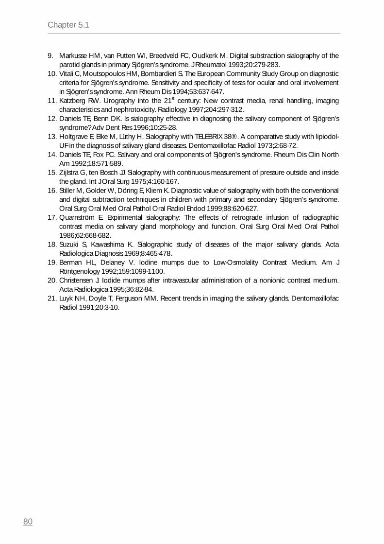
A BFigure 5.2.1 Stages of sialectasia in SS, aspresent on lateral parotid sialograms:
� punctate sialectasia (A): less than 1 mmin size;
� globular sialectasia (B): uniform ofshape and 1-2 mm in size;
� cavitary sialectasia (C): irregular ofshape and more than 2 mm in size;
� destructive sialectasia (not shown):complete loss of gland architecture.

SALIVARY IMAGING
87
R
If present, sialectasia (dilatations) were graded according to the description by Blatt:punctate if less than 1 mm in size; globular if uniform and 1-2 mm in size; andcavitary if irregular and more than 2 mm in size (figure 5.2.1). A destructive patternwas defined as complete destruction of the gland architecture, simulating aninvasive neoplastic process.12 Sialectasia were considered the only descriptionssuggestive for a diagnosis of SS. Presence of thin ducts was regarded as possiblyconsistent with sodium retention dysfunction syndrome or with sialoadenosis.34,35
Irregular and widened main ducts consistent with sialodochitis (salivary ductinflammation) were considered the prevalent feature in chronic-recurrentsialadenitis.36-39 A space-occupying lesion on a sialogram was considered suggestivefor a tumour compressing the gland.A consensus judgement whether or not a sialogram is in accordance with thediagnosis SS was based upon the majority of the individual descriptions of theobservers.
Statistical analysisData were submitted for statistical analysis using the Statistical Package for theSocial Sciences (SPSS), version 9.0. The following statistical procedures wereapplied: Cohen’s kappa as measure of inter- and intra-observer agreement (observerbias)40,41, and Pearson’s and Spearman’s coefficients as correlation tests. In theresults section it is stated which statistical test was applied in a specific situation. Asignificance level of 0.05 was pre-defined in all cases.
RESULTS
Study groupBy applying the revised European classification criteria for Sjögren’s syndrome (SS)on the studied cohort, 39 patients were categorised as SS (20 primary- and 19secondary SS; male/female ratio: 1/7; mean age of 54 years; SD 15; range 21 to 84)and 61 patients as non-SS (negative for SS) (male/female ratio: 1/14; mean age of54 years; SD 15; range 20 to 81). The latter were, based upon additional clinical andlaboratory tests, diagnosed as having sialoadenosis (n=18), sodium retentiondysfunction syndrome (n=18), medication induced xerostomia (n=11), or as havingno alternative disease directly related to salivary gland pathology (n=14). Meanduration of oral symptoms before referral was 35 months for SS- and 30 months fornon-SS patients (range: SS 0-180 months, non-SS 0-240 months).

Chapter 5.2
88
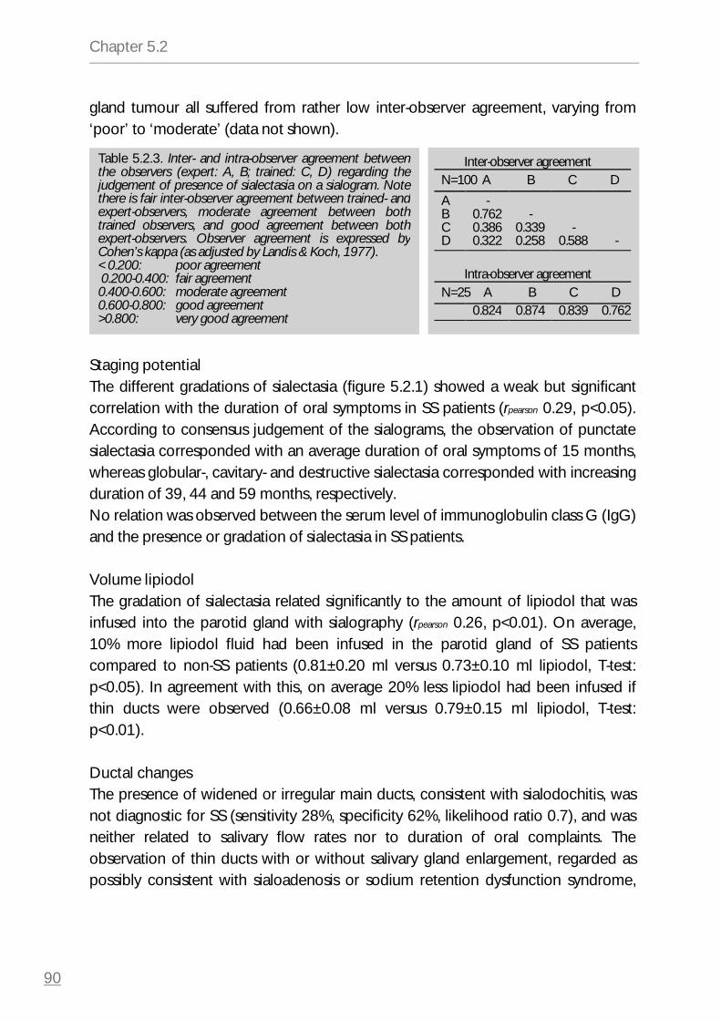
Figure 5.2.2Example of aparotidsialogram of aSS-patient,which couldgive rise fordoubt. Notethe presenceof initialsialectasia onbothprojections. Allobserversjudged thissialogram aspositive for SS(sialectasiapresent).
Figure 5.2.3Parotidsialogram of anon-SS patient.Note the smallradiodensitiesthat could beeasilymisinterpreted.Experienced-observersjudged thissialogram aspositive,whereasexpertobservers asnegative for SS(no sialectasiapresent).
Test accuracy for SSBy determining the presence of sialectasia as diagnostic indicator for SS, thesensitivity and specificity differed greatly between the trained- and expert-observers.Trained observers reached a sensitivity of 95 and a specificity of 33 percent,whereas expert-observers reached a sensitivity of 87 and a specificity of 84 percent(table 5.2.1). The large difference in specificity between trained- and expert-observers was mainly due to their decision when doubting between ‘noabnormality’ and ‘punctate sialectasia’. Examples of sialograms that gave rise fordoubt are illustrated in figures 5.2.2 and 5.2.3.
PALateral
PALateral

SALIVARY IMAGING
89
Expert-observers, reached a high specificity by choosing ‘no abnormality’ in case ofdoubt (observers A and B), whereas trained observers, who chose for ‘punctatesialectasia’ in the same situations, suffered from a major drop in specificity andgained only slight improvement of sensitivity (observers C and D). Consequently,the likelihood ratios also greatly differed between trained- and expert-observers,varying from 1.2 (not very useful as a test) to 5.0 (very useful as a test). Consensusjudgement based upon the majority of individual judgements reached anintermediate sensitivity and specificity for SS of 92 and 71 percent, respectively, anda likelihood ratio of 3.1. In 18 of the 61 non-SS patients, sialectasia was present onthe sialogram, according to consensus judgement (table 5.2.2).
Observer agreementInter- and intra-observer agreement was calculated for the four pathologicalconditions that were determined on the sialograms by each observer. With regardto the presence of SS (sialectasia as indicator), there was only ‘fair’ inter-observeragreement between trained- and expert-observers, whereas both expert-observersshowed ‘good’ agreement with one another, according to Cohen’s kappa. The intra-observer agreement was ‘good’ to ‘very good’ (table 5.2.3). The determination ofother sialographical alterations that occur in sialoadenosis, sialodochitis and salivary
Table 5.2.1. Sensitivity, specificity, positive predictive value (PPV), negative predictive value (NPV)and likelihood ratio (LR) of the four observers (expert: A, B; trained: C, D) for the diagnosis of SS on agroup of 100 patients by presence of sialectasia on the sialogram. Consensus judgement was basedupon the majority of individual judgements for each sialogram. Note the large differences betweenexpert- and trained observers regarding specificity and likelihood ratio.
N=100 A B C D consensusSensitivitySpecificityPPVNPVLR
87%71%65%90%3.0
82%84%76%88%5.0
95%33%47%91%1.4
92%23%43%82%1.2
92%71%67%94%3.1
Table 5.2.2. Judgements of 100 sialograms regarding the presence and grade of sialectasia by fourindividual observers (expert: A, B; trained: C, D) and by consensus. For each descriptive category thenumber of cases accordingly judged is given. Consensus judgement is based upon the majority ofindividual judgements for each sialogram. Note the large variability between expert- and trainedobservers regarding false positivity: the trained observers judged many sialograms from non-SSpatients as punctate.
N=100 A B C D consensusSialectasianonepunctateglobularcavitarydestructive
SS non-SS5 4313 511 64 36 4
SS non-SS7 5115 110 33 04 6
SS non-SS2 2011 244 515 87 4
SS non-SS3 1412 3615 57 52 1
SS non-SS3 4314 511 75 56 1
Chapter 5.2
90
gland tumour all suffered from rather low inter-observer agreement, varying from‘poor’ to ‘moderate’ (data not shown).
Staging potentialThe different gradations of sialectasia (figure 5.2.1) showed a weak but significantcorrelation with the duration of oral symptoms in SS patients (rpearson 0.29, p<0.05).According to consensus judgement of the sialograms, the observation of punctatesialectasia corresponded with an average duration of oral symptoms of 15 months,whereas globular-, cavitary- and destructive sialectasia corresponded with increasingduration of 39, 44 and 59 months, respectively.No relation was observed between the serum level of immunoglobulin class G (IgG)and the presence or gradation of sialectasia in SS patients.
Volume lipiodolThe gradation of sialectasia related significantly to the amount of lipiodol that wasinfused into the parotid gland with sialography (rpearson 0.26, p<0.01). On average,10% more lipiodol fluid had been infused in the parotid gland of SS patientscompared to non-SS patients (0.81±0.20 ml versus 0.73±0.10 ml lipiodol, T-test:p<0.05). In agreement with this, on average 20% less lipiodol had been infused ifthin ducts were observed (0.66±0.08 ml versus 0.79±0.15 ml lipiodol, T-test:p<0.01).
Ductal changesThe presence of widened or irregular main ducts, consistent with sialodochitis, wasnot diagnostic for SS (sensitivity 28%, specificity 62%, likelihood ratio 0.7), and wasneither related to salivary flow rates nor to duration of oral complaints. Theobservation of thin ducts with or without salivary gland enlargement, regarded aspossibly consistent with sialoadenosis or sodium retention dysfunction syndrome,
Table 5.2.3. Inter- and intra-observer agreement betweenthe observers (expert: A, B; trained: C, D) regarding thejudgement of presence of sialectasia on a sialogram. Notethere is fair inter-observer agreement between trained- andexpert-observers, moderate agreement between bothtrained observers, and good agreement between bothexpert-observers. Observer agreement is expressed byCohen’s kappa (as adjusted by Landis & Koch, 1977).< 0.200: poor agreement 0.200-0.400: fair agreement0.400-0.600: moderate agreement0.600-0.800: good agreement>0.800: very good agreement
Inter-observer agreementN=100 A B C D
ABCD
-0.7620.3860.322
-0.3390.258
-0.588 -
Intra-observer agreementN=25 A B C D
0.824 0.874 0.839 0.762
SALIVARY IMAGING
91
Ddid not relate significantly to any changes of salivary composition (e.g. sodium,potassium, amylase, total protein), nor to salivary flow rate.
DISCUSSION
It has become clear from this study that it is possible to achieve both a sensitive andspecific test result with parotid contrast sialography for diagnosing SS (likelihoodratio up to 5.0). This diagnostic accuracy, however, is very much dependant on theobserver involved, which implies that the technique lacks general applicability andrequires specific expertise.The four different gradations of sialectasia showed a weak but significant relation tothe duration of oral symptoms in SS patients, suggesting that sialectasia slowlyworsens (increases in number and size) during the disease course of SS. Previousstudies have already shown that, in SS patients, increasing gradations of sialectasiacorrespond with lower salivary flow rates3,15,42, as well as that salivary flow ratesdeteriorate with increasing duration of oral symptoms (§3.1).43 We thereforesuggest that SS can be subdivided into different sequential stages according to thetype of sialectasia on the sialogram, with a corresponding degree of hyposalivation.Though the use of oil-based contrast fluid has often been associated in literaturewith high rates of complications, we have experienced none of the complicationsassociated with oil-based contrast fluids during or after the one hundred sialogramsperformed. Since the use of oil-based contrast fluid does result in superior imagequality, we prefer oil-based contrast fluid above water-based alternatives for use insialography, in case of well-trained clinicians. Otherwise, a water-based contrast fluidis advisable. In case of iodine allergy, sialography should not be performed toprevent local and systemic allergic reactions. Alternative positive contrast materialsother than iodine that are currently in use are not suitable for sialography. Therefore,in cases of iodine allergy other imaging techniques such as scintigraphy orultrasonography should be used instead to visualise salivary gland involvement in SS.Conflicting results have been reported in literature regarding the use of CT and MRItechniques in diagnosing SS.4,5,8
Though some studies have reported abnormal parotid sialographic findings as afairly common finding in control subjects (up to 40%)8,44,45, sialography is generallyconsidered a very specific diagnostic test for SS.20-23 However, sialectasia, may alsooccur singly as result of chronic-recurrent parotitis, a condition unrelated to SS.38,39
The latter may perhaps account for at least some of the sialectasia we observed in30% of the non-SS patients. Furthermore, some of the observed sialectasia in non-SS

Chapter 5.2
92
A
patients probably has to be attributed to observer error, since the number of falsepositive cases varied markedly between trained- and expert-observers. Theobserver’s decision, especially when in doubt about recognising initial sialectasia atthe beginning of SS, reflects crucially upon the test specificity, i.e. the number offalse positive cases (tables 5.2.1 and 5.2.2, figures 5.2.2 and 5.2.3). Other imagingprocedures, however, may well suffer from the same human factor, i.e. subjectivityand varying expertise with interpreting the image.Since diagnostic testing for SS is performed in the second echelon, there is anincreased prior chance for SS compared to the general population. Furthermore,the diagnosis SS is based upon several diagnostic tests. Both the raised prior chancefor SS and the combined-test approach require diagnostic tests with emphasis onspecificity. For this reason, it is recommended that one chooses negatively when indoubt about the presence of sialectasia on a sialogram (as illustrated in figures 5.2.2and 5.2.3), thereby increasing the specificity of the test result. The diagnosticaccuracy of sialography might be further improved with a digital subtractiontechnique that eliminates osseous background structures, and thus offersinterference-free visualisation of glandular structures.10,22 Such enhancement ofimage quality might not only reduce the number of false positive test-results, butalso significantly improve the inter-observer agreement. Negative aspects of thisprocedure are its sensitivity to patient movement (swallowing, tongue movement)during contrast injection and the need for advanced X-ray equipment.
In conclusion, reading and interpreting a sialogram requires certain expertise withregard to the recognition and correct interpretation of first stage sialectasia,restricting its use as a diagnostic tool for incipient SS to expert-observers. In cases ofdoubt, one should therefore consider sending the (digitised) sialogram to an expertcentre. Despite limited general applicability, sialography still has its unique value inthe evaluation of SS. Its costs are low and, if interpreted properly, it is highlydiagnostic. Furthermore, it has a relatively low degree of invasiveness and it is arelatively simple and quick procedure (§5.1).46 The time-relation of the progressionof sialectasia renders sialography an especially valuable tool in SS with regard to theassessment of disease progression.
ACKNOWLEDGEMENTS
The advice and support of Dr. B. Stegenga (Oral and Maxillofacial Surgeon,Epidemiologist, University Hospital Groningen) and Dr. J. Schortinghuis (Research
SALIVARY IMAGING
93
RAssociate dept. of Oral and Maxillofacial Surgery, University Hospital Groningen)are gratefully acknowledged.
REFERENCES
1. Atkinson JC, Travis WD, Pillemer SR, Bermudez D, Wolff A, Fox PC. Major salivary gland functionin primary Sjögren’s syndrome and its relationship to clinical features. J Rheumatol 1990;17:318-322.
2. Atkinson JC. The role of salivary measurements in the diagnosis of salivary autoimmune diseases.Ann N Y Acad Sci 1993;694:238-251.
3. Vissink A, Panders AK, Nauta JM, Ligeon EE, Nikkels PGJ, Kallenberg CGM. Applicability of salivaas a diagnostic fluid in Sjögren’s syndrome. Ann N Y Acad Sci 1993;694:325-329.
4. Izumi M, Eguchi K, Ohki M, Uetani M, Hayashi K, Kita M, et al. MR imaging of the parotid gland inSjögren’s syndrome: a proposal for new diagnostic criteria. AJR Am J Roentgenol 1996;166:1483-1487.
5. Tonami H, Ogawa Y, Matoba M, Kuginuki Y, Yokota H, Higashi K, et al. MR sialography inpatients with Sjögren’s syndrome. AJNR Am J Neuroradiol 1998;19:1199-1203.
6. Napoli V, Tozzini A, Neri E, Calderazzi A, Gabriele M, Bonaretti S, et al. The imaging diagnosis ofSjögren’s syndrome: echography, sialography and scintigraphy compared in the study of thesalivary glands. Minerva Stomatol 1996;45:141-148.
7. Takashima S, Morimoto S, Tomiyama N, Takeuchi N, Ikezoe J, Kozuka T. Sjögren syndrome:comparison of sialography and ultrasonography. J Clin Ultrasound 1992;20:99-109.
8. de Clerck LS, Corthouts R, Francx L, Brussaard C, de Schepper A, Vercruysse HA, et al.Ultrasonography and computer tomography of the salivary glands in the evaluation of Sjögren’ssyndrome. Comparison with parotid sialography. J Rheumatol 1988;15:1777-1781.
9. Saito T, Fukuda H, Horikawa M, Ohmori K, Shindoh M, Amemiya A. Salivary gland scintigraphywith 99mTc-pertechnetate in Sjögren’s syndrome: relationship to clinicopathologic features ofsalivary and lacrimal glands. J Oral Pathol Med 1997;26:46-50.
10. Stiller M, Golder W, Döring E, Kliem K. Diagnostic value of sialography with both the conventionaland digital subtraction techniques in children with primary and secondary Sjögren’s syndrome.Oral Surg Oral Med Oral Pathol Oral Radiol Endod 1999;88:620-627.
11. Suzuki S, Kawasima K. Sialographic study of diseases of the major salivary glands. ActaRadiologica Diagnosis 1969;8:465-478.
12. Blatt IM, Rubin P, French AJ, Maxwell JH, Holt JF. Secretory sialography in diseases of the majorsalivary glands. Annals of Otology, Rhinology and Laryngology 1956;65:295-317.
13. Rubin P, Holt JF. Secretory sialography in diseases of the major salivary glands. American Journalof Roentgenology, Radium Therapy and Nuclear Medicine 1957;77:575-598.
14. Blatt IM. On sialectasis and benign lymphosialoadenopathy. Laryngoscope 1964;74:1684-1746.15. Saito T, Fukuda H, Arisue M, Matsuda A, Shindoh M, Amemiya A, et al. Relationship between
sialographic findings of parotid glands and histopathologic finding of labial glands in Sjögren'ssyndrome. Relation to clinical and immunologic findings. Oral Surg Oral Med Oral Pathol1991;72:675-680.
16. Vitali C, Tavoni A, Simi U, Marchetti G, Vigorito P, d’Ascanio A, et al. Parotid sialography andminor salivary gland biopsy in the diagnosis of Sjögren's syndrome. A comparative study of 84patients. J Rheumatol 1988;15:262-267.
Chapter 5.2
94
17. Vitali C, Monti P, Giuggioli C, Tavoni A, Neri R, Genovesi-Ebert F, et al. Parotid sialography and lipbiopsy in the evaluation of oral component in Sjögren's syndrome. Clin Exp Rheumatol1989;7:131-135.
18. Daniels TE, Benn DK. Is sialography effective in diagnosing the salivary component of Sjögren’ssyndrome? Adv Dent Res 1996;10:25-28.
19. Marx RE, Hartman KS, Rethman KV. A prospective study comparing incisional labial to incisionalparotid biopsies in the detection and confirmation of sarcoidosis, Sjögren's disease, sialosis andlymphoma. J Rheumatol 1988;15:621-629.
20. Pennec YL, Leroy JP, Jouquan J, Lelong A, Katsikis P, Youinou P. Comparison of labial andsublingual salivary gland biopsies in the diagnosis of Sjogren's syndrome. Ann Rheum dis1990;49:37-39.
21. Pennec YL, Letoux G, Leroy JP, Youinou P. Reappraisal of tests for xerostomia. Clin ExpRheumatol 1993;11:523-528.
22. Markusse HM, van Putten WI, Breedveld FC, Oudkerk M. Digital substraction sialography of theparotid glands in primary Sjögren's syndrome. J Rheumatol 1993;20-279-283.
23. Vitali C, Moutsopoulos HM, Bombardieri S. The European Community Study Group on diagnosticcriteria for Sjögren’s syndrome. Sensitivity and specificity of tests for ocular and oral involvementin Sjögren’s syndrome. Ann Rheum Dis 1994;53:637-647.
24. Vitali C, Bombardieri S, Moutsopoulos HM, Balestrieri G, Bencivelli W, Bernstein RM, et al.Preliminary criteria for the classification of Sjögren’s syndrome. Results of a prospective concertedaction supported by the European Community. Arthritis Rheum 1993;36:340-347.
25. Vitali C, Bombardieri S, Moutsopoulos HM, and the European Study Group on Diagnostic Criteriafor Sjögren’s Syndrome. The European classification criteria for Sjögren’s syndrome (SS). Proposalfor a modification of the rules for classification suggested by the analysis of the receiver operatingcharacteristic (ROC) curve of the criteria performance [abstract]. J Rheumatol 1997;24:38.
26. d'Agay MF, de Roquancourt A, Peuchmaur M, Janier M, Brocheriou C. Cystic benignlymphoepithelial lesion of the salivary glands in HIV-positive patients. Report of two cases withimmunohistochemical study. Virchows Arch A Pathol Anat Histopathol 1990;417:353-356.
27. Smith FB. Benign lymphoepithelial lesion and lymphoepithelial cyst of the parotid gland in HIVinfection. Prog AIDS Pathol 1990;2:61-72.
28. Schiødt M. HIV-associated salivary gland disease: a review. Oral Surg Oral Med Oral Pathol1992;73:164-167.
29. Bodenstein NP, Boustred RN, McIntosh WA. Cystic parotomegaly in an HIV-seropositive patient.A case report. S Afr J Surg 1992;30:24-26.
30. Lamey PJ, Felix D, Nolan A. Sialectasis and HIV infection. Dentomaxillofac Radiol 1993;22:159-160.
31. Som PM, Brandwein MS, Silvers A. Nodal inclusion cysts of the parotid gland and parapharyngealspace: a discussion of lymphoepithelial, AIDS-related parotid, and branchial cysts, cystic Warthin'stumors, and cysts in Sjögren’s syndrome. Laryngoscope 1995;105:1122-1128.
32. DiGiuseppe JA, Corio RL, Westra WH. Lymphoid infiltrates of the salivary glands: pathology,biology and clinical significance. Curr Opin Oncol 1996;8:232-237.
33. Kalk WWI, Vissink A, Spijkervet FKL, Bootsma H. Primary sialoangiectasia: A diagnostic pitfall inSjögren’s syndrome. Oral Surg oral Med Oral Pathol Oral Radiol Endod 1999;87:568-571.
34. Mandel L, Hamele Bena D. Alcoholic parotid sialadenosis. J Am Dent Assoc 1997;128:1411-1415.
35. Drage NA, Brown JE, Wilson RF, Shirlaw P. Sialographic changes in Sjögren’s and SOXsyndromes. Oral Surg Oral Med Oral Pathol Oral Radiol Endod 1998;86:104-109.
36. Gonzalez L, Mackenzie AH, Tarar RA. Parotid Sialography in Sjögren’s Syndrome. Radiology1970;97:91-93.

SALIVARY IMAGING
95
37. Dijkstra PF. Classification and differential diagnosis of sialographic characteristics in Sjögren’ssyndrome. Seminars in Arthritis and Rheumatism 1980;10:10-17.
38. van den Akker HP, Dijkstra PF. Plain radiography and sialography. In: Graamans K, van den AkkerHP, editors. Diagnosis of Salivary Gland Disorders. Dordrecht: Kluwer Academic Publishers;1991;5-26.
39. Maynard J. Recurrent swellings of the parotid gland, sialectasis and Mikulicz’s syndrome. J R SocMed 1979;72:591-598.
40. Altman D.G. Some common problems in medical research. In: Altman D.G., editor. Practicalstatistics for medical research. London: Chapman & Hall, 1997;396-439.
41. Landis JR, Koch GG. The measurement of observer agreement for categorical data. Biometrics1977;33:159-174.
42. Chisholm DM, Blair GS, Low PS, Whaley K. Hydrostatic sialography as an index of salivary glanddisease in Sjögren’s syndrome. Acta Radiologica Diagnosis 1971;2:577-585.
43. Kalk WWI, Vissink A, Spijkervet FKL, Bootsma H, Kallenberg CGM, Nieuw Amerongen AV.Sialometry & Sialochemistry: Diagnostic tools in Sjögren’s syndrome. Ann Rheum Dis [in press].
44. Whaley K, Blair S, Chisholm DM, Dick WC, Buchanan WW. Sialographic abnormalities inSjögren’s syndrome, rheumatoid arthritis, and other arthritides and connective tissue diseases. Aclinical and radiological investigation using hydrostatic sialography. Clin Radiol 1972;23:474-482.
45. Lindvall AM, Johnsson R. The salivary gland component of Sjögren’s syndrome: an evaluation ofdiagnostic methods. Oral Surg Oral Med Oral Pathol 1986;62:32-42.
46. Kalk WWI, Vissink A, Spijkervet FKL, Möller JM, Roodenburg JLN. Morbidity from parotidsialography. Oral Surg Oral Med Oral Pathol Oral Radiol Endod [in press].