Embed Size (px)
Citation preview
1
COURSE: ANATOMY
CLASS: MDI
SEMESTER: SPRING (May-August, 2013)
COORDINATOR: Associate Professor Dr. Reza Farhour
INSTRUCTORS: Associate Professor Dr. Hammoudi & Farhour
Email: [email protected]
COURSE DESCRIPTION:
Human gross anatomy is fundamental to medical education, providing students with their
most basic foundation for medical practice. This anatomical foundation is used throughout
the career of practitioners and in virtually every realm of medicine, from research to
practice to medical education.
Dissection of a human cadaver is key in preparing for clinical work. The importance of
dissection goes beyond learning anatomy. The study of gross anatomy helps students
transition to the habits-of-mind that clinicians use for a successful career. Clinical practice
and dissection both utilize processes of observation and history; that is, distinguishing
known structures (or conditions) from unknown ones, interpreting what you see with what
you expect to see (a form of differential diagnosis), and deciding on additional exploration
or dissection to narrow the possibilities to the correct one. During this process, students
use scholarship (consulting anatomy texts, atlases, notes from lectures), discussion among
members of the dissecting team, and team work to further understanding. Scholarship,
discussion, and team work are all important attributes for physicians. Furthermore, as
medical students, you are transitioning from being passive students to being active
professionals in charge of your own continuing education. Dissection involves working
independently, but in a manner that is mindful of your colleagues; that is, your fellow
students, who are relying on you to take the time and care to reveal structures without
destroying neighboring ones.
The SJSM anatomy course introduces students to the human body in two ways. First,
lectures and the textbook provide regional overviews of the human body. Second,
laboratory sessions provide students the privilege of dissecting the human body and its
anatomical structures and gaining direct experience about the structures discussed in
Saint James School of Medicine
2
lecture and encountered daily in medical practice. Always follow the dissection manual (as
you would surgical techniques) when dissecting. Both anatomy laboratory and lectures are
critical to success in a medical career.
Although points of clinical relevance are discussed and clinically-oriented lectures are
provided, the emphasis of this course is on normal human anatomy. An emphasis on
normal anatomy is necessary because diseases encountered in medical practice generally
represent departures from the fundamental pattern learned in this course. In addition,
knowledge about normal anatomy is shared across the curriculum in other courses.
Students are responsible for participating in both lecture and laboratory components.
Attendance in both components is maintained throughout the semester.
DISSECTION:
All students will be assigned to a group for dissection. Each student is required to have adissection manual and an atlas in laboratory. The dissection manual we use is Gray’sDissection Guide to Human Anatomy; second edition, Morton DA, Peterson KD, andAlbertine, KH (eds.); Churchill Livingstone: Elsevier. We require Gray’s Atlas, but manystudents prefer not to bring their new expensive atlases into the laboratory; therefore, werecommend that students purchase a cheaper, used medical-student-level atlas for use inthe laboratory. It does not need to be Gray’s.
Students who are not dissecting are expected to use the time studying the skeletal modelsto learn the bones, ligaments, and muscle attachments. Each student should be able todemonstrate the structures to others.
Students gain the most from dissection when they are well prepared for it. Consequentlyfor each laboratory, we require students to hand in a signed sheet of paper on which theyhave written and/or sketched the appropriate line of incision to be made and the structuresthat they anticipate uncovering and examining that day. No one who has not handed inthat sheet will be admitted to the dissection. As with lectures, no more than 20% of thedissections may be missed.
Laboratory coats (they may be purchased from SJSM) and closed-toe sandals/shoes arerequired for the laboratory. Open-toed sandals do not protect you from falling scalpels orcadaver fluids. Laboratory aprons are also helpful in preserving your clothes. The schoolprovides gloves and masks.
ABOUT THE CADAVERSHuman cadavers are available for dissection by the generosity of the donors and theirfamilies. You are one of a very small group allowed the opportunity to participate in thisunique learning experience. The privilege of dissecting a human cadaver should be takenseriously and can be revoked. Do not remove anatomical materials from the lab. Alwaysfollow the dissection manual as you would surgical techniques when dissecting. Alwaystreat the cadavers with respect and conduct yourself in the lab in a manner indicatingproper respect for the dead. Disrespectful behavioral will not be tolerated.
Saint James School of Medicine
3
COURSE OBJECTIVES:By the end of the course, the medical student will be able to:
A) KNOWLEDGE
At the end of the course the student shall be able to:
a) Comprehend the normal position, clinically relevant interrelationships, functional and
cross sectional anatomy of the various structures in the body
b) Identify the microscopic structure and correlate elementary ultra structure of various
organs and tissues and correlate the structure with the functions as a prerequisite for
understanding the altered state in the various disease processes
c) Comprehend the basic structures and connections of the central nervous system
d) Analyze the integrative and regulative functions of the organs and systems.
He/she shall be able to locate the site of gross lesions according to the deficits
encountered
e) Demonstrate the knowledge of the basic principles and sequential development of the
organs and systems; recognize the critical stages and development and effects of the
common teratogens, genetic mutations and environmental hazards. He/she shall be able
to explain the developmental basis of major variations and abnormalities.
B) SKILLS
At the end of the course the student shall be able to:
1. Identify and locate all the structures of the body and mark topography of the living
anatomy
2. Identify the organs and tissues of the body under microscope
3. Understand the principles of karyotyping and identify the gross congenital anomalies
4. Understand the principles of newer imaging techniques and interpretation of
Computerized Tomography
5. Understand the clinical basis of some common clinical procedures i.e.; intramuscular
and intravenous injection, lumbar puncture and renal biopsy etc.
C) INTEGRATION
From the integrated teaching of other basic sciences, the student shall be able to
comprehend the regulation and integration of the functions of the organs and systems in the
body and thus interpret the anatomical basis of disease process.
D) BEHAVIOUR
To demonstrate
a. Honesty and integrity in all interactionsb. Responsibility and trustworthiness in execution of dutiesc. The ability to accept criticism and to understand the limitations of ones own knowledge
and skillsd. Demonstrate a commitment to excellence and ongoing professional developmente. An understanding of the treats posed by conflicts of interest in the practice of medicine
and the performance of researchf. Dedication to life long learning and an appreciation of role of science in medical
advancesg. Dedication to continual enhancement of hands on skills.h.
4
STUDENT PERFORMANCE
Classroom behavior:
Students are expected to comport themselves with dignity and respect for others (classmates
and faculty). Demeanor in this class must follow the institution’s honor code and policies
regarding cheating, plagiarism, and other misconduct (see Student Handbook). Infractions by
students will be addressed appropriately in accordance with institutional policies.
Attendance:
Students must attend all lectures on a regular basis. Anyone with less than 80% attendance will
lose 20% on a block exam for a first transgression and not be allowed to take a block exam (i.e.,
receive a grade of zero) on a subsequent infraction. Details and a fuller explanation of the
institutional policies regarding attendance can be found in the Student Handbook.
Policy on the Use of Electronics:
Moodle is the mechanism for electronic communication between the faculty and students.
a) This includes professors posting assignments, announcements and information pertinentto the course (e.g., Powerpoint presentations, teaching aids, grades, etc.). Powerpointslides for an upcoming lecture will be posted for student access prior to thatpresentation. These Powerpoint files are for the exclusive use of the students as acomplement to the course and the information described in the book. That is, they arenot for posting or distribution.
b) Students will use Moodle to submit assignments and can use it to submit questions tothe faculty. This is by no means the sole basis for student-faculty interactions. Indeed,faculty members encourage students to talk with them in their office either in an inpromptu or scheduled manner. Faculty have posted office hours.
Out of respect for each other and the professors, students may not communicate electronically
during class with classmates, others, or the media without the explicit permission of the
instructor. Furthermore, students may not record any part of the lecture or other proceedings
without the explicit permission of the instructor. This includes audio and video recordings and
photographs. If a student breaks any of these policies, his/her equipment may be confiscated
for the remainder of the class, the block, or the semester and more severe disciplinary action
may be taken.
5
SUMMARY OF COURSE CONTENTS:
NO CHAPTER
1 INTRODUCTION TO CLINICALLY ORIENTED ANATOMY
2 BACK
3 THORAX
4 ABDOMEN
5 UPPER LIMB
6 PELVIS AND PERINEUM
7 LOWER LIMB
8 NECK
9 HEAD
6
DETAILS OF COURSE CONTENTS:
1. INTRODUCTION TO CLINICALLY ORIENTED ANATOMY
a. Approach to Study AnatomyI. Regiona, Systemic and Clinical Anatomy
b. Anatomical TerminologyI. Anatomical Position, Planes
II. Terms of Relationship and ComparisonIII. Terms of LateralityIV. Terms of Movement
c. Anatomical Variationd. Integumentary Systeme. Fascias, Fascial Compartment, Bursae, and Potential spacesf. Skeletal Systemg. Muscle tissue and Muscular Systemh. Cardiovascular System
I. Vasculature CircuitsII. Blood Vessels
i. Lymphoid Systemj. Nervous System
I. CNS, PNS, ANSk. Medical Imaging Techniques
2. BACKa. Overview of Back and Vertebral Columnb. Vertebrae
i. Structure and Function of Vertebraeii. Regional Characteristic of Vertebrae
iii. Variation in Vertebraec. Vertebral Column
i. Joints of Vertebral Columnii. Movement of Vertebral Column
iii. Curvature of Vertebral Columniv. Vasculature of Vertebral Columnv. Nerves of Vertebral Column
d. Muscles of Backi. Extrinsic Back Muscles
ii. Intrinsic Back Muscles
7
iii. Surface Anatomy of Back Musclese. Contents of Vertebral Canal
i. Spinal Cordii. Spinal Nerve Root
iii. Spinal Meninges and Cerebrospinal fluidiv. Vasculature of Spinal Cord and Spinal nerve Roots
3. THORAXa. OVERVIEW OF THE THORAXb. THORACIC WALL
i. Skeleton of Thoracic Wallii. Thoracic Apertures
iii. Joints of Thoracic Walliv. Movement of Thoracic Wallv. Muscles of Thoracic wall
vi. Fascia of Thoracic Wallvii. Nerves of Thoracic Wall
viii. Vasculature of Thoracic wallix. Breastsx. Surface Anatomy of Thoracic wall
c. VISCERA OF THORACIC CAVITYi. Pleaurae, Lungs, and Tracheobronchial Tree
ii. Overview of Mediastinumiii. Pericardiumiv. Heartv. Superior Mediastinum and Great Vessels
vi. Posterior Mediastinumvii. Anterior Mediastinum
viii. Surface Anatomy of Heart and Mediastinal Viscera
4. ABDOMENa. OVERVIEW, WALLS, CAVITIES, REGIONS, AND PLANESb. ANTEROLATERAL ABDOMINAL WALL
i. Fascia of Anterolateral Abdominal wallii. Muscles of Anterolateral Abdominal wall
iii. Neurovasculature of Anterolateral Abdominal walliv. Internal Surface of Anterolateral Abdominal wallv. Inguinal Region
8
vi. Spermatic Cord, Scrotum, and Testisvii. Surface Anatomy of Anterolateral Abdominal Wall
c. PERITONEUM AND PERITONEAL CAVITYd. ABDOMINAL VISCERA
i. Overview of Abdominal Viscera and Digestive Tractii. Esophagus
iii. Stomachiv. Small Intestinev. Large Intestine
vi. Spleenvii. Pancreas
viii. Liverix. Biliary Ducts and Gallbladderx. Kidneys, Ureters and Suprarenal Glands
xi. Summary of Innervation of Abdominal Viscerae. DIAPHRAGMf. POSTERIOR ABDOMINAL WALL
i. Fascia of Posterior Abdominal wallii. Muscles of Posterior Abdominal wall
iii. Nerves of Posterior Abdominal walliv. Vessels of Posterior Abdominal wall
g. SECTIONAL MEDICAL IMAGING OF ABDOMEN
5. UPPER LIMBa. OVERVIEWb. BONES OF UPPER LIMB
i. Clavicleii. Scapula
iii. Humerusiv. Bones of Forearmv. Bones of Hand
vi. Surface Anatomy of Upper Limb Bonesc. FASCIA, EFFERENT VESSELS, CUTANEOUS INNERVATION, AND
MYOTOMES OF UPPER LIMBi. Fascia of Upper Limb
ii. Venous Drainage of Upper Limbiii. Lymphatic Drainage of Upper Limbiv. Coetaneous Innervations of Upper Limbv. Myotomes of Upper Limb
9
d. PECTORAL AND SCAPULAR REGIONSi. Anterior Axioappendicular Muscles
ii. Posterior Axioappendicular and Scapulohumeral Musclesiii. Scapulohumeral ( Intrinsic Shoulder ) Musclesiv. Surface Anatomy of Pectoral, Scapular, and Deltoid Regions
e. AXILLAi. Axillary Artery
ii. Axillary Veiniii. Axillary Lymph Nodesiv. Brachial Plexus
f. ARMi. Muscles of Arm
ii. Brachial Arteryiii. Veins of Armiv. Nerves of Armv. Cubital Fossa
vi. Surface Anatomy of Arm and Cubital Fossag. FOREARM
i. Compartments of Forearmii. Muscles of Forearm
iii. Arteries of Forearmiv. Veins of Forearmv. Nerves of Forearm
vi. Surface Anatomy of Fore armh. HAND
i. Fascia and Compartment of Palmii. Muscle of Hand
iii. Long Flexor tendons and Tendon Sheet in Handiv. Arteries of Handv. Veins of Hand
vi. Nerves of Handvii. Surface Anatomy of Hand
i. JOINTS OF UPPER LIMBi. Sternoclavicular Joint
ii. Acromioclavicular Jointiii. Glenohumeral Jointiv. Elbow Jointv. Proximal Radio-Ulnar Joint
10
vi. Distal Radio-Ulnar Jointvii. Wrist Joint
viii. Intercarpal Jointix. Carpometacarpal and Intermetacarpal Jointsx. Metacarpophalangeal and Interphalangeal Joints
6. PELVIS AND PERINEUMa. INTRODUCTION TO PELVIS AND PERINEUMb. PELVIC GIRDLE
i. Bones and Features of Pelvic Girdleii. Orientation of Pelvic Girdle
iii. Joint and Ligaments of Pelvic Girdlec. PELVIC CAVITY
i. Walls and Floors of Pelvic Cavityii. Peritoneum and Peritoneal Cavity of Pelvis
iii. Pelvic Fasciad. NEUROVASCULAR STRUCTURE OF PELVIS
i. Pelvic Arteriesii. Pelvic Veins
iii. Lymph Nodes of Pelvisiv. Pelvic Nerves
e. PELVIC VISCERAi. Urinary Organs
ii. Rectumiii. Male Internal Genital Organsiv. Female Internal Genital Organsv. Lymphatic Drainage of Pelvic Viscera
f. PERINEUMi. Fascia and Pouches of Urogenital Triangle
ii. Feature of Anal Triangleiii. Male Urogenital Triangleiv. Female Urogenital Trianglre
g. SECTIONAL IMAGING OF PELVIS AND PERINEUM
7. LOWER LIMBa. OVERVIEW OF LOWER LIMBb. BONES OF LOWER LIMB
i. Arrangement of Lower Limb Bonesii. Hip Bone
11
iii. Femuriv. Tibia and Fibulav. Bones of Foot
vi. Surface Anatomy of Bones of Footc. FASCIA, VEINS, LYMPHATIC, EFFERENT VESSELS, AND CUTANEOUS
NERVES OF LOWER LIMBi. Subcutaneous Tissue and Fascia
ii. Venous Drainage of Lowe Limbiii. Lymphatic Drainage of Lower Limbiv. Cutaneous Innervations of Lower Limbv. Motor Innervation of Lower Limb
d. POSTURE AND GAITe. ANTERIOR AND MEDIAL REGIONS
i. Organization of Proximal Lower Limbii. Anterior Thigh Muscles
iii. Medial Thigh Musclesiv. Neurovascular Structures and Relationship in Anteromedial Thighv. Surface Anatomy of Anterior and Medial Regions of Thigh
f. GLUTEAL AND POSTERIOR THIGH REGIONSi. Gluteal Region: Buttocks and Hip region
ii. Muscles of Gluteal Regioniii. Posterior Thigh Regioniv. Neurovascular Structure of Gluteal and Posterior Thigh Regionsv. Surface Anatomy of Gluteal and Posterior Thigh Regions
g. POPLITEAL FOSSA AND LEGi. Popliteal region
ii. Anterior Compartment of legiii. Lateral Compartment of Legiv. Posterior Compartment of Legv. Surface Anatomy of Leg
h. FOOTi. Skin and Fascia of Foot
ii. Muscles of Footiii. Neurovascular Structure and Relationship in Footiv. Surface Anatomy of Ankle region and Foot
i. JOINTS OF LOWER LIMBi. Hip Joint
ii. Knee Jointiii. Tibiofibular Joint
12
iv. Ankle Jointv. Foot Joins
vi. Surface Anatomy of Joints of Knee, Ankle, and Foot
8. NECKa. OVERVIEWb. BONES OF NECK
i. Cervical Vertebraeii. Hyoid Bone
c. FASCIA OF NECKi. Cervical Subcutaneous Tissue and Platysma
ii. Deep Cervical Fasciad. CERVICAL REGIONS
i. Sternocleidomastoid Regionii. Posterior Cervical Region
iii. Lateral Cervical Regioniv. Anterior Cervical Regionv. Surface Anatomy of Cervical Regions and Triangle of Neck
e. DEEP STRUCTURE OF NECKi. Prevertebral Muscles
ii. Root of Neckf. VISCERA OF NECK
i. Endocrine Layer of Cervical Visceraii. Respiratory Layer of Cervical viscera
iii. Alimentary Layer of Cervical Visceraiv. Surface Anatomy of Endocrine and Respiratory Layers of
Cervical Viscerag. LYMPHATICS OF NECK
9. HEADa. OVERVIEWb. CRANIUM
i. Facial Aspect of Craniumii. Lateral Aspect of Cranium
iii. Occipital Aspect of Craniumiv. Superior Aspect of Craniumv. External Surface of Cranial Base
vi. Internal Surface of Cranial Base
13
vii. Walls of Cranial Cavityviii. Regions of Head
c. FACE AND SCALPi. Face, Scalp
ii. Muscles of Face and Scalpsiii. Nerves of Face and Scalpsiv. Superficial Vascvulature of Face and scalpv. Surface Anatomy of Face
d. CRANIAL MENINGESi. Dura Matter
ii. Arachnoid Matter and Pia Matteriii. Meningeal Spaces
e. BRAINi. Parts of Brain
ii. Ventricular System of Brainiii. Arterial Blood Supply of Brainiv. Venous Drainage of Brain
f. EYE, ORBIT, ORBITAL REGION AND EYEBALLi. Orbits,
ii. Eyelids and Lacrimal Apparatusiii. Eyeballiv. Extraocular Muscles of Orbitsv. Nerves of Orbit
vi. Vasculature of Orbitvii. Surface Anatomy of Eye and Lacrimal Apparatus
g. PAROTID AND TEMPORAL REGIONS, INFRATEMPORAL FOSSA, ANDTEMPOROMANDIBULAR JOINT
i. Parotid Regionii. Temporal Region
iii. Infratemporal Fossah. ORAL REGION
i. Oral Cavityii. Lips, Cheeks , and Gingivae
iii. Teethiv. Palate, Tonguev. Salivary Glands
i. PTERYGOPALATINE FOSSAi. Pterigopalatine Part of Maxillary Artery
ii. Maxillary Nerve
14
j. NOSEi. External Nose
ii. Nasal Cavitiesiii. Vasculature and Innervation of Noseiv. Paranasal Sinuses
k. EARi. External , Middle, and Inner Ear
TEACHING LEARNING METHODS:
The teaching learning activities during the pathology course will be carried out through
1. Lectures2. Practicals3. Tutorials and Integrated teaching4. Case based team work5. Self directed learning
ASSESSMENT:
The assessment format consists of 5 main sections:
1. Attendance: Students must attend all lectures on a regular basis. Minimum of 80%
attendance for every month is mandatory. Any student falling short of 80% attendance will
not be allowed to take the exam.
2. Assignment
3. Practical
4. Quiz
5. Exam
15
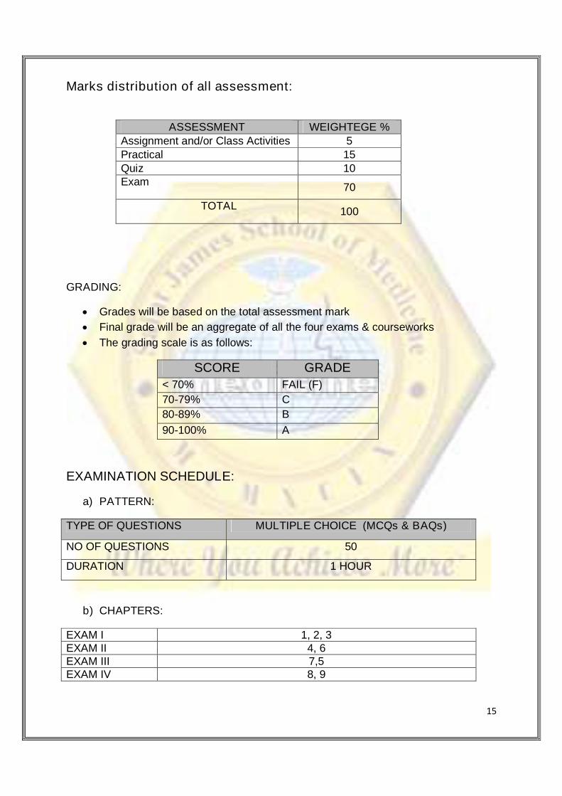
Marks distribution of all assessment:
ASSESSMENT WEIGHTEGE %
Assignment and/or Class Activities 5Practical 15Quiz 10Exam
70
TOTAL100
GRADING:
Grades will be based on the total assessment mark
Final grade will be an aggregate of all the four exams & courseworks
The grading scale is as follows:
SCORE GRADE
< 70% FAIL (F)
70-79% C
80-89% B
90-100% A
EXAMINATION SCHEDULE:
a) PATTERN:
TYPE OF QUESTIONS MULTIPLE CHOICE (MCQs & BAQs)
NO OF QUESTIONS 50
DURATION 1 HOUR
b) CHAPTERS:
EXAM I 1, 2, 3EXAM II 4, 6EXAM III 7,5EXAM IV 8, 9
16
c) EXAM DATES;
EXAM I 24 or 27 May 2013EXAM II 21 or 24 June 2013EXAM III 19 or 22 June 2013EXAM IV 21or 23 August 2013
TEXT BOOKS:
Required text:
1. Clinically Oriented Anatomy by Keith L. Moore et al., Lippincott Williams &
Wilkins, 2010
2. BRS Gross Anatomy, 7th edition by Kyung W. Chung. Published by Lippincott
Williams & Wilkins, 2012
3. Atlas of Human Anatomy, 4th edition by Frank H. Netter. Published by Saunders,
2006 or an equivalent atlas will be acceptable
4. Gray’s Dissection Guide for Human Anatomy, 2nd edition, David A. Morton, Kerry
D. Peterson, Kurt H. Albertine, Churchill Livingstone, 2007
Recommended Textbook
a. Gray's Anatomy for Students 2nd edition by Richard Drake, A. Wayne Vogal,
Adam.W. M. Mitchell, Churchill Livingstone, 2009
b. Clinical Anatomy by Region 9th edition by Richard S, Snell, Lippincott Williams &
Wilkins, 2011
c. Clinical Anatomy: Applied Anatomy for Students and Junior Doctors 12th editionby Harold Ellis, Vishy Mahadevan, 2010
d. Martini’s Atlas of the human body, Benjamin Cummings January 2003 | ISBN-10: 0130089060 , ISBN-13: 978-0130089069, Edition: 4th
e. Matt Hutchinson, Brief Atlas of the Human Body, January 3, 2006 , ISBN-10:080537373X , ISBN-13: 978-0805373738 . Edition: 2nd
17
f. Kaplan Medical , Anatomy Flashcards, January 6, 2009 , ISBN-10: 1427796947 ,ISBN-13: 978-1427796943
g. Kaplan Medical Anatomy Coloring Book, June 7, 2011 ,ISBN-10: 1419550403 ,ISBN-13: 978-1419550409 , Edition: 4
ON LINE RESOURCES:
1. http://www.dartmouth.edu/~humananatomy/2. http://www.med.umich.edu/lrc/coursepages/m1/anatomy2010/html/index.html3. http://www.anatomy.wisc.edu/courses/gross/ (dissections)4. http://www.wesnorman.com (Georgetown University’s anatomist, Wes Norman,
has his own site with images, information and quizzes)5. http://sinoemedicalassociation.org/anatomymed (Take Password from Dr.
Hammoudi)
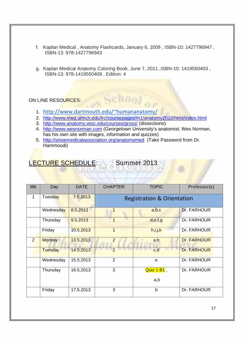
LECTURE SCHEDULE: Summer 2013
Wk Day DATE CHAPTER TOPIC Professor(s)
1 Tuesday 7.5.2013 Registration & Orientation
Wednesday 8.5.2013 1 a,b,c Dr. FARHOUR
Thursday 9.5.2013 1 d,e,f,g Dr. FARHOUR
Friday 10.5.2013 1 h,i,j,k Dr. FARHOUR
2 Monday 13.5.2013 2 a,b Dr. FARHOUR
Tuesday 14.5.2013 2 c,d Dr. FARHOUR
Wednesday 15.5.2013 2 e Dr. FARHOUR
Thursday 16.5.2013 3 Quiz 1 B1 ,
a,b
Dr. FARHOUR
Friday 17.5.2013 3 b Dr. FARHOUR
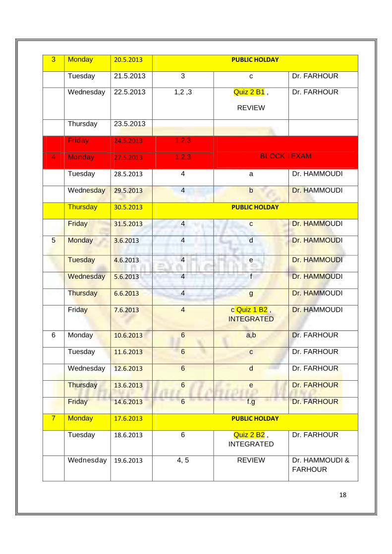
18
3 Monday 20.5.2013 PUBLIC HOLDAY
Tuesday 21.5.2013 3 c Dr. FARHOUR
Wednesday 22.5.2013 1,2 ,3 Quiz 2 B1 ,
REVIEW
Dr. FARHOUR
Thursday 23.5.2013
Friday 24.5.2013 1,2,3
BLOCK I EXAM4 Monday 27.5.2013 1,2,3
Tuesday 28.5.2013 4 a Dr. HAMMOUDI
Wednesday 29.5.2013 4 b Dr. HAMMOUDI
Thursday 30.5.2013 PUBLIC HOLDAY
Friday 31.5.2013 4 c Dr. HAMMOUDI
5 Monday 3.6.2013 4 d Dr. HAMMOUDI
Tuesday 4.6.2013 4 e Dr. HAMMOUDI
Wednesday 5.6.2013 4 f Dr. HAMMOUDI
Thursday 6.6.2013 4 g Dr. HAMMOUDI
Friday 7.6.2013 4 c Quiz 1 B2 ,
INTEGRATED
Dr. HAMMOUDI
6 Monday 10.6.2013 6 a,b Dr. FARHOUR
Tuesday 11.6.2013 6 c Dr. FARHOUR
Wednesday 12.6.2013 6 d Dr. FARHOUR
Thursday 13.6.2013 6 e Dr. FARHOUR
Friday 14.6.2013 6 f.g Dr. FARHOUR
7 Monday 17.6.2013 PUBLIC HOLDAY
Tuesday 18.6.2013 6 Quiz 2 B2 ,
INTEGRATED
Dr. FARHOUR
Wednesday 19.6.2013 4, 5 REVIEW Dr. HAMMOUDI &
FARHOUR
19
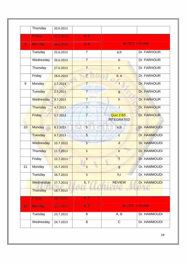
Thursday 20.6.2013
Friday 21.6.2013 4, 5
BLOCK II EXAM8 Monday 24.6.2013 4, 5
Tuesday 25.6.2013 7 a,b Dr. FARHOUR
Wednesday 26.6.2013 7 b Dr. FARHOUR
Thursday 27.6.2013 7 c Dr. FARHOUR
Friday 28.6.2013 7 d, e Dr. FARHOUR
9 Monday 1.7.2013 7 f Dr. FARHOUR
Tuesday 2.7.2013 7 g Dr. FARHOUR
Wednesday 3.7.2013 7 h Dr. FARHOUR
Thursday 4.7.2013 7 i Dr. FARHOUR
Friday 5.7.2013 7 Quiz 2 B3 ,
INTEGRATED
Dr. FARHOUR
10 Monday 8.7.2013 5 a,b Dr. HAMMOUDI
Tuesday 9.7.2013 5 c Dr. HAMMOUDI
Wednesday 10.7.2013 5 d Dr. HAMMOUDI
Thursday 11.7.2013 5 e Dr. HAMMOUDI
Friday 12.7.2013 5 f Dr. HAMMOUDI
11 Monday 15.7.2013 5 g Dr. HAMMOUDI
Tuesday 16.7.2013 5 h,i Dr. HAMMOUDI
Wednesday 17.7.2013 5, 7 REVIEW Dr. HAMMOUDI
Thursday 18.7.2013
Friday 19.7.2013 6, 7
BLOCK III EXAM12 Monday 22.7.2013 6, 7
Tuesday 23.7.2013 8 A, B Dr. HAMMOUDI
Wednesday 24.7.2013 8 C Dr. HAMMOUDI
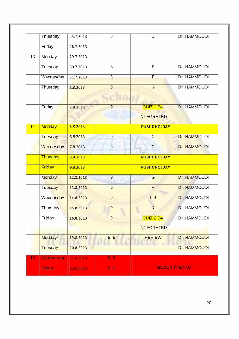
20
Thursday 25.7.2013 8 D Dr. HAMMOUDI
Friday 26.7.2013
13 Monday 29.7.2013
Tuesday 30.7.2013 8 E Dr. HAMMOUDI
Wednesday 31.7.2013 8 F Dr. HAMMOUDI
Thursday 1.8.2013 8 G Dr. HAMMOUDI
Friday 2.8.2013 9 QUIZ 1 B4,
INTEGRATED
Dr. HAMMOUDI
14 Monday 5.8.2013 PUBLIC HOLDAY
Tuesday 6.8.2013 9 C Dr. HAMMOUDI
Wednesday 7.8.2013 9 C Dr. HAMMOUDI
Thursday 8.8.2013 PUBLIC HOLDAY
Friday 9.8.2013 PUBLIC HOLDAY
Monday 12.8.2013 9 G Dr. HAMMOUDI
Tuesday 13.8.2013 9 H Dr. HAMMOUDI
Wednesday 14.8.2013 9 I, J Dr. HAMMOUDI
Thursday 15.8.2013 9 K Dr. HAMMOUDI
Friday 16.8.2013 9 QUIZ 2 B4,
INTEGRATED
Dr. HAMMOUDI
Monday 19.8.2013 8, 9 REVIEW Dr. HAMMOUDI
Tuesday 20.8.2013 Dr. HAMMOUDI
15 Wednesday 21.8.2013 8, 9
BLOCK IV EXAMFriday 23.8.2013 8, 9
21
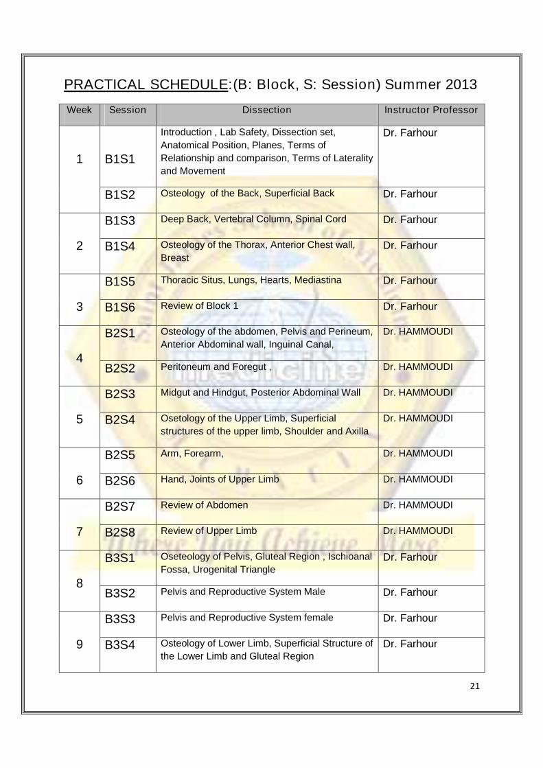
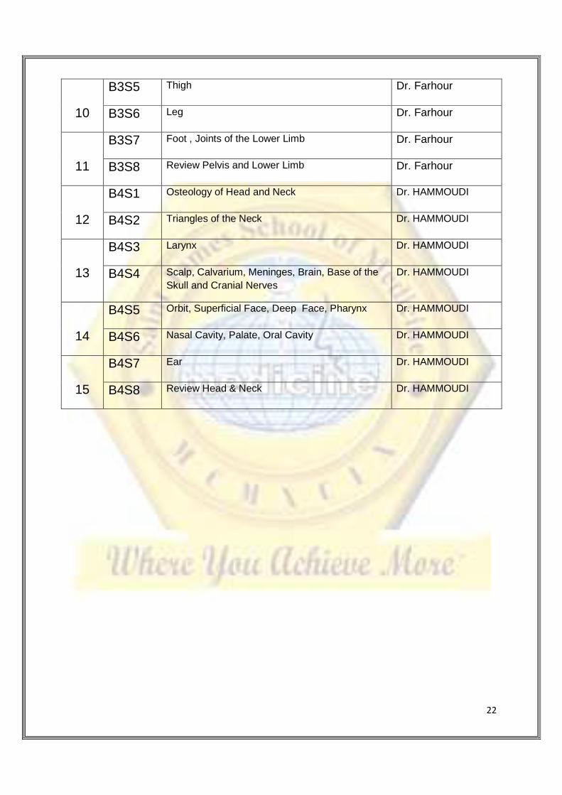
PRACTICAL SCHEDULE:(B: Block, S: Session) Summer 2013
Week Session Dissection Instructor Professor
1 B1S1
Introduction , Lab Safety, Dissection set,
Anatomical Position, Planes, Terms of
Relationship and comparison, Terms of Laterality
and Movement
Dr. Farhour
B1S2 Osteology of the Back, Superficial Back Dr. Farhour
2
B1S3 Deep Back, Vertebral Column, Spinal Cord Dr. Farhour
B1S4 Osteology of the Thorax, Anterior Chest wall,
BreastDr. Farhour
3
B1S5 Thoracic Situs, Lungs, Hearts, Mediastina Dr. Farhour
B1S6 Review of Block 1 Dr. Farhour
4
B2S1 Osteology of the abdomen, Pelvis and Perineum,
Anterior Abdominal wall, Inguinal Canal,
Dr. HAMMOUDI
B2S2 Peritoneum and Foregut , Dr. HAMMOUDI
5
B2S3 Midgut and Hindgut, Posterior Abdominal Wall Dr. HAMMOUDI
B2S4 Osetology of the Upper Limb, Superficial
structures of the upper limb, Shoulder and Axilla
Dr. HAMMOUDI
6
B2S5 Arm, Forearm, Dr. HAMMOUDI
B2S6 Hand, Joints of Upper Limb Dr. HAMMOUDI
7
B2S7 Review of Abdomen Dr. HAMMOUDI
B2S8 Review of Upper Limb Dr. HAMMOUDI
8
B3S1 Oseteology of Pelvis, Gluteal Region , Ischioanal
Fossa, Urogenital TriangleDr. Farhour
B3S2 Pelvis and Reproductive System Male Dr. Farhour
9
B3S3 Pelvis and Reproductive System female Dr. Farhour
B3S4 Osteology of Lower Limb, Superficial Structure of
the Lower Limb and Gluteal RegionDr. Farhour
22
10
B3S5 Thigh Dr. Farhour
B3S6 Leg Dr. Farhour
11
B3S7 Foot , Joints of the Lower Limb Dr. Farhour
B3S8 Review Pelvis and Lower Limb Dr. Farhour
12
B4S1 Osteology of Head and Neck Dr. HAMMOUDI
B4S2 Triangles of the Neck Dr. HAMMOUDI
13
B4S3 Larynx Dr. HAMMOUDI
B4S4 Scalp, Calvarium, Meninges, Brain, Base of the
Skull and Cranial Nerves
Dr. HAMMOUDI
14
B4S5 Orbit, Superficial Face, Deep Face, Pharynx Dr. HAMMOUDI
B4S6 Nasal Cavity, Palate, Oral Cavity Dr. HAMMOUDI
15
B4S7 Ear Dr. HAMMOUDI
B4S8 Review Head & Neck Dr. HAMMOUDI