Embed Size (px)
Citation preview
2923
Abstract. – OBJECTIVE: To determine the ap-propriate concentration of trypan blue (TB) for subretinal injection in a rat model and to provide a safety profile that limits retinal toxicity while maintaining dye visibility.
MATERIALS AND METHODS: Adult rats were subretinally injected with various concentra-tions of either TB or phosphate-buffered sa-line (PBS); rats which received sham injections served as an additional control. The inject-ed areas were visualized under a surgical mi-croscope. Electroretinography (ERG) was per-formed to measure retinal function. Animals were then sacrificed, and the eyes were sec-tioned and examined by light microscopy. Ter-minal deoxynucleotidy1 transferase dUTP nick-end labeling (TUNEL) was applied to determine retinal apoptosis.
RESULTS: One day after the subretinal in-jection, TB stains were visible under the surgi-cal microscope in the 0.2%, 0.08%, and 0.04% TB-injected groups, but not in the 0.02% TB-in-jected group. TB stain was detectable in the ret-ina and sclera of the 0.2%, 0.08%, and 0.04% TB-injected groups for over 2 weeks after in-jection. However, the amplitudes of ERGa- and b-waves were affected and became significant-ly lower in the 0.2% TB-injected group than the amplitudes in the PBS-, or sham-injected group. Moreover, TUNEL+ cells appeared in the outer nuclear layer (ONL), ganglion cell layer (GCL), and retinal pigment epithelium (RPE) layer of the 0.2% and 0.08% TB-injected groups at 1 and 7 days after subretinal injection. In contrast, very few TUNEL+ cells were found in the 0.04% TB- or PBS-injected group. Two weeks after injection,
the ONL was significantly thinner in the 0.2% TB-injected group than in the 0.04% TB-, PBS- or sham-injected group.
CONCLUSIONS: TB injection induces a dose-dependent neurotoxic effect on retinal cells. Subretinal injection of 0.04% TB is rela-tively safe and effective for subretinal staining.
Key Words:Trypan blue, Subretinal injection, Neurotoxicity,
Retinal cells, Rats.
Introduction
Subretinal injections of drugs1,2, gene con-structs3-6, or stem cells7-10 are emerging as com-mon procedures in experimental animals. The injected retinal area, however, is often difficult to locate, especially when it comes to preparing sections or analyzing the morphology. This is due to the quick recovery of the retinal detachment after subretinal injection. Researchers thus often use dyes to visualize the injected areas.
In clinical practice, vitreous or subretinal injection of dyes is used in vitreoretinal surgery that removes the internal limiting and epiretinal membranes11-18 or to localize retinal breaks19-22. The first dye to be used in eyes was indocyanine green (ICG). However, some studies23-26 showed that ICG exerted dose-dependent retinal tox-icity in both animals and humans. Other dyes
European Review for Medical and Pharmacological Sciences 2018; 22: 2923-2933
Y. FANG1, X.-Q. YAO1, L.-L. NIU1, J.-H. WU2, E.F. THEE3, D.-F. CHEN3,4, J.-Y. CHEN1, X.-H. SUN1,4,5
1Department of Ophthalmology and Visual Science, Eye & ENT Hospital of Fudan University, Shanghai, China2Experimental Research Center, Eye and ENT Hospital of Fudan University, Shanghai, China3Schepens Eye Research Institute, Massachusetts Eye and Ear, Department of Ophthalmology, Harvard Medical School, Boston, MA, USA4State Key Laboratory of Medical Neurobiology, Institutes of Brain Scienceand Collaborative Innovation Center for Brain Science, Fudan University, Shanghai, China5Key Laboratory of Myopia, NHFPC (Fudan University), and Shanghai Key Laboratory of Visual Impairment and Restoration (Fudan University), Shanghai, China
Yuan Fang and Xiaoqian Yao contributed equally to this work
Corresponding Author: Xinghuai Sun, MD; e-mail: [email protected] Junyi Chen, MD; e-mail: [email protected]
Safety evaluation of subretinal injection of trypan blue in rats
Y. Fang, X.-Q. Yao, L.-L. Niu, J.-H. Wu, E.F. Thee, D.-F. Chen, J.-Y. Chen, X.-H. Sun
2924
that have been investigated include trypan blue (TB)13,27,28, patent blue29, triamcinolone aceton-ide11, infracyanine green30,31, and brilliant blue G15,32,33.
TB is an anionic hydrophilic diazo dye of a relatively small molecular weight (960 Da). It can cross the membranes of dead cells, leading to distinctive blue staining of only dead cells34,35. TB staining is convenient and highly stable, without any background of autofluorescence36. It is thus also used in clinical practice to detect retinal breaks at concentrations ranging from 0.006-0.15%, with no reported complications19-22. However, some clinical studies12,14 reported that, after vitreous injection of 0.15% TB, the dye mi-grated from the vitreous into the subretinal space causing local chorioretinal atrophy and retinal pigment epithelium (RPE) changes. Currently, the safety profile of TB injection into the sub-retinal space is unknown. Therefore, the aim of this study is to determine the safe and effective concentrations of TB for subretinal injections through evaluating the histological and function-al effects of TB injection in rat eyes.
Materials and Methods
Animals, Anaesthesia, and MydriasisAll experimental procedures were performed
in accordance with the Institute’s Animal Care and Use Committee and adhered to the Statement for the Use of Animals in Ophthalmic and Vision Research. Male Sprague-Dawley rats (weighing 180-220 g) were maintained in a 12h light/12h dark cycle with free access to commercial rat food (Suzhou Laboratory animal feed Technol-ogy Co., Ltd. Suzhou, Jiangsu, China) and wa-ter. Rats were anesthetised by intraperitoneal injection of a mixture of 6.25 mg/kg xylazine (Sigma-Aldrich, St. Louis, MO, USA) and 10 mg/kg ketamine (Gutian Pharmaceuticals, FuJian, China). Pupils were dilated by a mixture of 5 mg/ml phenylephrine and 5 mg/mL tropicamide (Santen Pharmaceutical Co., Ltd., Osaka, Japan), and 4 mg/ml oxybuprocaine (Santen Pharmaceu-tical Co., Ltd) was applied as topical anaesthesia. The extent of mydriasis was checked under a sur-gical microscope (OM-10; Takagi Seiko Co., Ltd., Nagano, Japan). Anesthetization lasted for about 30-50 min and was sufficient for performing the subretinal injection. After the procedure, ofloxa-cin gel (Xing Qi Medical Company, Shenyang, China) was applied to prevent corneal opacifica-
tion and reduce the risk of infection. This study was approved by the Animal Ethics Committee of Fudan University Animal Center.
Subretinal Injection and Fundus Photography
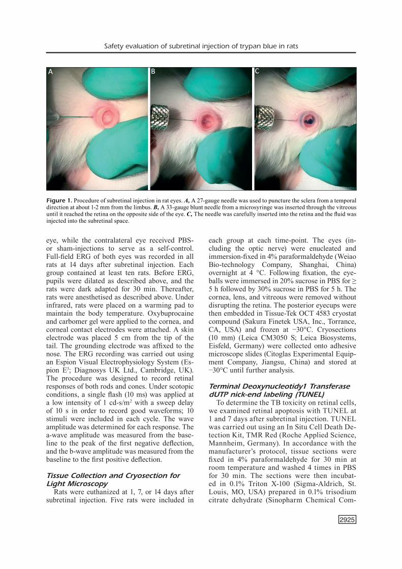
0.4% TB (Sigma-Aldrich, St. Louis, MO, USA) was diluted to final concentrations of 0.2%, 0.08%, 0.04%, and 0.02% in phosphate-buff-ered saline (PBS). Four µL of TB solution was slowly injected into the subretinal space of the rat eye. The contralateral eye that was injected with PBS was used as a control. Sham-injected and un-injected normal eyes served as additional control groups. Each group contained at least ten eyes. After pupil dilation, anaesthetized rats were placed under a surgical microscope in a lateral recumbent position. A 6-mm-diameter rubber ring was placed on the surface of the cornea, and one drop of carbomer eyedrops (Bausch & Lomb, Berlin, Germany) was applied within the ring to visualize the fundus. A 27-gauge needle (Meesawat Medical Industry Company, Shang-hai, China) was used to puncture the sclera from a temporal direction at about 1-2 mm from the lim-bus (Figure 1A). The needle was inserted with the bevel up at an angle of 45° to the posterior lens face and was advanced through the sclera into the vitreous humor. At least 50% of the bevel was pushed through the sclera to produce a hole suffi-cient to insert a blunt 33-gauge needle (Hamilton Company, Reno, NV, USA). The tip of the blunt needle was inserted through the puncture in the sclera, advanced into the vitreous avoiding dam-age to the lens, and then, aimed slightly towards the retina (Figure 1B). A feeling of slight resis-tance to the movement of the needle indicated when it had penetrated the retina and entered the subretinal space. Four µL of fluid was then slowly injected into the subretinal space over a period of 20 s (Figure 1C). This generated a visible detach-ment in about 1/4 circle of the retina (the inner boundary is about 1 mm from the optic nerve head) (Figure 2A-2D). The detachment lasted no longer than 24 h (Figure 2E-2H). The needle was gently withdrawn from the eye after subretinal injection. Fundus images were then obtained with a Kodak digital camera (FZ152; Eastman Kodak Company, Rochester, NY, USA).
Electroretinography (ERG)ERG was performed to assess retinal function.
The rats received a subretinal injection of TB at a concentration of 0.2%, 0.08%, or 0.04% in one
Safety evaluation of subretinal injection of trypan blue in rats
2925
eye, while the contralateral eye received PBS- or sham-injections to serve as a self-control. Full-field ERG of both eyes was recorded in all rats at 14 days after subretinal injection. Each group contained at least ten rats. Before ERG, pupils were dilated as described above, and the rats were dark adapted for 30 min. Thereafter, rats were anesthetised as described above. Under infrared, rats were placed on a warming pad to maintain the body temperature. Oxybuprocaine and carbomer gel were applied to the cornea, and corneal contact electrodes were attached. A skin electrode was placed 5 cm from the tip of the tail. The grounding electrode was affixed to the nose. The ERG recording was carried out using an Espion Visual Electrophysiology System (Es-pion E3; Diagnosys UK Ltd., Cambridge, UK). The procedure was designed to record retinal responses of both rods and cones. Under scotopic conditions, a single flash (10 ms) was applied at a low intensity of 1 cd-s/m2 with a sweep delay of 10 s in order to record good waveforms; 10 stimuli were included in each cycle. The wave amplitude was determined for each response. The a-wave amplitude was measured from the base-line to the peak of the first negative deflection, and the b-wave amplitude was measured from the baseline to the first positive deflection.
Tissue Collection and Cryosection for Light Microscopy
Rats were euthanized at 1, 7, or 14 days after subretinal injection. Five rats were included in
each group at each time-point. The eyes (in-cluding the optic nerve) were enucleated and immersion-fixed in 4% paraformaldehyde (Weiao Bio-technology Company, Shanghai, China) overnight at 4 °C. Following fixation, the eye-balls were immersed in 20% sucrose in PBS for ≥ 5 h followed by 30% sucrose in PBS for 5 h. The cornea, lens, and vitreous were removed without disrupting the retina. The posterior eyecups were then embedded in Tissue-Tek OCT 4583 cryostat compound (Sakura Finetek USA, Inc., Torrance, CA, USA) and frozen at −30°C. Cryosections (10 mm) (Leica CM3050 S; Leica Biosystems, Eisfeld, Germany) were collected onto adhesive microscope slides (Citoglas Experimental Equip-ment Company, Jiangsu, China) and stored at −30°C until further analysis.
Terminal Deoxynucleotidy1 Transferase dUTP nick-end labeling (TUNEL)
To determine the TB toxicity on retinal cells, we examined retinal apoptosis with TUNEL at 1 and 7 days after subretinal injection. TUNEL was carried out using an In Situ Cell Death De-tection Kit, TMR Red (Roche Applied Science, Mannheim, Germany). In accordance with the manufacturer’s protocol, tissue sections were fixed in 4% paraformaldehyde for 30 min at room temperature and washed 4 times in PBS for 30 min. The sections were then incubat-ed in 0.1% Triton X-100 (Sigma-Aldrich, St. Louis, MO, USA) prepared in 0.1% trisodium citrate dehydrate (Sinopharm Chemical Com-
Figure 1. Procedure of subretinal injection in rat eyes. A, A 27-gauge needle was used to puncture the sclera from a temporal direction at about 1-2 mm from the limbus. B, A 33-gauge blunt needle from a microsyringe was inserted through the vitreous until it reached the retina on the opposite side of the eye. C, The needle was carefully inserted into the retina and the fluid was injected into the subretinal space.
Y. Fang, X.-Q. Yao, L.-L. Niu, J.-H. Wu, E.F. Thee, D.-F. Chen, J.-Y. Chen, X.-H. Sun
2926
pany, Shanghai, China) in PBS at 4°C for 4 min. TUNEL reaction buffer was added at a dilution of 1:9 (solution 1:solution 2), and the slides were placed at 37°C in a thermo-stat (Yiheng Technology Company, Shanghai, China, DHP-9052) for 1 h. Retinal sections were then counterstained with a nuclear mark-er, Hoechst 33258 pentahydrate (1:1000; Life
Technology, Eugene, OR, USA) to reveal reti-nal laminar structures. Images were taken with a confocal fluorescence microscope (Leica SP8, Leica Microsystems, German). Afield size of 6.25 × 10⁻² mm2 per image was analyzed. Cells stained positive for TUNEL were counted, and a minimal 10 sections per rat, five rats per group, were analyzed.
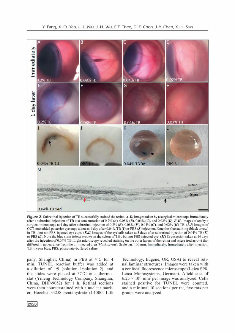
Figure 2. Subretinal injection of TB successfully stained the retina. A-D, Images taken by a surgical microscope immediately after a subretinal injection of TB at a concentration of 0.2% (A), 0.08% (B), 0.04% (C), and 0.02% (D). E-H, Images taken by a surgical microscope at 1 day after subretinal injection of 0.2% (E), 0.08% (F), 0.04% (G), and 0.02% (H) TB. (I,J) Images of OCT-embedded posterior eye cups taken as 1 day after 0.04% TB (I) or PBS (J) injection. Note the blue staining (black arrow) in TB-, but not PBS-injected eye cups. (K,L) Images of the eyeballs taken at 3 days after subretinal injection of 0.04% TB (K) or PBS (L). Note the blue stain (black arrow) on the sclera of TB-, but not PBS-injected eye. (M) Cryosection taken at 14 days after the injection of 0.04% TB. Light microscopy revealed staining on the outer layers of the retina and sclera (red arrow) that differed in appearance from the un-injected area (black arrow). Scale bar: 100 mm. Immediately: Immediately after injection; TB: trypan blue; PBS: phosphate-buffered saline.
Safety evaluation of subretinal injection of trypan blue in rats
2927
Haematoxylin and Eosin (H&E) Staining and Immunohistochemistry for Morphological Analysis
H&E staining was performed to assess the effects of TB on retinal morphology. Enucleated eyes (including the optic nerve) were cryosec-tioned. The slides containing a TB stained retinal area and the optic nerve head were selected and processed for H&E staining. Briefly, retinal sec-tions were immersed in 4% paraformaldehyde for 4 h and transferred to 70% ethanol. Individual samples were placed in processing cassettes, de-hydrated through a serial alcohol gradient, and embedded in paraffin. For immunofluorescence staining, retinal sections were deparaffinized in xylene, rehydrated through a decreasing ethanol gradient, and washed in PBS. The images of H&E stained retinal sections were acquired with a light microscope (Leica DM 4000B, Wetzhan, Germany). ONL thickness was quantified by counting the layers of photoreceptor nuclei in ret-inal sections at ~1 mm away from the optic nerve head. A minimal 10 sections per rat, five rats per group, were analyzed.
Statistical AnalysisThe mean number of TUNEL+ cells and ONL
thickness were compared among groups by one-way analysis of variance (ANOVA) with Tukey’s post-hoc test. To compare differences of ERG data between the left and right eyes, a paired Stu-dent’s t-test was used, and p < 0.05 was consid-ered statistically significant. Error bars represent the standard deviations. All statistical analyses were conducted using GraphPad 5 Prism 5 soft-ware (GraphPad, San Diego, CA).
Results
Tissue Staining by Subretinal Injection of TB
Based on previous reports, the concentrations of TB between 0.02%-0.2% were tested. Imme-diately after subretinal injection of 0.2%, 0.08%, 0.04% or 0.02% TB, blue stains were observed in the injected area with a surgical microscope (Figure 2A-2D). One day after injection, the blue staining remained detectable with surgical mi-croscopy (Figure 2E-2G) or in OCT-embedded eyecups in the 0.2%, 0.08%, and 0.04% (Figure 2I) TB-injected groups, but not in the 0.02% TB-injected group (Figure 2H) or PBS-injected group (Figure 2J). Three days after injection,
blue staining was visible in the sclera in the 0.2%, 0.08%, and 0.04% TB-injected groups (Figure 2K) and not in the PBS-injected group (Figure 2L). Moreover, one day after injection, the inject-ed region revealed a uniform color in the outer layers of the retina and sclera (red arrow in Figure 2M) of the 0.2%, 0.08%, and 0.04% TB-injected groups compared to the un-injected region under a light microscope (black arrow in Figure 2M). A blue stained area was visible in cryosections of the 0.04% (Figure 2M) and all other (data not shown) TB-injected groups even 14 days after injection. Thus, subretinal injections of TB at a concentration ≥ 0.04% effectively labeled the in-jected retinal areas for over 14 days. In contrast, 0.02% TB injection was inefficient in staining the retina and was thus excluded in the following experiments.
TB Impairs Retinal Function as Measured by ERG
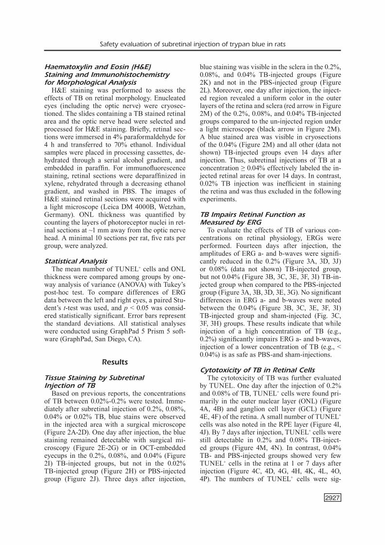
To evaluate the effects of TB of various con-centrations on retinal physiology, ERGs were performed. Fourteen days after injection, the amplitudes of ERG a- and b-waves were signifi-cantly reduced in the 0.2% (Figure 3A, 3D, 3J) or 0.08% (data not shown) TB-injected group, but not 0.04% (Figure 3B, 3C, 3E, 3F, 3I) TB-in-jected group when compared to the PBS-injected group (Figure 3A, 3B, 3D, 3E, 3G). No significant differences in ERG a- and b-waves were noted between the 0.04% (Figure 3B, 3C, 3E, 3F, 3I) TB-injected group and sham-injected (Fig. 3C, 3F, 3H) groups. These results indicate that while injection of a high concentration of TB (e.g., 0.2%) significantly impairs ERG a- and b-waves, injection of a lower concentration of TB (e.g., < 0.04%) is as safe as PBS-and sham-injections.
Cytotoxicity of TB in Retinal CellsThe cytotoxicity of TB was further evaluated
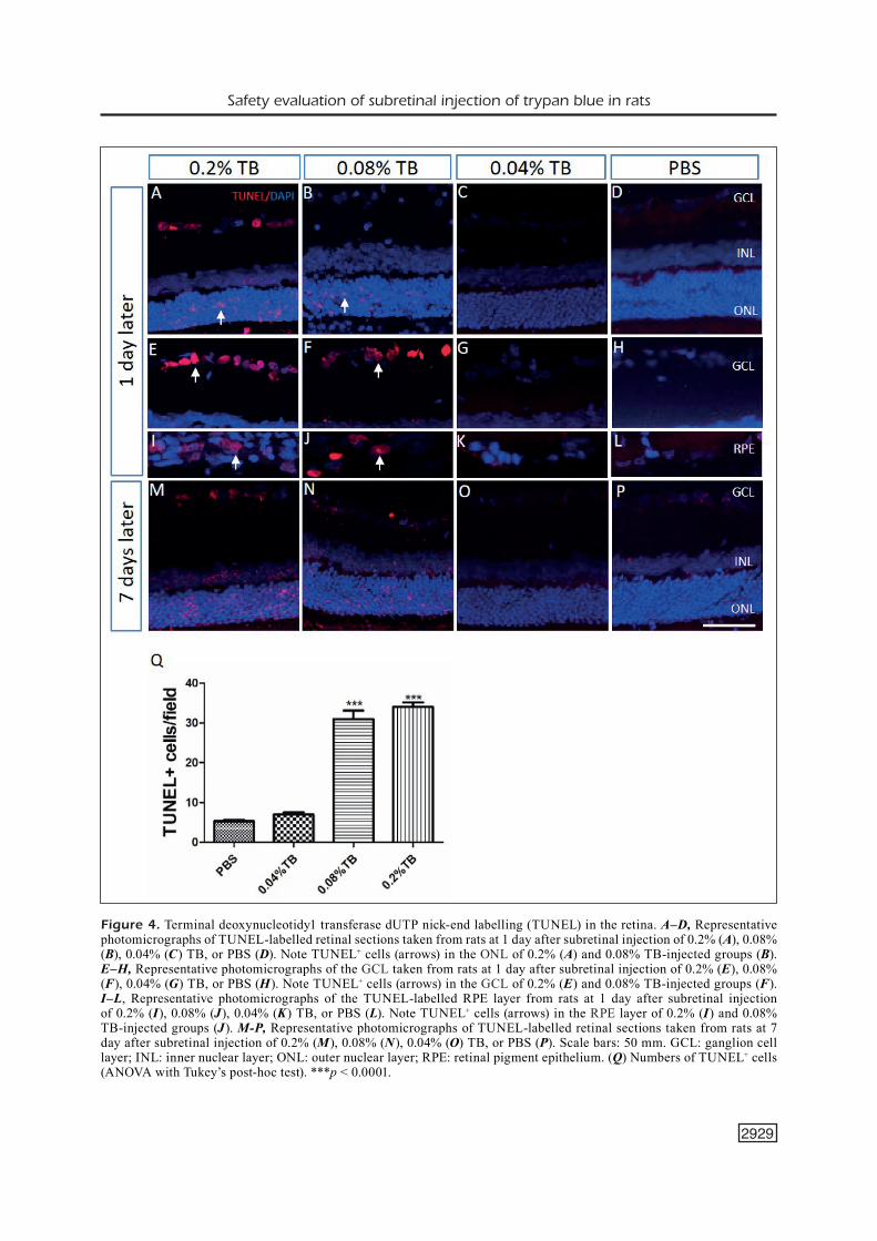
by TUNEL. One day after the injection of 0.2% and 0.08% of TB, TUNEL+ cells were found pri-marily in the outer nuclear layer (ONL) (Figure 4A, 4B) and ganglion cell layer (GCL) (Figure 4E, 4F) of the retina. A small number of TUNEL+ cells was also noted in the RPE layer (Figure 4I, 4J). By 7 days after injection, TUNEL+ cells were still detectable in 0.2% and 0.08% TB-inject-ed groups (Figure 4M, 4N). In contrast, 0.04% TB- and PBS-injected groups showed very few TUNEL+ cells in the retina at 1 or 7 days after injection (Figure 4C, 4D, 4G, 4H, 4K, 4L, 4O, 4P). The numbers of TUNEL+ cells were sig-
Y. Fang, X.-Q. Yao, L.-L. Niu, J.-H. Wu, E.F. Thee, D.-F. Chen, J.-Y. Chen, X.-H. Sun
2928
nificantly greater in retinal sections of 0.2% and 0.08% TB-injected groups than in those of 0.04% TB- and PBS-injected groups (Figure 4Q) (p < 0.0001). There were no significant differences in numbers of TUNEL+ cells between 0.2% and 0.08% TB-injected groups or between 0.04% TB- and PBS-injected groups (Figure 4Q). In conclu-sion, 0.04% TB injection is no more cytotoxic to the retina than PBS injection.
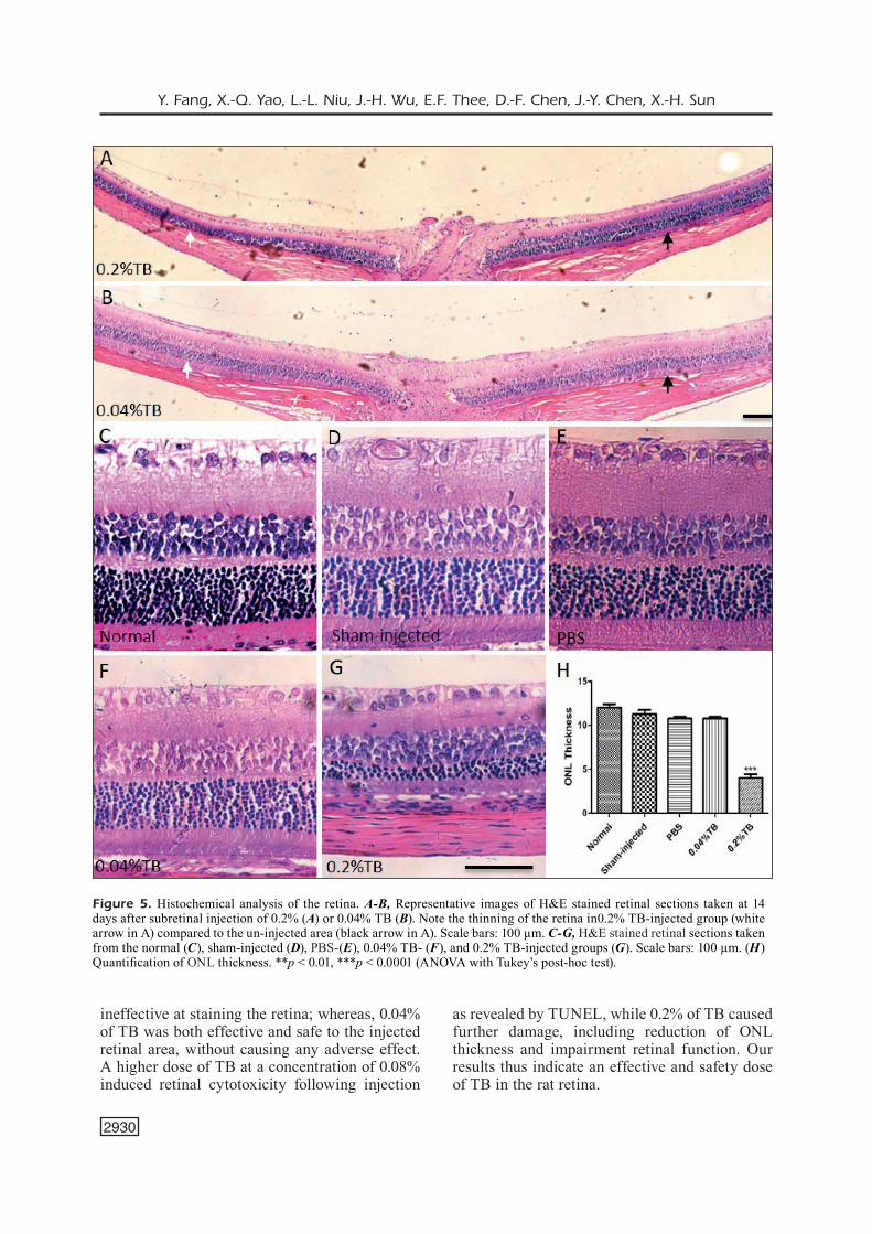
Histological Analysis of TB-Treated Retina
Histological analysis of TB-treated retina re-vealed that 14 days after TB injection, the inject-ed area (white arrow in Figure 5A) in the 0.2% TB-injected group exhibited apparently thinner retina, particularly the ONL, than the retina in the un-injected area (black arrow in Figure 5A). It supports a direct retinal cytotoxicity by 0.2% TB. In contrast, the retinas of the 0.04% TB-in-jected group exhibited no differences in retinal
morphology and retinal thickness between the injected (white arrow in Figure 5B) and un-in-jected area (black arrow in Figure 5B). Quanti-fication results showed no significant differences in ONL thickness among the normal (uninject-ed), sham-, PBS- and 0.04% TB-injected groups (Figure 5C-5F, 5H). An over 60% reduction of ONL thickness in the 0.2% TB-injected group compared to the normal retina was noted (Figure 5C, 5G, 5H) (p < 0.0001). The data indicate that 0.04% TB injection does not alter retinal mor-phology or ONL thickness.
Discussion
This study presents a series of in vivo experi-ments that analyzed the safety and tissue-staining efficacy of TB following a subretinal injection in the rat. Our data show that subretinal injection of TB at a concentration as low as 0.02% was
Figure 3. Retinal function measured by ERG at 14 days after subretinal injection. A-C, The amplitudes of a-waves in PBS-, 0.2% and 0.04% TB- and sham-injected groups. D-F, The amplitude of b-waves in PBS-, 0.2% and 0.04% TB- and sham-injected groups. G-J, Representative ERG waveforms taken from the PBS- (G), sham-(H), 0.04% TB- (I) and 0.2% TB-injected groups (J). Blue and red arrows indicate a-wave and b-wave, respectively. Black arrows illustrate how to measure time-to peak. *p < 0.05 and **p < 0.01 (paired Student’s t test). ERG: electroretinography.
Safety evaluation of subretinal injection of trypan blue in rats
2929
Figure 4. Terminal deoxynucleotidy1 transferase dUTP nick-end labelling (TUNEL) in the retina. A–D, Representative photomicrographs of TUNEL-labelled retinal sections taken from rats at 1 day after subretinal injection of 0.2% (A), 0.08% (B), 0.04% (C) TB, or PBS (D). Note TUNEL+ cells (arrows) in the ONL of 0.2% (A) and 0.08% TB-injected groups (B). E–H, Representative photomicrographs of the GCL taken from rats at 1 day after subretinal injection of 0.2% (E), 0.08% (F), 0.04% (G) TB, or PBS (H). Note TUNEL+ cells (arrows) in the GCL of 0.2% (E) and 0.08% TB-injected groups (F). I–L, Representative photomicrographs of the TUNEL-labelled RPE layer from rats at 1 day after subretinal injection of 0.2% (I), 0.08% (J), 0.04% (K) TB, or PBS (L). Note TUNEL+ cells (arrows) in the RPE layer of 0.2% (I) and 0.08% TB-injected groups (J). M-P, Representative photomicrographs of TUNEL-labelled retinal sections taken from rats at 7 day after subretinal injection of 0.2% (M), 0.08% (N), 0.04% (O) TB, or PBS (P). Scale bars: 50 mm. GCL: ganglion cell layer; INL: inner nuclear layer; ONL: outer nuclear layer; RPE: retinal pigment epithelium. (Q) Numbers of TUNEL+ cells (ANOVA with Tukey’s post-hoc test). ***p < 0.0001.
Y. Fang, X.-Q. Yao, L.-L. Niu, J.-H. Wu, E.F. Thee, D.-F. Chen, J.-Y. Chen, X.-H. Sun
2930
ineffective at staining the retina; whereas, 0.04% of TB was both effective and safe to the injected retinal area, without causing any adverse effect. A higher dose of TB at a concentration of 0.08% induced retinal cytotoxicity following injection
as revealed by TUNEL, while 0.2% of TB caused further damage, including reduction of ONL thickness and impairment retinal function. Our results thus indicate an effective and safety dose of TB in the rat retina.
Figure 5. Histochemical analysis of the retina. A-B, Representative images of H&E stained retinal sections taken at 14 days after subretinal injection of 0.2% (A) or 0.04% TB (B). Note the thinning of the retina in0.2% TB-injected group (white arrow in A) compared to the un-injected area (black arrow in A). Scale bars: 100 µm. C-G, H&E stained retinal sections taken from the normal (C), sham-injected (D), PBS-(E), 0.04% TB- (F), and 0.2% TB-injected groups (G). Scale bars: 100 µm. (H) Quantification of ONL thickness. **p < 0.01, ***p < 0.0001 (ANOVA with Tukey’s post-hoc test).
Safety evaluation of subretinal injection of trypan blue in rats
2931
A toxic effect of TB has been suggested pre-viously in in vitro and in vivo experiments. Al-though an acute exposure (less than or equal to 5 min) of RPE cells to TB seemed safe37,38, even at a relatively high concentration (0.30%)39, the safe-ty doses of TB to RPE cells became much lower (less than 0.05%) under the chronic exposure ( > 3 days)23. Retinal neurons, however, are more susceptible to TB toxicity than RPE cells. A short exposure (within 30 min) to 0.06% TB or a 3-day exposure to low concentrations (0.01%-0.025%) of TB induced cytotoxicity in RGCs40. In clinic, injection of 0.15% TB in patients caused retinal thinning and chorioretinal atrophy in areas show-ing TB migration, as revealed by optical coherence tomography12. Saeed and Heimann14 also noted an association between TB administration and retinal lesion that included increased retinal pig-mentation and RPE atrophy over a 2-week period after vitrectomy. In rabbit studies, Maia et al41 and Penha et al42 also showed that subretinal injection of 0.15% TB caused photoreceptor damage within 12 h after surgery, and the toxic effect of TB was observable even after 14 days. In the present study, we demonstrated that TB induced retinal damage in a dose-dependent manner in rats.
The reported safety dose of TB in vitro was slightly lower than that was seen in vivo. Likely, this is because the concentration of TB in the sub-retinal space is inaccurate or diluted due to fluid diffusion and outflow, while the TB concentration in cell cultures is more controllable. Second, cells embedded in the retinal structure might be more resistant to TB toxicity than dissociated cells in a culture dish. Presence of Müller glia and other cell types in the retina may serve to maintain retinal homeostasis and provide structural and trophic support to the surrounding cells.
In our work, 0.04% is a safe concentration for subretinal injection of TB. We assessed cell apop-tosis with TUNEL. A large number of TUNEL+ cells were detected in the ONL, INL, and GCL, as well as the RPE layer of 0.08% and 0.2% TB-injected groups, even at 7 days after injec-tion. These results are consistent with the ERG assessment, in which the amplitudes of both a- and b-waves were significantly reduced after ex-posure to high concentrations of TB. In contrast, TUNEL+ cells were barely detected in the 0.04% TB-injected group. Notably, subretinal injection of high concentrations of TB (0.08% and 0.2%) also showed toxic effects on RGCs in vivo. We suggested that TB might have leaked into the vitreous cavity through the retinal hole generated
by the syringe that entered via the vitreous. To-gether, the results of our study indicate that high concentrations of TB (0.08% and 0.2%) have a toxic effect on retinal cells, including ONL, INL, GCL, and RPE cells.
The cytotoxic effect of TB is likely to exist in humans. Currently, we do not yet know if the safety dose of TB in the retinas of humans and rats is the same. It is clear that low concentrations (e.g., less than or equal to 0.04%) of TB are likely safer than the higher concentrations (greater than 0.08%). Thus, if a vitreous or subretinal injection of a high concentration of TB (greater than or equal to 0.08%) is unavoidable, we suggest that it should be removed as soon as possible. While our study used only albino rats, it is most likely that TB induces a similar toxicity effect in the retinas of pigmented rats, just as it has been reported for many ophthalmic drugs in albino and pigmented rats43-45.
Conclusions
Since TB which has been initially introduced to the clinic, particularly vitreoretinal surgery, as a remarkable biocompatible dye, many research-es24,37,38,40 have reported a toxic effect of TB in the retina in a dose- and time-dependent manner. Our study in rats indicates that 0.04% TB is a safe and effective dose of TB for subretinal injection to localize the treated area under a light micro-scope. Currently, more long-term investigations are required for further verification of TB’s safety in the retina.
AcknowledgementsWe thank Rong Zhang for her valuable help in confocal im-ages analysis and Richard C Han for critical reading of the manuscript. This study was supported by the International Science & Technology Cooperation Program of China (No. 2015DFA31340) and the National Natural Science Founda-tion (NSFC81100667, NSFC81470623 and NSFC81430007).
Conflict of InterestThe Authors declare that they have no conflict of interests.
References
1) HayasHi a, Naseri a, PeNNesi Me, de JuaN eJ. Sub-retinal delivery of immunoglobulin G with gold nanoparticles in the rabbit eye. Jpn J Ophthalmol 2009; 53: 249-256.
Y. Fang, X.-Q. Yao, L.-L. Niu, J.-H. Wu, E.F. Thee, D.-F. Chen, J.-Y. Chen, X.-H. Sun
2932
2) you ys, Lee Cy, Li C, Lee sH, KiM K, JuNg H. An arched micro-injector (ARCMI) for innocuous subretinal injection. PLoS One 2014; 9: e104145.
3) BeNNett J, Maguire AM. Gene therapy for ocular disease. Mol Ther 2000; 1: 501-505.
4) FLaNNery Jg, ZoLotuKHiN s, Vaquero Mi, LaVaiL MM, MuZyCZKa N, HauswirtH ww. Efficient photorecep-tor-targeted gene expression in vivo by recombi-nant adeno-associated virus. Proc Natl Acad Sci U S A 1997; 94: 6916-6921.
5) BeNNett J, Maguire aM, CideCiyaN aV, sCHNeLL M, gLoVer e, aNaNd V, aLeMaN ts, CHirMuLe N, guP-ta ar, HuaNg y, gao gP, NyBerg wC, taZeLaar J, HugHes J, wiLsoN JM, JaCoBsoN sg. Stable trans-gene expression in rod photoreceptors after re-combinant adeno-associated virus-mediated gene transfer to monkey retina. Proc Natl Acad Sci U S A 1999; 96: 9920-9925.
6) CoNstaBLe iJ, BLuMeNKraNZ Ms, sCHwartZ sd, BaroNe s, Lai CM, raKoCZy eP. Gene therapy for age-relat-ed macular degeneration. Asia Pac J Ophthalmol (Phila) 2016; 5: 300-303.
7) staNZeL BV, Liu Z, BriNKeN r, BrauN N, HoLZ Fg, eter N. Subretinal delivery of ultrathin rigid-elastic cell carriers using a metallic shooter instrument and biodegradable hydrogel encapsulation. In-vest Ophthalmol Vis Sci 2012; 53: 490-500.
8) ParK Hy, KiM JH, suN KH, ParK CK. Stem cell-based delivery of brain-derived neurotrophic factor gene in the rat retina. Brain Res 2012; 1469: 10-23.
9) Koss MJ, FaLaBeLLa P, steFaNiNi Fr, PFister M, tHoM-as BB, KasHaNi aH, BraNt r, ZHu d, CLegg do, HiN-toN dr, HuMayuN Ms. Subretinal implantation of a monolayer of human embryonic stem cell-derived retinal pigment epithelium: a feasibility and safety study in Yucatan minipigs. Graefes Arch Clin Exp Ophthalmol 2016; 254: 1553-1565.
10) westeNsKow Pd, KuriHara t, BraVo s, FeiteLBerg d, sediLLo Za, aguiLar e, FriedLaNder M. Performing subretinal injections in rodents to deliver retinal pigment epithelium cells in suspension. J Vis Exp 2015; (95): 52247.
11) FaraH Me, Maia M, PeNHa FM, rodrigues eB. The use of vital dyes during vitreoretinal surgery-chro-movitrectomy. Dev Ophthalmol 2016; 55: 365-375.
12) gHosH s, issa s, eL gi, staNNard K. Subretinal mi-gration of trypan blue during macular hole and epiretinal membrane peel: an observational case series. Is there a safer method? Eye (Lond) 2010; 24: 1724-1727.
13) HaritogLou C, gaNdorFer a, sCHauMBerger M, Pri-gLiNger sg, MueLLer aJ, gass Ca, KaMPiK a. Trypan blue in macular pucker surgery: an evaluation of histology and functional outcome. Retina 2004; 24: 582-590.
14) saeed Mu, HeiMaNN H. Atrophy of the retinal pig-ment epithelium following vitrectomy with trypan blue. Int Ophthalmol 2009; 29: 239-241.
15) Badaro e, NoVais ea, PeNHa FM, Maia M, FaraH Me, rodrigues eB. Vital dyes in ophthalmology:
a chemical perspective. Curr Eye Res 2014; 39: 649-658.
16) Vote BJ, russeLL MK, JooNdePH BC. Trypan blue-as-sisted vitrectomy. Retina 2004; 24: 736-738.
17) Li K, woNg d, HisCott P, staNga P, groeNewaLd C, MCgaLLiard J. Trypan blue staining of internal lim-iting membrane and epiretinal membrane during vitrectomy: visual results and histopathological findings. Br J Ophthalmol 2003; 87: 216-219.
18) Perrier M, seBag M. Epiretinal membrane surgery assisted by trypan blue. Am J Ophthalmol 2003; 135: 909-911.
19) guPta d, oNg J, BurtoN rL. Trans-scleral dye in-jection during vitreous surgery to identify clinical-ly undetectable retinal breaks causing retinal de-tachment. Eye (Lond) 2011; 25: 1045-1049.
20) woNg r, guPta B, ayLward gw, LaidLaw da. Dye extrusion technique (DE-TECH): Occult retinal break detection with subretinal dye extrusion during vitrectomy for retinal detachment repair. Retina 2009; 29: 492-496.
21) JaCKsoN tL, KwaN as, LaidLaw aH, ayLward w. Iden-tification of retinal breaks using subretinal trypan blue injection. Ophthalmology 2007; 114: 587-590.
22) KHaNduJa s, siNHa s, gogia V, KaKKar a, VoHra r. Modified subretinal dye extrusion technique (MORE-DETECH): Subretinal diluted trypan blue for detecting occult retinal breaks in retinal de-tachment after endotamponade removal. Int Oph-thalmol 2013; 33: 729-732.
23) areVaLo JF, garCia ra. Macular hole surgery com-plicated by accidental massive subretinal indocy-anine green, and retinal tear. Graefes Arch Clin Exp Ophthalmol 2007; 245: 751-753.
24) KodJiKiaN L, riCHter t, HaLBerstadt M, BeBy F, FLueC-Kiger F, BoeHNKe M, garweg Jg. Toxic effects of in-docyanine green, infracyanine green, and trypan blue on the human retinal pigmented epithelium. Graefes Arch Clin Exp Ophthalmol 2005; 243: 917-925.
25) Hua L, LiN B, HoNg J, MiN HB, HaN wL, ZHou ty, ZHaNg Zq. Clinical research on one-third dose verteporfin photodynamic therapy in the treat-ment of chronic central serous chorioretinopathy. Eur Rev Med Pharmacol Sci 2018; 22: 278-284.
26) staNesCu-segaLL d, JaCKsoN tL. Vital staining with in-docyanine green: a review of the clinical and ex-perimental studies relating to safety. Eye (Lond) 2009; 23: 504-518.
27) de waard Pw, Budo CJ, MeLLes gr. Trypan blue capsular staining to “find” the leading edge of a “lost” capsulorhexis. Am J Ophthalmol 2002; 134: 271-272.
28) FaraH Me, Maia M, FurLaNi B, Bottos J, Meyer CH, LiMa V, PeNHa FM, Costa eF, rodrigues eB. Current concepts of trypan blue in chromovitrectomy. Dev Ophthalmol 2008; 42: 91-100.
29) LuKe C, LuKe M, siCKeL w, sCHNeider t. Effects of pat-ent blue on human retinal function. Graefes Arch Clin Exp Ophthalmol 2006; 244: 1188-1190.
Safety evaluation of subretinal injection of trypan blue in rats
2933
30) HaritogLou C, gaNdorFer a, gass Ca, KaMPiK a. His-tology of the vitreoretinal interface after staining of the internal limiting membrane using glucose 5% diluted indocyanine and infracyanine green. Am J Ophthalmol 2004; 137: 345-348.
31) CaCCiatori M, MCPHersoN r. Idiopathic macular hole surgery with low-concentration infracyanine green-assisted peeling of the internal limiting membrane. Am J Ophthalmol 2007; 143: 726, 727.
32) ueNo a, HisatoMi t, eNaida H, KagiMoto t, MoCHiZuKi y, goto y, KuBota t, Hata y, isHiBasHi t. Biocompati-bility of brilliant blue G in a rat model of subretinal injection. Retina 2007; 27: 499-504.
33) eNaida H, isHiBasHi t. Brilliant blue in vitreoretinal surgery. Dev Ophthalmol 2008; 42: 115-125.
34) Louis Ks, siegeL aC. Cell viability analysis using try-pan blue: manual and automated methods. Meth-ods Mol Biol 2011; 740: 7-12.
35) TraN sL, PuHar a, Ngo-CaMus M, raMarao N. Try-pan blue dye enters viable cells incubated with the pore-forming toxin HlyII of Bacillus cereus. PLoS One 2011; 6: e22876.
36) KuCsera J, yarita K, taKeo K. Simple detec-tion method for distinguishing dead and living yeast colonies. J Microbiol Methods 2000; 41: 19-21.
37) NarayaNaN r, KeNNey MC, KaMJoo s, triNH tH, sei-geL gM, reseNde gP, KuPPerMaNN Bd. Trypan blue: effect on retinal pigment epithelial and neurosen-sory retinal cells. Invest Ophthalmol Vis Sci 2005; 46: 304-309.
38) gaLe Js, ProuLx aa, goNder Jr, Mao aJ, HutNiK CM. Comparison of the in vitro toxicity of indocy-anine green to that of trypan blue in human ret-
inal pigment epithelium cell cultures. Am J Oph-thalmol 2004; 138: 64-69.
39) staLMaNs P, FeroN eJ, Parys-VaN gr, VaN LoMMeL a, MeLLes gr, VeCKeNeer M. Double vital staining us-ing trypan blue and infracyanine green in macular pucker surgery. Br J Ophthalmol 2003; 87: 713-716.
40) JiN y, uCHida s, yaNagi y, aiHara M, araie M. Neuro-toxic effects of trypan blue on rat retinal ganglion cells. Exp Eye Res 2005; 81: 395-400.
41) Maia M, PeNHa F, rodrigues eB, PriNCiPe a, diB e, Meyer CH, FreyMuLLer e, Moraes N, FaraH Me. Effects of subretinal injection of patent blue and trypan blue in rabbits. Curr Eye Res 2007; 32: 309-317.
42) PeNHa FM, Maia M, eid FM, PriNCiPe aH, FreyMuLLer eH, Maia a, MagaLHaes oJ, sMitH rL. Effects of subretinal injections of indocyanine green, trypan blue, and glucose in rabbit eyes. Ophthalmology 2007; 114: 899-908.
43) LeZMi s, roKH N, saiNt-MaCary g, PiNo M, saLLeZ V, tHeVeNard F, rooMe N, rosoLeN s. Chloroquine causes similar electroretinogram modifications, neuronal phospholipidosis and marked impair-ment of synaptic vesicle transport in albino and pigmented rats. Toxicology 2013; 308: 50-59.
44) roMaN d, VerHoeVe J, sCHadt H, ViCart a, waLKer uJ, turNer o, riCHardsoN ta, woLFord st, MiLLer Pe, ZHou w, Lu H, aKiMoV M, KLuwe w. Ocular toxicity of AUY922 in pigmented and albino rats. Toxicol Appl Pharmacol 2016; 309: 55-62.
45) aKuLa Jd, NooNaN er, di Nardo a, FaVaZZa tL, ZHaNg N, saHiN M, HaNseN rM, FuLtoN aB. Vigab-atrin can enhance electroretinographic respons-es in pigmented and albino rats. Doc Ophthalmol 2015; 131: 1-11.

![TheUseofIntravitrealRanibizumabfor ......(RD), subretinal fibrosis, choroidal neovascular membrane (CNVM) formation, and extraocular manifestations [2]. CNVMisfoundin2–15%ofVKHpatients[3].Wereporton](https://img.pdfslide.us/doc/110x75/6053729d1d5c5177055cef33/theuseofintravitrealranibizumabfor-rd-subretinal-ibrosis-choroidal.jpg)